Dual-Phase Cardiac Diffusion Tensor Imaging with Strain Correction
13
0
0
Texte intégral
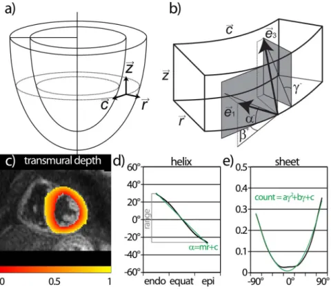
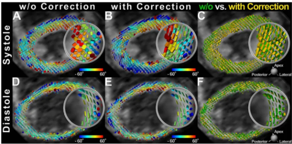
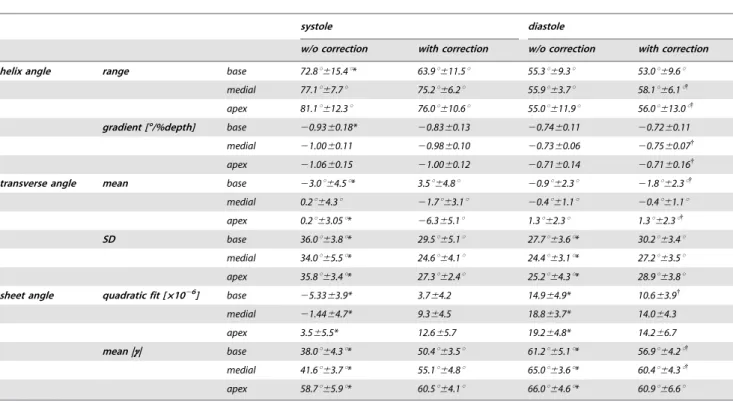
Figure

Documents relatifs
