HAL Id: hal-00267010
https://hal.archives-ouvertes.fr/hal-00267010
Submitted on 26 Mar 2008HAL is a multi-disciplinary open access archive for the deposit and dissemination of sci-entific research documents, whether they are pub-lished or not. The documents may come from teaching and research institutions in France or abroad, or from public or private research centers.
L’archive ouverte pluridisciplinaire HAL, est destinée au dépôt et à la diffusion de documents scientifiques de niveau recherche, publiés ou non, émanant des établissements d’enseignement et de recherche français ou étrangers, des laboratoires publics ou privés.
Sox9 regulates cell proliferation and is required for
Paneth cell differentiation in the intestinal epithelium.
Pauline Bastide, Charbel Darido, Julie Pannequin, Ralf Kist, Sylvie Robine,
Christiane Marty-Double, Frédéric Bibeau, Gerd Scherer, Dominique Joubert,
Frédéric Hollande, et al.
To cite this version:
Pauline Bastide, Charbel Darido, Julie Pannequin, Ralf Kist, Sylvie Robine, et al.. Sox9 regulates cell proliferation and is required for Paneth cell differentiation in the intestinal epithelium.. Journal of Cell Biology, Rockefeller University Press, 2007, 178 (4), pp.635-48. �10.1083/jcb.200704152�. �hal-00267010�
SOX9 REGULATES CELL PROLIFERATION AND IS REQUIRED FOR PANETH CELL DIFFERENTIATION IN THE INTESTINAL EPITHELIUM
Pauline Bastide1, Charbel Darido1, Julie Pannequin1, Ralf Kist2,*, Sylvie Robine3, Christiane Marty-Double4, Frédéric Bibeau5, Gerd Scherer2, Dominique Joubert1, Frédéric Hollande1, Philippe Blache1 and Philippe Jay1#
1
INSERM, U661, Montpellier, France; Department of Cellular and Molecular Oncology, Institut de génomique fonctionnelle, CNRS UMR5203, Université de Montpellier I and Université de Montpellier II, Montpellier, 141 rue de la Cardonille, F-34094, Montpellier, cedex 5, France; 2 Institute of Human Genetics and Anthropology; University of Freiburg, Breisacherstr. 33, D-79106, Freiburg, Germany;
3
Morphogenesis and Intracellular Signaling, Institut Curie-CNRS, 25 rue d'Ulm, 75248 Paris cedex 5, France; 4 Service d'anatomie-pathologie, CHU Carémeau, Place Prof. Robert Debré; 30000 Nimes, France ; 5 Service d'anatomie-pathologie, CRLC Val d’Aurelle, 326 rue des Apothicaires, Parc Euromédecine, 34298 Montpellier cedex 5, France ; * Present address: Institute of Human Genetics, International Centre for Life, University of Newcastle, Newcastle upon Tyne, NE1 3BZ, UK.
Condensed title: Sox9/Wnt interaction control intestinal homeostasis
#
Correspondence to Philippe Jay: philippe.jay@igf.cnrs.fr
Manuscript JCB 200704152, Jay (formerly 200608026), revised version: 21/06/2007 Character count: 38959
The HMG-box transcription factor Sox9 is expressed in the intestinal epithelium, specifically in stem/progenitor cells and in Paneth cells. Sox9 expression requires an active beta-catenin/Tcf complex, the transcriptional effector of the Wnt pathway. This pathway is critical for numerous aspects of the intestinal epithelium physiopathology but processes that specify the cell response to such multipotential signals still remain to be identified. Here, we inactivated the Sox9 gene in the intestinal epithelium to analyse its physiological function. Sox9 inactivation affected differentiation throughout the intestinal epithelium, with a disappearance of Paneth cells and a decrease of the goblet cell lineage. Additionally, the morphology of the colon epithelium was severely altered. We detected general hyperplasia and local crypt dysplasia in the intestine, and Wnt pathway target genes were upregulated. These results highlight the central position of Sox9 as both a transcriptional target and a regulator of the Wnt pathway in the regulation of intestinal epithelium homeostasis.
Introduction
The intestinal epithelium is constantly and rapidly renewing throughout the lifespan of vertebrates, thereby representing a major target for tumorigenesis. This epithelium can be divided into two functionally distinct compartments. The crypt of Lieberkühn constitutes the proliferative compartment and contains stem/progenitor cells, as well as, in the small intestine, terminally differentiated Paneth cells. Multipotent stem cells, located near the bottom of crypts, generate new cells, which migrate upwards while differentiating into enterocytes, goblet and enteroendocrine cells. Proliferation stops at the crypt-villus junction, and terminally differentiated cells are located on the neighboring villus, which constitute the differentiated compartment. In the small intestine, a fourth cell type, the Paneth cell, migrates downwards and settles at the bottom of the crypts as post-mitotic, differentiated, cells. The balance between proliferation, differentiation, migration and cell death must be tightly regulated to maintain homeostasis of this epithelium.
We reported the expression of Sox9, an HMG-box transcription factor, specifically in the rapidly proliferating stem/progenitor cells found at the bottom third of Lieberkühn crypts throughout the length of the intestine and in the Paneth cells of the small intestine, as well as in human tumors of the intestinal epithelium (Blache et al., 2004) . Sox9 was first identified as a key regulator of cartilage and male gonad development. Heterozygous Sox9 mutations are responsible for the campomelic dysplasia syndrome, a skeletal dysmorphology syndrome characterized by skeletal malformation of endochondral bones and by male to female sex reversal in the majority of genotypically XY individuals (Foster et al., 1994; Wagner et al., 1994). Sox9 has also been implicated in the development of cranial neural crest derivatives
(Spokony et al., 2002), in the neural stem cell switch from neurogenesis to gliogenesis (Stolt et al., 2003) and in heart (Akiyama et al., 2004a), hair (Vidal et al., 2005) and pancreas (Seymour et al., 2007) development. In each of these tissues, Sox9 expression is restricted to specific cell types, suggesting a complex transcriptional regulation. In addition, the currently identified Sox9 target genes, for instance in the cartilage and in the gonad, display tissue-specific expression (de Santa Barbara et al., 1998; Ng et al., 1997), indicating that Sox9 may regulate distinct sets of genes in the different tissues in which it is expressed.
In the intestinal epithelium, the function of Sox9 remains unresolved, although in vitro studies suggested a role in the control of cell differentiation (Blache et al., 2004). In vitro and in vivo data indicate that Sox9 is a transcriptional target of Wnt signalling. For instance, Sox9 expression is abrogated in Tcf4-null embryos, and it is strongly expressed in colorectal carcinoma cell lines containing activating mutations in components of the Wnt pathway (Blache et al., 2004).
The Wnt pathway plays a central role among the extracellular signals required to maintain the homeostasis of the intestinal epithelium. In particular, deletion of the gene encoding Tcf4, an other HMG-box transcription factor (Korinek et al., 1998), or overexpression of the inhibitor Dickkopf (Kuhnert et al., 2004; Pinto et al., 2003), resulted in a loss of the proliferative compartment and in impaired differentiation of secretory cell lineages. Conversely, mutation of the gene encoding APC, a negative regulator of the pathway, resulted in crypt expansion, abrogation of cell migration, and amplification of the Paneth cell population (Andreu et al., 2005; Sansom et al., 2004). In addition, deletion of the Wnt receptor Frizzled-5 revealed an essential role of the Wnt/Frizzled-5 pathway in the maturation of Paneth cells (van Es et al., 2005). The sorting process of epithelial cells along the crypt-villus axis also depends on the
Wnt pathway, via a modulation of Ephrin/Eph receptor interactions (Batlle et al., 2002). The Wnt signalling pathway can thus induce diverse cellular responses in the intestinal epithelium. In addition to these physiological functions, the Wnt pathway is centrally implicated in cancer, since mutations in components of this pathway have been identified in the majority of human colorectal carcinoma (Morin et al., 1997). Such mutations mimic activation of the pathway by Wnt ligants, i.e. stabilisation of beta-catenin, and result in constitutive transcriptional activity of the beta-catenin/Tcf4 complex and in aberrant expression of its target genes (Korinek et al., 1997). Despite the central importance of this pathway in the physiopathology of the intestinal epithelium, little is known about the molecular mechanisms involved in restricting this wide spectrum of potential functions to elicit a specific and adequate response from Wnt-stimulated cells.
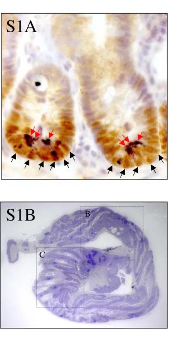
The fact that Sox9 is transcriptionally regulated by the beta-catenin/Tcf4 complex (Blache et al., 2004), together with the particular expression of Sox9 in the compartment of the intestinal epithelium that contains Wnt-stimulated cells, suggest distinct functions in proliferating stem/progenitor cells and in the postmitotic Paneth cells (see supplementary information, Fig. S1A online). To address the different aspects of Sox9 function during the turnover of the intestinal epithelium, including its possible role in specifying the cell response to Wnt signals, we specifically inactivated the corresponding gene in the intestinal epithelium.
Results
Generation of mice with a Sox9-deficient intestinal epithelium
In order to analyse the function of Sox9 in the turnover of the adult intestinal epithelium, Villin-Cre (vil-Cre) mice, in which the Cre recombinase is expressed specifically in the intestinal epithelium from 10.5 dpc onward (el Marjou et al., 2004) were crossed with Sox9flox/flox (Kist et al., 2002) mice, which have both Sox9 alleles flanked by loxP sequences. This generated Sox9flox/flox-vil-Cre mice, with an intestinal epithelium lacking Sox9 protein, indicating effective vil-Cre-mediated recombination of the Sox9flox allele (Fig. 1 a-d). The control villin-cre mice had no detectable phenotypic defect. Sox9flox/flox-vil-Cre mice developed as their control littermates (Sox9flox/flox or Sox9flox/wt-vil-Cre) to become healthy and fertile adult mice. No evidence for intestinal bleeding was found.
Sox9-inactivation causes aberrant morphology and decreased goblet cell
lineage in the colon
Histological analysis of the intestine from 2-6 months old adult Sox9flox/flox-vil-Cre mice revealed that, although the overall morphology of the small intestine seemed, at first sight, unaffected (Fig. 2 a, b), that of the colon was aberrant. The most striking feature of the Sox9flox/flox-vil-Cre mice colon was the folding of the epithelium into villus-like structures, protruding into the colon lumen, reminiscent of the small intestine morphology (Fig. 2, c-f, also see supplementary information, Fig. S1B, C, D online). Proliferation, however, was adequately restricted to the bottom half of the crypts in Sox9flox/flox-vil-Cre mice, as assessed by Ki67 staining (Fig. 2 g, h).
We then examined the differentiation pattern of the Sox9flox/flox-vil-Cre mice colon epithelium into the three main types of differentiated colon epithelial cells. Among these, mucus-producing goblet cells represent the largest population, and are responsible for epithelium protection and lubrication (Velcich et al., 2002). Most of the other cells are enterocytes, with few interspersed enteroendocrine cells. Alcian-blue and Muc2 stainings showed that the goblet cell population was strongly decreased in the colon of Sox9-deficient animals (Fig. 3 a-d). No differences were detected in the morphology or staining intensity between individual alcian-blue-positive goblet cells in Sox9flox/flox and Sox9flox/flox-vil-Cre mice, and no changes in cellular representation were found either for the scarce enteroendocrine cell population (Fig. 3 e, f), or for the Cdx2-expressing enterocyte population (Fig. 3 g, h). Thus, Sox9 is involved in defining the colon epithelium morphology and plays a specific role in the differentiation of the goblet cell lineage in the colon.
Sox9 is required for Paneth cell differentiation
When we examined the expression of markers representative of the four main cell lineages constituting the small intestinal epithelium, no major differences were found in enterocyte and enteroendocrine cell numbers, as shown by Alkaline Phosphatase and Chromogranin A staining (Fig. 4 a-d). However, both the goblet and Paneth cell lineages were significantly affected.
Alcian blue-positive goblet cells were found in the Sox9flox/flox-vil-Cre intestinal epithelium and appropriately expressed Muc2 but their number was reduced by 40% compared to control mice (Fig. 4 e-h, see also supplementary information, figure S2A, B online).
Paneth cells represent the fourth cell type found in the small intestine. They secrete a variety of products, including antimicrobial peptides, growth factors, phospholipase A2 and matrilysin. These cells are involved in regulating the interactions between epithelial cells and the indigenous microorganism population, which, in turn, is essential to elaborate the microvasculature underlying the epithelium (Stappenbeck et al., 2002; Wilson et al., 1999). Remarkably, morphological identification coupled with staining of small intestinal sections from Sox9flox/flox-vil-Cre mice for lysozyme, an early marker of Paneth cell differentiation, revealed that almost all the crypts were completely devoid of Paneth cells (Fig. 4 i, j). In Paneth cell-depleted crypts, the proliferative compartment expanded to occupy the whole crypt base, including the normal Paneth cell compartment (Fig. 4 k-n). As Paneth cells are located next to the putative stem cells, the replacement of Paneth cells by proliferating cells raises the possibility that the number of stem cells is altered in Sox9-deficiency mice. Indeed, the number of cells positively stained with Musashi-1, a putative marker of stem cells in the nervous system and the intestinal epithelium (Potten et al., 2003; Sakakibara et al., 1996), was increased in Sox9-deficient mice (Fig. 4 o, p). Sox9 is thus required for differentiation of the Paneth cell lineage, is involved in differentiation of the goblet cell lineage, and might be involved in the regulation of the stem cell number.
The observed decrease in Paneth and goblet lineages in Sox9-deficient mice was not due to increased apoptosis, since no differences were found in the apoptotic rates between Sox9flox/flox-vil-Cre and Sox9flox/flox mice (see supplementary information, Fig. S2C online).
In the healthy human body, Paneth cells are also found uniquely in the small intestinal crypts of Lieberkühn. In some pathological situations such as intestinal metaplasia, ectopic Paneth cells can also be found in the oesophagus (Barrett’s
oesophagus) or in the stomach (Schreiber et al., 1978). Thus, if Sox9 is required for the differentiation of the Paneth cell lineage, it should be expressed in Paneth cells found in such aberrant structures. To test this, we analysed biopsy sections from a patient with Barrett’s oesophagus. Paneth cells were detected using lysozyme expression and Sox9 expression was analysed on an adjacent section. Sox9 expression was found in most cells constituting crypt-like structures in the metaplasic area, including Paneth cells (Fig. 4 q, r). Thus, Sox9 expression also seems to be associated with Paneth cells in the pathological context of human intestinal metaplasia.
To gain insight into the mechanism underlying the Sox9-dependent differentiation of Paneth cells, we screened by real time PCR colon carcinoma cell lines for expression of such markers. All the tested cell lines (SW480, HT29Cl.16E, HCT116, DLD-1) had detectable expression of Paneth cell markers, and this expression was highest in HT29Cl.16E cells (data not shown), which were chosen for further analyses. A moderate (5-fold) overexpression of Sox9 in these cells resulted in an upregulation of expression of several Paneth cell markers, which was most prominent for Lysozyme, the matrix metalloproteinase MMP-7, and Angiogenin-4 (ANG-4) mRNAs (Fig. 4 s). Thus, Sox9 may regulate the differentiation of Paneth cells, at least in part, through the transcriptional regulation of several markers of these cells. This regulation might be direct or indirect but it likely contributes to the absence of identifiable Paneth cells in the intestinal epithelium of Sox9-deficient mice.
Increased cell proliferation and hyperplasia throughout the intestinal
We then asked whether the homeostasis of the epithelium would be conserved despite the extension of the proliferative compartment into the usual Paneth cell area and found that, in fact, the crypt size of Sox9flox/flox-vil-Cre mice seemed increased compared to Sox9flox/flox control mice (Fig. 5 a-d). When crypt diameters and BrdU incorporation rates were measured, an unambiguous increase of crypt size was found in Sox9-deficient mice (Fig. 5 e), and the ratio between BrdU-labelled cells and the total number of cells found in a crypt circumference was slightly, but reproducibly, increased in the Sox9-deficient mice (Fig 5 f). This was statistically significant (p<0.0001). The total number of cells in any crypt circumference increased according to the crypt size, indicating that the cell size was not affected (not shown). This indicates that the absence of Sox9 resulted in increased cell proliferation leading, in turn, to crypt hyperplasia throughout the small intestine. The epithelium from the proximal colon was also found to display hyperplastic features, but we were unable to perform accurate measurements because of its severely altered morphology in Sox9flox/flox-vil-Cre mice.
Focal dysplastic crypts spontaneously develop in the colon of Sox9flox/flox
-vil-Cre mice
In addition to the general mild hyperplasia found throughout the intestine of Sox9flox/flox-vil-Cre mice, extensive hyperplasia occurred, with occasional glandulo-cystic features, in the distal half of Sox9flox/flox-vil-Cre mice colon. Numerous crypts were enlarged and branched, and some were extensively dilated with a cystic appearance (Fig. 6 a, b). Proliferation was correctly restricted to the bottom of hyperplastic crypts (Fig. 6 c). Cells constituting cystic crypts also proliferated, albeit modestly, (Fig. 6 d) and were poorly differentiated (see supplementary information
Fig. S2D, E, F online). In addition, tubulo-villous micro-adenomas occasionally developed in hyperplastic areas of the epithelium with atypical tissue architecture (Fig. 6 e). Crypts with dysplastic features, including poor differentiation, pseudostratified nuclei, multiadenoid structures and numerous mitosis, spontaneously developed in several locations along the colon of Sox9-deficient mice (Fig. 6 f). Interestingly, slight crypt hyperplasia/dysplasia was also detectable in the colon of 3 weeks-old Sox9-deficient mice, indicating that these defects appear early but become more severe with time, likely as a consequence of increased cell proliferation (see supplementary information Fig. S2G, H, I, J online).
Some Sox9flox/flox-vil-Cre mice had no detectable lesions but also had a normal colon morphology and had Paneth cells in their small intestine (not shown). In such mice, Sox9 staining was invariably identical to that of wild-type mice (not shown), indicating inefficiency of the Cre recombinase. Thus, dysplastic-like lesions were always found in the colon of true Sox9-deficient mice. That we never observed true carcinoma in the intestine of deficient mice up to six months old suggests, in turn, that Sox9-deficiency may not be sufficient per se to induce cell transformation.
Hyperplastic and dysplastic-like crypts were found to strongly overexpress Wnt pathway-related genes such as c-Myc and cyclin-D1, suggesting an increase of Wnt-dependent transcriptional activity (Fig. 6, compare g with h, and i with j). This overexpression resulted from both an increase in the number of c-Myc- and Cyclin-D1-expressing cells, and increased staining intensities in individual positive cells (not shown). Expression of the Ki-67 proliferation marker was also affected (Fig. 6, compare k with l). This finding was confirmed by Western blot analysis of extracts from Sox9flox/flox and Sox9flox/flox-vil-Cre mice. Whereas little variations were found in the small intestine, likely because the few proliferating crypt cells are not sufficiently
represented in the whole epithelial cell population, a clear increase of c-Myc and Cyclin-D1 was evident in the Sox9-deficient colon (Fig 6 m). The increase in cell proliferation rate in Sox9-deficient mice (estimated to 15% from BrdU incorporation rates) was probably not sufficient to be clearly visualised with an anti-PCNA Western blot (Fig 6 m). An alternative explanation is that whereas crypt hyperplasia and increased BrdU intake are evident, the density of crypts is reduced in Sox9-deficient animals, which may compensate the increased proliferation observed in each crypt. That the overexpression of Wnt target genes is much more visible in the colon samples may reflect either the bigger size of the crypts relative to the entire epithelium in the colon compared to the small intestine, or the presence of dysplastic-like lesions, which strongly overexpress Wnt target genes, in the colon samples. We then asked whether these alterations in the small intestine and colon structure may impact on the mouse physiology. Indeed, the weight of Sox9flox/flox-vil-Cre animals was always reduced (not shown), compared to the related Sox9flox/flox control mouse. This reduction was modest (average 17%, n=10) but, despite the heterogeneity in the age and sex of the pairs of animals tested, reached a statistical significance (Student Test value < 0.05). In addition, stools from Sox9flox/flox-vil-Cre mice were more hydrated than that of control mice, indicating a partial impairment of the colonic epithelium function in Sox9-deficient animals (Fig 6 n).
Sox9 modulates Wnt pathway activity in vitro
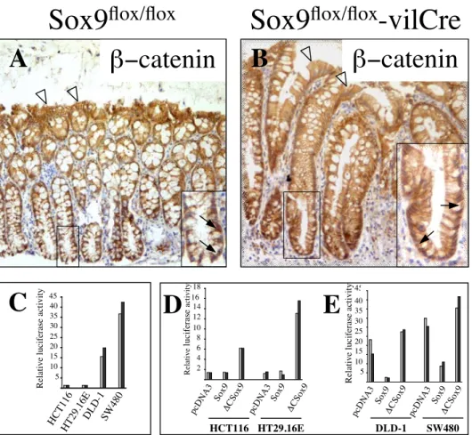
In order to understand the molecular bases of the observed upregulation of Wnt-target genes, we compared the expression of nuclear beta-catenin, the hallmark of Wnt signalling, in Sox9-deficient versus control mice. Comparable results were found in both situations, with a typical nuclear staining of some cells scattered through the
crypt bottom (Fig. 7 a, b; see also supplementary information Fig. S3A, B online), indicating that the overexpression of Wnt target genes found in Sox9-deficient animals was not due to an increased levels of beta-catenin, in the crypt nuclei of Sox9flox/flox-vil-Cre mice.
We and others have shown previously that the level of Sox9 expression regulates the transcriptional activity of the beta-catenin/Tcf4 complex in cultured HEK293 cells (Akiyama et al., 2004b; Blache et al., 2004). Physical interaction between Sox9 and beta-catenin has been reported, resulting in a competition between Sox9 and Tcf4 for binding to beta-catenin. Formation of the Sox9/beta-catenin complex results in degradation of the two proteins (Akiyama et al., 2004b). We thus hypothesized that the absence of Sox9 in crypt cells of Sox9flox/flox-vil-Cre mice, where Wnt signalling is physiologically active, might result in increased availability of the nuclear pool of beta-catenin for binding to Tcf4. To test this, we analysed the transcriptional activity of the beta-catenin/Tcf4 complex after manipulation of Sox9 expression in colon carcinoma cell lines containing a constitutive activation of the beta-catenin/Tcf transcriptional complex (Korinek et al., 1997). The basal beta-catenin/Tcf4 activity present in DLD-1 cells was efficiently inhibited by Sox9 and increased after overexpression of a dominant-negative version of Sox9 (Fig. 7 c). Comparable results were obtained in other colon carcinoma cell lines such as SW480, HCT116 and HT29Cl.16E (see supplementary information Fig. S3C, D, E online) demonstrating that even in colon carcinoma cells, in which nuclear beta-catenin accumulates constitutively, the level of Sox9 expression critically modulates the level of beta-catenin/Tcf transcriptional activity.
We used the HT29Cl.16E-Sox9 and HT29Cl.16E- CSox9 cell lines, inducibly overexpressing full-length or C-terminally truncated Sox9, respectively, to test
whether Sox9 overexpression would result in a downregulation of expression of endogenous Wnt pathway target genes. Doxycycline induction of Sox9 or CSox9 expression also resulted in down- or up-regulation, respectively, of the beta-catenin/Tcf complex activity (Fig. 7 d). Using real-time PCR, we found that induction of Sox9 expression resulted in a downregulation of c-Myc and Cyclin-D1 mRNA expression, whereas that of CSox9 resulted in an upregulation of these two Wnt target genes (Fig. 7 e). C-Myc and Cyclin-D1 protein expression changed accordingly (Fig. 7 f). This result demonstrates modulation of expression of key beta-catenin/Tcf target genes by Sox9, but the molecular mechanism underlying this regulation remained unclear since the C-Sox9 construct does not contain the domain thought to interact with beta-catenin (Akiyama et al., 2004b).
The previously reported physical interaction between Sox9 and beta-catenin had been detected after overexpression of tagged Sox9 and tagged beta-catenin in COS cells (Akiyama et al., 2004b). We reasoned that if this interaction was important to modulate Wnt signalling in colon carcinoma cells, then the endogenous complex should be readily detectable in these cells. Despite repeated efforts, no beta-catenin/Sox9 complex could be immunoprecipitated, although Tcf4 co-immunoprecipitated with catenin (not shown). We concluded that beta-catenin/Sox9 complexes are probably not abundant in colon carcinoma cells. In addition, the sub-cellular localisation of both beta-catenin and Tcf4 was unchanged after transient overexperession of Sox9 in SW480 cells (see supplementary information Fig. S4A-F online), and the level of beta-catenin expression was not decreased after induction of Sox9 expression in the HT29Cl.16E-Sox9 cells (see supplementary information Fig. S4G online).
In order to identify another possible mechanism, we performed mutational analysis of the Sox9-mediated inhibition of the beta-catenin/Tcf activity. The W143R point mutation (Fig. 8 a) was originally identified in a campomelic dysplasia patient and abolishes the DNA binding properties of Sox9 (Meyer et al., 1997). We introduced this mutation in both the Sox9 and CSox9 constructs. The resulting products did not have any transcriptional activity using Sox-luciferase reporters (Fig. 8 b) and failed to modify beta-catenin/Tcf activity (Fig. 8 c). This indicates that Sox9-mediated inhibition of the beta-catenin/Tcf activity requires an intact DNA-binding domain of Sox9, which raises the possibility that this inhibition might be at least partly due to transcriptional regulation. To test this, we used a Sox9 construct in which the C-terminal domain of Sox9, involved in transactivation, and, according to Akiyama et al., in the interaction with beta-catenin, is removed and replaced by the unrelated VP16 transactivating domain (Kamachi et al., 1999) (Fig. 8a). Transient transfection of this construct in colon carcinoma cells showed that the Sox9-VP16 chimeric protein potently activates transcription of a Sox-luciferase reporter gene (Fig. 8 d), and inhibits the beta-catenin/Tcf activity even more efficiently than wild-type Sox9 (Fig. 8 e). We conclude that Sox9-mediated inhibition of beta-catenin/Tcf activity involves transcriptional regulation.
We then aimed at identifying potential Wnt pathway inhibitors, such as the Inhibitor of beta-Catenin and Tcf (ICAT) (Tago et al., 2000) and Groucho-related (Grg/TLE) co-repressors (Cavallo et al., 1998; Roose et al., 1998), which might be transcriptionally regulated by Sox9. In vitro, the induction of Sox9 expression in HT29Cl.16E-Sox9 cells resulted in increased expression of the ICAT and TLE2-4 genes, whereas the expression of TLE1 was unaffected (Fig. 8 f). We then analysed the expression of the mouse homologues of these genes in Sox9flox/flox -vil-Cre mice and Sox9flox/flox mice,
and we found that, again, the expression of Grg2, 3 and 4 was significantly downregulated in Sox9-deficient mice, while that of Grg1 remained unchanged (Fig. 8 g). Icat expression seemed unchanged in Sox9-deficient mice but this result varied with the samples analysed (not shown). Although additional Wnt-pathway inhibitors may be involved, this result provides a basis to explain the increased expression of Wnt target genes observed in Sox9-deficient mice, despite the absence of an increase in nuclear beta-catenin expression.
Sox9 expression defines compartmentalisation of the crypt
When we stained small intestinal tissue for c-Myc and Cyclin-D1 protein expression, we found that both genes were expressed as expected in the stem/progenitor cell compartment but that their expression was strongly decreased in Paneth cells (Fig. 9 a, c). In contrast, the absence of Sox9 resulted in uniform expression of both c-Myc and Cyclin-D1 in the whole crypt bottom (Fig. 9 b, d), which then lacked Paneth cells. Thus, Sox9 expression may define a compartment in which nuclear beta-catenin expression results in a genetic program which includes low c-Myc and Cyclin-D1 expression, and in which differentiation of Paneth cells occurs. In the absence of Sox9, Paneth cells do not develop, and the genetic program characterised by high c-Myc and Cyclin-D1 expression expands to include the whole crypt base.
Discussion
This study shows that loss of Sox9 function affects the intestinal epithelium physiopathology at the level of (i) cell differentiation,(ii) tissue homeostasis, and (iii) colon tissue morphology. These different aspects of the phenotype were sex-independent.
Control of cell differentiation by Sox9 – relationship with the Wnt pathway
Interference with the Wnt pathway results in depletion of the Paneth cell lineage (Pinto et al., 2003) and, conversely, loss of the key negative regulator of the pathway, Apc, results in an expansion of the Paneth cell lineage (Andreu et al., 2005; Sansom et al., 2004). In addition, the terminal maturation of Paneth cells requires Wnt signalling through the Frizzled-5 receptor (van Es et al., 2005). Since we previously showed that Sox9 is a transcriptional target of Wnt signalling, the Sox9 requirement for the differentiation of the Paneth cell lineage is likely to reflect its role to specify this specific aspect of the Wnt pathway. In this view, Sox9 function would be expected to occur at an earlier stage than the Wnt/Frizzled-5 pathway, during the course of Paneth cell differentiation, since this pathway was previously shown to be involved in the terminal differentiation of already committed Paneth cells (van Es et al., 2005). Although less likely, an alternative in which Sox9 and Wnt signalling, as two distinct pathways, would control Paneth cell differentiation can not be formally excluded. The functional consequence of the loss of the Paneth cell lineage, a component of the innate immunity, is still a matter of debate. In this study, we did not find an increased mortality in the Sox9-deficient mice lacking Paneth cells. In agreement with this, a previous experiment with ablation of Paneth cells concluded that the turnover
of epithelial cells was not grossly perturbed. In addition, no major alteration of microorganism population along the crypt-villus axis was detected (Garabedian et al., 1997). On the other hand, a role of Paneth cells in regulating the villus angiogenesis (Stappenbeck et al., 2002) and in clearing bacterial infections (Wilson et al., 1999) have been described, although no major impact was reported at the physiopathological level.
The reduction of goblet cell numbers indicates that Sox9 is also involved in the differentiation of this lineage. This quantitative impact of the Sox9-deficiency on the goblet cell population is probably physiologically relevant given the key role of mucus in protecting the epithelium against the luminal content, well illustrated, for instance, by the colon epithelium tumorigenesis resulting from a deletion of the Muc2 gene (Velcich et al., 2002). This function of Sox9 in regulating the differentiation of goblet cells might also play a part in the cellular response to Wnt signals, since altered goblet cell differentiation was also found in mice in which the Wnt pathway has been impaired (Pinto et al., 2003).
That Sox9-deficiency results in the complete absence of Paneth cells but leads only to a reduction of the goblet cell lineage might indicate different Sox9 requirements for the differentiation of these two lineages, and suggests that additional factors might be involved in this process, at least for goblet cell lineage differentiation.
What is the function of Sox9 in goblet cell differentiation?
In a previous report, we showed that overexpression of Sox9 in the LS174T colon carcinoma cell line repressed the Muc2 gene (Blache et al., 2004). This apparently contrasts with the present finding that the absence of Sox9 in the intestinal epithelium causes a significant reduction of the number of goblet cells in vivo. The remaining
goblet cells express the Muc2 gene, which indicates their maturation into functional mucus-producing cells. The apparent discrepancy between the two experiments likely relies in the fundamentally different nature of the experimental models used, i.e. LS174T human colon carcinoma cells grown in vitro versus mouse intestinal epithelium in its physiological tissue context. We conclude that Sox9 definitely plays a role in the differentiation of the goblet cell lineage, and we hypothesize that this role occurs primarily at the level of cell fate choice, similar to what was shown previously in the developing nervous system (Spokony et al., 2002). In addition, as suggested by in vitro data and the respective expression patterns of Sox9 and Muc2, downregulation of Sox9 expression might be necessary to allow terminal maturation of committed goblet cells, although this hypothesis is difficult to test using the present gene inactivation model.
“Gatekeeper” role of Sox9 in modulating Wnt pathway activity?
Our in vitro and in vivo results both indicate that Sox9 down-regulates the transcriptional activity of the beta-catenin/Tcf4 complex on endogenous target promoters, such as the c-Myc and Cyclin-D1 gene promoters (see supplementary information, Discussion S5 online).
We could show that the transcriptional activity of Sox9 is involved in this inhibition, and genes encoding inhibitors of the beta-catenin/Tcf activity have been found to be regulated by Sox9 in vitro and in vivo. This finding might explain how many Sox proteins, which lack significant homology outside the HMG DNA-binding domain, have the conserved property of inhibiting beta-catenin/Tcf activity.
In vivo, inactivation of the Sox9 gene results in an increase in the rate of BrdU incorporation in crypt epithelial cells. This correlates with the development of
hyperplastic and dysplastic-like crypts, which overexpress Wnt target genes. The percentage of BrdU-positive cells obtained in this study with the Sox9flox/flox control mice (46-56% with a 2 hours BrdU pulse) is consistent with previous analyses showing a 3H labelling index of 29-33% after a 1 hour treatment with 3H-thymidine (Cheng and Bjerknes, 1982). The 16-32% increase in BrdU-positive cells in Sox9flox/flox -vil-Cre mice is therefore likely to significantly impact on epithelial homeostasis, given the rate of renewal of the intestinal epithelium. This increased proliferation rate likely explains the occurrence of Wnt target genes-overexpressing hyperplastic and dysplastic-like crypts in Sox9-deficient mice, whereas this was never observed in Sox9flox/flox animals. In agreement with this, an apparent increase of Musashi expressing cells was found in Sox9-deficient mice, suggesting that Sox9 might regulate the number of stem cells in the intestinal epithelium.
A role of Sox9 in regulating cell proliferation has already been proposed in chondrocytic cell lines, in which Sox9 promoted differentiation (Panda et al., 2001), as well as in breast and prostate tumor cell lines (Afonja et al., 2002; Drivdahl et al., 2004). Here, however, Sox9 is specifically expressed, in a normal physiological situation, in the proliferative compartment of the intestinal epithelium, and activity of the proto-oncogenic Wnt pathway is required for its expression (Blache et al., 2004). Thus, Sox9 might function as a gatekeeper in intestinal crypts to prevent over-activity of the Wnt-dependent transcriptional program.
Role of the Sox9-mediated inhibition of the Wnt pathway in the differentiation
of Paneth cells
Wnt signalling is known to be important for the maintenance of the proliferating compartment (Korinek et al., 1998; Kuhnert et al., 2004; Pinto et al., 2003) as well as
for the differentiation and maturation of Paneth cells (Andreu et al., 2005; Pinto et al., 2003; van Es et al., 2005). Here, we found that c-Myc, a central effector of the Wnt pathway, as well as Cyclin-D1, were expressed in the proliferative compartment of the small intestinal epithelium but not, or at a much lower level, in the Paneth cell compartment of wild-type mice; This was unexpected given that Paneth cells have been reported to express highest levels of nuclear beta-catenin (van Es et al., 2005). This suggests that distinct genetic programs are set, downstream to the Wnt pathway, in the stem/progenitor versus Paneth cell compartments, and this was reflected by a recent transcriptomic study (Van der Flier et al., 2007). The absence of Sox9 results in a single proliferative stem/progenitor compartment, in which all the cells express c-Myc and Cyclin-D1. This suggests that Sox9 is required to specify the Paneth cell compartment, in which the presence of nuclear beta-catenin results in a “Paneth cell” genetic program instead of a “stem/progenitor” program which includes c-Myc expression.
This also raises the question of the role of Sox9 in Wnt pathway-triggered tumorigenesis. Our data suggest that loss of Sox9 might promote tumorigenesis, either through the reduction of mucus protection, or through upregulation of the catenin/Tcf activity. It should be noted, however, that upregulation of the beta-catenin/Tcf complex activity by loss of Sox9 function is expected to occur only in cells in which Wnt pathway activity pre-exists, and Sox9 deletion might not be sufficient to cause neoplasia. Consistent with this, no evidence for spontaneous neoplasms was found in Sox9-deficient mice up to six months old.
This study reveals the dual role played by Sox9 in relation with the Wnt pathway: (i) it likely mediates part of the Wnt-dependent program, involved in Paneth and goblet cell differentiation. Sox9 thus provides the first example of a transcription factor involved in specifying the cellular response to Wnt signals, which potentially trigger very diverse cellular behaviours, including proliferation, differentiation, sorting of the cells along the crypt-villus axis, etc. (ii) Sox9 fine-tunes the transcriptional output of the canonical Wnt pathway in vitro, and inactivation of this gene causes an increase of BrdU incorporation rate, leads to hyperplasia and dysplasia of the intestinal epithelium and increased putative stem-cell numbers. (iii) Finally, Sox9 may also act independently of the Wnt pathway to control colon epithelium morphology, a role which had not been previously reported for the Wnt pathway, although architectural degeneration was described after Dickkopf-1 over-expression in the colon epithelium (Kuhnert et al., 2004).
Acknowledgements
The authors acknowledge Anne Cohen-Solal and Philippe Crespy for excellent technical help, members of the Cellular and Molecular Oncology Department and Vjeko Dulic for helpful suggestions, Daniel Fisher for critical reading of the MS, Drs. J.-N. Freund, I. van Seuningen, P. de Santa Barbara, and Pr. H. Okano for reagents. This work has been supported by Agence Nationale pour la Recherche (Jeune Chercheur), Association pour la Recherche contre le Cancer (ARC N°3570 and 3636), Ligue Nationale contre le Cancer (Equipe Labellisée), Fondation pour la Recherche Médicale and Groupement des Entreprises Françaises dans la Lutte contre le Cancer (GEFLUC). C.D. is supported by a CNRS-L fellowship. G.S. acknowledges support through grant Sche 194/15-1 and -2 by the Deutsche Forschungsgemeinschaft.
Materials and Methods
Mouse lines
The villin-cre strain (el Marjou et al., 2004) was in a nearly pure C57Bl/6 background (at least 14 backcrosses to this background). To generate Sox9 +/flox mice (Kist et al., 2002), exon 2 and 3 of the Sox9 gene were flanked by LoxP sequences. Cre recombination results in the deletion of the 2 last exons, which encode part of the HMG DNA-binding domain and the transactivation domain, thus resulting in a likely null allele. A peptide might still be produced from the first exon but this would not contain any known functional domain. Sox9 +/flox mice were originally on a 129P2/OlaHsd x C57BL/6 mixed genetic background, and have been backcrossed to C57BL/6 for three generations. These N3 mice were made Sox9 flox/flox by sister-brother mating before being crossed with the villin-cre strain.
Cells and cell culture conditions
Colorectal cancer cell lines HCT116, HT29.16E, DLD1 and SW480 were cultured at 37°C in Dulbecco’s Modified Eagle Medium (DMEM) supplemented with 10% fetal bovine serum (Eurobio, Les Ulis, France), 1% L-glutamine and penicillin/streptomycin. The HT29.16E -Sox9 and HT29.16E CSox9 cell lines have been described (Blache et al., 2004; Jay et al., 2005).
DNA constructs, transient transfections and luciferase assays
Full length human Sox9 and C-terminally truncated (dominant negative) human Sox9 constructs have been described (Südbeck et al., 1996), as well as the pTOP-FLASH and pFOP-FLASH reporter constructs (Morin et al., 1997). The Sox9-VP16 construct (Kamachi et al., 1999) was a kind gift by Professor Kondoh (Institute for Molecular
and cell biology,Osaka University, Osaka, Japan). The full-length human Sox9 (W143R) and the C-terminally truncated human Sox9 (W143R) expression constructs were constructed from a human Sox9(1-304) construct containing the W143R mutation, kindly provided by Doctor de Santa Barbara (Inserm, ER125, Muscle and Pathologies, Montpellier, France). DLD1, SW480, HCT116 and HT29-16E cells were cotransfected (EXGEN500, Euromedex) with 0,25 µg of pcDNA3, Sox9 or CSox9 DNA constructs, and 0,25 µg of TCF/LEF-1 reporter (pTOP-FLASH), or control vector (pFOP-FLASH), using standard procedures. pRLSV-Renilla (0,025 µg) was used as an internal control. Luciferase assays were performed with the Dual luciferase reporter assay system (Promega) according to the manufacturer’s instructions. Luciferase activities in cell lysates were normalized relative to the Renilla luciferase activity and the indicated activities represent the TOPFLASH/FOPFLASH ratio, indicative of the Tcf binding site-specific activity. Each experiment was carried out in duplicate, repeated several times and representative examples are shown.
RT-Quantitative PCR
Total RNAs were prepared using the Rneasy minikit (QIAGEN, Courtaboeuf, France) from HT29.16E-Sox9 or HT29.16E- CSox9 cells grown to 100% confluence and treated or not for 6 days with doxycycline. For mRNA quantification, 2.5 µg of total RNA was pretreated with DNAse RQ1 (Promega) for 30 min at 37°C and used for reverse transcription with M-MLV reverse transcriptase (Invitrogen). Quantitative PCR was carried out using the LightCycler FastStart DNA MasterPlus SYBR Green I kit (Roche Diagnostics, Meylan, France). Results were normalised with GAPDH expression. (see supplementary information table S5 online for primers sequences).
Immunohistochemistry and Western blot
Immunohistochemistry was performed essentially as described (Blache et al., 2004). Sections of human intestinal metaplasia were kindly provided by F. Bibeau (CRLC Val d’Aurelle Lamarque, Montpellier). Briefly, for preparation of mouse intestinal sections, the intestinal tract was dissected as a whole from 2-6 months old mice, and flushed gently with cold PBS to remove any faecal content. The small intestine and colon were rolled up into a compact circle and fixed in 4% PFA in PBS at room temperature for 4 hours, dehydrated, embedded in paraffin and sectioned at 5 µm, using standard procedures. Sections were dewaxed in a xylene bath and rehydrated in graded alcohols. Endogenous peroxidase activity was quenched with 1.5%H2O2 in methanol for 20 minutes and then washed in PBS. Antigen retrieval was performed by boiling slides in Na-citrate buffer (10 mM, pH 6.0) except for c-Myc and anti-cleaved Caspase3 antibodies for which antigen retrieval was performed by boiling slides 20 minutes in 100 mM TRIS, 12,6 mM EDTA (pH 9.0). Nonspecific binding sites were blocked with 1% BSA-3% NGS-0,2% triton in PBS for 45 minutes at room temperature for all antibodies staining except for anti-c-Myc (1% BSA in PBS) and anti-Ki67 (no blocking). Slides were incubated with the primary antibodies overnight at 4°C in PBS with 0.1% BSA. In all cases, Envision + (DakoCytomation) was used as a secondary reagent, stainings were developed with DAB (brown precipitate) or Vector Vip substrate (purple precipitate), and hematoxylin counterstain was used. After dehydration, sections were mounted in Pertex (Histolab). For alkaline phosphatase activity staining, sections were dewaxed and rehydrated as above and the alkaline phosphatase substrate (Vector red, Vector) was applied for 10 minutes. Sections were counterstained, dehydrated and mounted as above. For BrdU countings, mice were injected with a solution (0.1 mg per g of mouse body weight) of
Brdu diluted in PBS. Mice were sacrificed 2 hours after injection. For BrdU staining, the same method as explained above was used, excepted an additional step in HCl 2N 45 min, added after antigen retrieval. For the Musashi staining, an ABC kit (Vectastain) was used instead of the Envision+ system.
For Western blotting, an equal amount of protein, measured by the Bradford assay, was loaded on each lane of the gel. Protein lysates and immunoblotting were performed as described (Hollande et al., 2003). Proteins were visualized using ECL Plus (Amersham Biosciences) and the bands were quantified by densitometry using ImageJ 1.32J (NIH, Bethesda, MD).
Quantification of cMyc and Cyclin-D1 stainings
Immunofluorescence stainings were performed essentially as immunohistochemical stainings but a goat anti-rabbit secondary antibody (Alexa Fluor 488, Invitrogen) was used and nuclei were counterstained with Hoechst. Fluorescence quantifications were performed with the ImageJ software. Nuclei were selected uding the freehand selection tool and integrated densities were measured.
Antibodies
The Muc2 antibody (1:250) was kindly provided by I. Van Seuningen (INSERM U560, Lille, France), the Cdx2 antibody (1:200) was kindly provided by J.-N. Freund (INSERM U682, Strasbourg, France), the Musashi-biotin antibody (1/500) was kindly provided by H. Okano (Keio University, Japan) and the anti-Sox9 was described previously (de Santa Barbara et al., 1998). Beta-actin A5441 (Western blot, 1:5000) was purchased from Sigma-Aldrich, anti-Ki-67 (1:200) and anti-Lysozyme (1:1000) were purchased from DakoCytomation, anti-PCNA (1:100) and anti-Cyclin-D1 (1:100)
was purchased from Neomarker, anti-c-Myc (1:50) was purchased from Santa-Cruz, anti-E-Cadherin (1:150) and anti-beta-Catenin (1:50) were purchased from Transduction Laboratories, Claudin2 (1:100) was from Zymed, anti-Chromogranin A (1:300) was purchased from Immunostar, anti-BrdU (1:200) was from Novocastra and anti-Caspase 3 (1:100) was purchased from Cell Signalling.
Analysis of stool hydration
To determine stool hydration, freshly isolated stool was weighted before and after overnight incubation at 50°C. Four mice of each genotype were analyzed. Average and standard deviation values, as well as statistical significance (Student-T-test) were calculated using the Microsoft excel software.
Measurements of crypt diameter and of the number of BrdU-positive cells
Fields containing crypt transverse sections were selected randomly at several locations along the rostro-caudal axis of the small intestine. Only sections with several BrdU-positive cells and an apparent lumen were considered to avoid large variation in the position of the section in the crypt-villus axis. 6-9 fields, each containing 6-40 measured crypts were analysed by two individuals, which were blinded to the mouse genotypes and two mice of each genotype were analysed. Average and standard deviation values, as well as statistical significance (Student-T-test) were calculated using the Microsoft excel software.
Image acquisition and manipulation
Immunohistochemistry images were acquired at RT using either an Axiophot microscope (Carl Zeiss MicroImaging, Inc.), 10 x 0.3 Plan Neofluar or 40 x 1.0 Plan
Apochromat lenses (Carl Zeiss MicroImaging Inc.) and a camera (model DXM1200; Nikon), or an Eclipse 80i microscope (Nikon Instruments), with Plan Fluor 10 x 0.3, 20 x 0.5, 40 x 0.75 and 60 x 0.5-1.25 lenses (Nikon Instruments) and a camera (Q-Imaging Retiga 2000R with a Q-(Q-Imaging RGB Slider). Images were acquired with Nikon ACT-1 or Q-Capture Pro softwares and manipulated with the Adobe Photoshop software. For acquisition of immunofluorescence experiments, an Axiophot2 (Zeiss) microscope was used, with a 63 x 1.4 Plan-Apochromat objective (oil) and a Coolsnap (Photometrics) camera driven by the Metavue software.
Legends to figures
Figure 1 : Absence of Sox9 protein expression in the intestinal epithelium of
Sox9-deficient mice.
In Sox9flox/flox mice, the Sox9 protein is expressed in the bottom of small intestinal (a) and colon (c) crypts. The absence of specific Sox9 staining in the small intestine (b) and colon (d) from Sox9flox/flox -vil-Cre mice demonstrate efficient villin-cre-mediated recombination. Arrows and arrowheads indicate Sox9-positive and Sox9-negative nuclei, respectively. Bar: 150 µm for the panels, 50 µm for the inserts.
Figure 2 : Morphological alterations in the colon of Sox9-deficient mice.
Histological analysis (hematoxylin staining) of the intestine of Sox9flox/flox (a, c, e) and Sox9flox/flox -vil-Cre mice (b, d, f). The gross morphology of the small intestine of the Sox9flox/flox -vil-Cre mice is not affected (a, b), whereas that of the colon is strongly altered. The surface of the colon is normally flat (arrows in c and e) but villus-like structures protrude into the lumen of the colon of Sox9flox/flox-vil-Cre mice (arrows in d and f). Immunohistochemical staining with the Ki-67 proliferation in Sox9flox/flox mice (g) and Sox9flox/flox -vil-Cre mice (h) indicates appropriate crypt-restricted proliferation in the colon of Sox9-deficient animals. Bar: 150µm.
Figure 3 : Altered goblet cell differentiation in the colon of Sox9-deficient mice.
Immunohistochemical analysis of Sox9flox/flox mice (a, c, e, g) and Sox9flox/flox -vil-Cre mice (b, d, f, h). Alcian blue staining (a, b) and Muc2 immunoreactivity (c, d) reveal a strong reduction of the goblet cell population in the colon epithelium of Sox9flox/flox -vil-Cre mice. Sox9flox/flox and Sox9flox/flox-vil-Cre mice have comparable populations of
chromogranin A-positive enteroendocrine cells (e, f), and Cdx-2-positive enterocytes (g, h). Bar: 150µm.
Figure 4 : Sox9 controls differentiation of the Paneth and goblet cell lineages.
Sox9flox/flox (a, c, e, g, i, k, m) and Sox9flox/flox-vil-Cre (b, d, f, h, j, l, n) mice are compared. Differentiation along the enterocyte (a, b) and enteroendocrine (c, d) lineages is not affected. The goblet cell population, revealed by alcian blue staining for acidic mucins (e, f) and Muc2 immunostaining (g, h), is reduced in the small intestinal epithelium of Sox9-deficient mice. Paneth cells (arrows in control mice) are completely absent (arrowheads) in Sox9-deficient crypts (i, j). Instead, the proliferative compartment (arrows in k, l, m, n) expands to occupy the whole crypt bottom in mutant animals, where more Musashi-1-expressing putative stem cells are found (arrows in o, p). Ectopic Paneth cell found in a human patient with Barrett’s oesophagus (intestinal metaplasia in the oesophagus) also express Sox9, as shown by staining of adjacent sections with Lysozyme (q) and Sox9 (r) antibodies. Expression analysis of the Paneth cell markers Lyzozyme, Mmp7 and Angiogenin-4, in the HT29Cl.16E-Sox9 cell line, before and after doxycycline induction of exogenous Sox9 expression (s).
Bars: 120 µm, except for panels m, n: 30 µm and panels o, p : 40µm.
Figure 5 : Generalized hyperplasia develops in the absence of Sox9
Difference of crypt size in the small intestines of Sox9flox/flox (a, c) and Sox9flox/flox -vil-Cre (b, d) mice. a, b: crypt cross sections; c, d: crypt longitudinal sections. Bars: 75µm.
e : histogramm showing average crypt diameters along the small intestines of three Sox9flox/flox and three Sox9flox/flox-vil-Cre mice. Standard deviations are indicated. Student T-test values: < 0.001.
f : histogramm showing the BrdU incorporation rates (number of BrdU-positive cells/total number of cells) in crypts from three Sox9flox/flox and three Sox9flox/flox-vil-Cre mice. Standard deviations are indicated. Student T-test values: < 0.001.
Figure 6 : Sox9-deficiency causes hyperplasia and dysplasia.
In the colon epithelium of Sox9flox/flox-vil-Cre mice, hyperplastic lesions are found (hematoxylin staining) with branched and enlarged crypts (a). Some crypts have a cystic appearance (b). Such structures proliferate (c, d, arrows). Tubulo-villus micro-adenomas spontaneously developed in hyperplastic areas of Sox9flox/flox-vil-Cre mice (e). Typical example of the dysplastic-looking crypts (arrows) found in the distal half of the colon of Sox9-deficient mice. Arrowheads point at muti-adenoid crypts (f). Compared with normal tissue (g, i, k), hyperplastic and dysplastic crypts of Sox9 deficient mice (h, j, l) overexpress Wnt pathway target genes such as c-Myc (compare panels g and h) and cyclin-D1 (compare panels i and j), and accordingly have elevated staining for the Ki67 proliferation marker (compare panels k and l). Inserts in g-l show enlarged pictures of the indicated area of the panel and arrows point at typical staining pictures. Western blot analysis of c-Myc, Cyclin-D1 and PCNA in the small intestine and colon of Sox9flox/flox and Sox9flox/flox-vil-Cre mice. Beta-actin expression is shown as a loading control (m). Stool hydration is increased in Sox9-deficient mice (n). Bars: 150 µm for the panels; 50 µm for the inserts.
Figure 7 : Sox9 fine tunes the activity of the beta-catenin/Tcf4 transcriptional
effector of the Wnt signalling pathway. No alteration of the number of cells
containing nuclear beta-catenin (arrows) is found in Sox9-deficient animals (a, b), Bars: 40 µm. Analysis of the transcriptional activity of the beta-catenin/Tcf4 complex, using a luciferase reporter system (Morin et al., 1997) (c, d). Endogenous transcriptional activity of the beta-catenin/Tcf4 complex. Transient overexpression of Sox9 inhibits the beta-catenin/Tcf4 activity, whereas overexpressing CSox9 increases it (c). Similarily, induction of Sox9 overexpression in the HT29-16E-Sox9 cell line causes inhibition of beta-catenin/Tcf4 transcriptional activity whereas inducing overexpression of CSox9 in the HT29-16E- CSox9 cell line resulted in a significant increase of this activity (d). Regulation of the beta-catenin/Tcf4 transcriptional activity impacted on c-Myc and Cyclin-D1 mRNA expression. The amounts of mRNA after doxycycline induction are indicated as % of the non-induced state (dashed line) (e). These variations at the mRNA level were reflected at the protein level (f).
Figure 8: Sox9 transcriptionally activates expression of inhibitors of the
beta-catenin/Tcf activity. Structure-function analysis of the inhibitory function of Sox9 on
the beta-catenin/Tcf activity (a, b, c, d, e) in DLD-1 cells. Diagram showing the different constructs used in this study (a). Transcriptional activity of Sox9 and its mutated or truncated versions on a Sox-luciferase reporter system (b). Inhibition of the beta-catenin/Tcf activity by Sox9 and its truncated or mutated versions (c). Transcriptional activity of the Sox9-VP16 chimeric protein compared to that of Sox9 on a Sox-luciferase reporter system (d). Inhibition of the beta-catenin/Tcf activity by Sox9 and Sox9-VP16 (e). Real-time RT-PCR analysis of the expression of the ICat
and Groucho-related inhibitors of the beta-catenin/Tcf activity in HT29Cl.16E-Sox9 cells before and after induction of exogenous Sox9 expression (f) and in the intestinal mucosa of Sox9flox/flox versus Sox9flox/flox-vil-Cre mice (g). Results are expressed relative to the non-induced or non-recombined states. Standard deviations are indicated.
Figure 9: Alteration of c-Myc and Cyclin-D1 expression in the bottom of
Sox9-deficient crypts. C-Myc and Cyclin-D1 expression was analysed by immunohistochemistry in the intestine of Sox9flox/flox (a, c) and Sox9flox/flox-vil-Cre (b, d) mice. Arrows indicate the Paneth cell compartment in Sox9flox/flox control mice (a, c) and the equivalent location in the Sox9flox/flox-vil-Cre mice (b, d). Bars: 50 µm.
References
Afonja, O., B.M. Raaka, A. Huang, S. Das, X. Zhao, E. Helmer, D. Juste, and H.H. Samuels. 2002. RAR agonists stimulate SOX9 gene expression in breast cancer cell lines: evidence for a role in retinoid-mediated growth inhibition. Oncogene. 21:7850-60.
Akiyama, H., M.C. Chaboissier, R.R. Behringer, D.H. Rowitch, A. Schedl, J.A. Epstein, and B. de Crombrugghe. 2004a. Essential role of Sox9 in the pathway that controls formation of cardiac valves and septa. Proc Natl Acad Sci U S A. 101:6502-7.
Akiyama, H., J.P. Lyons, Y. Mori-Akiyama, X. Yang, R. Zhang, Z. Zhang, J.M. Deng, M.M. Taketo, T. Nakamura, R.R. Behringer, P.D. McCrea, and B. de Crombrugghe. 2004b. Interactions between Sox9 and beta-catenin control chondrocyte differentiation. Genes Dev. 18:1072- 87.
Andreu, P., S. Colnot, C. Godard, S. Gad, P. Chafey, M. Niwa-Kawakita, P. Laurent-Puig, A. Kahn, S. Robine, C. Perret, and B. Romagnolo. 2005. Crypt-restricted proliferation and commitment to the Paneth cell lineage following Apc loss in the mouse intestine. Development. 132:1443-51.
Batlle, E., J.T. Henderson, H. Beghtel, M.M.W. van den Born, E. Sancho, G. Huls, J. Meeldijk, J. Robertson, M. van de Wetering, T. Pawson, and H. Clevers. 2002. ß-catenin and TCF mediate cell positioning in the intestinal epithelium by controlling the expression of EphB/EphrinB. Cell. 111:251-263.
Blache, P., M. Van De Wetering, I. Duluc, C. Domon, P. Berta, J.N. Freund, H. Clevers, and P. Jay. 2004. SOX9 is an intestine crypt transcription factor, is regulated by the Wnt pathway, and represses the CDX2 and MUC2 genes. J Cell Biol. 166:37-47.
Cavallo, R.A., R.T. Cox, M.M. Moline, J. Roose, G.A. Polevoy, H. Clevers, M. Peifer, and A. Bejsovec. 1998. Drosophila Tcf and Groucho interact to repress Wingless signaling activity. Nature. 395:604-608.
Cheng, H., and M. Bjerknes. 1982. Whole population cell kinetics of mouse duodenal, jejunal, ileal, and colonic epithelia as determined by radioautography and flow cytometry. Anat Rec . 203:251-64.
de Santa Barbara, P., N. Bonneaud, B. Boizet, M. Desclozeaux, B. Moniot, P. Sudbeck, G. Scherer, F. Poulat, and P. Berta. 1998. Direct interaction of SRY-related protein Sox9 and steroidogenic factor 1 regulates transcription of the human anti-mullerian hormone gene. Molecular and Cellular Biology. 18:6653-6665.
Drivdahl, R., K.H. Haugk, C.C. Sprenger, P.S. Nelson, M.K. Tennant, and S.R. Plymate. 2004. Suppression of growth and tumorigenicity in the prostate tumor cell line M12 by overexpression of the transcription factor SOX9. Oncogene. 23:4584-93.
el Marjou, F., K.P. Janssen, B.H. Chang, M. Li, V. Hindie, L. Chan, D. Louvard, P. Chambon, D. Metzger, and S. Robine. 2004. Tissue-specific and inducible Cre-mediated recombination in the gut epithelium. Genesis. 39:186-93.
Foster, J.M., M.A. Dominguez-Steglich, S. Guioli, C. Kwok, P.A. Weller, M. Stevanovic, J. Weissenbach, S. Mansour, I.D. Young, P.N. Goodfellow, J.D.
Brook, and A.J. Schafer. 1994. Campomelic dysplasia and autosomal sex reversal caused by mutations in an SRY-related gene. Nature. 372:525- 530. Garabedian, E.M., L.J.J. Roberts, M.S. McNevin, and J.I. Gordon. 1997. Examining
the role of Paneth cells in the small intestine by lineage ablation in transgenic mice. The Journal of Biological Chemistry. 272:23729-23740.
Hollande, F., D.J. Lee, A. Choquet, S. Roche, and G.S. Baldwin. 2003. Adherens junctions and tight junctions are regulated via different pathways by progastrin in epithelial cells. J Cell Sci. 116:1187-97.
Jay, P., P. Berta, and P. Blache. 2005. Expression of the carcinoembryonic antigen gene is inhibited by SOX9 in human colon carcinoma cells. Cancer Res. 65:2193-8.
Kamachi, Y., K.S. Cheah, and H. Kondoh. 1999. Mechanism of regulatory target selection by the SOX high-mobility-group domain proteins as revealed by comparison of SOX1/2/3 and SOX9. Mol Cell Biol. 19:107-20.
Kist, R., H. Schrewe, R. Balling, and G. Scherer. 2002. Conditional inactivation of Sox9: a mouse model for campomelic dysplasia. Genesis. 32:121-123.
Korinek, V., N. Barker, P. Moerer, E. van Donselaar, G. Huls, P.J. Peters, and H. Clevers. 1998. Depletion of epithelial stem-cell compartments in the small intestine of mice lacking Tcf-4. Nature Genetics. 19:379-383.
Korinek, V., N. Barker, P.J. Morin, D. van Wichen, R. de Weger, K.W. Kinzler, B. Vogelstein, and H. Clevers. 1997. Constitutive transcriptional activation by a b-catenin-Tcf complex in APC-/- colon carcinoma. Science. 275:1784-1787. Kuhnert, F., C.R. Davis, H.T. Wang, P. Chu, M. Lee, J. Yuan, R. Nusse, and C.J.
Kuo. 2004. Essential requirement for Wnt signaling in proliferation of adult small intestine and colon revealed by adenoviral expression of Dickkopf-1. Proc Natl Acad Sci U S A. 101:266- 71.
Meyer, J., P. Südbeck, M. Held, T. Wagner, M.L. Schmitz, F. Dagna Bricarelli, E. Eggermont, U. Friedrich, O.A. Haas, A. Kobelt, J.G. Leroy, L. Van Maldergem, E. Michel, B. Mitulla, R.A. Pfeiffer, A. Schinzel, H. Schmidt, and G. Scherer. 1997. Mutational analysis of the SOX9 gene in campomelic dysplasia and autosomal sex reversal: lack of genotype/phenotype correlations. Human Molecular Genetics. 6:91-98.
Morin, P.J., A.B. Sparks, V. Korinek, N. Barker, H. Clevers, B. Vogelstein, and K.W. Kinzler. 1997. Activation of b-catenin-Tcf signaling in colon cancer by mutations in b-catenin or APC. Science. 275:1787-1790.
Ng, L.-J., S. Wheatley, G.E.O. Muscat, J. Conway-campbell, J. Bowles, E. Wright, D.M. Bell, P.P.L. Tam, K.S.E. Cheah, and P. Koopman. 1997. SOX9 binds DNA, activates transcription, and coexpresses with type II collagen during chondrogenesis in the mouse. Developmental Biology. 183:108- 121.
Panda, D.K., D. Miao, V. Lefebvre, G.N. Hendy, and D. Golzman. 2001. The transcription factor SOX9 regulates cell cycle and differentiation genes in chondrogenic CFK2 cells. The Journal of Biological Chemistry. 276:41229-41236.
Pinto, D., A. Gregorieff, H. Begthel, and H. Clevers. 2003. Canonical Wnt signals are essential for homeostasis of the intestinal epithelium. Genes Dev. 17:1709-13. Potten, C.S., C. Booth, G.L. Tudor, D. Booth, G. Brady, P. Hurley, G. Ashton, R.
Clarke, S. Sakakibara, and H. Okano. 2003. Identification of a putative intestinal stem cell and early lineage marker; musashi-1. Differentiation. 71:28-41.
Roose, J., M. Molenaar, J. Peterson, J. Hurenkamp, H. Brantjes, P. Moerer, M. van de Wetering, O. Destrée, and H. Clevers. 1998. The Xenopus Wnt effector XTcf-3 interacts with Groucho-related transcriptional repressors. Nature. 608:608-612.
Sakakibara, S., T. Imai, K. Hamaguchi, M. Okabe, J. Aruga, K. Nakajima, D. Yasutomi, T. Nagata, Y. Kurihara, S. Uesugi, T. Miyata, M. Ogawa, K. Mikoshiba, and H. Okano. 1996. Mouse-Musashi-1, a neural RNA-binding protein highly enriched in the mammalian CNS stem cell. Dev Biol. 176:230-42.
Sansom, O.J., K.R. Reed, A.J. Hayes, H. Ireland, H. Brinkmann, I.P. Newton, E. Batlle, P. Simon-Assmann, H. Clevers, I.S. Nathke, A.R. Clarke, and D.J. Winton. 2004. Loss of Apc in vivo immediately perturbs Wnt signaling, differentiation, and migration. Genes Dev. 18:1385- 90.
Schreiber, D.S., M. Apstein, and J.A. Hermos. 1978. Paneth cells in Barrett's esophagus. Gastroenterology. 74:1302- 4.
Seymour, P.A., K.K. Freude, M.N. Tran, E.E. Mayes, J. Jensen, R. Kist, G. Scherer, and M. Sander. 2007. SOX9 is required for maintenance of the pancreatic progenitor cell pool. Proc Natl Acad Sci U S A. 104:1865-70.
Spokony, R.F., Y. Aoki, N. Saint-Germain, E. Magner-Fink, and J.P. Saint-Jeannet. 2002. The transcription factor Sox9 is required for cranial neural crest development in Xenopus. Development. 129:421-32.
Stappenbeck, T.S., L.V. Hooper, and J.I. Gordon. 2002. Developmental regulation of intestinal angiogenesis by indigenous microbes via Paneth cells. Proc Natl Acad Sci U S A. 99:15451-5.
Stolt, C.C., P. Lommes, E. Sock, M.C. Chaboissier, A. Schedl, and M. Wegner. 2003. The Sox9 transcription factor determines glial fate choice in the developing spinal cord. Genes Dev. 17:1677-89.
Südbeck, P., L. Schmitz, P.A. Baeuerle, and G. Scherer. 1996. Sex reversal by loss of the C-terminal transactivation domain of human SOX9. Nature Genetics. 13:230-232.
Tago, K., T. Nakamura, M. Nishita, J. Hyodo, S. Nagai, Y. Murata, S. Adachi, S. Ohwada, Y. Morishita, H. Shibuya, and T. Akiyama. 2000. Inhibition of Wnt signaling by ICAT, a novel beta-catenin-interacting protein. Genes Dev. 14:1741-9.
Van der Flier, L.G., J. Sabates-Bellver, I. Oving, A. Haegebarth, M. De Palo, M. Anti, M.E. Van Gijn, S. Suijkerbuijk, M. Van de Wetering, G. Marra, and H. Clevers. 2007. The Intestinal Wnt/TCF Signature. Gastroenterology. 132:628-32.
van Es, J.H., P. Jay, A. Gregorieff, M.E. van Gijn, S. Jonkheer, P. Hatzis, A. Thiele, M. van den Born, H. Begthel, T. Brabletz, M.M. Taketo, and H. Clevers. 2005. Wnt signalling induces maturation of Paneth cells in intestinal crypts. Nat Cell Biol. 7:381-6.
Velcich, A., W.C. Yang, J. Heyer, A. Fragale, C. Nicholas, S. Viani, R. Kucherlapati, M. Lipkin, K. Yang, and L. Augenlicht. 2002. Colorectal cancer in mice genetically deficient in the Mucin Muc2. Science. 295:1726-1729.
Vidal, V.P., M.C. Chaboissier, S. Lutzkendorf, G. Cotsarelis, P. Mill, C.C. Hui, N. Ortonne, J.P. Ortonne, and A. Schedl. 2005. Sox9 is essential for outer root sheath differentiation and the formation of the hair stem cell compartment. Curr Biol. 15:1340-51.
Wagner, T., J. Wirth, J. Meyer, B. Zabel, M. Held, J. Zimmer, J. Pasantes, F.D. Bricarelli, J. Keutel, E. Hustert, U. Wolf, N. Tommerup, W. Schempp, and G.
Scherer. 1994. Autosomal sex reversal and campomelic dysplasia are caused by mutations in and arround the SRY-related gene SOX9. Cell. 79:1111-1120. Wilson, C.L., A.J. Ouellette, D.P. Satchell, T. Ayabe, Y.S. Lopez-Boado, J.L.
Stratman, S.J. Hultgren, L.M. Matrisian, and W.C. Parks. 1999. Regulation of intestinal alpha-defensin activation by the metalloproteinase matrilysin in innate host defense. Science. 286:113- 7.