Characterization of an immortalized human granulosa cell line (COV434)
8
0
0
Texte intégral
(2) Immortalized human granulosa cell line. Table I. The primers used for reverse transcription–polymerase chain reaction studies and DNA sequencing analyses Gene. Primers (a: upstream, b: downstream). Bcl-Xl. a. ATGTCTCAGAGCAACCGGGAG b. GGCCAGGAACGCTTCAACCGCTGAGAT a. GAAGATCTCATATGGCTTCGGGGCAAGGCC b. CTGAACTTGGGCAATGGTCCCTGAGGATCCCG a. CGGGATCCACACCGTTGTCAGCACTTGG b. GGAGGAGATTGACACAATGGGAATTCCC a. GGAATTCCATATGGCGGCTCCCCAAGAC b. CAGGTTGGATGGAACACAGTGTCGACGAATTCC a. GGAATTCCATATGGAAGGTGTGGAACTTAA b. GACTGTTCACTGCTGTACTGGTCGACGAATTCC a. GGGAATTCAGCCGAGTGAGCAGGAAGAC b. AGAGCGCAGGCACTGCAACAGGATCCCG a. TTCAATGGGACGACACTGACTT b. TTCAGGAGCACATCGGGGTG. Bak Bag Nip1 Nip2 Bad LH receptor. Predicted length. Reference. Restriction endonuclease analysis. 631. Boise et al., 1993. SmaI. 752. Farrow et al., 1995. PstI. 282. Takayama et al., 1995. PvuII. 823. Boyd et al., 1994. PvuII. 1138. Boyd et al., 1994. BamHI. 482. Yang et al., 1995. PvuII. 458. Minegishi et al., 1990. Table II. The antibodies used in Western blotting for study of apoptosis-associated gene expression Protein. Source. First antibodies. Bcl-2 Bcl-Xl Bax Cpp32 p10 Cpp32 p20 Tx ICH-IL Bad. Idun, San Diego, CA, USA Idun, San Diego, CA, USA Pharmingen, San Diego, CA, USA Idun, San Diego, CA, USA Idun, San Diego, CA, USA Santa Cruz, CA, USA Transduction Laboratories, KY, USA Santa Cruz, CA, USA. Monoclonal mouse anti-human Bcl-2 Monoclonal mouse anti-human Bcl-Xl Polyclonal rabbit anti-human Bax Monoclonal rabbit anti-human Monoclonal rabbit anti-human Polyclonal goat anti-human Tx p20 Mouse anti-ICH-1L mAb Goat polyclonal IgG Bad (C20). cells did not possess a functional follicle stimulating hormone (FSH) receptor. This communication describes some endocrine and structural characteristics of a cell line of human immortalized granulosa cells originating from a granulosa cell tumour (Van den Berg-Bakker et al., 1993).. Materials and methods The line of immortalized granulosa cells (COV434) was established from a primary human granulosa cell tumour and was donated by P.I.Schrier of the Department of Oncology of the University of Leiden, The Netherlands (Van den Berg-Bakker et al., 1993). The cells were collected and established in 1984 from a 27 year old female suffering from a metastatic granulosa cell carcinoma. The experiments described in this report were all done with cells from passage no. 24. In order to augment the number of cells available for experimental purposes the granulosa cells were cultured in Dulbecco’s modified Eagle’s medium (DMEM) supplemented with 10% (v/v) fetal calf serum (FCS; Gibco, Basel, Switzerland). The culture medium was also supplemented with penicillin/streptomycin (50 µg/ml), with L-glutamine (3 mmol/l) and with L-asparagine (1 mmol/l) (Van den Berg-Bakker et al., 1993). Two or three times a week, the cells were harvested by pipetting or trypsinization, then centrifuged for 5 min at 400 g and subjected to experiments or stored at –80°C in 15% (v/v) dimethylsulphoxide (DMSO) in culture medium for 24 h. Before preparation for light and electron microscopy, the COV434 granulosa cells were thawed and cultured for 18 h. For long-term storage the cells were kept in liquid nitrogen. During additional 14 passages the properties of the COV434 granulosa cells did not undergo significant changes.. Collection of granulosa cells from IVF or ICSI patients Initially, it was planned for control experiments to collect granulosa cells from single follicles in untreated menstrual cycles. However, for the purpose of the experiments outlined below, insufficient numbers of granulosa cells were aspirated (⬍107 per follicle) and pooling of single follicles was not possible. Therefore, control granulosa cells were aspirated from patients treated with IVF or intracytoplasmic sperm injection (ICSI) by transvaginal ultrasoundguided puncture of mature ovarian follicles. These patients had been treated hormonally with human menopausal gonadotrophins (HMG, Pergonal; Serono, Aubonne, Switzerland), with recombinant FSH (Gonal F, 75 IU per ampoule; Serono) and with 10 000 IU of human chorionic gonadotrophin (HCG, Profasi; Serono) after previous desensitization of endogenous gonadotrophin secretion with 3.75 mg of triptorelin (Decapeptyl Retard; Ferring, Kiel, Germany). After identification and removal of the cumulus oophorus–oocyte complexes (COC) the freshly collected follicular aspirates were centrifuged for 5 min at 400 g. The pellet was disolved in 4 ml of PBS and centrifuged on 6 ml of Ficoll paque PLUS (Pharmacia, Uppsala, Sweden) for 20 min at 400 g. The granulosa cells were visible in the interphase layer, isolated by pipetting, washed twice in 10 ml of PBS and centrifuged again at 400 g for 5 min for final collection of the cells. The infertile patients’ granulosa cells were prepared for light and electron microscopy after their collection. Light microscopy COV434 granulosa cells and granulosa cells of infertile patients treated for IVF or ICSI were fixed with 3% glutaraldehyde on glass slides and stained according to Papanicolaou. Thereafter, they were observed at ⫻630 magnification with a Leica DM LB microscope. 147.
(3) H.Zhang et al. (Wetzlar, Germany). The number of lipid droplets per cell was counted in 100 of each of both cell types in four sections. The functional capacity of the COV434 granulosa cells was studied by co-culture with immature COC, collected from ovarian follicles with a diameter of 5–10 mm by transvaginal ultrasound-guided aspiration in four patients treated with IVF or ICSI. Cultured COV434 granulosa cells were stimulated overnight with FSH (200 ng/ml in culture medium, each well containing 5⫻106 cells). Five cumulus-enclosed human oocytes originating from immature follicles of one patient and two immature COC from another patient were layered upon these cultured COV434 granulosa cells and evaluated daily at ⫻200 magnfication with an inverted microscope (Leica DM IRB) using Hoffmann contrast optics over a period of nine days. Transmission electron microscopy COV434 granulosa cells or infertile patients’ granulosa were harvested to a pellet, fixed with 3% glutaraldehyde in PBS buffer for 20 min, postfixed with 1% OsO4 in cacodylate buffer for 30 min and then dehydrated in increasing concentrations of ethanol. Thereafter, the pelleted cells were embedded in Epon 512 and heat-polymerized at 65°C for 24 h. Blocks were sectioned on an ultramicrotome (Reichert Ultracut) and imaged in the transmission electron microscope (Zeiss EM 900, Oberkochen, Germany).. Figure 1. Light microscopic images of COV434 granulosa cells and of granulosa cells collected from an in-vitro fertilization (IVF) patient. The cells were stained fixed, stained according to Papanicolaou and photographed at ⫻640 magnification. The granulosa cells of IVF patients contained more lipid droplets.. Scanning electron microscopy COV434 granulosa cells were co-cultured with four COC on sterilized 12 mm diameter cover glasses. After 2 days, the cover glasses were washed in PBS buffer, then rapidly rinsed with distilled water and fixed in 3% aqueous glutaraldehyde for 20 min. After an additional wash in distilled water, the probes were blotted and frozen in liquid nitrogen. The probes were then transferred to a cryo-SEM (Jeol 6300, Balzers SCU020) for imaging. Confocal laser scanning microscopy (CLSM) Two co-cultured COV434 granulosa–COC were grown on sterilized 12 mm diameter cover glasses. After 2 days these cell complexes. Figure 2. The detailed morphological structure of COV434 granulosa cells and cells collected from in-vitro fertilization patients. The surface of the immortalized granulosa cells of the COV434 granulosa cells appeared smoother as fewer microvilli were present. Some immortalized granulosa cells were able to construct intercellular junctions with others (inset).. 148.
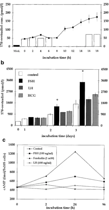
(4) Immortalized human granulosa cell line. Figure 4. Scanning electron microscopic image of immortalized COV434 granulosa cells in the presence of an intact oocyte. Long, slender, neurite-like intercellular connections are formed between granulosa cells. These structures did not appear in cultures of COV434 granulosa cells without an oocyte.. Figure 3. Multiple intercellular connections were formed between COV434 granulosa cells, and cells of a cumulus oophorus were formed only in the presence of an oocyte (A), but not if the cumulus complex did not contain an oocyte (B) (photographed after 24 h of culture, at ⫻400 magnification, light microscopy, inverted microscope).. were fixed with 3% glutaraldehyde for 60 min and mounted with MOWIOL mounting medium. The aldehyde-induced autofluorescence was imaged in a CLSM (NORAN Odyssey XL) in the rhodamine channel with ⫻63 magnification oil immersion objective lenses. Production of 17β-oestradiol and of cAMP For assessment of aromatase activity, immortalized COV434 granulosa cells (105 per ml) were plated in a 24-well tissue culture plate in DMEM culture medium containing 10% v/v FCS supplemented with 4-androstene-3,17-dione (10 µmol/l; Sigma, Steinheim, Germany) in the presence of recombinant FSH (100 ng/ml or 3⫻10–4 IU/ml, Gonal F; Serono) of recombinant LH (100 ng/ml or 3⫻10–4 IU/ml, LHadi; Serono) or of urinary HCG (100 ng/ml or 3⫻10–4 IU/ml, Profasi; Serono). At different periods after initiation of the culture 500 µl of the supernatant were collected from each well for measurement of the concentrations of 17β-oestradiol using commercial assay kits (Cobas Core, Roche, Basel). The production of cAMP was measured in the presence of FSH (100 ng/ml), of LH (100 ng/ml) and of forskolin (1 mmol/l) using a commercial enzyme immunoassay kit (Amersham, Buckinghamshire, UK).. RNA extraction and reverse transcription–polymerase chain reaction (RT–PCR) analysis Total RNA was extracted from 1⫻107 granulosa cells using a commercially available RNeasy Total RNA kit from Qiagen (Hilden, Germany). The quantity of RNA was assessed by measuring the optical density at A260 nm. Total RNA (1 µg) from granulosa cells was reverse transcribed into single strand cDNA using the cDNA synthesis kit (Boehringer Mannheim, Mannheim, Germany). All primers were synthetized by Microsynth, Balgath, Switzerland (Table I). The single strand cDNA was used for polymerase chain reaction (PCR). Then the single strand cDNA was subjected to 40 cycles of PCR amplification using one of the primer sets (50 s denaturation at 94°C, 50 s annealing at 60°C, and 30 s extension at 72°C). The amplified products were separated on 1% agarose gels. The PCR products were analysed by single restriction endonucleases (Table I; Biolab, Beverly, MA, USA) and DNA sequencing (ABI, PE Applied Biosystems, Foster City CA, USA) following the prediction based on their molecular size. Blanks served as a negative control. Protein extraction and Western blotting Granulosa cells (1⫻107 cells) were lysed in a lysis buffer (10 mmol/ l Tris, 1 mM EDTA, 1% Triton, protease inhibitor cocktail) for 10 min in ice and were sonicated twice for 10 s. For the assessment of Bcl-2 and of Bcl-Xl the samples were sonicated and quantified directly with a protein assay (Biorad, Hercules, USA). For the assessment of Bax, CPP32, Tx, ICH-IL and Bad the samples were first centrifuged for 10 min at 4°C at 12 000 g and only supernatants were quantified. Samples were boiled for 10 min and 20–30 µg of total protein was loaded on 15% sodium dodecyl sulphate– polyacrylamide gel electrophoresis. They were then were electrotransferred to a membrane (Millipore, Bedford, MA, USA), immunoblotted with different specific first antibodies (Table II) and developed with NBT/BCIP (Sigma, Steinheim, Germany). All experiments outlined above were presented to and approved by the Ethics Committee of the Medical Department of the University of Basel, Switzerland. All patients involved were informed about the rationale of the experiments and consented to the use of some of their biological material for experimental purposes. Statistical analyses were performed with the Kruskal–Wallis and Wilcoxon tests. The level of statistical significance was set at P ⬍ 0.05.. 149.
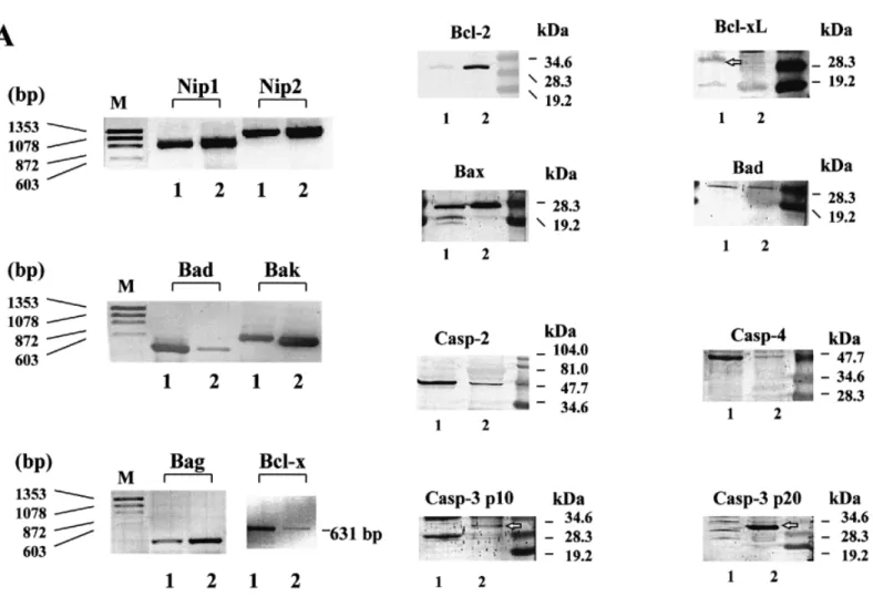
(5) H.Zhang et al.. Figure 5. Confocal images of COV434 granulosa cells stimulated by an oocyte (A) and in the absence of an oocyte (B), projected in x–y (main image), x–z (bottom) and y–z (right) directions. The COV434 granulosa cells formed in the presence of an oocyte extended fine fibrillar connections that did not form in the absence of an oocyte.. Results Morphological characterization of the human granulosa cell line The cultured COV434 granulosa cells grew in the culture medium as small aggregates on the bottom of the culture dish or flask and they formed colonies within 6 h. The granular appearance of the COV434 granulosa cells, as visible with light microscopy, was very similar to that of granulosa cells aspirated from IVF or ICSI patients (Figure 1). Some cells formed intercellular junctions with other cells (Figure 2, inset). The freshly collected granulosa cells of the patients displayed numerous microvilli, which were virtually absent in the COV434 granulosa cells (Figure 2). Lipid droplets were observed with light and electron microscopy both in the granulosa cells collected from infertile patients and in the COV434 granulosa cells, but the number of the lipid droplets in the former was much higher than in the latter. The mean number of lipid droplets counted in granulosa cells from IVF or ICSI patients was 104 [95% confidence interval (CI): 10.6], whereas the median number of droplets visible in a COV434 granulosa cell was only 6 (95% CI: 0.7, P ⬍ 0.0001). Co-culture of COV434 granulosa cells with COC The development of intercellular interactions between COV434 granulosa cells and COC was observed in culture medium, supplemented with FCS (10%) and FSH (200 ng/ml or 6⫻10–4 IU/ml). One day after initiation of the co-culture, multiple intercellular connections between the cumulus cells and the COV434 granulosa cells became apparent at high magnification and remained until day 8 of the co-culture (Figure 3). Intercellular connections remained absent between COV434 granulosa cells and the cells of one cumulus oophorus, which contained no oocyte as confirmed both with the stereomicroscope and the inverted microscope (Figure 3). Images of the intercellular connections between the immortalized COV434 granulosa cells and COC are presented in Figure 4 (scanning electron microscopy) and in Figure 5 (confocal light microscopy). Evaluation of endocrine function The effects of FSH on the secretion of 17β-oestradiol in the supernatant medium by incubated COV434 granulosa cells 150. and of LH, FSH and HCG on the secretion of 17β-oestradiol during prolonged culture of the COV434 granulosa cells are shown in Figure 6. Approximately 8 h after the addition of 100 ng/ml FSH to the culture medium the COV434 granulosa cells started to secrete 17β-oestradiol, whereas in the control samples lacking FSH there was no secretion of 17β-oestradiol (Figure 6A). During prolonged culture in the presence of FSH, higher levels of 17β-oestradiol were secreted than during prolonged culture in presence of LH and HCG (Figure 6B). The production of cAMP by COV434 granulosa cells was enhanced by FSH (100 ng/ml) and by forskolin (1 mmol/l), but not by LH (100 ng/ml, Figure 6C). Under the experimental conditions outlined above, the secretion of progesterone was not altered by the addition of FSH, LH or HCG to the in-vitro culture of COV434 granulosa cells (data not shown). Since the COV434 granulosa cells did not respond to stimulation with LH or HCG, the expression of LH receptor was examined by RT–PCR (Table I). Whereas LH receptor mRNA was readily identified by RT–PCR in granulosa cells from patients treated with IVF or ICSI, LH receptor mRNA was undetectable in COV434 granulosa cells cultured in culture medium devoid of FSH (Figure 7). Apoptosis-associated gene expression in COV434 granulosa cells The expression of several pro-apoptotic genes including bax, bad, bak, Casp-2, Casp-3 and Casp-4 were analysed together with anti-apoptotic genes such as bcl-2, bcl-Xl, Bag and genes encoding for Bcl-2 interacting proteins, Nip1 and Nip2. Gene expression in COV434 granulosa cells was evaluated by RT– PCR and Western blotting and the results were compared to those in granulosa cells collected from IVF and ICSI patients. The mRNA expression of nip1, nip2 (Boyd et al., 1994), bcl-Xl (Boise et al., 1993), bak (Farrow et al., 1995), bad (Yang et al., 1995) and bag (Takayama et al., 1995) were observed both in COV434 granulosa cells and in granulosa cells from infertile patients (Table I and Figure 8A). Western blot analysis revealed that Bax, Casp-2 (ICH-1L) and Bad proteins were similarly expressed in both granulosa cell types. However, Bcl-2 and Casp-3 (CPP32) proteins were expressed in granulosa cells from IVF and ICSI patients but not in.
(6) Immortalized human granulosa cell line. Figure 7. Expression of luteinizing hormone receptor (LHR) was examined by reverse transcription–polymerase chain reaction in pooled granulosa cells of 10 patients treated for in-vitro fertilization or intracytoplasmic sperm injection (1) and in COV434 granulosa cells (2). The arrow indicates the expected size of LHR at 458 bp, which was confirmed by DNA sequencing.. COV434 granulosa cells. On the other hand, Bcl-Xl and Casp-4 (TX) proteins were only expressed in COV434 granulosa cells but not in granulosa cells from IVF and ICSI patients (Table II and Figure 8B).. Discussion. Figure 6. (A) Secretion of 17β-oestradiol in the supernatant culture medium during incubation of COV434 granulosa cells (105/ml) in presence of follicle stimulating hormone (FSH) (100 ng/ml). The experiment was repeated three times and the results are presented as mean values ⫾ SEM. The black column represents the concentration of 17β-oestradiol in the culture medium, whereas the white columns represent the fluctuation in 17β-oestradiol concentration produced by COV434 granulosa cells in the absence of FSH. (B) Synthesis and secretion of 17β-oestradiol by immortalized COV434 granulosa cells (105 per ml) during prolonged culture in the presence of FSH, luteinizing hormone (LH) or human chorionic gonadotrophin (HCG) (100 ng/ml each) or in non-supplemented culture medium (control). The concentration of 17β-oestradiol in the supernatant was significantly higher in the presence of FSH than in controls lacking FSH (*P ⬍ 0.05). The height of each column indicates the mean ⫾ SEM. (C) Production of cAMP of immortalized COV434 granulosa cells (105 per ml) in the presence of FSH or LH (100 ng/ml each) or of forskolin (1 mM). Culture medium without these additives was used as a control. The production of cAMP was enhanced in the presence of FSH and forskolin, but not of LH.. Granulosa cells play a key role in the functional maturation of the follicle and the oocyte and display a high degree of structural change. These changes occur in the presence of numerous hormonal and paracrine stimuli and consist of a complex balance of proliferation and programmed cell death. The molecular pathways responsible for the differentiation of granulosa cells in parietal granulosa cells, which tend to be the main hormonally active portion of the granulosa cells, or in cumulus or coronal granulosa cells, which surround and nurse the oocyte (Buccione et al., 1990), are still largely unknown. We have investigated whether an immortalized granulosa cell line can be produced to mimic the complex processes of follicular development. To our knowledge, the cell line isolated by Van den Berg-Bakker et al. (1993) is the first ever described human immortalized granulosa cell line collected from a granulosa cell tumour still displaying functional receptivity to FSH. The following features were considered essential for the definition of normal functionality of the COV434 granulosa cells: increased synthesis and secretion of 17β-oestradiol after stimulation of cytochrome P450 aromatase with FSH (Erickson and Hsueh, 1978), establishment of intercellular connections between the immortalized granulosa cells and cells of a cumulus oophorus (Eppig, 1979; Bachvarova et al., 1980; Schultz, 1985; Sirard and Bilodeau, 1990) and the potential response to similar inducers of proliferation and apoptosis as compared to natural granulosa cells (Kaipia and Hsueh, 1997). Various experiments demonstrate that several of the properties considered essential for normal granulosa cell function are present in the immortalized human granulosa cell line COV434. First, the production and secretion of 17β-oestradiol in the supernatant culture medium could be stimulated with FSH. Secondly, the proliferation of the COV434 granulosa cells was stimulated by the addition of FSH in culture medium supplemented with FCS. These observations accord with a recent communication which demonstrated the presence of intact FSH receptors in cells collected from granulosa cell tumours (Fuller et al., 1998). Although the granulosa cells 151.
(7) H.Zhang et al.. Figure 8. Expression of apoptosis-associated genes in COV434. (A) mRNA expression of six apoptosis-associated genes was analysed by reverse transcription–polymerase chain reaction of total RNA. (B) protein expressions were examined by Western blotting. RNA and protein samples were extracted from either COV434 granulosa cells (1) or granulosa cells from patients treated with in-vitro fertilization or intracytoplasmic sperm injection (2).. also proliferated and secreted 17β-oestradiol in the absence of FSH, their activity was significantly enhanced by FSH. A similar observation was made recently (Fo¨ ldesi et al., 1998) using cultured luteinized granulosa cells obtained from IVF patients. Thirdly, within hours, intercellular connections between the COV434 granulosa cells and cells of a cumulus oophorus containing an oocyte became visible. Interestingly, these intercellular connections were not formed with cells of a cumulus oophorus not containing an oocyte, indicating that the proximity of an intact oocyte is essential for normal granulosa cell function. Recent findings have stressed the role of the oocyte by demonstrating that expansion of the cumulus oophorus depends on the presence of an intact oocyte (Buccione et al., 1990) and that the oocyte participates in suppressing the expression of the LH receptor mRNA in the surrounding granulosa cells (Eppig et al., 1997). It is also important to note that the intercellular connections were formed exclusively in the presence of a viable oocyte. Their appearance is more similar to the oocyte-derived structures involved in early folliculogenesis (Dong et al., 1996). The exact nature of this type of intercellular connection is currently being investigated. 152. Considering apoptosis-associated gene expression, some differences were found between the COV434 granulosa cells and granulosa cells from patients treated with gonadotrophins for IVF or ICSI. Whereas Nip1, Nip2, Bak, Bad (protein but not mRNA), Bag, Bax and Casp-2 (ICH-1L) were similarly observed in both granulosa cell types, it was demonstrated that others, Bcl-2 and Casp-3 (CPP32), were expressed only in granulosa cells from treated patients. Bcl-Xl and Casp-4 were detected only in COV434 granulosa cells. Whether these differences in apoptosis-associated gene expression between both granulosa cell types are caused by immortalization or by the treatment with exogenous gonadotrophins, mainly HCG, which was given 35 h prior to follicular aspiration for final maturation of the oocytes, remains to be clarified experimentally. The differences in apoptosis-associated gene expression may also be caused by the different availability of matrix metalloproteinases among both granulosa cell types. Matrix metalloproteinases are known to be involved in tissue remodelling during ovulation and may have been more abundant among the luteinized granulosa cells collected from the IVF and ICSI patients (Hulboy et al., 1997). Some of the morphological.
(8) Immortalized human granulosa cell line. differences between both granulosa cell types such as the presence of microvilli and lipid droplets may also be caused by gonadotrophin treatment. The density of microvilli on the outer surface of the granulosa cells and of lipid droplets within the cytoplasm are related to the action of FSH (Amsterdam and Rotmensch, 1987). Further studies are necessary to determine the optimal concentration of FSH to obtain the normal morphological appearance of the COV434 granulosa cells. A constantly available immortalized granulosa cell line with well-defined biological characteristics may be used for the development of in-vitro maturation of immature oocytes. In recent years, increased efforts have been devoted to this technique both in the human (Cha et al., 1991; Trounson et al., 1994) and in the animal (Eppig and O’Brien, 1996). Altogether, in-vitro maturation of human oocytes has only been successful to a limited extent (Cha et al., 1991; Trounson et al., 1994; Jaroudi et al., 1997; Russell et al., 1997). The low developmental capacity of in-vitro matured oocytes may be caused by the lack of support by surrounding and nursing granulosa cells (Bachvarova et al., 1980) and a constantly available pool of granulosa cells may be used for this purpose. In such a setting, the inability of the COV434 granulosa cells to luteinize could be considered an important advantage. An immortalized human granulosa cell line may also become valuable for other purposes including the establishment of standardized bioassays of FSH or for basic research concerning the balance between apoptotic processes and hormonal actions on the syncytial complex surrounding the maturing oocyte.. Acknowledgements We thank Dr Peter I.Schrier from the Department of Clinical Oncology of the University Hospital of Leiden in The Netherlands for providing the human granulosa cell line COV434 cells. We are also grateful to Prof. F.Gudat of the Institute of Pathology and M.Du¨ ggelin and D.Mathys of the SEM laboratory at the University of Basel and to Priv. Doz. Peter Huber, Head of the Hormone Laboratory of the University Hospital. This work was supported by grant Nr 31– 49834.96 of the Swiss National Fund.. References Amsterdam, A. and Rotmensch, S. (1987) Structure–function relationships during granulosa cell differentiation. Endocr. Rev., 8, 309–337. Bachvarova, R., Baran, M.M. and Tejblum, A. (1980) Development of naked growing mouse oocytes in vitro. J. Exp. Zool., 211, 159–169. Boise, L.H., Gonzalez-Garcia, M., Postema, C.E. et al. (1993) Bcl-x, a bcl2-related gene that functions as a dominant regulator of apoptotic cell death. Cell, 74, 597–608. Boyd, J.M., Malstrom, S., Subramanian, T. et al. (1994) Adenovirus E1B 19 kDa and Bcl-2 proteins interact with a common set of cellular proteins. Cell, 79, 341–351. Buccione, R., Schroeder, A.C. and Eppig, J.J. (1990) Interactions between somatic cells and germ cells throughout mammalian oogenesis. Biol. Reprod., 43, 543–547. Cha, K.Y., Koo, J.J., Choi, D.H. et al. (1991) Pregnancy after in vitro fertilization of human follicular oocytes collected from nonstimulated cycles, their culture in vitro and their transfer in a donor oocyte program. Fertil. Steril., 55, 109–113. Chedrese, P.J., Rodway, M.R., Swan, C.L. and Gillio-Meina, C. (1998) Establishment of a stable steroidogenic porcine granulosa cell line. J. Mol. Endocrinol., 20, 287–292. Dong, J., Albertini, D.F., Nishimori, K. et al. (1996) Growth differentiation factor-9 is required during early ovarian folliculogenesis. Nature, 383, 531–535.. Eppig, J.J. (1979) A comparison between oocyte growth in coculture with granulosa cells and oocytes with granulosa cell–oocyte junctional contact maintained in vitro. J. Exp. Zool., 209, 345–353. Eppig, J.J. and O’Brien, M.J. (1996) Development in vitro of mouse oocytes from primordial follicles. Biol. Reprod., 54, 197–207. Eppig, J.J., Wigglesworth, K., Pendola, F. and Hirao, Y. (1997) Murine oocytes suppress expression of luteinizing hormone receptor messenger ribonucleic acid by granulosa cells. Biol. Reprod., 56, 976–984. Erickson, G.F. and Hsueh, A. J.W. (1978) Stimulation of aromatase activity by follicle stimulating hormone in rat granulosa cells in vivo and in vitro. Endocrinology, 102, 1275–1282. Farrow, S.N., White, J.H., Martinou, I. et al. (1995) Cloning of a bcl-2 homologue by interaction with adenovirus E1B19K. Nature, 374, 731–733. Fo¨ ldesi, I., Breckwoldt, M. and Neulen, J. (1998) Oestradiol production by luteinized human granulosa cells: evidence of the stimulatory action of recombinant follicle stimulating hormone. Hum. Reprod., 13, 1455–1460. Fuller, P.J., Verity, K., Shen, Y. et al. (1998) No evidence of a role for mutations or polymorphisms of the follicle-stimulating hormone receptor in ovarian granulosa cell tumors. J. Clin. Endocrinol. Metab., 83, 274–279. Gougeon, A. (1996) Regulation of ovarian follicular development in primates: facts and hypotheses. Endocr. Rev., 17, 121–155. Hosokawa, K., Dantes, A., Schere-Levy, C. et al. (1998) Induction of Ad4BP/ SF1, steroidogenic acute regulatory protein, and cytochrome P450scc enzyme system expression in newly established human granulosa cell lines. Endocrinology, 139, 4679–4687. Hulboy, D.L., Rudolph, L.A. and Matrisian, L.M. (1997) Matrix metalloproteinases as mediators of reproductive function. Mol. Hum. Reprod., 3, 27–45. Jaroudi, K.A., Hollanders, J.M.G., Sieck, U.V. et al. (1997) Pregnancy after transfer of embryos which were generated from in-vitro matured oocytes. Hum. Reprod., 12, 857–859 Kaipia, A. and Hsueh, A.J. (1997) Regulation of ovarian follicle atresia. Annu. Rev. Physiol., 59, 349–63. Kananen, K., Markkula, M., Rainio, E. et al. (1995) Gonadal tumorigenesis in transgenic mice bearing the mouse inhibin α-subunit promotor/Simian virus T-antigen fusion gene: characterization of ovarian tumors and establishment of gonadotropin-responsive granulosa cell lines. Mol. Endocrinol., 9, 616–627. Lerner, A.A., Salamone, D.F., Chiappe, M.E. and Baranao, J.L. (1995) Comparative studies between freshly isolated and spontaneously immortalized bovine granulosa cells: protein secretion, steroid metabolism, and responsiveness to growth factors. J. Cell Physiol., 164, 395–403. Li, R., Phillips, D.M., Moore, A. and Mather, J.P. (1997) Follicle-stimulating hormone induces terminal differentiation in a predifferentiated rat granulosa cell line (ROG). Endocrinology, 138, 2648–2657. Lie, B.-L., Leung, E., Leung, P.C.K. and Auerspreg, N. (1996) Longterm growth and steroidogenic potential of human granulosa–lutein cells immortalized with SV40 large T antigen. Mol. Cell. Endocrinol., 120, 169–176. Minegishi, T., Nakamura, K., Takakura, Y. et al. (1990) Cloning and sequencing of human LH/hCG receptor cDNA. Biochem. Biophys. Res. Commun., 172, 1125–1130. Russell, J.B., Knezevich, K.M., Fabian, K.F. and Dickson, J.A. (1997) Unstimulated immature oocyte retrieval: early versus midfollicular endometrial priming. Fertil. Steril., 67, 616–620. Schultz, R.M. (1985) Roles of cell-to-cell communication in development. Biol. Reprod., 32, 27–42. Sirard, M.A. and Bilodeau, S. (1990) Effects of granulosa cell co-culture on in-vitro meiotic resumption of bovine oocytes. J. Reprod. Fertil., 89, 459–465. Takayama S., Sato, T., Krajewski, S. et al. (1995) Cloning and functional analysis of BAG-1: a novel Bcl-2-binding protein with anti-cell death activity. Cell, 80, 279–284. Trounson, A., Wood, C. and Kausche, A. (1994) In vitro maturation and the fertilization and developmental competence of oocytes recovered from untreated polycystic ovarian patients. Fertil. Steril., 62, 353–362 Van den Berg-Bakker, C.A., Hagemeijer, A., Franken-Postma, E.M. et al. (1993) Establishment and characterization of 7 ovarian carcinoma cell lines and one granulosa tumor cell line: growth features and cytogenetics. Int. J. Cancer, 53, 613–620. Yang, E., Zha, J., Jockel, J. et al. (1995) Bad, a heterodomeric partner for Bcl-XL and Bcl-2 displaces Bax and promotes cell death. Cell, 80, 285–291. Received on May 27, 1999; accepted on October 28, 1999. 153.
(9)
Figure
+3
Documents relatifs