European Heart Journal (1984) 5 (Supplement F), 85-93
Myosin isoenzymes in human hypertrophic hearts.
Shift in atrial myosin heavy chains and in ventricular
myosin light chains.
M. C. SCHAUB, C. R. TUCHSCHMID, T. SRIHARI AND H. O. HIRZEL
Department of Pharmacology, University of Zurich and Department of Cardiology, University Hospital, Zurich,
Switzerland
KEY WORDS: Myosin isoenzymes, human cardiac hypertrophy, atrial myosin, ventricular myosin. The myosin light chain complement and proteolytic peptide patterns of myosin heavy chains were studied by two-dimensional and one-two-dimensional electrophoretic techniques respectively, in a total of 57 samples from ventricular and atrial tissues of normal and hypertrophied human hearts. Hypertrophies were classified haemodynamically as due to pressure-overload and volume-overload. In addition to the occurrence of ventricular light chains in hypertrophied atria we also observed the atrial light chain-1 (ALC-1) in hypertrophied ventricular tissues. On average over 6% of total light-chain-1 comprised ALC-1 in pressure-overloaded ventricles and around 3% in volume-overloaded ventricles. In single cases of pressure-overload ALC-1 amounted up to over 20% of total light chain-1. With regard to the myosin heavy chains limited digestion by two different proteinases produced over 200 clearly resoluble peptides. The absence of any detectable differences in the peptide patterns between myosin heavy chains from normal and hypertrophic tissues of left or right ventricle is in line with the findings of J. J. Schier and R. S. Adelstein (J Clin Invest 1982; 69: 816-825). In atrial tissues however, reproducible qualitative differences in the peptide patterns indicated that during hypertrophy a different type of myosin heavy chains becomes expressed. No differences were seen between the myosin heavy chains from normal left and right atria.
Introduction myosin has the highest and V3 the lowest
ATPase activity'9'10'.
In cardiac muscle the contractile protein myosin is Based on comparison of electrophoretic mobilities dimeric and consists of two heavy chains (HC) with in normal human ventricle the main isoenzyme is an approximate molecular weight of 200000 dalton thought to be V3 with its low ATPase activity'611',
and 4 light chains (LC), a couple of each with a The enzymatic centre is known to be comprised molecular weight of around 21000 and 18000 within the HC only and the LC seem not to affect dalton'11. The existence of myosin isoenzymes in the ATPase of isolated myosin"2'1 3'. Thus shifts in
cardiac muscle has been postulated to explain the the composition of the HC isoforms are expected to correlation between myosin ATPase activity as be accompanied by changes in ATPase activities, measured in vitro and altered myocardial contractile However, a decline in myofibrillar ATPase activity properties in disease states'2-3'. Furthermore, shifts has been reported for human ventricles in congestive
in myosin isoenzyme composition with regard to heart failure'14'and severe hypertrophic states"5'1 6',
both HC and LC, have also been observed during No change in myosin or myofibrillar ATPase ontogenetic development of the heart of man and activities nor in the structure of the myosin HC as animals'4"7'. In heart ventricles of rodents two types judged from their peptide pattern, has been observed
of myosin HC have been described which are able to in ventricles of patients with hypertrophic form homodimeric and heterodimeric isoenzymes obstructive cardiomyopathy'17-18'. So in this latter
designated in order of decreasing electrophoretic disease there seems to occur no transition in the mobility as Vj = 2 x HC-alpha, V2 = HC- expression of the isoforms of HC. The lower
alpha + HC-beta and V3 = 2 x HC-beta(8'. Vi myofibrillar ATPase activities observed in
trophies induced by pressure- or volume-overload is difficult to explain on the grounds of changes in the composition of HC isoforms, since the normal heart ventricle already contains almost exclusively V3 myosin with the lowest ATPase activity. Either a new additional type of myosin HC may become expressed in some forms of heart diseases or else the LC complement, if its composition changes, might still affect the enzymatic properties of the intact myofibril. Recently it has been reported for skeletal muscle myosin that the LC indeed play a role in the subtle interaction of the myosin head portions where the LC are located, with their reaction partner actin and the calcium-sensitive regulatory proteins
troponin and tropomyosin(19). Therefore
examination of both HC and LC is required. Transition in the LC complement both in human atria and ventricles have been reported recently to occur in response to cardiac pressure overload'20'211. Thereby LC characteristic for ventricle appear in the atrium under pathologic conditions and vice versa. The atrial LC, (ALC,) which occurs in the ventricle of pathologically hypertrophied hearts can also be observed in man and animals to be present in early developmental stages in the foetal ventricle and to disappear after birth'5-22'23'. Apparently both heart chambers have retained the ability of transition of their LC complement in the adult. We have therefore undertaken a structural study of both LC and HC in ventricles and atria of normal and pathologically hypertrophied human hearts. It was possible to quantify the occurrence of the atrial ALC, in hypertrophic ventricles in response to pressure- and volume-overload separately. In the right ventricle of children fewer ALC, emerged in hypertrophy due to pressure-overload. But in an infant at the age of 7 months a large portion of ALC,, which is present normally in the foetal ventricle, was retained in the hypertrophic right ventricle. In contrast, no structural transition was apparent in the myosin HC from hypertrophic ventricles, but we report here for the first time a change in the peptide pattern of HC in pathologically hypertrophic atria.
Methods
Left ventricular endomyocardial biopsy material was obtained from 15 patients by the transseptal route, whereby the King's College Bioptome was advanced through a French-11-5-Brockenbrough catheter from the right femoral vein to the left ventricle. The samples were derived mainly from the
lateral apical portion of the left ventricle. Samples from pathological and normal ventricular or atrial tissues were taken during surgery or from autoptic material within 8 h after death. Cases with left ventricular hypertrophy were classified on haemo-dynamic grounds as due to pressure-overload with maximal and average pressure gradients above 30 mmHg and valve opening areas below 1-5 cm2, or as due to volume-overload with regurgitation fractions above 40%.
Tissue fragments were stored at — 20°C or below until usage. Samples of 1-10 mg were homogenized in the appropriate buffer system and directly applied to electrophoresis. Protein concentrations were determined according to Lowry and coworkers(24). One-dimensional polyacrylamide gel electrophoresis in the presence of sodium dodecyl sulphate (SDS) was carried out according to Laemmli and Favre(25) by using 3% stacking (3x18 cm) and 12-5% separating gels (13 x 18 cm) of 1-5 mm thickness and applying around 25 pig protein per sample. For two-dimensional gel electrophoresis isoelectric focusing was beforehand carried out in the first dimension according to O'Farrell'26' by using a (4:1, v/v) mixture of ampholines (1-75%) of the pH range 5-8 and 3-5—10 respectively. Polyacrylamide gels (3-64%) of 11 -5 cm length were prepared in glass tubes (3 mm inner diameter and 17 cm long). For isoelectro-focusing around \20 fig of each sample were applied to the top of the pre-electrophoresed gels. The protein in all different gels were visualized by staining in 0-25% Coomassie Brillant Blue R-250. Destaining of gels and determination of pH gradient in isoelectric focusing gels were done as before'27'.
For isolation of myosin HC, tissue samples were homogenized in 10 vol of 40 mM sodium pyro-phosphate (pH 7-5 adjusted with HC1) containing 2 mM dithiothreitol and 5 mM ethylene-glycol-bis (beta-amino ethylether) N,N-tetraacetic acid1281. Immediately before electrophoresis, 4 mM N-ethyl-maleimide were added to the sample containing between 200 and 400 ng of protein, which were loaded on gels composed of 3% acrylamide and 0-24% bis-acrylylcystamine'29). Chamber and gel buffer, pH 8-4, were composed of tris-(hydroxy-methyl)aminomethanol (Tris) and (N,N-bis(2-hydroxyethyl)glycine) (Bicine) containing 0-1 % SDS as described elsewhere'30'. After the electrophoretic run, gels were stained with Coomassie Brillant Blue R-250, subsequently soaked for 15 min in 20% methanol followed by 15 min in the chamber buffer Tris-Bicine pH 8-4. Then bands representing myosin
Myosin isoenzymes 87
HC were excised and the gel slices dissolved by addition of 3-4% beta-mercaptoethanol. To these samples were added 0-5 vol. bringing the final concentrations similar to those described by Whalen and coworkers(31): 70 mM Tris, 70 mM Bicine,
0-23% SDS, 3-5% beta-mercaptoethanol, 150 mM NaCl, 003 mM MgCl2, 48 mM phosphate buffer pH
6-5. For digestion of 80-100 ng of myosin HC per sample, proteinase from Staphylococcus aureus V8 (EC 3.4.21.19) (Miles, Elkhart, Indiana, USA) and papain (EC 3.4.22.2) (Boehringer, Mannheim, West Germany) were used. The molar ratio of both proteinases to myosin HC of 200000 dalton molecular weight range from 0-001 up to 005 based on the molecular weights of 27 700 dalton for the proteinase V8 and 23 350 dalton for proteinase papain. Digestion was performed for 30 min at 37°C and the reaction stopped by addition of around 100-fold molar excess of phenylmethylsulphonyl fluoride in the case of the proteinase V8 and by heating samples for 3 min at 100°C in the case of proteinase papain. The protein digest was then resolved on usual one-dimensional gel electrophoresis in SDS(25).
All peptide patterns were stained by Coomassie Brillant Blue R-250, and in many cases they were, after destaining in 40% methanol plus 10% acetic acid, stained again with silver'32' in order to increase
sensitivity for visualization of peptides occurring in small quantities.
One-dimensional stained gels and positive films of them were scanned and recorded either by the Gilford Spectrophotometer System 2600 (Gilford Instr. Lab. Inc., Oberlin, Ohio, USA) at 560 nm or by the LKB 2202 UltroScan Laser Densitometer (LKB-Produkter AG, Bromma, Sweden) at 633 nm. In two-dimensional electrophoresis, firstly, the central absorbance of the spots on the stained gels and on their films was measured with the McBeth TD 504 Densitometer (McBeth Color & Photo-metry Div., Newburgh, New York, USA). Secondly, the spots were excised from enlarged prints and weighed. With purified tropomyosin for calibration the average of the two measurements yielded a relationship that was linear from 0-5 up to 14 ^tg of protein per spot. In this way the myosin LC were quantified in each two-dimensional gel relative to tropomyosin. No allowance was made for possible differences in dye-uptake by these proteins. For calculation of the stoichiometry the chemical molecular weights of 21 000 dalton for LC,, 18000 for LC2 and 66 000 for tropomyosin from both atrial
and ventricular tissues were taken. This needs to be
mentioned for the apparent molecular weights of the LC in SDS gel electrophoresis are different: 27400 dalton for LC, and 22600 for LC2 from atrium and
26000 dalton for LC, and 21000 for LC2 from
ventricle'22'. The myosin HC and sometimes also
actin were not reliably resolved in the isoelectro-focusing run and could therefore not be quantified.
Results
VARIATION IN LIGHT CHAIN COMPLEMENT
In support of the finding of Cummins'20' we also
observed the occurrence of additional myosin LC in hypertrophied atria which exhibit identical electro-phoretic mobilities to those of the ventricle VLC, and VLC2. Such a ventricular VLC2 was
consistently found in atrial tissues from severely hypertrophic hearts. It mostly appeared as double spot due to partial phosphorylation. But occasionally a faint, more acidic, third spot was also visible which is thought to represent a second type of VLC2(2O). Such a second VLC2 type has also been
described in several vertebrate species beside man'33'.
The VLC, type occurring in hypertrophic atria was variable and in all cases much reduced in comparison to its VLC2 counterpart. Quantification
was therefore not attempted. Quantification of the VLC2 occurring in atria neither proved to be useful
since the total amount of the LC2 type varied in
both atrial and ventricular tissues of normal as well as hypertrophic hearts. Thereby the LC2 type
displayed the tendency to be reduced variably in comparison to the LC, type.
On the other hand, the LC, type exhibited a remarkable constancy in amount related to that of tropomyosin (Table 1). Fig. 1 shows the occurrence
Table I Occurrence of atrial ALCX in hypertrophic and normal left ventricles. Total LCS content is given as molar ratio to tropomyosin and ALCX as percentage thereof. The occurrence of ALCl is significant in both forms of hypertrophy at the level of P<00l in comparison to normal
ventricles (mean values are given)
Age in Number Total LC,
Condition years of cases per tropomyosin %ALC, Normal Pressure-overload Volume-overload 23-66 35-79 32-72 7 15 10 2-7 3-4 2-5 0-4 6-3 2-8
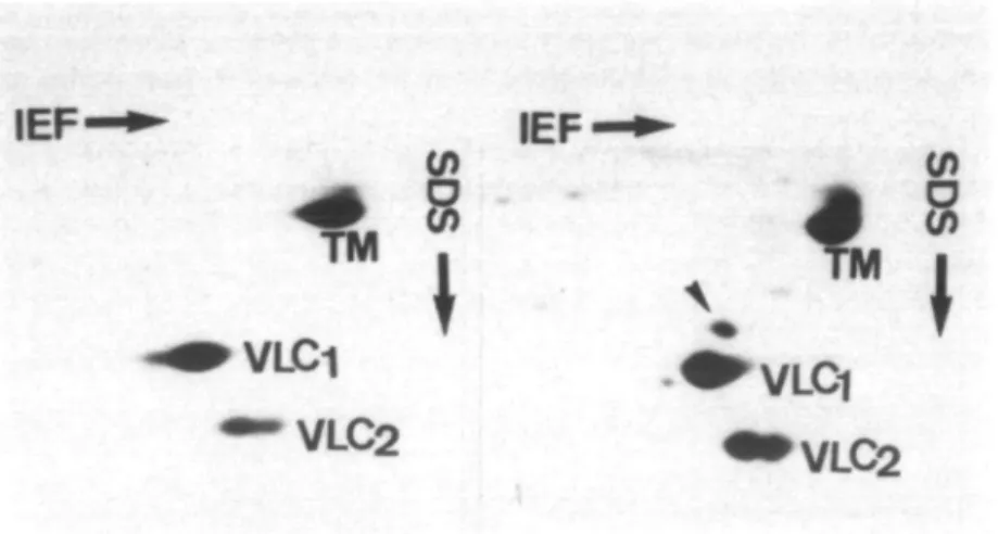
VLC1
VLC2
Figure I Two-dimensional electrophoresis of total tissue homogenate from left
ventricles. Gels are presented with the basic pH range in isoelectrofocusing (IEF) to the left and decreasing molecular weights from top to bottom in SDS electrophoresis. Left side, normal ventricle of a women at the age of 41 years. Right side, ventricle of a man at the age of 39 years with pressure-overload. TM, mainly alpha-tropomyosin with some little beta-tropomyosin on top; VLC, and VLC2, ventricular myosin LC; arrow indicates the
ALC, in the hypertrophic ventricle.
of ALC, in the left hypertrophic ventricle of an adult in comparison to a normal case.
Quantification of the atrial ALC, occurring in hypertrophic ventricles under pathological conditions seemed useful and led to the results given in Table 1. The total amount of LC, type is expressed in moles of LC, per mole of tropomyosin with a molecular weight of 66 000 dalton as described in the methods section. The content of the total amount of LC, did on average not vary significantly between normal and hypertrophic ventricular tissues. There was a slight tendency of higher values of total LC, in some cases of severe pressure-overload, reaching extremes of 40 and 9-6 in two single instances. In these latter cases however, the portion of ALC, did not exceed 5 and 6%, respectively. On the other hand, the additional ALC, was significantly increased in hypertrophic (/><0-0l) when compared with normal tissue. In pressure-overload the portion of ALC, varies between 1 and 27% of total LC, whereas in volume-overload it ranged between 1 and 5% only. In normal ventricular tissue ALC, was seen occasionally as a very faint spot which never exceeded 0-5% of total LC,.
In four children at the age of 2-7 years with pressure-overload of the right ventricle due to congenital malformation (tetralogy or trilogy of Fallot), the portion of ALC, in the ventricular tissue
ranged between 0-5 and 2-7%. In an infant at the age of 7 months the portion of ALC, in the ventricle attained 29%. At this age in normal infants the relative content of ALC, in the ventricle is around 1-5% and decreases continuously until, at the age of around one year, it has disappeared completely. Price and coworkers(5) have reported a value of 5-7% for the portion of ALC, in the normal ventricle of an infant at the age of 8 months.
In two cases with pressure-overload samples were taken separately from both subendocardial and subepicardial muscle layers, in one case of the left ventricle from an adult and in the other from the right ventricle of a child. In both cases the inner and outer muscle layers displayed almost identical LC patterns. In particular, the ventricular ALC, did not vary significantly between the two layers. Further-more, in co-electrophoresis of the two muscle layers of one heart, but also in co-electrophoresis of the hearts of the two patients, the ventricular ALC, always appeared as one single co-migrating spot.
VARIATION IN HEAVY CHAIN STRUCTURE
Limited digestion of isolated myosin HC either with proteinase V8 or with proteinase papain were performed in the presence of 0-23% SDS under carefully controlled conditions. Examined were 5 cases of left ventricles and 3 cases of left atria from
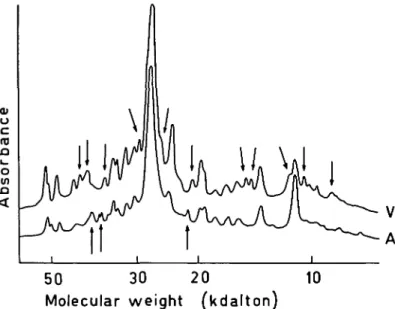
Myosin isoenzymes 89 0) c 03 X I 50 30 20
Molecular weight (kdalton) 10
Figure 2 Densitometric traces of electrophoretograms with peptide patterns
of myosin HC from normal left atrial and ventricular tissues after digestion with proteinase V8 (ratio to myosin HC, 005). Coomassie staining. V, ventricle; A, atrium; arrows indicate qualitative differences (see text in results section).
pressure-overloaded hearts of adults, 2 cases of right ventricles from pressure-overloaded hearts of children, 3 cases of normal left ventricles, 4 cases of normal left atria and 2 cases of normal right atria. In each digestion experiment one or two pathological samples were processed at a time strictly in parallel with a normal one. In separate experiments two normal samples or, alternatively, two or three pathological samples were processed together. Each experiment consisted of a series of parallel digestions with varying concentrations of the proteinases in relation to the concentration of the myosin HC as indicated in the methods section. Peptide patterns were thus obtained in one-dimensional electro-phoretic resolution with both proteinases, in which the majority of peptide bands spread predominantly over the high (80-200 kdalton), middle (30-120 kdalton) or low (10-50 kdalton) molecular weight ranges. In this way well over 200 peptide bands could clearly be distinguished per sample and compared with those of the experimental companions.
Densitometric evaluation was only justified when the patterns to be compared indicated equal degrees of digestion that is the staining intensity of most peptide bands was very similar. As soon as there
were different amounts of undigested material between the parallels left at the top of the gel slab, or if in some regions variable staining intensities indicated unequal digestion between them, such experiments were rejected. Particular caution had to be exerted if differences appeared among the protein bands in the high molecular weight range of samples which were processed in parallel. Repetitive experi-ments proved that in this region transiently occurring peptide bands were occasionally seen in single digestions which could not be reproduced. However, in the middle and low molecular weight ranges differences or, alternatively, no differences at all, between parallels, were clearly recognizable and also reproducible. No differences at all could be detected with either proteinase between the peptide patterns of myosin HC from right or left ventricles nor between those originating in the outer, middle or inner muscle layers of the left ventricular wall or in the papillary muscles.
The peptide patterns of myosin HC from atrium and ventricle of a normal adult heart in Fig. 2 show a large number of differences in the low molecular weight range after digestion with proteinase V8. The differences pointed out in Fig. 2 concern peptide bands which occur in one of the two digestion
o c X) o in
80 40 20
Molecular weight (kdalton)
Figure 3 Densitometric traces of electrophoretograms with peptide patterns of myosin HC from normal and hypertrophic left ventricles after digestion with papain (ratio to myosin HC, 002). Coomassie staining. V, normal ventricle of a man at the age of 29 years; VH, ventricle of a man at the age of 79 years with pressure-overload.patterns only and do not reflect merely quantitative differences of bands existing in both patterns. Fig. 3 comprises peptide patterns of myosin HC after digestion with papain from normal and hyper-trophied left ventricles of adults. The two patterns are almost identical. On close inspection only minute quantitative variation in some of the smallest peptide bands can be detected. In no parallel digestion of myosin HC from normal and hypertrophic ventricles did we ever see reproducible qualitative differences. This confirms the results of Schier and Adelstein(I8) who did neither find differences in the peptide patterns of ventricular myosin from normal and hypertrophic cardiomyopathic hearts. In their case intact native myosin was digested with alpha-chymotrypsin and papain.
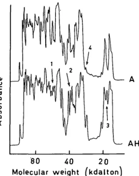
In contrast, qualitative differences could be detected between the peptide patterns of myosin HC from normal and hypertrophic atrial tissues. The samples shown in Fig. 4 both stem from left atria and were digested with papain. Beside the qualitative differences pointed out by the arrows numbered 1-3, much larger qualitative variations in single bands can be seen than in the two patterns from ventricular myosin HC in Fig. 3. Such a quantitative variation of a particular band is marked with the arrow
o c n o in 80 40 2 0 Molecular weight [kdalton]
Figure 4 Densitometric traces of electrophoretograms with peptide patterns of myosin HC from normal and hypertrophic left atria after digestion with papain (ratio to myosin HC, 005). Silver staining. A, normal atrium of a woman at the age of 41 years; AH, atrium of a woman of the age of 55 years with pressure-overload; arrows 1—3 indicate qualitative differences; arrow 4 points to a quantitative difference (see text in results section).
number 4, but immediately to the higher molecular weight side of arrow number 1 is a triplet of bands exhibiting also considerable quantitative variation between the two patterns. Thus unlike the myosin HC from ventricle, those from atrium do become changed in hypertrophy. Of the 3 hypertrophic left atria examined, two displayed identical peptide patterns between themselves while the third one differed. The patterns of all normal atrial tissues looked identical, even those from right and left atria. Discussion
The present study indicates that transitions in the myosin LC complement occur in both atria and ventricles with pathological hypertrophies. In left atria an additional VLC2 is found predominantly while in the ventricles an ALC, shows up. Thus in the former case we observed a partial transition of LC in an atrial-ventricular, and in the latter, in
Myosin isoenzymes 91
contrast, in a ventricular-atrial direction. This is somewhat unexpected. It has been speculated the primitive cardiac tube may synthesize mainly an atrial-related type of myosin(5). Under the stimulus of increasing pressure and workload experienced by the newly developing ventricular chamber, ventricular myosin will then come to be produced. In the atrium this sequence of events seems to be maintained when under pathological work-overload ventricular LC become expressed. In the ventricle however, despite an even greater workload, the genetic expression switches back to the primordial type of atrial-like LC. It appears then that with regard to the LC complement which seems to undergo opposing transitions, the isoenzyme expression follows some pre-determined pattern rather than to obey the functional stimulus.
As expounded in the results section quantification of the LC, type alone led to sensible results. The LC2 type varied in both atria and ventricles in an unpredictable way. It has been reported that this LC2 type characteristic of myosins with low ATPase activity, always yields variable recoveries by various extraction procedures'30'. The appearance of ALC, is highly significant for a pathologically increased workload of the left ventricle. Its appearance is further more pronounced under pressure-overload than under volume-overload. The relatively low level of ALC, compared to the normal VLC, content may explain why Klotz and coworkers'34' failed to detect variations in the primary structure of the LC in human hypertrophied left ventricles. In children with pressure-overload of the right ventricle the ventricular-atrial transition of the LC, type remains even more moderate. However, within the first year of age, where some little ALC, is still being produced under normal conditions, under pressure-overload the amount of ALC, in the right ventricle is very large indeed. At this young age the large proportion of the atrial ALC, in the right ventricle affected by congenital malformation reflects the persistence of its otherwise declining normal expression.
The total content of the LC, type in ventricular tissue amounts on average to around 3 moles per mole of tropomyosin. This latter was found to be the most reliable reference protein. In sarcomeric muscles the molar ratio of myosin to tropomyosin was determined to be very close to unity<35). Isolated ventricular myosin contains two moles of LC, and LC2 each per mole'36'. Thus two moles of LC, type would be expected per mole of tropomyosin.
In total ventricular tissue homogenates we found a constant amount of LC, type just below three moles per mole of tropomyosin (Table 1), which is very close to the expected value. If the ratio of myosin to tropomyosin is indeed one, then almost all LC, seem to be bound to myosin. The myosin LC are known to have in heart muscle a somewhat slower turnover rate than the HC'37', so there might be a small surplus in the pool of LC in the tissue in order to allow all myosin HC to combine with the appropriate LC to form intact molecules. We observed a tendency of total LC, type to increase in ventricles with hypertrophy due to pressure-overload. The quantitative analyses of the individual cases indicate however, that this increase in total LC, type was never caused specifically by the additional ALC, in the ventricle. It rather results from a general stimulation of synthesis of the LC, type. Should the content of total LCt be related to that of HC, then myosin would on average not vary drastically in hypertrophic ventricles.
Our new finding with regard to the myosin HC concerns the occurrence of a HC isoform in hypertrophied left atria from patients with pressure-overload. In normal hearts the HC structure as revealed in the peptide patterns was identical in right and left atria. Ventricular myosin HC are different from normal and pathological atrial myosin HC. In ventricles we never found a difference between normal and hypertrophied tissues. Under the stringent criteria laid down in the results section this means that in the absence of qualitative differences the primary structures of the HC from normal and pathological ventricles have an extremely high degree of homology or they may indeed be identical. The presence of a new HC species in hypertrophy in appreciable amounts is excluded by our findings, since a single additional peptide band from such a new species amounting to, or exceeding, 5% of total protein would not be overlooked in electrophoresis. The peptide patterns of HC from ventricles could in principle result from a mixture of HC isoforms. But again such a mixture would have to remain constant in proportion in normal and pathological ventricles since we did not even find significant quantitative differences between them. Electrophoresis of intact native myosin and immunochemical tests have shown that normal human ventricle in fact
comprises mainly one isoenzyme, the V3
myosin'6'1".
On the other hand, the qualitative differences in the peptide patterns of HC from normal and
pathological atria indicate the occurrence of a new isoform in hypertrophy which is not present in the normal right or left atria. In atrium again a mixture of more than one HC isoform could be present normally, and the additional quantitative differences under pathological conditions would then indicate furthermore, a shift in the composition of the basic isoenzyme pattern. Or else, if the normal atrium contains only one HC isoform the quantitative changes may simply reflect its relative decline as the new species arises in hypertrophy.
Alpert and coworkers(38) have demonstrated that
hypertrophying heart tissue adapts to slow, economical tension development in response to pressure-overload. In small rodents this is accompanied by a transition of isoenzymes from the normally present V! myosin with high ATPase activity to V3 with low activity'9'. In man(6>11) as
well as in larger animals V3 represents the main
isoenzyme in normal ventricles'6-20' and as shown
here for man, no transition in HC isoforms occurs under pathological workload. In atria however, whose myosin has a higher ATPase activity than its ventricular counterpart'39', adaptation to the
increased workload by expression of a new isoform of HC is still possible. Presumably this new myosin isoenzyme has a lower ATPase activity, though this remains to be proven.
In conclusion, we have shown that the expression of isoforms of myosin HC seems to follow functional adaptation in human atria under increased workload while no such isoenzymatic transition takes place in ventricles. On the other hand, the LC complement is regulated independently of the HC and changes in hypertrophied atria towards the ventricular, and in hypertrophied ventricles towards the atrial composition. Presently we are examining the LC transitions in various forms of heart disease and it seems that some diagnostic value could be derived from the knowledge of the LC pattern.
We are particularly indebted to Professor M. Turina, Department of Surgery, University Hospital, Zurich, for 23 heart samples taken during surgery and to Dr J. Schneider, Department of Pathology, University Hospital, Zurich, for 19 heart samples from autopsy. This work was supported by the Swiss National Science Foundation grant 3.535.79 and 3.152.81, the Swiss Foundation of Cardiology and the Fritz Hoffmann-LaRoche Foundation.
References
(!) Weeds AG, Frank G. Structural studies on the light chains of myosin. Cold Spring Harbor Symp Quant Biol 1972; 37: 9-14.
(2) Scheur J, Bhan AK.. Cardiac contractile proteins: activity and physiological function. Circ Res 1979; 45:
1-12.
(3) Jacob R, Rupp H, Ebrecht G, Holubarsch C, Kissling G. The significance of the isoenzyme pattern of myosin for myocardial mechanics and energetics. Relevance to the definition of cardiac contractility. Z Kardiol 1982; 71: 553-65.
(4) Whalen RG, Sell SM. Myosin from fetal hearts contains the skeletal muscle embryonic light chain. Nature 1980; 286: 731-3.
(5) Price KM, Littler A, Cummins P. Human atrial and ventricular light-chain subunits in the adult and during development. Biochem J 1980; 191: 571-80.
(6) Lompre AM, Mercadier JJ, Wisnewsky C. et al. Species- and age-dependent changes in the relative amounts of cardiac myosin isoenzymes in mammals. Develop Biol 1981; 84: 286-90.
(7) Chizzonite RA, Everett AW, Clark WA, Jakovcic S, Rabinowitz M, Zak R. Isolation and characterization of two molecular variants of myosin heavy chain from rabbit ventricle. Change in their content during normal growth and after treatment with thyroid hormone. J. Biol Chem 1982; 257: 2056-65.
(8) Hoh JFY, Yeoh GPS, Thomas MAW, Higginbottom L. Structural differences in the heavy chains of rat ventricular myosin isoenzymes. FEBS Letters 1979; 97: 330-4.
(9) Hoh JFY, McGrath PA, Hale PT. Electrophoretic analysis of multiple forms of rat cardiac myosin. Effects of hypophysectomy and thyroxine replacement. J Molec Cell Cardiol 1978; 10: 1053-76.
(10) Pope B, Hoh JFY, Weeds A. The ATPase activities of rat cardiac myosin isoenzymes. FEBS Letters 1980;
118:202-8.
(11) Clark WA, Chizzonite RA, Everett AW, Rabinowitz M, Zak R. Species correlations between cardiac isomyosins. A comparison of electrophoretic and immunological properties. J Biol Chem 1982; 257: 5449-54.
(12) Wagner PD, Giniger E. Hydrolysis of ATP and reversible binding to F-actin by myosin heavy chains free of all light chains. Nature 1981; 292: 560-2. (13) Wagner PD, Weeds AG. Studies on the role of myosin
alkali light chains. Recombination and hybridisation of light chains and heavy chains in subfragment-1 preparation. J Mol Biol 1977; 109: 455-73.
(14) Alpert NR, Gordon MS. Myofibrillar adenosine triphosphatase activity in congestive heart failure. Am J Physiol 1962; 202: 940-6.
(15) Leclerq JF, Swinghedauw B. Myofibrillar ATPase, DNA and hydroxyproline content of human hyper-trophic heart. Eur J Clin Invest 1976; 6: 27-33. (16) Swinghedauw B, Schwartz K, Leger JJ. Cardiac
myosin. Phylogenic and pathologic changes. Basic Res Cardiol 1977; 72: 254-60.
(17) Maron BJ, Ferrans VJ, Adelstein RS. Isolation and characterization of myosin from subjects with
Myosin isoenzymes 93
asymmetric septal hypertrophy. Circ Res 1977; 40: 468-73.
(18) Schier JJ, Adelstein RS. Structural and enzymatic comparison of human cardiac muscle myosins isolated from infants, adults and patients with hypertrophic cardiomyopathy. J Clin Invest 1982; 69: 816-25. (19) Wagner PD, Stone DB. Calcium-sensitive binding of
heavy meromyosin to regulated actin requires light chain-2 and the head-tail junction. Biochemistry 1983; 22: 1334-42.
(20) Cummins P. Transitions in human atrial and ventricular myosin light-chain isoenzymes in response to cardiac-pressure-overload-induced hypertrophy. Biochem J 1982; 205: 195-204.
(21) Tuchschmid CR, Srihari T, Hirzel HO, Schaub MC. Structural variants of heavy and light chains of atrial and ventricular myosins in hypertrophied human hearts. In: Cardiac adaptation to hemodynamic overload, training and stress. Steinkopff-Verlag, 1983:
123-8.
(22) Srihari T, Tuchschmid CR, Hirzel HO, Schaub MC. Electrophoretic analyses of atrial and ventricular cardiac myosins from foetal and adult rabbits. Comp Biochem Physiol 1982; 72B: 353-7.
(23) Srihari T, Tuchschmid CR, Schaub MC. Isoforms of heavy and light chains of cardiac myosins from rat and rabbit. Basic Res Cardiol 1982; 77: 599-609.
(24) Lowry OH, Rosebrough NJ, Farr AL, Randall RF. Protein measurement with the Folin phenol reagent. J Biol Chem 1951: 193: 265-75.
(25) Laemmli UK, Favre M. Maturation of the Head of bacteriophage T4. I. DNA packaging events. J Mol Biol 1973; 80: 575-99.
(26) O'Farrell PH. High resolution two-dimensional electro-phoresis of proteins. J Biol Chem 1975; 250: 4007-21. (27) Srihari T, Wiehrer W, Pette D, Harris BG.
Electro-phoretic analysis of myofibrillar proteins from the body wall muscle of Ascaris suum. Mol Biochem Parasitol 1981; 3: 71-82.
(28) Rupp H. The adaptive changes in the isoenzyme
pattern of myosin from hypertrophied rat myocardium as a result of pressure overload and physical training. Basic Res Cardiol 1981; 76: 79-88.
(29) Hansen JN. Electrophoresis of ribonucleic acid on a polyacrylamide gel which contains disulfide-crosslinkages. Anal Biochem 1976; 76: 37-44. (30) Weeds AG. Light chains from slow-twitch muscle
myosin. Eur J Biochem 1976; 66: 157-73.
(31) Whalen RG, Schwartz K, Bouveret P, Sell SM, Gros F. Contractile protein isozymes in muscle development; identification of an embryonic form of myosin heavy chain. Proc Nat Acad Sci USA 1979; 76: 5197-201.
(32) Wray W, Boulikas T, Wray VP, Hancock R. Silver staining of proteins in polyacrylamide gels. Analyt Biochem 1981; 118: 197-203.
(33) Westwood SA, Perry SV. Two forms of the P-light chain of myosin in rabbit and bovine hearts. FEBS Letters 1982; 142: 31-4.
(34) Klotz C, Leger JJ, Elzinga M. Comparative sequence of myosin light chains from normal and hypertrophied human hearts. Circ Res 1982; 50: 201-9.
(35) Potter JD. The content of troponin, tropomyosin, actin and myosin in rabbit skeletal muscle myofibrils. Arch Biochem Biophys 1974; 162: 436-41.
(36) Pfister M, Schaub MC, Watterson JG, Knecht M, Waser PG. Radioactive labeling and location of specific thiol groups in myosin from fast, slow and cardiac muscles. Biochim Biophys Acta 1975; 410:
193-209.
(37) Zak R. Metabolism of myofibrillar proteins in the normal and hypertrophic heart. Basic Res Cardiol
1977; 72: 235-40.
(38) Alpert NR, Mulieri LA, Litten RC. Functional significance of altered myosin adenosine triphosphatase activity in enlarged hearts. Am J Cardiol 1979; 44: 947-53.
(39) Yazaki Y, Ueda S, Nagai R, Shimada K. Cardiac atrial myosin adenosine triphosphatase of animals and humans. Circ Res 1979; 45: 522-7.