HAL Id: hal-02396085
https://hal.archives-ouvertes.fr/hal-02396085
Submitted on 30 Nov 2020
HAL is a multi-disciplinary open access
archive for the deposit and dissemination of
sci-entific research documents, whether they are
pub-lished or not. The documents may come from
teaching and research institutions in France or
L’archive ouverte pluridisciplinaire HAL, est
destinée au dépôt et à la diffusion de documents
scientifiques de niveau recherche, publiés ou non,
émanant des établissements d’enseignement et de
recherche français ou étrangers, des laboratoires
Modification of both functional neurological symptoms
and neuroimaging patterns with a good anatomoclinical
concordance: a case report
Silvio Galli, Selma Aybek, Sylvie Chokron, Thierry Moulin, Eloi Magnin
To cite this version:
Silvio Galli, Selma Aybek, Sylvie Chokron, Thierry Moulin, Eloi Magnin. Modification of both
func-tional neurological symptoms and neuroimaging patterns with a good anatomoclinical concordance:
a case report. BMC Neurology, BioMed Central, 2019, 19 (1), pp.5-14. �10.1186/s12883-019-1475-3�.
�hal-02396085�
Charcot’s “psychodynamic lesions”
Modification of both functional neurological symptoms and
neuroimaging patterns with a good anatomoclinical concordance: a case
report.
Silvio Galli1, Selma Aybek2, Sylvie Chokron3, Thierry Moulin1, Eloi Magnin1
1 Department of neurology, University Hospital Besancon, Besancon, France 2 Neurology University Clinic, Inselspital, 3010 Bern, Switzerland.
3 Laboratory of Psychology of Perception, UMR 8242, CNRS and Paris Descartes University and Vision and Cognition Unity, Fondation Ophtalmologique Rothschild, Paris, France.
Author for correspondence: Dr Silvio Galli, Department of Neurology, University Hospital of Besançon, 3 bd alexandre fleming, Besançon 25030, France. E-mail: sgalli@chu-besancon.fr
Phone number: +33381668236 Fax number: +33381668470
Author’s mail: selma.aybek@insel.ch, schokron@fo-rothschild.fr, thierry.moulin@univ-fcomte.fr, eloi.magnin@univ-fcomte.fr Grants or other financial support: None.
Abstract:
Background
In the nineteenth century, Jean Martin Charcot explained functional neurological disorder (formerly called conversion disorder) as a “psychodynamic” lesion. Numerous advances in neuroimaging have permitted identification of the neural underpinnings of this disorder.
Case presentation
Herein we describe a case of functional neurological disorder (FND) with initial left sensorimotor deficit, ataxiain-coordinated limb movements, neglect, clouded consciousness, slurred speech and a semiology of cortical visual impairment. A single photon emission computed tomography (SPECT) showed a right thalamic
hypoperfusion, which is rather concordant with the initial semiology. Later, the semiology changed, presenting with a predominantly neurovisual complex presentation. The second SPECT showed no more thalamic abnormalities but an hypoperfusion in the right temporo-occipital junction, right inferior parietal lobe and left superior frontal lobe, which is also rather concordant with the changing semiology.
Conclusions
This case illustrates the evolving proteiform neuroimaging patterns of FND but also the concordance between semiology and neuroimaging findings in FND.
Keywords:
Background
Functional neurological disorder (FND) is a medical condition in which patients present neurological symptoms with no other identifiable neurological diseasewith no organic explanationin the absence of neurological disease. This pathology is a complex entity, as evidenced by the multiple terminologies used (conversion, somatoform, dissociative or somatization disorder) and the various proposed classifications. According to the Diagnostic and statistical manual of mental disorders 5ed (DSM-V), FND is classified by somatic symptoms (formerly called somatoform
disorders), instead of International classification of diseases 10ed (ICD-10) where it is
included as a dissociative disorder. These classifications are based on different theoretical approaches.
Diagnosing this condition in clinical practice is difficult because of a polymorphic and sometimes fluctuating symptomatology such as sensorimotor deficits, movement disorder, sensorial symptom or psychogenic non-epileptic seizure (PNES). Furthermore, patients can present several symptoms simultaneously with a “chameleon” presentation, as it was already described in the seventeenth century by Sydenham in a patient with a complex protean semiology (1).
Neuroimaging has allowed a better understanding of the pathophysiological mechanisms of FND, demonstrating potential neural networks associated with clinical signs. Different cortical areas like primary motor cortex in functional motor deficit or visual cortex in functional blindness may show an altered activation or metabolism (2–4). In addition, other cortical or subcortical structures may simultaneously exhibit abnormal pattern activation like frontal areas, basal ganglia or hippocampus, eliciting altered emotional and limbic functioning interfering with primary functions.
Here, we present a case of FND with “proteiform”variable semiology, including sensorimotor deficit, unilateral spatial neglect, slurred speech and cortical visual impairments that evolved during the follow-up. Neuroimaging and clinical follow-up
reveals a concordance between clinical symptoms and anatomical correlates at the initial phase, as well as when the semiology evolved during the follow-up.
Case presentation
A 43 year-old right-handed woman with medical history of episodic depression, hypertension, headache, a pelvis fracture in a work accident, a gastric band surgery, hiatal hernia and esophagitis was explored for an atypical presentation. She worked in the ordering service of an optician. She was divorced three years ago and she lived alone with her two children (14 and 18 years-old). She had a normal education. She had one older sister and a twin brother who committed suicide at 22 years old. Her grandmother died by hanging herself.
After an episode of a gastric bleeding due to eating disorders, she complained of transient memory and attentional deficits. She also reported episodes of lacunar amnesia and another kind of episode with sudden loss of consciousness and hypotonia, lasting for 3 or 4 minutes, which may correspond to sleep attacks. Three months after symptoms-onset, one of these transient episodes induced a car accident. She presented with fibula and rib fractures and with a minor head trauma with a (normal score in the initial Glasgow Coma Scale) (15/15), and no retrograde and post-traumatic amnesia. The day after the accident, partial visual loss of the left eye, left tinnitus, partial left hearing loss and paresis weakness of the left arm occurred. Ophthalmologic and ENT assessment, cerebral tomodensitometry (CT) and magnetic resonance imaging (MRI) showed no abnormalities.
Two months later, after the reception of a letter from her insurance specifying that expenses of her accident would not be covered, a speech disorder immediately occurred. She was therefore addressed to the neurology department for a speech disorder. Medical examination showed an atypical neurological presentation including permanent dysarthria, left hypoesthesia including the face and the lower limb, and a left paresis weakness with positive clinical signs; Hoover sign and drift without pronation (5,6), in-coordinate limb movements that were both variable and distractible and ataxia of upper and lower limb with global progressive worsening. Again, she complained of memory impairment and wakefulness disorder.
Mis en forme : Barré
Mis en forme : Barré
Mis en forme : Barré
Mis en forme : Barré Mis en forme : Barré Mis en forme : Barré Mis en forme : Barré
Mis en forme : Barré
Cerebrospinal fluid, blood examination, electroencephalogram,
electroneuromyogram, CT, MRI and positron emission tomography (PET) of whole body showed no significant abnormalities. An electroneuromyogram showed a left fibular nerve involvement in its sensitive portion (sequelar of her an old fibula fracture). Sleep explorations found an obstructive sleep apnea syndrome with an apnea-hypopnea index of 21.6 per hour.
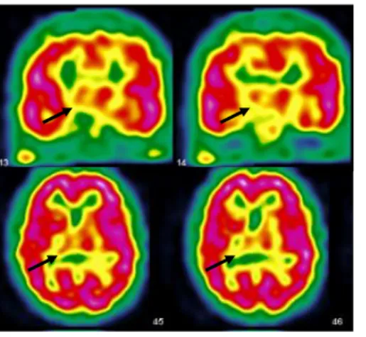
A first cerebral single photon emission computed tomography (SPECT, 99mTc-hexamethylpropylene-amineoxime) showed a hypoperfusion of the right thalamus (figure 1).
Logopedic assessmentLanguage examination showed a language disorder with anomia, dysprosodia, spasmodic dysphonia, and stuttering. Picture-naming tests were impossible due to visual deficit mimicking cerebral visual impairment, whereas reading and writing were not affected. Neuropsychological assessment showed attentional fluctuation (especially in the visual domain), memory impairment and dysexecutive syndrome with working memory impairment, flexibility disorders, and sensitivity to interference. A complex and atypical cerebral visual impairment was observed with a simultanagnosia, a left unilateral spatial neglect and mental imagery deficit. She only processed local information when presented with a visual scene. As a compensatory strategy, she followed the line of Rey’s figure point by point with her finger and hid some parts of the figure to limit visual interferences to perform the copy. Although during the copy of the Rey complex figure she was not able to process the global information, her reproduction did not display any spatial disorganization.
Psychiatric evaluation showed elements of histrionic personality disorder, such as seduction and immature behavior. Emotional numbing and affective discordance was reported, but no personality test was performed. No depression, hallucination or delusion was observed. She felt anxiety according to her accident without claiming any benefit or financial compensation. By resuming her medical history, an episode of atypical neurological disorder with hemiparesis in her adolescence was reported by a psychiatrist and labeled as FND. However, no more details were available and she has few memories of this episode.
Mis en forme : Barré
Mis en forme : Barré Mis en forme : Barré
Inconsistency, variability and evolution of symptoms without medical explanation permitted to diagnose FND according to the DSM-V criteria. Furthermore, risk factors were present, such as psychological and physical trauma (brother’s suicide and car accident) or elements of personality disorder (immaturity, seduction) not satisfying criteria of borderline or histrionic personality disorder.
At home, her daily life was virtually unimpaired. She only stopped driving and needed some help for shopping. One year after the symptoms onset, sleep attacks and speech disorders disappeared. However, the patient presented with atypical ‘blind’ behaviors. She used a cane, but she was able to apply make-up correctly and cleaned her apartment without assistance.
Visuoperceptive impairment was confirmed by speech and neuropsychological assessment. An atypical letter alexia without any deficit for words or sentences reading was observed. To recognize objects, she sometimes needed to manipulate them or she identified them from a detail (identification of the match box when seeing matches). Copying of geometric figure (Rey complex figure test) and tests involving visuospatial and graphomotor skills (crossing of test, Trail Making Test or BEC’s triangle) were not assessable because she reported an unbearable visual discomfort. She avoided seeing some objects, such as a striped shirt, because she reported a too “aggressive” feeling, which is suggestive of a visual intolerance to complex visual patterns and visual fatigability.
Ophthalmologic assessment with visual acuity, pupil reaction, ocular and fundoscopic examination, optical coherence tomography (OCT) of macula and optic nerve were normal. Static and kinetic visual field perimetric examinations were non interpretable because of the attention deficit. A second cerebral MRI was performed and again, no morphological lesion was visible.
A second SPECT two years after the symptoms-onset, while she presented with episodes of blindness and atypical cerebral visual impairments, showed hypoperfusion in the right temporo-occipital junction, known to be involved in global processing, right inferior parietal lobe, explaining the spatial and attentional deficits, and left superior frontal lobe (figure 2).
Mis en forme : Barré Mis en forme : Barré
Mis en forme : Barré
One year later, the cortical visual impairments persisted, as well as a clumsinessataxia
of left upper and lower limb. Whereas clinical state remained quite stable, the neurological follow-up is ongoing in order to not misdiagnose any neurological disease.
Figure 1: Cerebral SPECT showing a right thalamic hypoperfusion at the initial period of the patient history.
Figure 2: Second cerebral SPECT showing hypoperfusion in the right temporo-occipital junction, right inferior parietal lobe and left superior frontal lobe.
Discussion and conclusion
This case illustrates the variable symptomatology encountered in FND with the association of sensorimotor deficits, speech impairment, unilateral spatial neglect, a complex cerebral visual impairment and sleep attacks, probably corresponding to PNES. Frequently, FND semiology associates different domains and can vary over time with cognitive, behavioral, affective and even motor events, mainly movement disorders or PNES. This protean complex semiology, which corresponds to Sydenham’s “chameleon” syndrome, is sometimes difficult to diagnose. Furthermore, coexistence with an organic other neurological disorder pathology is not uncommon, especially PNES and epilepsy (7). Recent studies focus on “positive” clinical signs, such as Hoover sign, for paresis of lower limb: hip extension deficit disappears during contralateral hip flexion. They are based on internal inconsistency or incompatibility with recognized neurological or medical disease, and show good specificity and reliability (5,8). This is in agreement with recent modifications of the DSM-V regarding the definition of FND, including “positive signs” (9). However, the rate of misdiagnosis is still about 4 % (10).
The initial semiology of our patient, including sensorimotor deficit, ataxia in-coordinate limb movements, dysarthria, hemineglect, dysprosodia, dysarthria, sleep attacks, cortical visual impairments and unilateral spatial neglect, was concordant with a right thalamic syndrome (11,12). The initial SPECT showing a right thalamic hypoperfusion was coherent with this initial clinical presentation.
Subsequently, semiology and neuroimaging findings changed. Cortical visual impairments increased, while other symptoms were no longer present, suggesting a posterior cortical syndrome (13). A second SPECT examination showed a concordant right occipitoparietal hypoperfusion and no further right thalamic abnormalities.
Usually, paraclinical examinations have to be strictly normal in FND, but functional neuro-imaging can elicit abnormal cerebral pattern activation or metabolism. During the last decades, the number of publications increased; showing a growing interest of the scientific community for this clinical presentation. The first was in the nineties, showing hypoperfusion in the sensorimotor cortex contralateral to a functional hemiparesis (2,3). Since then, many functional dysfunctions have been reported in FND in various cortical and subcortical structures, including basal ganglia (14). The authors reported simultaneous anterior cingulate and medial prefrontal cortex hyperperfusions, suggesting an increased inhibition of the motor cortex, probably under influence of the limbic system, including motivational and emotional processes. Basal ganglia seem to also play a key role in FND. The physiological role of basal ganglia in the elaboration of the movement was found to be modified during FND, with an exaggerated monitoring of the limbic system interfering with motor pattern
(15,16). Numerous hypotheses emerged from neuroimaging studies, such as emotional dysregulation, memory suppression, and abnormal self-monitoring or sense of agency (17–21). In FND, amygdala is no more activated for fearful than happy stimuli; that is the rule for normal subjects and an impaired habituation for repetitive and similar stimuli, hippocampus activity decreased during recall of traumatic events and prefrontal cortex could be the witness of abnormal self-monitoring (17,19,22).
Furthermore mechanism of dissociation is commonly admitted in FND because it is associated with a high prevalence of dissociative disorders and symptoms (1,23,24). Dissociation is a process in which mental function as memory, consciousness… is disrupted. Both share common points in neuroimaging with cortical and subcortical structures implicated in emotion regulation such as prefrontal region (25,26).
However, no common neuroimaging pattern is identified in FND. This is probably because of the complexity of this disorder and the polymorphicproteiform clinical presentation. These findings support Jean-Martin Charcot’s point of view (1825-1893). He developed the concept of a “psychodynamic” lesion which is invisible in a macroscopic way and which can evolve, resulting in a varied symptomatology. A previous study showed modification of cerebral perfusion with an hypoperfusion in the thalamus and basal ganglia contralateral to functional sensorimotor deficit which disappeared after recovery (16). Our case illustrates an evolving and dynamic lesion with a proteiform variable neuroimaging pattern. First we had a right thalamic
hypoperfusion concordant with right thalamic syndrome and then, later, an evolving semiology with predominant neurovisual disorders with a concordant hypoperfusion in the occipito-parietal cortex.
Another originality of our report is the unusual complex central neurovisual disorders, suggesting posterior cortical dysfunction, which occurred during the follow-up (13). To our knowledge, this is the first report of a higher visual dysfunction of dorsal and ventral pathways in functional visual disorders (FVD), instead of the classical loss of acuity or a vision field loss. FVD represents 1-5 % of patients in ophthalmology clinics with loss of acuity, visual field abnormalities, diplopia, oscillopsia, ptosis and blepharospasm (27). The most frequent complaints are monocular vision loss, concentric loss of peripheral vision and spasm of near reflex in motor ocular disorder. They share the same demographic characteristics with other FNDs.
One study compares cerebral pattern activation of FVD versus healthy subjects and showed a reduced activation in the primary visual cortex, with increased activation in the posterior cingular cortex, basal ganglia, insula, and prefrontal cortex (4). This pattern differs from patients with neuro-organic opthalmologic lesion (28). Another study do not find FVD patterns in neuroimaging, but rather a neurophysiological modification of N1 event-related potential response during blindness that disappeared after recovery. (29). N1 response comes from the intraparietal sulcus and plays a role in spatial sustained attention to locate stimuli in the visual peripheral field. Misused dysfunction of these mechanisms may be involved in FVD. In our case, the fatigability and discomfort in front of complex visual stimuli, such as visual patterns, may correspond to visual attention disorders inducing visual detection deficit. Abnormal top-down control for spatial attention could contribute to the deficit, which is supported by hypoperfusion in occipito-parietal cortex seen on SPECT in our case.
Our patient presented with simultanagnosia and abnormalities seen in the right parietal lobe, which is concordant with the “dorsal stream” of the visual processing network (30,31). It is involved in the treatment of spatial data and movement. Its dysfunction leads to Balint syndrome including ataxia, ocular motor apraxia and simultanagnosia (32).
Our patient presented with visual associative agnosia, with preserved forms and color descriptions, but difficulties in the identification of visual objects requiring the use of another sensorial modality (tactile modality) to recognize items (33). Furthermore, she also presented with an atypical alexia without agraphia, which is frequently associated with visual agnosia. Alexia was considered as atypical because the deficit was on preponderant letter recognition and less impaired for words, which is usually the opposite (words more impaired than letters) or both items are impaired. Alexia without agraphia involved the left occipitotemporal pathway, including the visual word form area in the left fusiform gyrus specialized in word form recognition. The occipito-temporal hypoperfusion observed in our case was quite concordant with this agnosic and alexic semiology with a quite preserved left occipitotemporal pathway, which may explain the preserved word recognition. All these abnormalities lead to a dysfunction in the “ventral stream” of the visual processing, which is involved in the identification of objects (30,31).
Interestingly, our patient worked in an ordering service of an optician and developed neurovisual symptoms. According to Jean Martin Charcot and Pierre Janet, the notion of suggestibility is closely related to FND history. For a long time, social and cultural process was considered as a huge influencing factor in the ethiopathogenesis of FND and high degree of suggestibility seemed to be a characteristic. Other studies showed the opposite and the distinction between feigners and FND is not easy (34,35).
Our case raises the question about differential diagnosis such as malingering (36). Its definition is a patient who deliberately produces, feigns or exaggerates physical and/or psychological symptoms, motivated by external incentives such as avoiding military duty, work or obtaining financial compensation (9). Another possibility is factitious disorder, where there is no direct benefit except the medical condition itself
(9). For practitioners who are not familiar with these entities, highlighting clinical differences could be difficult. In FND, there is reproducible clinical signs like Hoover sign or drift without pronation, which are based on incompatibility between the symptom and recognized neurological or medical conditions (5,9). In functional movement disorder, electrophysiological criteria were identified based on variability, entrainment (i.e. external synchronization), distractibility and the presence of “bereitschaftpotential”, a cortical event seen before a voluntary movement, can be an
argument (37,38). In neuropsychological assessment, tests exist in order to distinguish feigners, measuring effort, response bias and symptom validity. These items are gathered in the battery of Symptom Validity Test (SVT). The capacity of these tests to distinguish conversion, factitious disorder and malingering are debated (39). The most pertinent test seems to be the forced choice SVT, where performances could be worse than expected. A negative response bias constitutes evidence for a strategic avoidance of correct answers. It shows intentionality in symptom production and is in favor of malingering. In FND, performances are inhomogeneous and fluctuant, and a negative bias response could be found at a lower level (40,41). Clinical signs, electrophysiological criteria and neuropsychological tests, as well as general context, help physicians to differentiate FND, malingering, factitious or organic disorder. However, there remains a lack of validated biomarkers of FND.
A functional neuroimaging study compares feigners and FND in sensorimotor deficit. A Go/no Go paradigm elicited an activation of the right inferior frontal gyrus during simulated weakness in feigners compared with motor FND (42). Other studies found an implication of the supplementary motor area, premotor cortex, cingulate cortex and right and medial prefrontal cortex during preparation and execution of the movement (43–46). Neural underpinning of feigners is clearly different from FND one, but it is not well described, possibly due to various paradigms and a weak number of subjects. No functional imaging defect was reported during malingering or feigners, supporting the diagnosis of FND and, in our case, presenting with hypoperfusions. These results suggest that functional imaging (especially hypoperfusions) may be an interesting biomarker to support diagnosis.
Obviously discussion based on a single subject study should be handle with caution. The SPECT could be over-readed considering non-specific hypoperfusions but anatomoclinical concordance in term of laterality, cortical and subcortical structures support our findings. Secondly, symptom validity test (SVT) was not performed initially so neuropsychological assessment should be interpreted with caution. However SVT failure was found in FND especially in PNES (47).
In conclusion, our report highlights 1/ the evolving proteiform neuroimaging patterns concordant with the variableproteiform semiology of FND, suggesting the charcot’s
Commenté [ss1]: Sweet et abuse, not financial, predicts non credible… drane, clin neurophyio 2012
“psychodynamic lesion”, 2/ the possibility of complex central neurovisual disorders suggesting posterior cortical dysfunction of dorsal and ventral pathways instead of more primary visual disorders (loss of acuity or visual field defect) in FVD and 3/ the potential role of functional neuroimaging as biomarkers in FND.
List of abbreviations
CT: Cerebral tomodensitometry
DSM: Diagnostic and statistical manual of mental disorders FND: Functional neurological disorder
FVD: Functional visual disorder
ICD: International classification of diseases MRI: Magnetic resonance imaging PET: Positron emission tomography PNES: Psychogenic non-epileptic seizure
SPECT: Single photon emission computed tomography SVT: Symptom validity test
Declarations
Ethics approval and consent to participate
Not applicable. Consent for publication
Written informed consent was obtained from the patient for publication of this case report and many accompanying images. A copy of the written consent is available for review by the Editor-in-Chief of his journal.
Availability of data and material
All data generated or analysed during this study are included in this published article. Competing interests
The authors declare that they have no competing interests. Funding
Not applicable. Authors’ contributions
SG was a major contributor in writing the manuscript. SG and EM gave the clinical information containing medical history, neurological and SPECT image findings. SA,
SC, TM and EM reviewed the manuscript. All authors read and approved the final manuscript.
Acknowledgments Not applicable.
Bibliography
1. Magnin E, Thomas-Antérion C, Sylvestre G, Haffen S, Magnin-Feysot V, Rumbach L. Conversion, dissociative amnesia, and Ganser syndrome in a case of “chameleon” syndrome: Anatomo-functional findings. Neurocase. 2014 Jan 2;20(1):27–36.
2. Marshall JC, Halligan PW, Fink GR, Wade DT, Frackowiak RS. The functional anatomy of a hysterical paralysis. Cognition. 1997;64(1):B1–8.
3. Tiihonen J, Kuikka J, Viinamäki H, Lehtonen J, Partanen J. Altered cerebral blood flow during hysterical paresthesia. Biol Psychiatry. 1995;37(2):134– 5.
4. Werring DJ, Weston L, Bullmore ET, Plant GT, Ron MA. Functional magnetic resonance imaging of the cerebral response to visual stimulation in medically unexplained visual loss. Psychol Med. 2004;34(04):583–9. 5. Daum C, Hubschmid M, Aybek S. The value of “positive” clinical signs for
weakness, sensory and gait disorders in conversion disorder: a systematic and narrative review. J Neurol Neurosurg Psychiatry. 2014 Feb
1;85(2):180–90.
6. Daum C, Aybek S. Validity of the “Drift without pronation” sign in conversion disorder. BMC Neurol. 2013;13(1):1–3.
7. Baroni G, Piccinini V, Martins WA, de Paola L, Paglioli E, Margis R, et al. Variables associated with co-existing epileptic and psychogenic
nonepileptic seizures: a systematic review. Seizure. 2016 Apr;37:35–40. 8. Daum C, Gheorghita F, Spatola M, Stojanova V, Medlin F, Vingerhoets F, et al.
Interobserver agreement and validity of bedside “positive signs” for functional weakness, sensory and gait disorders in conversion disorder: a pilot study. J Neurol Neurosurg Psychiatry. 2015 Apr;86(4):425–30. 9. American Psychiatric Association. Diagnostic and statistical manual of
mental disorders (5 ed). American Psychiatric Publishing. Washington DC; 2013.
10. Stone J. Systematic review of misdiagnosis of conversion symptoms and “hysteria.” BMJ. 2005 Oct 29;331(7523):989–0.
11. De Witte L, Brouns R, Kavadias D, Engelborghs S, De Deyn PP, Mariën P. Cognitive, affective and behavioural disturbances following vascular thalamic lesions: A review. Cortex. 2011 Mar;47(3):273–319. 12. Melo TP, Bogousslavsky J, Moulin T, Nader J, Regli F. Thalamic ataxia. J
Neurol. 1992;239(6):331–7.
13. Crutch SJ, Schott JM, Rabinovici GD, Murray M, Snowden JS, van der Flier WM, et al. Consensus classification of posterior cortical atrophy. Alzheimers Dement. 2017 Aug;13(8):870–84.
14. Boeckle M, Liegl G, Jank R, Pieh C. Neural correlates of conversion disorder: overview and meta-analysis of neuroimaging studies on motor conversion disorder. BMC Psychiatry [Internet]. 2016 Dec [cited 2016 Oct 31];16(1). Available from:
http://bmcpsychiatry.biomedcentral.com/articles/10.1186/s12888-016-0890-x
15. Schrag AE, Mehta AR, Bhatia KP, Brown RJ, Frackowiak RSJ, Trimble MR, et al. The functional neuroimaging correlates of psychogenic versus organic dystonia. Brain. 2013 Mar 1;136(3):770–81.
16. Vuilleumier P, Chicherio C, Assal F, Schwartz S, Slosman D, Landis T. Functional neuroanatomical correlates of hysterical sensorimotor loss. Brain. 2001 Jun 1;124(6):1077–90.
17. Aybek S, Nicholson TR, Zelaya F, O’Daly OG, Craig TJ, David AS, et al. Neural correlates of recall of life events in conversion disorder. JAMA Psychiatry. 2014 Jan;71(1):52–60.
18. Delange F, Roelofs K, Toni I. Increased self-monitoring during imagined movements in conversion paralysis. Neuropsychologia. 2007;45(9):2051–8.
19. Voon V, Brezing C, Gallea C, Ameli R, Roelofs K, LaFrance WC, et al. Emotional stimuli and motor conversion disorder. Brain. 2010 May 1;133(5):1526–36.
20. Voon V, Gallea C, Hattori N, Bruno M, Ekanayake V, Hallett M. The involuntary nature of conversion disorder. Neurology. 2010 Jan 19;74(3):223–8.
21. Saj A, Arzy S, Vuilleumier P. Functional Brain Imaging in a Woman With Spatial Neglect Due to Conversion Disorder. JAMA. 2009;302(23):2552–4. 22. Vuilleumier P. Brain circuits implicated in psychogenic paralysis in
conversion disorders and hypnosis. Neurophysiol Clin Neurophysiol [Internet]. 2014 Jan [cited 2014 Jun 11]; Available from: http://www.em-consulte.com/en/article/875278
23. Bell V, Oakley DA, Halligan PW, Deeley Q. Dissociation in hysteria and hypnosis: evidence from cognitive neuroscience. J Neurol Neurosurg Psychiatry. 2011 Mar 1;82(3):332–9.
24. Şar V, Akyüz G, Kundakçı T, Kızıltan E, Doğan O. Childhood trauma, dissociation, and psychiatric comorbidity in patients with conversion disorder. Am J Psychiatry. 2004;161(12):2271–6.
25. van der Kruijs SJ, Bodde NM, Carrette E, Lazeron RH, Vonck KE, Boon PA, et al. Neurophysiological correlates of dissociative symptoms. J Neurol Neurosurg Psychiatry. 2014;85(2):174–9.
26. Şar V, Seher U, Erdinc O. Frontal and occipital perfusion changes in dissociative identity disorder. Psychiatry Res Neuroimagin. 2007;156:217– 23.
27. Egan R, LaFrance W. Functional Vision Disorder. Semin Neurol. 2015 Oct 6;35(05):557–63.
28. Werring DJ, Bullmore ET, Toosy AT, Miller DH, Barker GJ, MacManus DG, et al. Recovery from optic neuritis is associated with a change in the
distribution of cerebral response to visual stimulation: a functional magnetic resonance imaging study. J Neurol Neurosurg Psychiatry. 2000;68(4):441–9.
29. Schoenfeld MA, Hassa T, Hopf J-M, Eulitz C, Schmidt R. Neural Correlates of Hysterical Blindness. Cereb Cortex. 2011 Oct 1;21(10):2394–8.
30. Milner AD. How do the two visual streams interact with each other? Exp Brain Res [Internet]. 2017 Mar 2 [cited 2017 Mar 21]; Available from: http://link.springer.com/10.1007/s00221-017-4917-4
31. Milner AD, Goodale MA. The visual brain in action. Oxford University Press; 2006. (Oxford psychology series).
32. Pisella L, Biotti D, Vighetto A. Combination of attentional and spatial working memory deficits in Bálint-Holmes syndrome: Attention versus spatiotemporal integration. Ann N Y Acad Sci. 2015 Mar;1339(1):165–75. 33. Lissauer H. Ein Fall von Seelenblindheit nebst einem Beitrage zur Theorie
derselben. Arch Für Psychiatr Nervenkrankh. 1890 Jun 1;21(2):222–70. 34. Foong J, Lucas PA, Ron MA. Interrogative suggestibility in patient with
conversion disorders. J Psychosom Res. 1997;43(3):317–21.
35. Gudjonsson GH. Historical background to suggestibility: How interrogative suggestibility differs from other types of suggestibility. Personal Individ Differ. 1987;8(3):347–55.
36. Galli S, Tatu L, Bogousslavsky J, Aybek S. Conversion, Factitious Disorder and Malingering: A Distinct Pattern or a Continuum? In: Neurologic-Psychiatric Syndromes in Focus-Part II. Karger Publishers; 2018. p. 72–80. 37. Apartis E. Clinical neurophysiology of psychogenic movement disorders:
How to diagnose psychogenic tremor and myoclonus. Neurophysiol Clin Neurophysiol. 2014 Oct;44(4):417–24.
38. Schwingenschuh P, Saifee TA, Katschnig-Winter P, Macerollo A, Koegl-Wallner M, Culea V, et al. Validation of “laboratory-supported” criteria for functional (psychogenic) tremor: Laboratory-Supported Criteria for Functional Tremor. Mov Disord. 2016 Apr;31(4):555–62.
39. Delis D, Wetter S. Cogniform Disorder and Cogniform Condition: Proposed diagnoses for excessive cognitive symptoms. Arch Clin Neuropsychol. 2007 Jun;22(5):589–604.
40. Cragar DE, Berry DTR, Fakhoury TA, Cibula JE, Schmitt FA. Performance of Patients with Epilepsy or Psychogenic Non-Epileptic Seizures on Four Measures of Effort. Clin Neuropsychol. 2006 Sep;20(3):552–66.
41. Kemp S, Coughlan AK, Rowbottom C, Wilkinson K, Teggart V, Baker G. The base rate of effort test failure in patients with medically unexplained symptoms. J Psychosom Res. 2008 Oct;65(4):319–25.
42. Cojan Y, Waber L, Carruzzo A, Vuilleumier P. Motor inhibition in hysterical conversion paralysis. NeuroImage. 2009 Sep;47(3):1026–37.
43. Hassa T, de Jel E, Tuescher O, Schmidt R, Schoenfeld MA. Functional networks of motor inhibition in conversion disorder patients and feigning subjects. NeuroImage Clin. 2016;11:719–27.
44. Spence SA, Crimlisk HL, Cope H, Ron MA, Grasby PM. Discrete
neurophysiological correlates in prefrontal cortex during hysterical and feigned disorder of movement. The Lancet. 2000;355(9211):1242–3.
45. Stone J, Zeman A, Simonotto E, Meyer M, Azuma R, Flett S, et al. fMRI in Patients With Motor Conversion Symptoms and Controls With Simulated Weakness: Psychosom Med. 2007 Nov;69(9):961–9.
46. van Beilen M, Vogt BA, Leenders KL. Increased activation in cingulate cortex in conversion disorder: What does it mean? J Neurol Sci. 2010 Feb;289(1-2):155–8.
47. Williamson DJ, Holsman M, Chaytor N, Miller JW, Drane DL. Abuse, not financial incentive, predicts non-credible cognitive performance in patients with psychogenic non-epileptic seizures. Clin Neuropsychol.