HAL Id: hal-01824408
https://hal.umontpellier.fr/hal-01824408
Submitted on 27 Jun 2018HAL is a multi-disciplinary open access archive for the deposit and dissemination of sci-entific research documents, whether they are pub-lished or not. The documents may come from teaching and research institutions in France or abroad, or from public or private research centers.
L’archive ouverte pluridisciplinaire HAL, est destinée au dépôt et à la diffusion de documents scientifiques de niveau recherche, publiés ou non, émanant des établissements d’enseignement et de recherche français ou étrangers, des laboratoires publics ou privés.
Olivier Cazorla, A Freiburg, M. Helmes, T Centner, M Mcnabb, Yiming Wu,
K Trombitás, S Labeit, Henk Granzier
To cite this version:
Olivier Cazorla, A Freiburg, M. Helmes, T Centner, M Mcnabb, et al.. Differential Expression of Cardiac Titin Isoforms and Modulation of Cellular Stiffness. Circulation Research, American Heart Association, 2000, 86 (1), pp.59-67. �10.1161/01.RES.86.1.59�. �hal-01824408�
Differential expression of cardiac titin isoforms and
modulation of cellular stiffness.
(short title: Cazorla et al. Titin isoforms and cellular stiffness)
Cazorla, O.
1, Freiburg, A
2, Helmes, M.
1, Centner, T.
2, McNabb, M.
1, Wu, Y.
1,
Trombitás, K.
1, Labeit, S.
2, and Granzier, H.
11Department of Veterinary and Comparative Anatomy, Pharmacology and Physiology Washington State University, Pullman, WA, 99164-6520, USA
2European Molecular biology Laboratory, Meyerhofstrasse 1, P.O.Box 102209, 69012 Heidelberg, Germany.
Correspondence and reprint requests: Henk Granzier
Department of Veterinary and Comparative Anatomy, Pharmacology and Physiology Wegner Hall, 205
Washington State University, Pullman, WA, 99164-6520 USA
Phone: (509)-335-3390 Fax: (509)-335-4650 E-mail: granzier@wsunix.wsu.edu
Abstract
Extension of the I–band segment of titin gives rise to part of cardiac muscle’s diastolic force. Previous studies of human cardiac titin transcripts suggested a series of differential splicing events in titin`s I–band segment leading to the so-called N2A and N2B isoform transcripts. Here we investigated titin expression at the protein level in a wide range of mammalian species. Results indicate that the myocardium co-expresses two distinct titin isoforms: a smaller isoform containing the N2B element only (N2B titin), and a larger isoform with both the N2B and N2A elements (N2BA titin).
The expression ratio of large N2BA to small N2B titin isoforms was found to vary greatly in different species, e.g. in the left ventricle the ratio is ~0.05 in mouse and ~1.5 in pig. Differences in the expression ratio were also found between atria and ventricles, and between different layers of the ventricular wall. Immunofluorescence experiments with isoform-specific antibodies suggest that co-expression of these isoforms takes place at the single myocyte level. The diastolic properties of single cardiac myocytes isolated from various species expressing high levels of the small (rat and mouse) or large (pig) titin isoform were studied. On average pig myocytes are significantly less stiff than mouse and rat myocytes. Gel analysis indicates that this result can not be explained by a varying amounts of titin in mouse and pig myocardium. Rather, low stiffness of pig myocytes can be explained by its high expression level of the large isoform: the longer extensible region of this isoform results in a lower fractional extension for a given sarcomere length and hence a lower force. Implications of our findings to cardiac function are discussed.
Key words: Compliance, passive tension, diastolic force, mechanical properties, myocyte, connectin.
Introduction
During diastole, the myocardium stretches and passive force is generated. A major contributor to this force is the giant protein titin, spanning the half sarcomere from Z-band to M-line. When sarcomere length (SL) increases during diastole, titin’s I-band region extends and passive force (F) develops. The shape of titin’s F–SL relation is expected to influence ventricular filling during diastole and ventricular emptying during systole. In addition to influencing ventricular filling, titin also helps to maintain the structural integrity of the contracting sarcomere (for reviews see 1- 5).
Titin’s force arises from its extensible I-band region which consists of two main segment types: 1) a segment rich in proline (P), glutamate (E), valine (V) and lysine (K) residues (the so-called PEVK segment) and 2) serially linked immunoglobulin-like domains (tandem Ig segments) flanking this PEVK segment.6 Several titin isoforms are now known, all of which contain PEVK and tandem Ig segments. In addition to these common segments the extensible region of the N2B isoform contains the N2B element (3 Ig domains and a 572- residue unique sequence) while the N2A class of isoforms contains the N2A element (4 Ig domains and a 106- residue unique sequence). N2B titin is found exclusively in cardiac muscle and contains within its central I-band region a 163 residue PEVK segment and tandem Ig segments with 37 Ig domains. N2A titins are found in skeletal muscles with PEVK and tandem Ig segments that differ in length in different muscles.6 Heart muscle expresses not only N2B transcripts but also N2A transcripts; the latter are associated with a larger PEVK segment than the N2B transcripts.6 Here we investigated cardiac titin isoform expression by high resolution SDS-PAGE and by various immuno-labeling techniques (western blotting, immunoelectron microscopy and immunofluorescence) with isoform-specific antibodies.
In slack sarcomeres titin’s extensible segment has a short end-to-end length as a result of thermally-induced bending motions that lead to a state of maximal entropy.7, 8 Straightening of the extensible region by extending the sarcomere lowers the conformational entropy and results in a force, known as entropic force. The extensible region of titin does not straighten uniformly, rather tandem Ig and PEVK segments extend sequentially.9-11 In addition, it was recently found that the 572 residue unique sequence of the N2B element is extensible as well, explaining why the extensible region of N2B titin can be stretched to lengths much longer than the combined contour length (end-to-end-length when completely straight) of the tandem Ig and PEVK segments.12,13 Variation in the length of the extensible region is predicted to influence the developed force upon extension. Cardiac myocytes that express a larger isoform (referred to below as N2BA titin) have
a longer extensible region and are predicted to develop less force than those that express N2B-titin, as for a given SL the fractional extension (end-to-end length of extensible region divided by its contour length) of N2BA titin will be less than that of the N2B isoform (for details see Discussion).
Here we investigated titin expression at the protein level. A number of mammalian species were surveyed for the expression of different cardiac titin isoforms. SDS-PAGE revealed that in most species both small and large isoforms are co-expressed, but in widely varying ratios. Western blot studies with N2B and N2A antibodies suggest that the smaller isoform contains the N2B element (N2B titin), whereas the larger isoform contains both the N2B and N2A elements (N2BA titin). To test the hypothesis that the expression of the N2BA isoform leads to a decrease in cell stiffness, mechanical experiments were performed on single cardiac myocytes isolated from hearts showing a high or low N2BA expression level. The results indicate that expressing small and large titin isoforms at different ratios is a means to modulate cardiac myocyte stiffness.
Material and Methods
Cardiac myocyte isolation. Ventricular and atrial myocytes were isolated from rat, mouse,
and pig by digesting their extracellular matrix with collagenase and hyaluronidase. The protocol to isolate cells from rat has been explained earlier14, the protocol for mouse and pig was adapted from Wolska 15. For isolating pig atrial cells we used the blending method developed by Moss 16. The myocytes were chemically skinned as previously described.14 To prevent degradation, all solutions contained protease inhibitors (see 14). See Material and Methods online.
Gel electrophoresis. Myocardial and skeletal muscle samples were quick-frozen in liquid
nitrogen, pulverized and then rapidly solubilized.14 The samples were analyzed with SDS-PAGE (2-9.5% acrylamide gradient gels) and Coomassie Blue stained. Titin and myosin heavy chain (MHC) bands were analyzed as previously described 14 with some adaptations (see Material and
Methods online).
Western blotting. Western blotting was performed with affinity purified anti-titin antibodies
(raised in rabbit) specific to the N-terminal end of titin (Z1/Z2 and Zr), the C-terminal end of titin (T51), the C-terminus of the PEVK segment (bk283/4), N2A (X105-X106) and N2B (X150-X151, X214-X215 and X216-X217).8 (For details see Fig. 3A and Material and Methods online.)
Immunofluorescence. Single or double labeling experiments with PEVK (9D10) 17 and N2B
(X151-X151) or N2A (X105-X106) antibodies were performed on cardiac myocytes. (For details see Material and Methods online.)
Passive tension – SL measurement of cardiac myocytes. For technical details of force and
on-line sarcomere length measurement see 12,14. The passive force – SL relationship of the cells was measured by imposing a slow ramp stretch-release (0.1 length/s) on cells in relaxing solution.
Electron microscopy. Electron microcopy was used to obtain cross-sections of skinned cells to
determine the myofibrillar fractional area. Immunoelectron microscopy was used to study rat and pig left ventricular cells labeled with N2A (X105-106) and N2B (X150-151) antibodies 8,18.
Results
SDS-PAGE analysis. Comparative gel electrophoresis of human skeletal muscle (soleus) and
myocardium of rat, rabbit, bovine and human confirmed earlier findings and provided novel results as well. Confirming earlier findings, skeletal muscle and rat myocardium contain a single major titin band at the top of the gel (T1 in Fig. 1, left two lanes), representing the full-length titin molecule 8. T1 mobility greatly varies, reflecting the difference in molecular mass of the 3.7 MDa soleus titin and the 2.97 MDa rat cardiac titin.6 The samples also reveal a minor band with higher mobility (T2) derived from a large titin degradation product that contains the A-band segment of titin.8 The T2 band is barely detectable in samples carefully prepared to minimize degradation (Fig. 1). Novel findings were obtained when using myocardium of rabbit, bovine and human. These species contain two T1 bands, a bottom band which co-migrates with the T1 band of rat and a top band with clearly reduced mobility (Fig. 1). The top T1 is just visible in rabbit (Fig. 1 inset) but easily detectable in bovine and human. The fact that two T1 bands in cardiac muscle have gone unnoticed in our earlier works, is because our previous studies focussed on rat and rabbit cardiac titin and in these species the top T1 band is minor or absent. Limited gel resolution is another explanation why two T1 bands may have been missed in our previous studies. Only large pore gels with high spatial resolution allow the two T1 bands in bovine and human to be clearly separated.
To follow up on these findings we surveyed additional species, examined ventricular and atrial myocardium in some of them, and determined the ratio of the top to bottom T1 bands using quantitative densitometry. Two T1 bands were consistently found in many species, but the density of the bands varied greatly. Rat, rabbit and mouse myocardium contain barely detectable levels of the top T1 band, in dog and human the two bands are present at similar levels, and in pig and cow the top T1 band dominates (Figs. 2A and B). Furthermore, in the two species in which we examined both left ventricular and left atrial myocardium, the expression ratio of top to bottom band is much higher in the atrium than in the ventricle (Figs. 2A and B). For example, in the left ventricle of the cow the top T1 band is 1.8 times more abundant than the bottom band, while in the left atrium the top band is 21 times more abundant. Finally, we also studied titin expression in the sub-endocardial, mid-wall and sub-epicardial regions of the ventricular free wall of the pig. Significantly higher levels of the large isoform were found in the sub-endocardium (Fig. 2C).
Immuno-labeling experiments. Using the mobility of human soleus T1 (Mr 3.7 MDa) and rat cardiac titin T1 (Mr 2.97 MDa) as standards suggests that the two T1 bands of pig differ by ~0.35 MDa in mass. To test whether both T1 bands represent full-length titin molecules (and not degradation products), western blot experiments were performed with antibodies against the terminal ends of the molecule (Fig. 3A). The presence of the N-terminal end of titin was investigated with Z1/Z2 and Zr antibodies (Z1/Z2 labels the 200 first N-terminal amino acid residues and Zr labels epitopes ~ 500 residues from the N-terminus, 19) and the presence of titin’s C-terminal end with T51 antibody (labels ~200 residues from the C-terminus, 20). Western blot results show that all antibodies reacted with the two T1 bands (Fig. 3B). Thus it is likely that the two T1 bands represent distinct full-length titin molecules.
Cardiac muscle differentially expresses transcripts, which code for the N2B and N2A elements, respectively.6 To determine the identity of the titin bands seen on SDS gels, western blot studies were performed using a library of antibodies raised against sequences from the N2B and N2A elements (see Fig. 3A). These antibodies were first tested on skeletal muscle titin (human soleus) which is known to contain only N2A titin 6 and, as expected, the N2A-element specific antibody reacted with skeletal muscle titin, while the N2B-element specific antibodies did not (Fig. 3C). Western blot experiments were also performed using pig myocardium, because it contains high levels of both T1 bands (Fig. 2). The top T1 band reacted with both N2A and N2B specific antibodies while the bottom T1 band reacted only with the N2B antibody (Fig. 3D). These results are consistent with the top T1 band representing a titin isoform containing both the N2B and N2A elements (referred as N2BA cardiac titin) and the bottom band an isoform that contains only the N2B element (N2B cardiac titin).
To investigate titin in the sarcomere, immunoelectron microscopy was performed on pig and rat ventricular cells using N2A- and N2B-specific antibodies. Pig cells were positive for both N2B and N2A antibodies and rat cells only for the N2B-specific antibodies (Fig. 3E). These results are consistent with the conclusion that pig myocardium expresses the N2BA isoform and rat myocardium predominately the N2B isoform. Furthermore, in the pig cells the N2B epitope was on average closer to the Z-line than the N2A epitope. For example at a SL of 2.45 µm the distance between the epitope and the middle of the Z-line was 175 ± 21 nm (n=9) for the N2B epitope and 279 ± 15 nm (n=10) for the N2A epitope. Thus, in the N2BA isoform the N2B element is
N-terminal of the N2A element. This result is consistent with recent sequencing work on cardiac titins (Freiburg A. and Labeit S., unpublished results, 1999).
Immuno-fluorescence (IF). The SDS-PAGE and westernblot results were performed at the
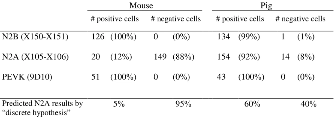
tissue level and the results may be explained either by co-expression of isoforms in cells, or by discrete expression in cells (i.e., cells either express N2B or N2BA titin, but not both) with species variation in the ratio of cells that are N2B or N2BA pure. To distinguish between co-expression and discrete expression, IF studies were carried out on single cardiac myocytes. Considering that only N2BA titin reacts with the N2A-element specific antibodies, discrete expression predicts that a certain fraction of cells (N2B cells) will not stain with this antibody. This fraction can be estimated from the expression ratio of N2B and N2BA titins determined by solubilizing a large number of cells from the cell suspension used for IF, followed by electrophoresis and quantitative densitometry. Results indicate that 60 ± 5% of total titin in pig left ventricle is N2BA titin and 40 ± 7% is N2B titin (Table 1). Thus, the discrete expression hypothesis predicts that only 60% of the pig cells stain with the N2A antibodies. Antibodies specific for the N2B element are predicted to stain all pig cells. For a comparison mouse myocytes were studied as well. Considering that mouse myocardium contains a barely detectable N2BA band (Fig. 2), discrete expression predicts that only few mouse cells are positive for the N2A antibodies while all cells are predicted to be positive for the N2B-element specific antibodies.
Experiments with the N2B antibodies revealed that all pig and mouse cells were positive (Table 1; Figs. 4A and B), consistent with the results of the above discussed western blot studies. When using the N2A antibodies, we obtained positive cells (Figs. 4C, E), and cells that contained a faint background-fluorescence level without a clear banding pattern (Figs. 4D, F) similar to that obtained in control experiments in which the primary antibodies were omitted from the labeling protocol (no-primary antibody control). To test whether absence of labeling with the N2A antibodies may have resulted from titin degradation or inability of the N2A antibody to penetrate some of the cells, experiments with the anti-titin antibody 9D10 were performed. 9D10 labels the PEVK region of titin 11, a site which is sensitive to degradation.21 Furthermore, 9D10 is an IgM type antibody 17, which is the largest of all antibody types and, therefore, 9D10 is expected to diffuse slowly into the cells. Results revealed that all cells were 9D10 positive (Figs. 4G, I; Table
1), suggesting that antibodies can fully penetrate the cells and that all cells contain intact titin. Therefore, cells that gave rise to a fluorescence similar to that of ‘no-primary antibody control cells’ were scored as N2A negative cells.
It was found that 92% of the pig cells are positive for the N2A antibody (Table 1), a value much higher value than the 60 ± 5 % positive cells predicted by the discrete expression hypothesis (cells express only one of the isoforms). On the other hand, the co-expression model (all cells co-express isoforms at the same ratio) predicts that all pig cells would be N2A positive while we found that 8% of pig cells are N2A-negative. These findings may be explained by co-expression that varies from cell-to-cell with a small fraction of the cells expressing predominantly one of the isoforms.
Diastolic properties of cardiac myocytes. To investigate the functional significance of
co-expressing titin isoforms, mechanical experiments were performed on single cardiac myocytes and their passive force - SL relation was measured. The force generated by the intermediate filament network was determined by extracting the thin and thick filaments from the myocytes using KCl and KI containing relaxing solutions (see 14). Extraction removes titin as a passive force generator as the extraction-induced depolymerization of actin and myosin removes titin’s anchors in the sarcomere. The force after extraction was subtracted from the force before extraction, resulting in data that represent the force produced by titin alone. This force was converted to tension (T) by dividing it by the cross-sectional area of the cell. The cells studied had been isolated by either blending myocardial tissue (cf. 16) or by enzymatic digestion of the heart (see captions of Figures). When ventricular cells isolated by blending or digestion were compared (data not shown), no statistically significant differences were found in either the titin content or the titin-based stiffness of the cells.
Titin-based T-SL curves of 15 mouse and 10 pig ventricular cells are shown in figure 5. Results indicate that the average stiffness of the mouse myocytes is much higher than that of the pig (inset Fig. 5). To test for statistical differences between mouse and pig cells, the tension developed at a SL of 2.4 µm was compared using a Mann-Whitney test (this test was used because results were not normally distributed). This revealed that mouse ventricular cells are significantly stiffer than pig myocytes (p<0.05). To determine whether the differences in stiffness may have resulted from differences in the fractional area of myofibrils, cells were prepared for electron microscopy and their cross-sections were analyzed. Results revealed that this explanation is unlikely because mouse and pig cells contain similar fractional areas of myofibrils (Table 2). We also studied
whether differences in the cellular content of titin could explain the mechanical differences between pig and mouse cells. The ratio of titin relative to myosin heavy chain (MHC) was determined using quantitative densitometry (for details see Methods). It was found that the titin/MHC ratios of mouse and pig cells are not significantly different (Table 2). The titin/MHC ratio was also used to determine the number of titin molecules per half-thick filament (for details see Methods). Results indicate 6.0 ± 0.3 and 6.5 ± 0.4 titin molecules per half-thick filament in mouse and pig cells, respectively (Table 2). Thus a difference in the titin content of mouse and pig cells is an unlikely explanation for their different passive properties.
It may be noted from Fig. 5 that a few of the pig cells are as stiff as most of the mouse cells while one of the mouse cells is as compliant as most pig cells. Although experimental error may underlie part of this variation, differences in cell behavior may also result from cell to cell variation in the expression ratio of N2B and N2BA titins. For example, the compliant mouse cells may express high levels of N2BA titin. To further explore cell-to-cell variation in cell stiffness, cells isolated from pig atrium and rat ventricle were also studied. Results indicate that most of the pig atrial cells are compliant (i.e. their T-SL curve are shallow) and that a few cells are rigid and behaved like the average mouse cell (Fig. 6 red curves). In contrast, rat cardiac cells were all stiff (Fig. 6). Considering that the rat expresses predominantly N2B titin, cell-to-cell variation appears more prominent in cells isolated from tissue that co-expresses both titin isoforms.
Discussion
We found that the myocardium co-expresses two titin isoforms. One of the isoforms contains the N2B element and the other both the N2B and N2A elements. The expression ratio of these isoforms varies in different species and in different locations within the heart. Furthermore, results suggest that co-expression of isoforms takes place at the level of the single cell. The mechanical properties of single cardiac myocytes were measured and this revealed that on average cells isolated from a species that contains high levels of N2BA titin (pig) are significantly less stiff than those from species that contain high levels of N2B titin (rat and mouse). Below we discuss these findings and their functional significance.
Cardiac titin isoforms. Previous RT-PCR studies of human cardiac titin revealed the
existence of N2A and N2B isoform transcripts.6 Here we studied isoform expression at the protein level. SDS-PAGE indicates that myocardium of many species contains two T1 bands, a top band which reacts with N2A- and element specific antibodies and a bottom band with only N2B-specific antibodies. That the two T1 bands represent full-length titin molecules is supported by western blot experiments with antibodies against the N-terminal end (Z1/Z2 and Zr) and the C-terminal end (T51) of titin. This showed that both T1 bands of pig are full-length molecules and not degradation products. Absence of titin degradation is also supported by the IEM data. Degradation results in retraction of titin towards the Z-line and the A-band 22, and no such retraction was found in the cells that we investigated. Hence we conclude that the two T1 bands represent distinct isoforms. This conclusion is in accordance with recent RT-PCR studies using rabbit cardiac mRNA which revealed that differential splicing gives rise to two main classes of cardiac titin isoforms: the N2B and N2BA titins; so named because they contain either the N2B element or both the N2B and N2A elements (Freiburg, A. and Labeit S., unpublished results, 1999). Cardiac N2B titin is the smallest isoform known to date and the small size results from skipping the I20-I75 segment and splicing together the I19 to I76 encoding exons. The splicing of the exon coding for I19 to exons in the I47-I60 segment leads to a family of isoforms, all including the N2A segment, and thus these isoforms are referred to as N2BA titins.
Due to the sequence differences, the molecular mass of the cardiac titin isoforms varies from ~3.3 MDa for the N2BA titin isoform to ~3.0 MDa for N2B titin (6; Freiburg, A. and Labeit S., unpublished results, 1999). Our current work indicates that this mass difference is large enough to allow separation of N2B and N2BA titins on high resolution gels (Figs. 1-3). This enables
studies of the expression levels of the isoforms in different species and in different locations within the heart. Results establish that co-expression of isoforms takes places in a wide range of species, but at widely varying ratios, and that atria express higher N2BA levels than the ventricles. The quantitative densitometric study of titin in mouse and pig (expressing high levels of N2B and N2BA titin, respectively) reveals that while the expression ratio of titin isoforms varies greatly, the number of titin molecules per half-thick filament appears constant (Table 2). Thus, titin’s stoichiometry is well controlled while the isoform expression ratio is variable.
Co-expression of titin isoforms at the single cardiac myocyte level. The titin isoform
co-expression results were obtained with SDS-PAGE and western blot techniques using myocardial tissue as starting material. Co-expression of isoforms at the tissue level can be explained by assuming (1) that individual cells express only one of the isoforms (discrete expression hypothesis) and (2) that the ratio of N2B cells to N2BA cells determines the isoform expression ratio of the myocardial tissue. However, we found that the ratio of positive and negative cells is inconsistent with a discrete expression of isoforms (Table 1, see Results). Instead results are consistent with a co-expression of titin isoforms within individual cells. The small fraction of negative cells can be explained by cell-to-cell variation in the co-expression ratios resulting in some cells with predominantly one isoform. This ‘variable co-expression’ hypothesis is also consistent with recent immunoelectron microscopical studies that revealed co-expression of isoforms within the same I-band and cell-to-cell variation in the co-expression level (Trombitás, K., Labeit, S., Granzier, H., 1999. Unpublished data). In summary, our findings indicate that the titin isoform co-expression seen at the myocardial level results from co-expression at the level of the single cardiac myocyte.
Diastolic properties of cardiac myocytes. Whether diastolic properties of N2B cells are
predicted to be different from those of N2BA cells can be ascertained from the molecular mechanism of titin-based passive force development. The molecular mechanism that underlies titin’s force has been investigated in dynamic light scattering studies on titin in solution 23, mechanical studies on single titin molecules 24;-27, and immunoelectron microscopic studies on skeletal and cardiac muscle titin 11; 28; 29. From these, a model has emerged in which the tandem-Ig segments (containing folded tandem-Ig domains) and the PEVK segment (acting largely as an unfolded polypeptide) behave as serially-linked entropic springs. In short sarcomeres, these springs are in a contracted state with high entropy and upon sarcomere extension the springs straighten, lowering
their conformational entropy and resulting in a force, known as entropic force. This force increases with the fractional extension of the segment (end-to-end length divided by the contour length). (see 8).
The serially-linked entropic springs model of passive force development may be applied to size variants of titin's elastic segment by adapting the entropic forces to the fractional extensions multiplied by the contour lengths of the size variant's tandem Ig and PEVK segments. The contour lengths of tandem Ig and PEVK segments are ~100 and ~250 nm longer, respectively, in N2BA than in N2B titin (assuming a 5 nm repeat per Ig domain and 3.8Å per PEVK residue, see 11, 12). It follows that at a given SL the fractional extension of tandem Ig and PEVK segments is considerably less for N2BA titin than for N2B titin and, therefore, passive force will be lower. This prediction is qualitatively unaffected by the extensibility of the unique N2B sequence 12 because both cardiac titin isoforms contain this sequence. In conclusion, the serially-linked entropic springs model of passive force development predicts that the passive force-SL relation increases less steeply (i.e., the compliance is higher) for N2BA containing cells than for N2B cells.
We studied the passive tension – SL relation of single cardiac myocytes isolated from mouse, rat and pig myocardium. The mouse and rat were chosen because their cardiac myocytes are predicted to be stiff (they express high levels of N2B titin), and the pig because its cells are predicted to be compliant (high levels of N2BA titin). Consistent with the predicted tension differences, pig myocytes are on average significantly more compliant than rat and mouse cells (Figs. 5 and 6). Considering the similar fractional cell areas of myofibrils and the similar number of titin molecules per half thick filament (Table 2), it is unlikely that these mechanical results can be explained by differences in the amount of titin per unit cross-sectional cell area. Rather it is likely that these mechanical differences result from within titin itself. It is also worthwhile to point out that studies performed more than 20 years ago by Fabiato and Fabiato 30 are consistent with our findings. In this earlier work, it was shown that dog myocytes are more compliant than rat myocytes, consistent with the high levels of N2BA titin that we found in dog myocardium and high levels of N2B titin in rat (Fig. 2). Finally, Brady 31 has reported the passive stiffness modulus (normalized to the cross-sectional area) of myocytes from several species and this revealed lower stiffness in rabbit than in rat. This finding is in agreement with the higher N2BA levels of rabbit (Fig. 3).
Mechanical results of cells isolated from the same ventricle often showed considerable variation (Figs. 5 and 6). Although some of this variation may result from experimental error (such as errors in the cross-sectional area measurement), it seems more likely that additional sources of variation exist as well. It is also worth noting that variation in mechanical properties has been reported in intact guinea-pig ventricular cells by Gannier et al 32 and Cazorla et al 33 with a tendency of the cells to separate into stiff and compliant sub-populations. Variation in cellular stiffness is consistent with the variable co-expression hypothesis that was derived from the immunofluorescence study (see above). Variation in the titin isoform co-expression ratio of cardiac myocytes is expected to give rise to variation in the level of passive tension for a given SL with an increase in cell compliance as the N2BA expression level increases.
In summary, our mechanical studies reveal cell-to-cell variation in cell stiffness, but on average cells isolated from myocardium that expresses high levels of N2B titin are stiff and those from myocardium that expresses N2BA titin are more compliant. Thus, the diastolic properties of cardiac myocytes isolated from different species are not the same, but instead they vary with the expression ratio of the titin isoforms.
Functional significance. Work on rat cardiac muscle has shown that both titin and collagen
are major contributors to passive stiffness of the myocardium, with titin’s contribution dominating at the short to mid-range of the physiological SL range and collagen contributing more at longer lengths 14. Although comparative muscle studies in different species remain to be done, it is likely that variation in titin-based stiffness of the myocytes will translate into variation in stiffness at the level of the myocardium. Thus, we speculate that as the expression level of N2BA titin increases and cell stiffness decreases, myocardial stiffness will decrease as well. Variation in myocardial stiffness is expected to influence filling of the heart. For example, lower myocardial stiffness will allow for faster filling and larger end-diastolic volumes for a given filling pressure. The variation in expression levels of titin isoforms in different species may thus be related to variation in filling rate and/or filling volume of the heart. We speculate that the cell-to-cell stiffness variation within species is related to strain equalization of muscle fibers in different layers of the wall. Consistent with this explanation is the variation in the N2BA/N2B isoform expression ratio that was found in different layers of the ventricular wall (Fig. 2C).
In conclusion, this work revealed that the myocardium co-expresses titin isoforms and that the expression ratio of the isoforms varies between species and within a species in different locations
of the heart. Co-expressing titin isoforms at different levels modulates cellular stiffness and we hypothesize that this influences filling of the heart. Research at the multicellular and organ levels will be required to test this hypothesis and to fully elucidate the functional significance of the present findings.
Acknowledgments.
This work was supported by grants from NIHLBI (HL61497 and HL62881) to HG, the Human Frontier Science Program (SL) the Deutsche Forschungsgemeinschaft (La668/5-1 to SL) and a postdoctoral fellowship awarded to OC from the American Heart Association (Washington State Affiliate, #98-WA-115). HG is an Established investigator of the American Heart Association.
References
1. Trinick J. Titin as a scaffold and spring. Cytoskeleton. Curr Biol. 1996;6:258-260.
2. Wang K. Titin/connectin and nebulin: giant protein rulers of muscle structure and function.
Adv Biophys J. 1996;33:123-134.
3. Labeit S, Kolmerer B, Linke WA. The giant protein titin. Emerging roles in physiology and pathophysiology. Circ Res. 1997;80:290-294.
4. Maruyama K. Connectin/titin, giant elastic protein of muscle. FASEB J. 1997;11:341-345. 5. Gregorio C, Trombitas K, Kolmerer B, Stier G, Granzier H, Kunke K, Suzuki K, Obermayr
F, Herrmann B, Sorimachi H, Labeit S. The NH2-terminus of titin spans the Z-disc: its interaction with a nevel19-kDa ligand (T-cap) is required for sarcomeric integrity. J Cell
Biol. 1999;143:1013-1027.
6. Labeit S, Kolmerer B. Titins: giant proteins in charge of muscle ultrastructure and elasticity.
Science. 1995;270:293-296.
7. Trombitas K, Jin JP, Granzier H. The mechanically active domain of titin in cardiac muscle.
Circ Res. 1995;77:856-861.
8. Granzier H, Helmes M, Trombitas K. Nonuniform elasticity of titin in cardiac myocytes: a study using immunoelectron microscopy and cellular mechanics. Biophys J. 1996;70:430-442.
9. Linke W.A., Ivemeyer M, Olivieri N, Kolmerer B, Ruegg JC, Labeit S. Towards a molecular understanding of the elasticity of titin. J Mol Biol. 1996;261: 62 - 71.
10. Gautel M, Goulding D. A molecular map of titin/connectin elasticity reveals two different mechanisms acting in series. FEBS Lett. 1996;385:11-14.
11. Trombitas K, Greaser M, Labeit S, Jin JP, Kellermayer M, Helmes M, Granzier H. Titin extensibility in situ: entropic elasticity of permanently folded and permanently unfolded molecular segments. J Cell Biol. 1998;140:853-859.
12. Helmes M, Trombitas K, Centner T, Kellermayer M, Labeit S, Linke A, Granzier H. Mechanically driven contour-length adjustment in rat cardiac titin`s unique N2B sequence: titin is an adjustable spring. Cir Res. 1999;84:1339-1352.
13. Linke WA, Rudy DE, Centner T, Gautel M, Witt C, Labeit S, Gregorio CC. I-band titin in cardiac muscle is a three-element molecular spring and is critical for maintaining thin filament structure. J. Cell Biol. 1999;146:631-644.
14. Granzier HL, Irving TC. Passive tension in cardiac muscle: contribution of collagen, titin, microtubules, and intermediate filaments. Biophys J. 1995;68:1027-1044.
15. Wolska BM, Solaro RJ. Method for isolation of adult mouse cardiac myocytes for studies of contraction and microfluorimetry. Am J Physiol. 1996;271:H1250-H1255.
16. Hofmann PA, Hartzell HC, Moss RL. Alterations in Ca2+ Sensitive Tension Due to Partial Extraction of C-Protein from Rat Skinned Cardiac Myocytes and Rabbit Skeletal Muscle Fibers. J. Gen. Physiol. 1991;97:1141-1163.
17. Wang SM, Greaser ML. Immunocytochemical studies using a monoclonal antibody to bovine cardiac titin on intact and extracted myofibrils. J Muscle Res Cell Motil. 1985;6:293-312.
18. Trombitas K, Baatsen PHWW, Kellermayer MSZ, Pollack GH. Nature and origin of gap filaments in striated muscle. Journal of Cell Science. 1991;100:809-814.
19. Gregorio C, Granzier H, Sorimachi H, Labeit S. Muscle assembly: a titanic achievement?
Current opinion in Cell Biology. 1998;11:18-25.
20. Obermann WM, Gautel M, Steiner F, van der Ven PF, Weber K, Furst DO. The structure of the sarcomeric M band: localization of defined domains of myomesin, M-protein, and the 250-kD carboxy-terminal region of titin by immunoelectron microscopy. J Cell Biol. 1996;134:1441-1453.
21. Helmes M, Trombitas K, Granzier H. Titin develops restoring force in rat cardiac myocytes.
Circ Res. 1996;79:619-626.
22. Trombitas K, Greaser ML, Pollack GH. Interaction between titin and thin filaments in intact cardiac muscle. J Muscle Res Cell Motil. 1997;18:345-351.
23. Higuchi H, Nakauchi Y, Maruyama K, Fujime S. Characterization of beta-connectin (titin 2) from striated muscle by dynamic light scattering. Biophys J. 1993;65:1906-1915.
24. Rief M, Gautel M, Oesterhelt F, Fernandez JM, Gaub HE. Reversible unfolding of individual titin immunoglobulin domains by AFM. Science. 1997;276:1109-1112.
25. Kellermayer MS, Smith SB, Granzier HL, Bustamante C. Folding-unfolding transitions in single titin molecules characterized with laser tweezers [published erratum appears in Science 1997 Aug 22;277(5329):1117]. Science. 1997;276:1112-1116.
26. Tskhovrebova L, Trinick J. Direct visualization of extensibility in isolated titin molecules. J
27. Kellermayer MSZ, Smith SB, Bustamante C, Granzier HL. Complete unfolding of the titin molecule under external force. J Struct Biol. 1998;122:197-205.
28. Granzier H, Kellermayer M, Helmes M, Trombitas K. Titin elasticity and mechanism of passive force development in rat cardiac myocytes probed by thin-filament extraction.
Biophys J. 1997;73:2043-2053.
29. Linke WA, Stockmeier MR, Ivemeyer M, Hosser H, Mundel P. Characterizing Titin's I-Band IG Domain Region as an Entropic Spring. J. Cell Science. 1998;111:1567-1574.
30. Fabiato A, Fabiato F. Myofilament-Generated Tension Oscillations during Partial Calcium Activation and Activation Dependence of the Sarcomere Length-Tension Relation of Skinned Cardiac Cells. J. Gen. Physiol. 1978;72:667-699.
31. Brady AJ. Length dependence of passive stiffness in single cardiac myocytes. Am. J. Physiol. 1991;260:H1062-H1071.
32. Gannier F, White E, Lacampagne A, Argibay JA, Garnier D, Leguennec J-Y. Streptomycin reverses a large stretch-induced increase in [Ca2+] in isolated guinea-pig ventricular myocytes. Cardiovasc. Res. 1994;28:1193-1198.
33. Cazorla O, Pascarel C, Garnier D, Le Guennec JY. Resting tension participates in the modulation of active tension in isolated guinea pig ventricular myocytes. J Mol Cell Cardiol. 1997;29:1629-1637.
34. Squire J. Muscle: design, diversity and disease. The Benjamin/Cummings Publishing
Captions
Fig. 1: Comparative SDS-PAGE analysis of left ventricular myocardium (rat, rabbit, bovine, and human) and skeletal muscle (human soleus). Myocardium contains a prominent T1 band that has a similar mobility in all species. In addition a lower mobility T1 band is clearly present in bovine and human, just detectable in rabbit, but absent from rat. The inset shows an enlargement of the titin bands of rat and rabbit. (Human soleus was included as a titin molecular mass marker (Mr 3.7 Mda.))
Fig. 2. A) SDS-PAGE based analysis of titin expression in left ventricle and right atrium of various species. Dog, human, pig, and cow contain two T1 bands, while mouse, rat, and rabbit contain a strong bottom T1 band and a barely detectable top band. The atrium contains predominately the top T1 band. B) Densitometric quantification of the T1 bands. The ratio of top to bottom T1 bands varies greatly in the different species. In mouse a faint top band was sometimes detectable by eye but the band was at the detection limit of our gel scanner and the obtained result (asterisk) has a limited accuracy. For the rat, no top band was detectable. C) Analysis of titin expression across the left ventricular wall of pig. The ratio of top to bottom bands in the sub-endocardium (endo) is significantly higher than the ratio in the mid-wall and the sub-epicardium (epi) (P<0.05, ANOVA, n= 3 pigs). Values are the mean ± SD.
Fig. 3: Immuno-labeling of pig cardiac titin. A) Layout of the titin molecule in the half-sarcomere with localization of the epitopes of the antibodies used. B) Western blot analysis of the pig myocardial proteins with antibodies raised against the N-terminal end of titin (Z1/Z2 and Zr) and the C-terminal end of titin (T51). All antibodies react with the two T1 bands, indicating that both bands represent full-length titin molecules. C) and D) Western blot analysis of human soleus muscle titin (C) and pig cardiac titin (D) with antibodies specific to the N2-sequences. Skeletal muscle titin reacts with N2A antibodies only. The top T1 band of pig cardiac titin reacts with all N2A and N2B antibodies and the bottom band with the N2B antibodies only. E) Immunoelectron micrographs of rat myocardium (left) and pig myocardium (right) double-labeled with the N2B antibody (X150-X151), and the N2A antibody (X105-X106). The rat contains a single epitope derived from the N2B antibody (as concluded from single antibody labeling experiments; results
not shown). The pig, however, reveals two epitopes with the epitope closer to the Z-line derived from the N2B antibody and the farther from the N2A antibody (as concluded from single antibody labeling experiments; results not shown).
Fig. 4: Immunofluorescence of cardiac myocytes from mouse and pig left ventricles. A) Typical example of pig cell labeled with the N2B (X150-X151) antibody that reveals strong labeling in narrow repeating bands. B) Comparison of phase contrast image (top) and immuno-fluorescence (bottom) of pig myofibril labeled with N2A (X105-X106) antibody. Note that labeling is restricted to the I-band region of the sarcomere. C)-F): Cells labeled with N2A antibodies. Examples are shown of positive cells (C, E) and negative cells that only contain a faint background fluorescence (D, F). G) and H) Examples of cells labeled with the 9D10 antibody (labels the PEVK segment). Scale bar denotes 10 µm.
Fig. 5: Passive tension (T) – sarcomere length (SL) relations of mouse and pig left ventricular myocytes. T–SL curves of 15 mouse (black) and 10 pig ventricular cells (red) are shown superimposed. Contribution of intermediate filaments (IFs) to tension was subtracted from total measured tension (see Results) and only the titin-based tension is shown. Inset shows the mean ± SEM of the cells (black symbols: mouse; red symbols: pig). Mean IF-based tension is shown in the inset as well. IF results from mouse and pig superimpose. (Cells obtained by enzymatic digestion of the ventricles.)
Fig. 6: Passive tension (T) – sarcomere (SL) relation of rat left ventricular myocytes (black, n= 9 cells) and pig right atrial myocytes (red, n= 8 cells). The average T-SL relation of mouse ventricular cells is also shown (orange open circles; same data as in Fig. 5). Inset shows the mean ± SEM of the cells (black symbols: rat; red symbols: pig). See text for further details. (Cells were isolated by blending of myocardial muscle tissue.)
Table 1: Immunofluoresence of cardiac myocytes.
Mouse Pig
# positive cells # negative cells # positive cells # negative cells
N2B (X150-X151) 126 (100%) 0 (0%) 134 (99%) 1 (1%)
N2A (X105-X106) 20 (12%) 149 (88%) 154 (92%) 14 (8%)
PEVK (9D10) 51 (100%) 0 (0%) 43 (100%) 0 (0%)
Predicted N2A results by
“discrete hypothesis” 5% 95% 60% 40%
Table 2: Cross-sectional analysis and titin content of cardiac myocytes.
Mouse Pig Cross section (µm2) 271 ± 18 (n=16) 329 ± 10 (n=10) % myofibrillar area 68 ± 3 (n=7) 73 ± 7 (n=3) N2BA:N2B ratio < 0.05 (n=6) 1.48 ± 0.11 (n=8) Titin / MHC ratio (1) 0.29 ± 0.02 (n=5) 0.34 ± 0.02 (n=5) # titin molecules / half-thick filament (2) 6.0 ± 0.3 (n=5) 6.5 ± 0.4 (n=5) Values are expressed as mean ± SEM.
(1) To determine the amount of titin relative to that of MHC, gels were scanned and the total OD (optical density) of the MHC and of all titin peaks was determined for a range of loadings. The slope of the linear range of the OD vs. loading relation of titin and MHC was measured and the slope ratio was taken as a measure of relative amount of titin in the samples.
(2) This relative amount was converted to the number of titin molecules per half thick filament assuming 150 myosin molecules per half-thick filament, a molecular mass of 0.205 MDa for MHC 34, 2.97 MDa for N2B titin and 3.35 MDa for N2BA 6. For myocardium that co-expresses titin isoforms, the average molecular mass was calculated from the isoform expression ratio.