HAL Id: hal-02503407
https://hal.sorbonne-universite.fr/hal-02503407
Submitted on 9 Mar 2020HAL is a multi-disciplinary open access
archive for the deposit and dissemination of sci-entific research documents, whether they are pub-lished or not. The documents may come from teaching and research institutions in France or abroad, or from public or private research centers.
L’archive ouverte pluridisciplinaire HAL, est destinée au dépôt et à la diffusion de documents scientifiques de niveau recherche, publiés ou non, émanant des établissements d’enseignement et de recherche français ou étrangers, des laboratoires publics ou privés.
Artery Fibromuscular Dysplasia and Clinical
Characteristics
Sébastien Savard, Olivier Steichen, Arshid Azarine, Michel Azizi, Xavier
Jeunemaitre, Pierre-François Plouin
To cite this version:
Sébastien Savard, Olivier Steichen, Arshid Azarine, Michel Azizi, Xavier Jeunemaitre, et al.. Associ-ation Between 2 Angiographic Subtypes of Renal Artery Fibromuscular Dysplasia and Clinical Char-acteristics. Circulation, American Heart Association, 2012, 126 (25), pp.3062-3069. �10.1161/CIRCU-LATIONAHA.112.117499�. �hal-02503407�
Savard. Uni- and multifocal renal artery FMD
Sébastien Savarda MD MSc, Olivier Steichenb,c MD MSc, Arshid Azarined MD MSc, Michel Azizia,f MD PhD, Xavier Jeunemaitree,f,g MD PhD, Pierre-François Plouina,f MD
a
Assistance Publique-Hôpitaux de Paris, Hôpital Européen Georges-Pompidou, Hypertension Unit, Paris, F-75015, France;
b
Assistance Publique-Hôpitaux de Paris Hôpital Tenon, Department of Internal Medicine, Paris, F-75020, France;
c
Université Pierre et Marie Curie Paris 6, Faculté de Médecine, Paris, F-75006, France;
d
Assistance Publique-Hôpitaux de Paris, Hôpital Européen Georges-Pompidou, Department of Vascular Radiology, Paris, F-75015, France;
e
Assistance Publique-Hôpitaux de Paris, Hôpital Européen Georges-Pompidou, Department of Genetics and Reference Centre for Rare Vascular Diseases, Paris, F-75015, France;
f
Université Paris-Descartes, Faculté de Médecine, Paris, F-75006, France;
g
INSERM, UMRS 970, Paris Cardiovascular Research Center, Paris, F-75015, France
Corresponding author:
Pierre-François Plouin, Hypertension Unit, Hôpital Européen Georges-Pompidou, 20 rue Leblanc, 75908 Paris cedex 15, France.
Phone: +33.1.56.09.37.71. Fax: +33.1.56.09.37.91. Email: pierre-francois.plouin@egp.aphp.fr
Total word count: 5362
Journal Subject Codes: [14] other hypertension; [17] peripheral vascular disease; [20] Other
Abstract
Background: Initially based on histology, the diagnosis of renal artery fibromuscular dysplasia
(FMD) is now mostly based on angiographic appearance because arterial tissue samples are
rarely available. This retrospective cross-sectional study aimed to assess clinical relevance of a
binary angiographic classification of FMD lesions (unifocal or multifocal) based on computed
tomographic or magnetic resonance angiography.
Methods and results: Adult patients diagnosed with FMD in a single tertiary care center for
hypertension management were identified by screening electronic files. FMD lesions were
reviewed and classified according to computed tomography or magnetic resonance
angiography as multifocal if there were at least two stenoses in the same arterial segment;
otherwise, they were classified as unifocal. Of 337 patients with established renal artery FMD,
276 (82%) were classified as multifocal. Patients with unifocal and multifocal lesions differed
significantly in median age at diagnosis of FMD (30 and 49 years) and hypertension (26 and 40
years), sex distribution (female:male ratio 2:1 and 5:1), initial blood pressure (157/97 and
146/88 mmHg), current smoking (50 and 26%), prevalence of unilateral renal artery lesions (79
and 38%), presence of kidney asymmetry (33 and 10%), renal revascularization procedures
(90% and 35%), and hypertension cure rates in patients who underwent revascularization (54%
and 26%).
Conclusions: A binary angiographic classification into unifocal or multifocal renal artery FMD
is straightforward and discriminates two groups of patients with different clinical phenotypes.
Introduction
Fibromuscular dysplasia (FMD) is a heterogeneous group of idiopathic, non-atherosclerotic,
relatively rare vascular diseases, leading to the narrowing of medium-sized arteries, mostly the
renal and internal carotid arteries 1, 2. Renal artery FMD is the second most frequent cause of
renovascular hypertension 3. Pathologic classifications of FMD were proposed in the sixties 4-6
and consensus classifications were subsequently proposed by Harrison & McCormack 7 and
Stanley et al 8. They identified three main types of FMD according to the dominant arterial
wall layer involved: intimal fibroplasia, present in less than 10% of cases; medial fibroplasia,
the most frequent type of FMD (rarer medial FMD lesions are perimedial fibroplasia and
medial hyperplasia) and adventitial or periarterial fibroplasia, which is the rarest FMD subtype
1, 7, 8
.
Surgery for renal artery FMD makes pathologic specimens available and allows the use of
pathologic classifications. In recent years, however, most patients with renal artery FMD
needing intervention undergo percutaneous angioplasty rather than surgical reconstruction. A
recent meta-analysis of interventions for renal artery FMD after 1995 identified only one
published surgical series but 24 angioplasty series 9. Consequently, contemporary classification
needs to be based on the angiographic appearance of FMD lesions. Pathologic-angiographic
correlation studies indicate that a string-of-beads appearance is associated with medial
fibroplasia 4-8, whereas other angiographic aspects, either focal or tubular, are not associated
with any specific type of FMD (Table 1).
In the present study, we investigated whether a binary angiographic classification of FMD into
unifocal and multifocal types discriminated between distinct clinical phenotypes.
Methods
We screened our institution’s electronic medical record database and the computerized
collection of minutes of the multidisciplinary meetings for adult patients for whom the
diagnosis of FMD was considered between January 1st 1986 and November 30th 2011. We also
cross-checked research databases used in the Hypertension Unit and in the Department of
Genetics. The weekly meetings are attended by hypertension specialists, vascular radiologists
and vascular surgeons. We accepted cases as FMD without further verification if the diagnosis
was confirmed at multidisciplinary meetings and if angiographic reports mentioned a typical
string-of-beads appearance. Other records and imaging studies were reviewed by at least two of
us (S.S., P.-F.P., A.A.) to ascertain FMD diagnosis. Our diagnostic pathway meets the recent
recommendations of a European consensus panel 10. Echo-Doppler is not specific enough to
diagnose renal artery FMD and was only used for screening. Computed tomography
angiography (CTA), magnetic resonance angiography (MRA) and catheter-based angiography
were used for diagnostic confirmation. CTA and MRA are non-invasive and specific enough to
rule in FMD, but not sensitive enough to rule it out in case of high clinical suspicion.
Catheter-based angiography was performed: (i) when revascularization was medically justified; (ii)
when the diagnosis remained uncertain after CTA or MRA. The procedure for the inclusion of
patients was consistent with French institutional guidelines.
Diagnostic Criteria
In accordance with current definitions 1, 7, 8, we considered the diagnosis of renal artery FMD in
patients with non-atherosclerotic stenosing lesions affecting the trunk or branches of the renal
arteries in the absence of aortic wall thickening or biochemical evidence of inflammation and
in the absence of known syndromic arterial disease, such as type-1 neurofibromatosis,
Angiographic classification was based on imaging studies performed before any renal artery
intervention. Cases with confirmatory CTA or MRA performed before 1990 were not retained
to ensure the quality and availability of imaging studies.
CTA of the renal arteries and aorta were performed up to 2005 on a 4-row multislice CT
scanner (Somatom, Siemens AG, Erlangen, Germany) with a slice thickness of 1 mm, and
subsequently on a GE LightSpeed VCT 64-slice scanner (GE Medical Systems, Milwaukee,
Wisconsin) with a slice thickness of 0.625 mm. Non-ionic contrast medium (100 mL of
Xenetix, 350 mg of iodine/mL; Guerbet, Roissy, France) was injected with a power injector
into a peripheral vein at a rate of 4-5 mL/s followed by a 30-mL saline flush. The helical
acquisition was initiated after the bolus reached the abdominal aorta using a triggering system.
MRA examinations were performed on a 1.5 T scanner (Excite up to 2005, currently Excite
HDx, General Electric Medical Systems, Waukesha, Wisconsin) with multichannel body array
coil. Bolus tracking was used to monitor the arrival of contrast agent to the abdominal aorta.
Gadoterate meglumine (Dotarem; Guerbet, Roissy, France) was injected at 1.8 mL/s followed
by a 30-mL saline flush, using an automated power injector (Optistar, Mallinckrodt). The
sequence parameters for the image acquisition varied as follows: For coronal orientation,
parameters included repetition time msec/echo time msec, 3–6/1–2; flip angle, 25°–35°;
number of signals acquired, 0.5–1; section thickness, 1.8–2.2 mm; pixel size, 1.2–0.8 & 1.5–
1.1 mm; and overall acquisition time, 25 seconds or less. The vascular field of view was
tailored to each patient to include the kidneys, celiac trunk, superior and inferior mesenteric
arteries, common and external iliac arteries. CTA and MRA data were analyzed on a computer
workstation (Advantage Workstation, AW4.4, GE Medical Systems).
We limited inclusion to cases with renal artery FMD, with or without FMD lesions in other
vascular beds. FMD lesions were classified according to their radiological appearance as either
(presence of two or more stenoses on a given vessel segment, with or without the typical
string-of-beads appearance, Figure 1 C, D, E and F) 5, 6, 8, 11. Patients who had unifocal FMD lesions
on a segment of renal arteries but multifocal FMD lesions on another segment or another
vascular bed were classified as having multifocal FMD. The presence of renal artery
dissections or aneurysms without direct evidence of an FMD stenosing lesion was not
considered sufficient to diagnose FMD 12. The extent of renal artery FMD lesions was scored 1
to 4 depending on the presence of unilateral or bilateral lesions involving renal artery trunk,
branches, or both as previously published 13: 1, trunk or branch(es), but not both, affected on
one side; 2, trunk and branch(es) affected on one side; 3, trunk or branch(es), but not both,
affected on both sides; 4, trunk and branch(es) affected on both sides. Renal asymmetry was
defined as a difference >20 mm in the bipolar length between the two kidneys on ultrasound 14.
Hypertension cure was defined as a blood pressure <140/90 mmHg in the absence of any
antihypertensive drug.
Clinical data retrieval
We extracted clinical and biological data collected at the first visit in our unit. For patients
referred after FMD had been diagnosed elsewhere, we considered clinical and biochemical data
collected at the first visit to our unit if it occurred within 1 year of the diagnosis of FMD and if
there had been no renal artery intervention during this period. We estimated creatinine
clearance using the Cockcroft-Gault formula 15 normalized for body surface area considering
intrinsic limits of each equation 16 and the fact that most creatinine measurements were not
done with current standards.
Statistical methods
Comparisons were performed between unifocal and multifocal FMD. Quantitative variables are
reported as medians, 25th and 75th centiles and were compared with the Mann Whitney test.
with Fisher's exact test and the trend Chi² test, respectively. Due to the large difference in
distribution of age and sex between the two groups, we tested if p-values < 0.05 for unadjusted
comparisons remained < 0.05 after adjusting for age and sex. Adjusted comparisons were
performed with ANOVA for continuous variables and with the Mantel-Haenzel test for binary
variables. Age at evaluation (FMD diagnosis) was dichotomized into year old or > 40-year old for these adjustments. Reported p-values are two-sided and, due to the exploratory
nature of the study, no adjustment was made for multiple comparisons. Stata 9.2 (Stata-Corp,
College Station, Texas, USA) was used for statistical analyses.
Results
Patient screening and selection
Querying databases found 700 patient records in which the diagnosis of FMD was mentioned
at least once. Among all these potential FMD patients, 363 were excluded for the reasons
shown in Figure 2, and 337 were considered to have conclusive diagnosis of renal artery FMD.
Overall, 56 of these patients (7 with unifocal and 49 with multifocal FMD) had been diagnosed
elsewhere within the year preceding the first visit to our unit, without undergoing any renal
artery intervention in the meantime.
Characteristics of FMD patients at first visit
There were less than 20% missing data for all baseline variables except estimated creatinine
clearance (21% missing data), renal asymmetry (28% missing data) and other vascular bed
imaging. Indeed, we systematically assess extra-renal arteries in patients with renal artery FMD
since 2009. Before that, extra-renal vascular beds were mostly assessed in patients with
neurological symptoms or complications, or in patients with digestive symptoms or intermittent
127/276 patients with multifocal FMD; digestive arteries were assessed in 16 and 97 patients
respectively, and ilio-femoral arteries in 22 and 123 patients, respectively.
Table 2 reports characteristics at presentation of patients with unifocal and multifocal FMD.
Multifocal FMD was the most frequent type (82%). Almost all patients were referred for
hypertension, including twenty-two patients following a recent neurological event (stroke,
vertebral or carotid artery dissection, or intracranial aneurysm rupture) related to cervical artery
FMD (19 with multifocal FMD, 3 with unifocal FMD). Patients with unifocal FMD were
younger (age distribution is shown in Figure 3), were more frequently current smokers and had
higher blood pressure levels at presentation than patients with multifocal FMD. Kidney
asymmetry was more frequent in patients with unifocal FMD. FMD more frequently affected
the right renal artery in both subtypes. Patients with multifocal FMD more frequently had
bilateral lesions and had a higher renal artery score than patients with unifocal FMD. P-values
adjusted for sex and age at FMD diagnosis remained < 0.05 for the following variables:
diastolic BP (p=0.04), kidney asymmetry (p=0.03), unilateral disease (p<0.001), renal artery
score (p<0.001). Although cervical FMD lesions were sought in less than one patient in two,
they tended to be more frequently diagnosed in patients with multifocal renal artery FMD than
in those with unifocal FMD.
Renal artery interventions and characteristics at follow-up
Most (53/61; 87%) patients with unifocal FMD, but fewer than half (105/276; 38%) of those
with multifocal FMD had undergone a renal artery intervention at any time (nephrectomy,
surgical reconstruction or percutaneous angioplasty) (unadjusted and adjusted p<0.001). For
those patients who had at least one intervention, the median number of interventions was 1 [1,
2] for both FMD subtypes (p = 0.49 for the difference).
Among patients who had a clinical follow-up visit in our unit more than 365 days after the
had undergone renal artery intervention (Table 3, adjusted and unadjusted p<0.001). The
decreases from baseline to follow-up in systolic blood pressure and in the number of
antihypertensive agents administered were larger for patients with unifocal FMD than with
multifocal FMD. When the comparison was restricted to patients who had undergone renal
artery interventions, those with unifocal and multifocal FMD had similar drops in systolic
blood pressure (–29 [–51, –14] and –29 [–48, –19], p = 0.86). However, the proportion of
patients cured of hypertension was 15/28 (54%) for unifocal FMD and 13/50 (26%) for
multifocal FMD (p = 0.03). This difference remained significant after adjusting for sex and age
(p = 0.02).
Discussion
Since the advent of renal artery percutaneous angioplasty, few patients with FMD undergo
primary surgical revascularization 9; furthermore, any surgery usually follows one or more
attempts at percutaneous angioplasty 10. Consequently, unaltered pathologic samples are rarely
available so the diagnosis and classification of FMD has to be based on the angiographic
appearance of stenoses and exclusion criteria (the disease affects medium-sized arteries, is
neither atherosclerotic nor inflammatory, and there is no known syndromic vascular disease).
Previous and current angiographic classifications
Several series published in the sixties and seventies described comparisons of pathology and
catheter-based angiography. These studies led to an angiographic classification, in which the
beaded aspect was a specific but not sensitive correlate of the medial-type FMD (see Table 1).
Several later series used a pathologic terminology (e.g. intimal, medial or subadventitial
fibroplasia), although pathologic specimens were not available 17-21. More recent series
dichotomized renal artery FMD into two subtypes according to various criteria: multifocal or
appearance 13, 22-24. Other series considered FMD to be present only where angiography showed “beads” or a “string-of-pearls” 25-27
. Twenty-four of the 47 studies reporting percutaneous
angioplasty in patients with FMD did not indicate the criteria used for diagnosing FMD 9. To
avoid these confusing considerations, we propose a binary angiographic classification into
multifocal or unifocal disease. This classification is possible from CTA or MRA, which are
minimally invasive imaging tests. In the present angiographic classification, unifocal FMD
includes focal or tubular FMD, sometimes called non-medial FMD. Multifocal FMD is defined
by the presence of multiple stenoses on a given vessel segment with or without the
string-of-beads appearance. Patients with or without the typical string-of-string-of-beads pattern did not differ in
clinical presentation, distribution of lesions, or number of interventions and we therefore
considered them altogether (Supplemental Table 1). This finding strengthens the view that
patients with multifocal FMD stenoses represent a relatively homogeneous subgroup, whether
stenoses alternate with arterial dilatations or not. Our data show that this binary angiographic
classification, at least at the renal artery level, distinguishes patients who also differ by several
clinical traits, making them two distinct entities.
Comparison between unifocal and multifocal FMD
In our series of adult patients with established renal artery FMD, 61 of the 337 (18%) patients
showed the unifocal pattern. This proportion is similar to that reported in the
angiographic/pathologic series by McCormack et al 6 (31%), Kincaid et al 5 (11%) and Stanley
8
(16%) (See Table 1), and in a recent meta-analysis of angioplasty in FMD 9 (30%). However,
non-medial FMD accounted for only 8.6% of 302 cases in the US registry for FMD 2. The
difference between the proportion of unifocal FMD in the present series and the proportion of
non-medial FMD in the US registry may have several explanations: The spectrum of patients
differs since the US registry dealt with FMD at any vascular bed whereas patient with cervical
angiographic criteria used to define medial-type and non-medial-type FMD across the 9
participating US centers were not reported; and the type of FMD was not recorded in 145
patients from this registry. Using a similar terminology and similar diagnostic methods should
help to compare observations across centers and conclude whether the observed difference in
the prevalence of unifocal (or non-medial) FMD is linked to diagnostic criteria, patient
presentation or the vascular beds involved.
In addition to the angiographic appearance, various clinical characteristics differed between
unifocal and multifocal FMD patients. A higher proportion of unifocal than multifocal FMD
patients were men, and unifocal FMD was diagnosed almost 20 years earlier than multifocal
FMD. Bilateral lesions were less frequent in unifocal than multifocal FMD patients. At referral,
blood pressure was higher and renal asymmetry more frequent for unifocal FMD than
multifocal FMD patients. Among patients with a follow-up over 365 days, unifocal FMD
patients more frequently underwent renal artery intervention but this may be because the
degree of unifocal stenosis is more easily evaluated by non-invasive angiography and unifocal
lesions are more amenable to angioplasty. There was also a larger drop in blood pressure and in
the number of antihypertensive agents in patients with unifocal FMD; this was explained by a
higher proportion of renal artery interventions because the blood pressure drop was similar in
both subgroups if only revascularized patients are considered. The overall cure rate was 36%
after angioplasty, entirely consistent with published results 9. However, our results suggest that
hypertension cure rates are higher in patients with unifocal FMD (54%) than in those with
multifocal FMD (26%). Patients with unifocal FMD were younger, had a shorter duration of
hypertension at diagnosis of FMD, and displayed renal asymmetry more frequently than
patients with multifocal FMD. All these findings increase the probability of true renovascular
hypertension rather than associated essential hypertension. Moreover quantifying stenosis
hemodynamically significant multifocal FMD and associated essential hypertension may be
advised to undergo angioplasty, resulting in a disappointing BP outcome.
The few previous papers comparing clinical characteristics between FMD subtypes are
consistent with our results. McCormack et al 6 reported that 9 of 14 patients with intimal FMD
and 3 of 15 patients with medial FMD were male. Stewart et al 21 and Stanley et al 8 stated that
intimal fibroplasia (with a non-beaded aspect) occurs more frequently in children and young
adults. Meaney et al 19 and Goncharenko et al 17 reported that patients with non-beaded renal
artery FMD more frequently had progressive stenoses. In a previous study of sporadic and
apparently familial FMD 13, we found the same age and sex difference between unifocal and
multifocal FMD as in the present study. These various findings suggest that unifocal and
multifocal FMD are distinct entities. The unifocal FMD type (with no predictable histological
substrate) appears to be a precocious, severe and progressive form of FMD, whereas the
multifocal type (mostly reflecting medial fibroplasia) presents as a relatively diffuse but milder
arterial disease. Our data concerning extra-renal FMD should be considered with caution
however, because until recently, we did not systematically investigate cervical arteries in
patients with renal artery FMD.
Strengths and weaknesses of the study
Our study is based on a large number of carefully selected and well-characterized patients with
FMD. It relies on contemporary, easily available imaging tests, CTA and MRA. We performed
multiple comparisons so some of the p values of <0.05 may have been due to chance alone.
However, most differences were highly significant (p <0.001) and can therefore be regarded
with confidence. Our study is retrospective and exposed to referral biases. The retrospective
design entails limitations, including missing data, especially for extra-renal vascular imaging.
Consequently, robust conclusions cannot be drawn about the prevalence of concomitant FMD
in patients with FMD has been underestimated 2 and this is probably also the case in this series.
Indeed, only hypertensive patients are referred to our center, leaving out those who present
with signs or symptoms suggesting cervical artery FMD but who do not have high blood
pressure. By contrast, the computerized and structured data recording for clinical care resulted
in there being very few missing data for other variables. All our patients had renal artery FMD
and most were referred on the basis of resistant hypertension or hypertension diagnosed at a
young age. This may explain why the median age at referral for the present series was lower
than those for series that included patients with cervical artery FMD 28. Finally, CTA and MRA
allow FMD to be diagnosed reliably but not to quantify stenosis grade. Therefore, we did not
attempt to relate blood pressure levels or renal asymmetry to the severity of renal artery
stenosis. Studying such relationships requires either well-standardized renal artery duplex
Doppler studies or invasive angiographies with intravascular ultrasound investigations or
transtenotic pressure gradient determination.
Perspectives
An essential step towards collaborative studies on rare diseases is to establish a consensus for
definition and classification. Our results indicate that minimally invasive angiography reliably
distinguishes two types of FMD. Collecting prospective data from a large number of patients
with FMD in international databases should help elucidate the pathophysiology of FMD and
confirm whether the natural history correlates with the angiographic phenotype.
Conclusion
Our analysis suggests that a binary angiographic classification into unifocal and multifocal
types of FMD is clinically relevant. Unifocal and multifocal types of FMD have differing
operated patients, the angiographic classification may be used for all FMD patients. It should
facilitate future pathophysiological, epidemiological, clinical, and genetic studies.
Acknowledgments
The authors are indebted to Dr Michael Frank, Dr Jacques Julien, Dr Antoine Chedid and Dr
Béatrice Fiquet for their help with FMD patient recruitment and data collection.
Funding Sources
Sébastien Savard has received a financial support from la Société Québécoise d’Hypertension
Artérielle, la Société Québécoise de Néphrologie, La Faculté de Médecine de l’Université
Laval (McLaughlin Scholarship Program), and from La Fondation du Centre Hospitalier
Universitaire de Québec for his post-doctoral fellowship.
Disclosures
References
1. Slovut DP, Olin JW. Fibromuscular dysplasia. N Engl J Med. 2004;350:1862-1871
2. Olin JW, Froehlich J, Gu X, Bacharach JM, Eagle K, Gray BH, Jaff MR, Kim ES,
Mace P, Matsumoto AH, McBane RD, Kline-Rogers E, White CJ, Gornik HL. The united
states registry for fibromuscular dysplasia: Results in the first 447 patients. Circulation.
2012;125:3182-3190
3. Safian RD, Textor SC. Renal-artery stenosis. N Engl J Med. 2001;344:431-442
4. Harrison EG, Jr., Hunt JC, Bernatz PE. Morphology of fibromuscular dysplasia of the
renal artery in renovascular hypertension. Am J Med. 1967;43:97-112
5. Kincaid OW, Davis GD, Hallermann FJ, Hunt JC. Fibromuscular dysplasia of the
renal arteries. Arteriographic features, classification, and observations on natural history of
the disease. Am J Roentgenol Radium Ther Nucl Med. 1968;104:271-282
6. McCormack LJ, Poutasse EF, Meaney TF, Noto TJ, Jr., Dustan HP. A
pathologic-arteriographic correlation of renal arterial disease. Am Heart J. 1966;72:188-198
7. Harrison EG, Jr., McCormack LJ. Pathologic classification of renal arterial disease in
renovascular hypertension. Mayo Clin Proc. 1971;46:161-167
8. Stanley JC, Gewertz BL, Bove EL, Sottiurai V, Fry WJ. Arterial fibrodysplasia.
Histopathologic character and current etiologic concepts. Arch Surg. 1975;110:561-566
9. Trinquart L, Mounier-Vehier C, Sapoval M, Gagnon N, Plouin PF. Efficacy of
revascularization for renal artery stenosis caused by fibromuscular dysplasia: A systematic
review and meta-analysis. Hypertension. 2010;56:525-532
10. Persu A, Touze E, Mousseaux E, Barral X, Joffre F, Plouin PF. Diagnosis and
management of fibromuscular dysplasia: An expert consensus. Eur J Clin Invest.
2012;42:338-347
12. Stanley JC. Renal artery fibrodysplasia. In: Novick AC, Scable J, Hamilton G, eds.
Renal vascular disease. London: WB Saunders; 1996:21-23.
13. Pannier-Moreau I, Grimbert P, Fiquet-Kempf B, Vuagnat A, Jeunemaitre X, Corvol P,
Plouin PF. Possible familial origin of multifocal renal artery fibromuscular dysplasia. J
Hypertens. 1997;15:1797-1801
14. Bofinger A, Hawley C, Fisher P, Daunt N, Stowasser M, Gordon R. Increased
severity of multifocal renal arterial fibromuscular dysplasia in smokers. J Hum Hypertens.
1999;13:517-520
15. Cockcroft DW, Gault MH. Prediction of creatinine clearance from serum creatinine.
Nephron. 1976;16:31-41
16. Earley A, Miskulin D, Lamb EJ, Levey AS, Uhlig K. Estimating equations for
glomerular filtration rate in the era of creatinine standardization: A systematic review. Ann
Intern Med. 2012;156:785-795
17. Goncharenko V, Gerlock AJ, Jr., Shaff MI, Hollifield JW. Progression of renal artery
fibromuscular dysplasia in 42 patients as seen on angiography. Radiology. 1981;139:45-51
18. Luscher TF, Keller HM, Imhof HG, Greminger P, Kuhlmann U, Largiader F,
Schneider E, Schneider J, Vetter W. Fibromuscular hyperplasia: Extension of the disease and
therapeutic outcome. Results of the university hospital zurich cooperative study on
fibromuscular hyperplasia. Nephron. 1986;44 Suppl 1:109-114
19. Meaney TF, Dustan HP, McCormack LJ. Renal arterial disease and hypertension. An
analysis of selective renal arteriography in 1500 consecutive patients. Minn Med.
1968;51:1115-1120
20. Pohl MA, Novick AC. Natural history of atherosclerotic and fibrous renal artery
21. Stewart BH, Dustan HP, Kiser WS, Meaney TF, Straffon RA, McCormack LJ.
Correlation of angiography and natural history in evaluation of patients with renovascular
hypertension. J Urol. 1970;104:231-238
22. Beregi JP, Louvegny S, Gautier C, Mounier-Vehier C, Moretti A, Desmoucelle F,
Wattinne L, McFadden E. Fibromuscular dysplasia of the renal arteries: Comparison of
helical ct angiography and arteriography. AJR Am J Roentgenol. 1999;172:27-34
23. Sang CN, Whelton PK, Hamper UM, Connolly M, Kadir S, White RI, Sanders R,
Liang KY, Bias W. Etiologic factors in renovascular fibromuscular dysplasia. A case-control
study. Hypertension. 1989;14:472-479
24. Willoteaux S, Faivre-Pierret M, Moranne O, Lions C, Bruzzi J, Finot M, Gaxotte V,
Mounier-Vehier C, Beregi JP. Fibromuscular dysplasia of the main renal arteries:
Comparison of contrast-enhanced mr angiography with digital subtraction angiography.
Radiology. 2006;241:922-929
25. Gowda MS, Loeb AL, Crouse LJ, Kramer PH. Complementary roles of color-flow
duplex imaging and intravascular ultrasound in the diagnosis of renal artery fibromuscular
dysplasia: Should renal arteriography serve as the "gold standard"? J Am Coll Cardiol.
2003;41:1305-1311
26. Neymark N. Assessing the quality of economic evaluations of health care
interventions. Ann Oncol. 2000;11:1513-1515
27. Vasbinder GB, Nelemans PJ, Kessels AG, Kroon AA, Maki JH, Leiner T, Beek FJ,
Korst MB, Flobbe K, de Haan MW, van Zwam WH, Postma CT, Hunink MG, de Leeuw PW,
van Engelshoven JM, Renal Artery Diagnostic Imaging Study in Hypertension Study G.
Accuracy of computed tomographic angiography and magnetic resonance angiography for
28. Touze E, Oppenheim C, Trystram D, Nokam G, Pasquini M, Alamowitch S, Herve D,
Garnier P, Mousseaux E, Plouin PF. Fibromuscular dysplasia of cervical and intracranial
Figure legends
Figure 1
Images of renal artery fibromuscular dysplasia lesions.
Multislice computed tomography angiographies (A, C, E) and digital substracted
angiographies (B, D, F) of renal arteries with unifocal (A, B) and multifocal (C, D, E, F)
FMD lesions. On panel (E), irregularities (arrow) of the arterial wall suggested multifocal
FMD that was confirmed by selective angiography (F) clearly showing at least three
diaphragms (heads of arrow). FMD, fibromuscular dysplasia.
Figure 2
Flow chart showing the selection of patients with fibromuscular dysplasia.
*Takayasu arteritis, 8; Alagille syndrome, 4; renal artery spasm related to
pheochromocytoma, 3; pseudo-xanthoma elasticum, 2; Ehlers-Danlos syndrome, 2; Williams
syndrome, 1.
FMD, fibromuscular dysplasia; CTA, computed tomography angiography; MRA, magnetic
resonance angiography.
Figure 3
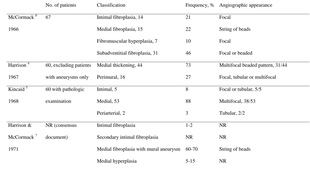
Table 1. Pathologic classifications of renal artery fibromuscular dysplasia and corresponding angiographic appearance
No. of patients Classification Frequency, % Angiographic appearance
McCormack 6
1966
67 Intimal fibroplasia, 14 21 Focal
Medial fibroplasia, 15 22 String of beads
Fibromuscular hyperplasia, 7 10 Focal
Subadventitial fibroplasia, 31 46 Focal or beaded
Harrison 4
1967
60, excluding patients
with aneurysms only
Medial thickening, 44 73 Multifocal beaded pattern, 31/44
Perimural, 16 27 Focal, tubular or multifocal
Kincaid 5
1968
60 with pathologic
examination
Intimal, 5 8 Focal or tubular, 5/5
Medial, 53 88 Multifocal, 38/53 Periarterial, 2 3 Tubular, 2/2 Harrison & McCormack 7 1971 NR (consensus document) Intimal fibroplasia 1-2 NR
Secondary intimal fibroplasia NR NR
Medial fibroplasia with mural aneurysm 60-70 String of beads
Perimedial fibroplasia 15-25 May be beaded
Periarterial fibroplasia <1 NR
Stanley 8
1975
177 (25 children), 86
specimens suitable for
classification
Intimal fibroplasia ~5 Focal or tubular
Medial hyperplasia ~1 Focal
Medial fibroplasia ~85 A continuum of disease angiographically
Perimedial dysplasia ~10 Focal, occasionally multiple constrictions
Table 2. Past history and characteristics at diagnosis of fibromuscular dysplasia in patients with unifocal or multifocal renal artery lesions
Unifocal FMD (n = 61) Multifocal FMD (n = 276)
No.* Values No.* Values p
Male sex 61 19 (31) 276 47 (17) 0.02
Personal history of hypertension 61 60 (98) 276 258 (93) 0.22
Age at diagnosis of hypertension, years 60 26 [21, 36] 256 40 [32, 49] <0.001
Age at diagnosis of FMD, years 61 30 [25, 39] 276 49 [42, 58] <0.001
History of diabetes 59 1 (2) 269 12 (4) 0.48
History of hypercholesterolemia 59 10 (17) 270 78 (29) 0.07
Current smoker 58 29 (50) 268 69 (26) <0.001
Systolic blood pressure, mmHg 55 157 [137, 174] 234 146 [128, 162] 0.006
Diastolic blood pressure, mmHg 55 97 [87, 110] 234 88 [76, 100] <0.001
No. of antihypertensive agents 55 1 [1, 2 ] 234 2 [1, 3] 0.74
Body mass index, kg/m² 54 22 [20, 24] 232 23 [21, 27] <0.001
Renal asymmetry >20 mm 48 16 (33) 195 19 (10) <0.001
FMD site: right, unilateral 61 29 (48) 276 85 (31)
<0.001 left, unilateral 61 19 (31) 276 20 (7)
bilateral 61 13 (21) 276 171 (62)
Median renal artery score 61 1 [1, 2] 276 3 [1, 3] <0.001
Presence of renal artery aneurysms 61 7 (11) 276 31 (11) 1
Presence of renal artery dissections 61 4 (7) 276 12 (4) 0.50
Presence of cervical arteries FMD 24 6 (25) 127 65 (51) 0.03
Presence of ilio-femoral arteries FMD 22 2 (9) 123 30 (24) 0.16
Presence of digestive arteries FMD 16 5 (31) 97 50 (52) 0.18
The number of patients available for analysis is shown for each variable. Values are numbers of patients (percentage) for binary
variables and median [25th centile, 75th centile] for quantitative variables. FMD, fibromuscular dysplasia. * Missing data mostly
Table 3. Characteristics at follow-up over 365 days for patients with unifocal or multifocal renal artery FMD with first visit before October 1st 2010
Unifocal (n = 52) Multifocal (n = 213)
No. Values No. Values p
Patients with clinical follow-up >365 days 52 31 (60) 213 141 (66) 0.42
Follow-up duration, years 31 4 [2, 8] 141 4 [3, 9] 0.39
Renal artery intervention during follow-up 31 28 (90) 141 50 (35) <0.001
Current smoker, at last follow-up 31 9 (29) 139 24 (17) 0.14
Body mass index at last follow up, kg/m² 30 23 [22, 26] 130 25 [22, 28] 0.25
Systolic blood pressure at last follow-up, mmHg 31 123 [114, 130] 139 124 [114, 135] 0.54
Change in systolic blood pressure, mmHg 31 -30 [-53, -14] 138 -20 [-37, -5] 0.03
Diastolic blood pressure at last follow-up, mmHg 31 77 [74, 85] 139 74 [67, 82] 0.06
Change in diastolic blood pressure, mmHg 31 -19 [-35, -3] 138 -14 [-27, -2] 0.14
Number of antihypertensive agents at last follow-up 31 0 [0, 1] 140 2 [1, 3] <0.001
Change in number of antihypertensive agents 31 -1 [-1, 0] 139 0 [-1, 1] 0.006
m/min/1.73m²
Change in estimated creatinine clearance,* ml/min/1.73m² 25 42 [24, 53] 98 41 [27, 56] 0.96
The number of patients available for analysis (No.) is shown for each variable. Values are numbers of patients (percentage) for binary
variables and median [25th centile, 75th centile] for quantitative variables. FMD, fibromuscular dysplasia. *Using the Cockcroft-Gault