HAL Id: inserm-02907291
https://www.hal.inserm.fr/inserm-02907291
Submitted on 27 Jul 2020
HAL is a multi-disciplinary open access
archive for the deposit and dissemination of
sci-entific research documents, whether they are
pub-lished or not. The documents may come from
teaching and research institutions in France or
abroad, or from public or private research centers.
L’archive ouverte pluridisciplinaire HAL, est
destinée au dépôt et à la diffusion de documents
scientifiques de niveau recherche, publiés ou non,
émanant des établissements d’enseignement et de
recherche français ou étrangers, des laboratoires
publics ou privés.
replication fork remodeling and restart
Matteo Berti, Federico Teloni, Sofija Mijic, Sebastian Ursich, Jevgenij Fuchs,
Maria Dilia Palumbieri, Jana Krietsch, Jonas Schmid, Edwige Garcin,
Stéphanie Gon, et al.
To cite this version:
Matteo Berti, Federico Teloni, Sofija Mijic, Sebastian Ursich, Jevgenij Fuchs, et al.. Sequential role
of RAD51 paralog complexes in replication fork remodeling and restart. Nature Communications,
Nature Publishing Group, 2020, 11 (1), pp.3531. �10.1038/s41467-020-17324-z�. �inserm-02907291�
Sequential role of RAD51 paralog complexes in
replication fork remodeling and restart
Matteo Berti
1,4
, Federico Teloni
2,4
, So
fija Mijic
1
, Sebastian Ursich
1
, Jevgenij Fuchs
1
,
Maria Dilia Palumbieri
1
, Jana Krietsch
1
, Jonas A. Schmid
1
, Edwige B. Garcin
3
, Stéphanie Gon
3
,
Mauro Modesti
3
, Matthias Altmeyer
2
✉
& Massimo Lopes
1
✉
Homologous recombination (HR) factors were recently implicated in DNA replication fork
remodeling and protection. While maintaining genome stability, HR-mediated fork
remo-deling promotes cancer chemoresistance, by as-yet elusive mechanisms. Five HR cofactors
–
the RAD51 paralogs RAD51B, RAD51C, RAD51D, XRCC2 and XRCC3
– recently emerged as
crucial tumor suppressors. Albeit extensively characterized in DNA repair, their role in
replication has not been addressed systematically. Here, we identify all RAD51 paralogs while
screening for modulators of RAD51 recombinase upon replication stress. Single-molecule
analysis of fork progression and architecture in isogenic cellular systems shows that the
BCDX2 subcomplex restrains fork progression upon stress, promoting fork reversal.
Accordingly, BCDX2 primes unscheduled degradation of reversed forks in BRCA2-defective
cells, boosting genomic instability. Conversely, the CX3 subcomplex is dispensable for fork
reversal, but mediates ef
ficient restart of reversed forks. We propose that RAD51 paralogs
sequentially orchestrate clinically relevant transactions at replication forks, cooperatively
promoting fork remodeling and restart.
https://doi.org/10.1038/s41467-020-17324-z
OPEN
1Institute of Molecular Cancer Research, University of Zurich, Winterthurerstrasse 190, 8057 Zurich, Switzerland.2Department of Molecular Mechanisms of
Disease, University of Zurich, Winterthurerstrasse 190, 8057 Zurich, Switzerland.3Cancer Research Center of Marseille; CNRS; Inserm; Institut
Paoli-Calmettes; Aix-Marseille Université, 27 Bd Leï Roure, CS 30059, 13273 Marseille, France.4These authors contributed equally: Matteo Berti, Federico Teloni.
✉email:[email protected];[email protected]
123456789
P
rotecting the integrity of replicating chromosomes is crucial
to maintain genome stability and to avoid cellular
trans-formation. Mutations impairing the replication stress
response predispose to cancer
1. However, the same mutations
often impair the cellular responses to genotoxic treatments,
offering important windows of opportunity for effective cancer
chemotherapy by DNA replication interference
2.
Replication fork reversal—i.e., the active conversion of
repli-cation forks into four-way junctions—has recently emerged as a
global and genetically controlled response to various forms of
endogenous and exogenous replication stress
3–5. These transient
replication intermediates can be efficiently restarted
6,7and their
function was proposed to slow down replication forks under
unfavorable conditions, thereby limiting ssDNA accumulation
and fork breakage
8–10. Replication fork reversal requires
specia-lized enzymes, such as the DNA translocases SMARCAL1,
ZRANB3, and HLTF
11–14, and the central recombinase RAD51
5.
Genetic modifications affecting fork reversal typically result in
unrestrained fork progression upon genotoxic stress, highlighting
the relevance of fork remodeling for active fork slowing
5,12,15.
RAD51 function in regulating fork progression and remodeling
extended the role of homologous recombination (HR) factors
from classical double-stranded break (DSB) repair to the
repli-cation stress response
16. Accordingly, other HR factors—like
BRCA1 and BRCA2—protect stalled forks from excessive
nucleolytic
degradation
by
promoting
efficient RAD51
loading
9,17,18. This function in replication stress is genetically
uncoupled from their canonical role in DSB repair
14,17,19, is
associated with the exquisite chemosensitivity of BRCA-deficient
cells
20and has been recently extended to additional HR factors
21.
Importantly, this deregulated degradation of nascent DNA is also
genetically dependent on fork remodeling, as regressed arms are
mandatory entry points for DNA degrading enzymes
13,14,22,23.
Moreover, the HR protein RAD52 was recently shown to
mod-ulate fork stability and restart, by limiting
SMARCAL1-dependent fork reversal or regulating reversed fork processing
by MRE11
14,24. Collectively, these recent observations have
implicated the RAD51 recombinase and other HR factors both in
the formation and in the stability of reversed replication forks
5,14.
The latter function is BRCA2-dependent and reflects protection
of the DNA end at the fourth regressed arm. However, the role of
RAD51 in reversed fork formation is still enigmatic, as fork
reversal does not require BRCA2, nor stable RAD51
nucleofila-ment formation
14, thereby challenging canonical models of
RAD51 function
16. Molecular understanding of these clinically
relevant transactions on replicating chromosomes requires
fur-ther mechanistic investigation, particularly on the role of
alter-native HR mediators and on their functional interaction
with RAD51.
Besides RAD51 and its meiotic counterpart DMC1,
five
addi-tional mammalian paralogs of bacterial RecA were discovered two
decades ago, either by DNA sequence alignments (RAD51B,
RAD51C, and RAD51D) or by functional complementation of
radiation sensitive Chinese hamster ovary (CHO) mutant cells
(XRCC2 and XRCC3)
25. These proteins display limited sequence
homology to each other and to RAD51, and are generally
reported as classical RAD51 paralogs. The Shu complex, which
regulates HR in mitosis and meiosis, was recently shown to
include a sixth RAD51 paralog, i.e., SWSAP1
25–27. Classical
RAD51 paralogs were proposed to form two biochemically and
functionally distinct subcomplexes, i.e. the
RAD51B-RAD51C-RAD51D-XRCC2 complex (BCDX2) and the RAD51C-XRCC3
complex (CX3), showing common, but also distinct biochemical
properties
28–32. Ablation of these proteins in mice resulted in
embryonic lethality and functional studies on these factors were
thus largely based on gene inactivation in p53-deficient CHO cells
and chicken DT40 B-lymphocytes
33–39. Overall, these studies
uncovered important roles for these proteins in genome stability
and DSB response, via modulation of RAD51. Recent biochemical
data on the dimeric RAD51 paralog complexes in S. cerevisiae and
C. elegans—Rad55/Rad57 and RFS-1/RIP-1,
respectively—sup-ported this concept, by showing that RAD51 paralogs protect
RAD51 nucleofilaments from antirecombinase activities
40and
remodel
these
filaments to promote strand exchange
reactions
41,42. However, whether similar biochemical properties
can be associated with the different subcomplexes of human
RAD51 paralogs is still unknown.
siRNA-mediated depletion of the human proteins suggested
different roles for the two RAD51 paralog subcomplexes at early
(BCDX2) versus late (CX3) stages of HR-mediated DSB repair
43–45.
Very recently, a panel of RAD51 paralog gene ablations was
produced by CRISPR-Cas9 in cancerous and non-transformed
human cell lines
46. While RAD51B inactivation lead to
com-paratively mild phenotypes, genetic ablation of any of the other
paralogs resulted in drastic impairment of HR-mediated DSB
repair proficiency in all tested cell lines
46.
Mutations in several RAD51 paralog genes have been
asso-ciated with increased susceptibility to breast and ovarian
can-cer
47–51. Moreover, hypomorphic mutations in RAD51C and
XRCC2 were linked to Fanconi anemia, a rare human disorder
linked to defective replication-coupled repair of specific DNA
lesions in the bone marrow
52,53. These recent
findings revived the
interest in these accessory HR factors, promoting new
mechan-istic studies to unravel their precise role in DNA replication and
genome stability. Interestingly, all human RAD51 paralogs were
shown to associate with nascent DNA
54, but the mechanistic role
(s) of these factors in replication were not investigated
system-atically. CHO or DT40 cell lines carrying different mutations in
individual genes—i.e., RAD51C, XRCC2, and XRCC3—displayed
specific defects in replication fork progression and stability
54–56.
Furthermore, based on shRNA-mediated downregulation, human
XRCC2 was recently proposed to modulate fork progression also
in human cells, as a specific response to limited nucleotide
availability
57. Overall, our mechanistic understanding of the role
of human RAD51 paralogs at replication forks is still incomplete
and requires a systematic analysis of these factors in isogenic
backgrounds.
Here, all classical human RAD51 paralogs are identified in a
high-content microscopy screen for RAD51 modulation upon
mild replication stress. Combining these cytological data with
single-molecule investigations on replication intermediates in
multiple cellular systems, we
find that the BCDX2 complex assists
RAD51 in reversed fork formation, limiting fork speed upon
DNA damage and mediating stalled fork degradation in
BRCA2-defective cells. Conversely, the CX3 complex—albeit dispensable
for fork reversal and fork protection—is found to mediate
effi-cient restart of previously reversed forks.
Results
A screen for RAD51 foci in CPT-treated cells identi
fies RAD51
paralogs. RAD51 is readily detected on chromatin as nuclear foci
in human osteosarcoma U2OS cells even during unperturbed
replication. To identify key modulators of the RAD51
recombi-nase at challenged replication forks, we subjected U2OS cells to a
short (45 min) treatment with low dose of the Topoisomerase I
inhibitor camptothecin (CPT, 50 nM), which was previously
shown to induce marked fork slowing and frequent fork
rever-sal
58. As in these conditions induction of DSBs is undetectable
58,
RAD51 foci likely mark HR-mediated replication fork remodeling
events occurring at stalled, yet-unbroken forks (Fig.
1a). We then
designed a targeted small interfering RNA (siRNA) library, using
three individual siRNAs against each target gene and covering
approximately 450 factors that were found to be enriched at
replication forks by immunoprecipitation of newly synthesized
DNA and mass spectrometric analysis of the associated
pro-teins
59–61. CPT-treated cells were labeled with EdU and analysed
by high-content microscopy for quantitative image-based
cyto-metry (QIBC)
62,63, which allows simultaneous assessment of
cell-cycle distribution, replication competence, as well as number and
intensity of RAD51 foci. We selected an early time point after
siRNA transfection (48 h), in order to limit cell-cycle effects due
to prolonged depletion of essential factors. In these hypomorphic
conditions, only depletion of key replication factors (e.g., PCNA)
led to replication impairment, while most other siRNAs did not
impact replication competence and cell proliferation, including
a
b
g
c
d
e
z -scoreRAD51 foci counts per nucleus
z
-score
RAD51 foci counts per nucleus
Gene name (ranked)
RAD51 counts UT CPT IR U2OS RAD51 counts UT CPT IR RPE-1
h
f
Gene name (ranked) 3 2.5 2 1.5 1 0.5 0 –0.5 –1 –1.5 –2 –2.5 3 2.5 2 1.5 1 0.5 0 –0.5 –1 –1.5 –2 –2.5 RAD51 counts
RAD51 RAD51 foci count per nucleus
DAPI
Total intensity per nucleus (a.u. x 104)
DAPI
Total intensity per nucleus (a.u. x 104)
60 50 40 30 20 10 0 Max Min Average CPT (50 nM) EdU 0′ 15′ 45′
Gene name (alphabetically)
3 2.5 3 2.5 2 1.5 1 0.5 0 –0.5 –1 –1.5 –2 –2.5 3.5 BRIP1 FAAP24 FANCI FANCL FIGNL1 FANCM POLA1 RFWD3 RAD54L SLX4 WDHD1 CDC73 CTR9 BRCA1 BARD1BRCA2 CHEK1 CHAF1A PAF1 NHP2L1 PCNA PALB2 RAD51CSF3B3 DSS1 RAD51 RNF168 PRPF8 z -score
RAD51 foci counts per nucleus
z
-score
RAD51 foci counts per nucleus
Gene name (alphabetically) 2 1.5 1 0.5 0 –0.5 –1 –1.5 –2 –2.5 3.5 BRCA2 RAD51 RAD51C RAD51D RAD51B XRCC2 XRCC3 BRCA1 siCtrl siCtrl siRAD51C siRAD51C siRAD51B siRAD51B siBRCA2 siRAD51D siRAD51D siXRCC2 siXRCC2 siXRCC3 siXRCC3
siCtrl siBRCA2siRAD51BsiRAD51CsiRAD51DsiXRCC2siXRCC3
3.5 20 150 3.5 DAPI RAD51 EdU Merge 60 30 15 0 20 150 20 150 20 150 20 150 20 150 20 150 20 150 20 150 20 150 20 150 RAD51 counts
EdU Mean intensity per nucleus (a.u.)
60 30 15 0 Untreated CPT (50 nM) 1000 400 100 40 10 4 1 0.4 20 40 60 20 40 60 Max Min Average #1 #2 #1 #2 #1 #2 #1 #2 #1 #2 :oligo
those targeting essential HR factors like BRCA1 (Supplementary
Fig. 1a). Reassuringly, when ranked according to their deviation
from the mean (i.e., z-score), known positive (BRCA1, BRCA2,
BARD1,
PALB2)
and
negative
regulators
(RAD54L)
of
RAD51 scored among the top hits at either end of the distribution
(Fig.
1b, c, Supplementary Data 1 and 2), with all three siRNAs
showing consistent results (Supplementary Data 2). However,
presumably because of the hypomorphic conditions chosen for
the screen, siRNAs targeting genes of other known
anti-recombinase activities (i.e., RADX/CXorf57, FBH1/FBXO18,
RECQ5, and PARI/C12orf48) did not show strong effects on
RAD51 accumulation (Supplementary Fig. 1b–c; Supplementary
Data 1 and 2). Furthermore, although RAD51 has been previously
involved in different steps of fork remodeling and protection,
siRNAs targeting genes for key fork remodeling factors (i.e.,
SMARCAL1, ZRANB3, HLTF, and RECQ1/RECQL) did not
sig-nificantly affect RAD51 foci number and intensities in our
screening conditions (Supplementary Fig. 1b–c). Besides the
possibility of incomplete downregulation, this result may also
reflect intrinsic ambiguities associated with RAD51 foci
mea-surements as sole readout. Impairing fork remodeling activities
may not only negatively affect RAD51 binding to forks, but can
also induce fork breakage even at these minimal CPT doses
5,58,
thereby enhancing RAD51 accumulation in foci.
Despite these potential caveats and its inherently
non-comprehensive nature, our screen identified several new or
uncharacterized modulators of RAD51 chromatin binding upon
replication stress, showing strong positive (>1.5) or negative
(<1.5) z-scores (Fig.
1c, Supplementary Data 1). Here, we focused
our attention on the
five classical RAD51 paralogs, which all
displayed a negative z-score in the experimental conditions of our
screen (Fig.
1d, e). SWSAP1—encoding another noncanonical
RAD51 paralog
25–27—was not among the targeted genes in our
siRNA library, but downregulation of its interaction partner
SWS1 led to defects in RAD51 foci comparable to those observed
for the other RAD51 paralogs (Supplementary Fig. 1b–c,
Supplementary Data 1), in agreement with the recently reported
involvement of the Shu complex in the replication stress
response
64.
In light of the established clinical relevance of the classical
RAD51 paralogs and their elusive role in the replication stress
response, we decided to focus on and explore this set of genes. We
validated the screen results in U2OS cells using two individual
siRNAs, testing also—besides low CPT treatments—untreated
conditions and ionizing radiation. In these analyses, short-term
(48 h) depletion of all classical RAD51 paralogs consistently led to
reduced RAD51 foci counts in replicating cells, while not
significantly affecting cell cycling and replication competence
(Fig.
1f, g and Supplementary Fig. 1d). In agreement with
previous reports
45, we noticed that XRCC3 downregulation
showed milder effects on RAD51 foci, compared to inactivation
of the other RAD51 paralogs. This difference was also observed
when performing validation experiments in untransformed
human retinal epithelial cells (RPE-1, Fig.
1h) and is particularly
evident in S phase cells upon mild CPT treatment
(Supplemen-tary Fig. 1e). As XRCC3 and RAD51C form a specific protein
complex (CX3), while all other factors assemble in a second
multimeric RAD51 paralog complex (BCDX2), these results
pointed to separate functional roles of the two complexes in the
replication stress response, as suggested for DSB repair
45.
BCDX2 promotes fork slowing and remodeling upon mild
genotoxic stress. To investigate the functional role of BCDX2 and
CX3 in the replication stress response, we
first monitored the
stability of the two complexes upon single-component
down-regulation in U2OS cells by two independent siRNAs (Fig.
2a, b).
In keeping with previous reports
45, RAD51C downregulation
affected protein levels of all components of both complexes, thus
potentially abolishing both BCDX2 and CX3 activities. In
con-trast, RAD51D downregulation did leave CX3 levels unaffected,
thus allowing the assessment of the specific functional role of
BCDX2. Conversely, XRCC3 downregulation had only minor
effects on RAD51C levels and should thus specifically affect CX3
function (Fig.
2a, b). We therefore focused our functional analysis
on these three protein depletions, to explore specific roles of the
two complexes upon mild replication interference (Fig.
2a). We
first used an established nascent DNA labeling protocol to
monitor by DNA
fiber spreading active replication fork slowing
upon mild treatment with CPT
58. RAD51C and RAD51D
downregulation by two independent siRNA sequences drastically
impaired active fork slowing, leading to unrestrained fork
pro-gression in the presence of CPT, both as measured by
fiber track
length and as ratios between the differentially labeled nascent
DNA (Fig.
2c, and Supplementary Fig. 2a–c). Conversely, XRCC3
downregulation did not significantly affect CPT-induced fork
slowing (Fig.
2c, and Supplementary Fig. 2a–c). We then
per-formed similar experiments in U2OS cells where the same three
genes had been knocked-out (KO) by CRISPR-Cas9
46. Compared
to RAD51 paralog downregulation by siRNA (Supplementary
Fig. 1a), all KO cells—including XRCC3-KO—displayed a more
drastic reduction of endogenous and drug-induced RAD51 foci
(Supplementary Fig. 2d), likely reflecting full and prolonged
inactivation of these factors. Nonetheless, all tested KO cells led to
very similar observations when compared to downregulated cells
in terms of protein level interdependency and replication fork
progression phenotypes, with RAD51C-KO and RAD51D-KO
cells, but not XRCC3-KO cells, displaying unrestrained fork
progression in CPT (Fig.
2d, e). Importantly, unrestrained fork
progression in RAD51C-KO and RAD51D-KO cells was readily
complemented by stable expression of a FLAG-tagged version of
the missing protein (Fig.
2d, e). Finally, we confirmed very similar
effects on CPT-induced fork slowing by siRNA-mediated
down-regulation of RAD51C, RAD51D, and XRCC3 in the
non-transformed RPE-1 human cell line (Supplementary Fig. 2e).
Overall, these data strongly suggest that BCDX2, but not CX3,
mediates active fork slowing upon mild genotoxic stress.
Active fork slowing is linked to replication fork reversal
(Fig.
2f) and depends on the RAD51 recombinase and multiple
Fig. 1 QIBC-based screen identifies RAD51 paralogs as important modulators of RAD51 foci formation upon mild replication stress. a QIBC-based
screen layout. Left: screen experimental condition. U2OS cell were treated with 50 nM CPT for 45 min and replicating cells were concomitantly labeled
with EdU for 30 min. Center: representative immunofluorescence image (scale-bar, 50 μm). Right: representative scatter plot showing cell-cycle
distribution (based on DNA content and EdU incorporation) of RAD51 foci in untreated and CPT-treated U2OS cells. The experiment shown was performed
twice yielding similar results.b Screen gene ranking based on the RAD51 foci z-score. c z-score plot of positive (z-score <−1.5) and negative (z-score > 1.5)
RAD51 modulators (marked in gray). Color code as ind, e. d, e z-score for thefive classical RAD51 paralogs (marked in purple) and positive controls
(marked in red).f Validation of the RAD51 paralogs influence on RAD51 foci formation upon 50 nM CPT. g, h Differential effect of RAD51 paralogs on
RAD51 foci formation in untreated (UT), CPT- (50 nM for 45 min) and IR- (5 Gy, 2 h recovery) treated U2OS and RPE-1 cells. Source data for a-f are
a
b
c
f
e
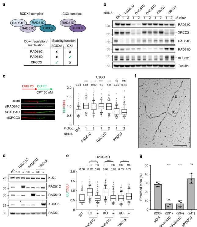
BCDX2 complex CX3 complex RAD51B RAD51C RAD51D RAD51C Downregulation/ inactivation BCDX2 CX3 Stability/function RAD51C RAD51D XRCC3 CldU 25′ ldU 25′ CPT 50 nM RAD51C XRCC3 RAD51B RAD51D XRCC2 RAD51C XRCC3 RAD51B RAD51D XRCC2 Tubulin siRNA: * 1 2 1 2 1 2 1 2 1 2d
RAD51C KU70 RAD51D XRCC3 RAD51 KO +KO + KO +g
U2OS 1 2 1 2 1 2 # oligo: siRNA: 1.5 1.0 0.5 0 2.0 0.74 1.04 0.99 1.0 1.0 0.75 0.74 **** **** **** **** ns ns U2OS-KO KO WT WT + KO + KO + ldU / CldU ldU / CldU 1.5 1.0 0.5 0 2.0 0.66 0.92 0.62 0.92 0.63 0.63 0.72 **** ns **** ns ns ns **** **** *** D D R P Reversed forks (%) 40 30 20 10 0 ns *** *** * siCtrl siRAD51C siRAD51D siXRCC3 siCtrlsiRAD51C siRAD51D siXRCC3
35 35 35 35 35 55 70 35 35 35 35 50 (230) (231) (234) (241) XRCC3 XRCC2 :# oligo RAD51C RAD51D XRCC3 RAD51C Ctrl Ctrl XRCC3 RAD51D RAD51CRAD51DXRCC3
Fig. 2 BCDX2 paralog complex, but not CX3, promotes replication fork slowing and reversal upon mild CPT treatment. a Rationale for focusing on RAD51C, RAD51D, and XRCC3 downregulation, to perform functional studies on BCDX2 and CX3 complexes during replication stress. Top: schematic of the
two main RAD51 paralog complexes. Bottom: differential effects ofRAD51C, RAD51D, and XRCC3 inactivation on paralog complex stability/function.
b Western blot analysis of RAD51 paralogs upon 48 h downregulation with two different siRNAs. Ctrl, control siRNA; Tubulin, loading control. asterisk,
specific band. In b and d multiple gels/blots were processed in parallel, ensuring equal and comparable loading across gels. The experiment was performed
twice yielding similar results.c DNAfiber analysis of U2OS cells 48 h after transfection with a control siRNA (siCtrl) or with siRNAs targeting RAD51C,
RAD51D, and XRCC3. Top-left: schematic of the CldU/IdU pulse-labeling protocol used to evaluate fork progression upon 50 nM CPT treatment.
Bottom-left: representative DNAfibers from each genetic condition. Left: IdU/CIdU ratio is plotted as a readout of fork progression. In c and e, the numbers indicate
the mean value; a minimum of 150 forks was scored in two independent experiments yielding similar results. Bounds of box are 25–75th percentile, center
shows the median, whiskers indicate the 10–90 percentiles, data points outside this range are drawn as individual dots. Statistical analysis: Kruskal–Wallis
test; ns not significant; ****p-value < 0.0001, ***p-value = 0.0003. d Western blot analysis of RAD51 paralogs in Knock-Out (KO) U2OS cells and in cells
reconstituted of the respective protein (+). KU70, loading control. The experiment was performed twice yielding similar results. e DNA fiber analysis of KO
and reconstituted (+) U2OS cells labeled as in c. f, g Frequency of reversed replication forks in U2OS cells transfected with control siRNA (Ctrl) or with
siRNAs targetingRAD51C, RAD51D, or XRCC3 for 48 h and treated with 50 nM CPT (1 h). f Electron micrograph of a representative reversed replication fork
from CPT-treated U2OS cells (P parental duplex, D daughter duplexes, R regressed arm; scale-bar, 100 nm).g Graph-bar showing mean and SD from three
independent EM experiments. In brackets, total number of molecules analyzed per condition. Statistical analysis: one-way ANOVA followed by Bonferroni
DNA translocases
5,12,65. Using an established EM method to
visualize replication intermediates
66, we indeed confirmed that
downregulation of RAD51C and RAD51D, but not of XRCC3,
markedly impairs CPT-induced replication fork reversal (Fig.
2g,
Supplementary Table 1a).
BCDX2 primes stalled fork degradation in BRCA2-defective
cells. We then used the same validated cellular systems to further
investigate the role of BCDX2 and CX3 in fork stability, using an
established DNA
fiber protocol where double-labeled ongoing
forks experience prolonged stalling by HU-induced nucleotide
depletion (Fig.
3a)
14,17. As reported, BRCA2-depleted cells
dis-played nascent DNA degradation at stalled forks, monitored
as reduced ratio between second (IdU) and
first (CldU) track
length (Fig.
3a, b). Despite effective depletion of protein levels
(Supplementary Fig. 3a)—which were sufficient to induce
unrestrained fork progression in CPT (Fig.
2c)—downregulation
of none of the tested RAD51 paralogs led to detectable fork
degradation (Fig.
3a, b). However, downregulation of RAD51C or
RAD51D—but not of XRCC3—with two different siRNA
sequences fully restored nascent DNA stability in
BRCA2-depleted cells (Fig.
3a, b and Supplementary Fig. 3a). Very similar
results were obtained in RAD51C-KO and in RAD51D-KO U2OS
cells, and re-expression of the missing protein restored the fork
degradation phenotype upon BRCA2 depletion (Fig.
3c). Also in
this cellular system, genetic inactivation of XRCC3 had no
detectable effects on nascent DNA stability, in either
BRCA2-proficient or -deficient cells (Fig.
3c). Consistently,
down-regulation of RAD51C or RAD51D—but not XRCC3—fully
restored stalled fork stability also in BRCA2-depleted RPE-1 cells
(Supplementary Fig. 3b–c). On one hand, these observations
suggest that inactivation of any of the RAD51 paralogs does not
a
d
HU 4 mM 5 hb
U2OS ldU / CldU 1 2 1 2 1 2 1 2 1 2 1 2 siBRCA2 # oligo: siRNA: 0 0.5 1.5 1.0 2.0 1.00 1.00 1.01 0.96 0.97 0.99 0.95 0.73 1.03 0.97 0.94 0.97 0.73 0.74 **** **** **** ns ns ns ns ns ns ns ns ns nsc
U2OS-KO ldU / CldU siCtrlsiBRCA2 siRAD51C siXRCC3
siCtrl KO siBRCA2 siCtrl + KO + KO + KO WT WT + KO + KO + 0 0.5 1.5 1.0 2.0 siCtrl siRAD51C siRAD51D siXRCC3 siCtrl siBRCA2 0.93 0.98 0.95 0.98 0.99 0.99 0.94 0.73 0.99 0.73 0.98 0.73 0.72 0.72 ns ns ns ns ns ns **** ns **** ns **** **** **** ns ns ns **** **** ns Reversed forks (%) Mirin: – + – + – + – + ns *** ns *** *** ns ns (222) (219) (254) (233) (219) (235) (224) (226) RAD51C Ctrl Ctrl RAD51D XRCC3 RAD51C RAD51D XRCC3 RAD51C RAD51D XRCC3 RAD51C RAD51D XRCC3 ns ns *** CldU 20′ ldU 20′ 30 20 10 0 40
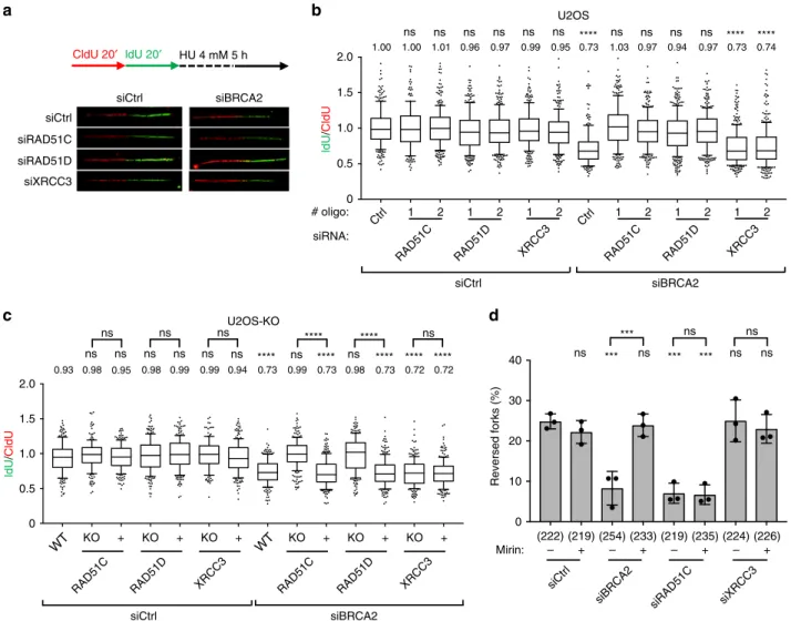
Fig. 3 BCDX2 complex, but not CX3, promotes reversed fork degradation in BRCA2-depleted U2OS cells. a, b DNAfiber analysis of U2OS cells double
transfected with a control siRNA (Ctrl) or with siRNAs targetingRAD51C, RAD51D, and XRCC3 for 60 h and with BRCA2 siRNA for 48 h. a Top: schematic
CldU/IdU pulse-labeling protocol to evaluate fork degradation upon HU treatment (4 mM, 5 h). Bottom: representative DNAfibers from each genetic
condition.b IdU/CIdU ratio is plotted as a readout of fork degradation. In b and c, the numbers indicate the mean value; a minimum of 150 forks was scored
in two independent experiments yielding similar results. Bounds of box are 25–75th percentile, center shows the median, whiskers indicate the 10–90
percentiles, data outside this range are drawn as individual dots. Statistical analysis: Kruskal–Wallis test; ns not significant; ****p-value < 0.0001. c DNA
fiber analysis of KO and reconstituted (+) U2OS cells transfected with a control siRNA (Ctrl) or with BRCA2 siRNA for 48 h and consecutively labeled and
HU-treated as ina. IdU/CIdU ratio is plotted as a readout of fork degradation. d Frequency of reversed replication forks in U2OS cells transfected with
control siRNA (Ctrl) or with siRNAs targetingBRCA2, RAD51C, or XRCC3 for 48 h and treated for 5 h with HU 4 mM; where indicated 50 μM Mirin was
added 1 h before HU treatment (6 h total treatment). Graph-bar depicts mean and SD from three independent EM experiments. In brackets, total number of
molecules analyzed per condition. Statistical analysis: one-way ANOVA followed by Bonferroni test; ns not significant; ***p-value = 0.0003. Source data
affect stalled fork integrity in human cells. On the other hand, in
light of the data in Fig.
2, they also suggest that BCDX2—but not
CX3—mediates the formation of reversed forks, which serve then
as entry points for nucleolytic degradation of nascent DNA in
BRCA2-defective cells. EM visualization confirmed this
hypoth-esis: as reported in different cellular systems
13,14,22,
BRCA2-depleted U2OS cells displayed a marked reduction in
HU-induced reversed fork frequency, which was readily suppressed by
MRE11 inhibition using mirin (Fig.
3d, Supplementary Table 1b).
Conversely, mirin did not restore reversed fork frequency upon
RAD51C depletion and XRCC3 depletion had no significant
effects (Fig.
3d, Supplementary Table 1b). These EM data on
RAD51C closely resemble previous observations obtained by
RAD51 depletion
14and, combined with the data in Fig.
3b–c,
strongly suggest that BCDX2—but not CX3—is required for fork
reversal, upstream of BRCA2-mediated stabilization of reversed
forks.
CX3 mediates ef
ficient restart of reversed forks after stalling.
We next tested by DNA
fiber spreading whether RAD51 paralogs
assist the restart of stalled replication forks, measuring number and
length of replicated tracks after HU removal (Fig.
4a). In our
experimental conditions (24 h of siRNA-mediated downregulation),
RAD51 inactivation did not affect the efficiency of fork restart and
slightly increased the velocity of restarting forks (Fig.
4a and
Sup-plementary Fig. 4a). This likely reflects impaired fork remodeling
associated with RAD51 depletion, as non-reversed forks may be
faster in restarting DNA synthesis once nucleotide levels are
restored, possibly by efficient replication fork repriming events.
Accordingly, downregulation of RAD51C or RAD51D—which are
also required for fork reversal (Figs.
2
and
3)—led to similar mild
effects on fork restart (Fig.
4a and Supplementary Fig. 4a).
Conversely, XRCC3 depletion drastically increased the fraction of
forks failing to restart DNA synthesis and delayed progression of
the restarting forks (Fig.
4a and Supplementary Fig. 4a). Very
similar effects on efficiency and velocity of fork restart were
obtained in U2OS cells carrying genetic ablation of these genes
and all effects were fully complemented by re-expression of the
missing protein (Supplementary Fig. 4b–c). As XRCC3 is not
required for fork reversal (Figs.
2
and
3), we reasoned that
XRCC3-depleted cells might be specifically impaired in their
ability to restart forks that had previously been reversed. Indeed,
co-depletion of RAD51 in XRCC3-depleted U2OS cells fully
rescued efficiency and velocity of fork restart (Fig.
4b and
Sup-plementary Fig. 4d). Similarly, XRCC3 depletion impaired fork
restart in ZRANB3-proficient, but not in ZRANB3-deficient cells
(Fig.
4c and Supplementary Fig. 4e). Moreover, also RAD51D
depletion rescued fork restart efficiency and velocity in
XRCC3-depleted cells (Fig.
4d, Supplementary Fig. 4f), further supporting
the role of BCDX2 in fork reversal, upstream of XRCC3-mediated
restart. Finally, as XRCC3 is destabilized in RAD51C-depleted
cells (Fig.
2b), RAD51C downregulation itself represents a third
condition that impairs fork reversal and rescues fork restart in the
absence of XRCC3 (Fig.
4a). Overall, these data establish a specific
role for the CX3 complex in replication fork restart, downstream
of RAD51-, BCDX2-, and ZRANB3-dependent fork reversal.
BCDX2 fuels chromosomal instability in BRCA2-defective
cells. As reversed forks are the entry points for unscheduled fork
degradation in BRCA2-depleted cells, we
finally tested whether
genetic impairment of RAD51C and RAD51D—which are
required for fork reversal—may restore genome stability in
BRCA2-defective cells. In metaphase spreads, depletion of
RAD51 paralogs per se did not detectably increase chromosomal
abnormalities (Fig.
5a). However, depletion of RAD51C or
RAD51D—but not of XRCC3—suppressed the chromosomal
instability associated with BRCA2 depletion (Fig.
5a), suggesting
that efficient fork remodeling by BCDX2 engages BRCA2 in a
crucial genome maintenance function.
Discussion
The recent discoveries that mutations in classical RAD51 paralog
genes predispose to cancer and Fanconi anemia
47–53encouraged
new mechanistic studies on these HR accessory factors,
particu-larly focused on the human proteins and their possible role in the
replication stress response. Human RAD51 paralogs were
first
expressed and purified over two decades ago, leading to the
identification of two main complexes: BCDX2 (composed of
RAD51B, RAD51C, RAD51D, XRCC2) and CX3 (composed of
RAD51C and XRCC3). However, biochemical insights have
remained relatively scarce to date. Furthermore, until very
recently, genetic investigations on these proteins were also largely
limited to single siRNAs or mutated cell lines in specific model
systems, calling for more systematic genetic investigations of
these proteins in isogenic human systems, particularly with
respect to their role upon replication stress.
Here, we selectively inactivated the BCDX2 complex (via
RAD51D inactivation), the CX3 complex (via XRCC3 inactivation),
or both complexes (via RAD51C inactivation) in U2OS and RPE-1
human cells, and performed single-molecule investigations on
replication intermediates. We show that the BCDX2 complex drives
reversed replication fork formation, presumably by assisting the
central recombinase RAD51, which was previously shown to
mediate this step of fork remodeling
5,14. Differently from
replica-tion fork protecreplica-tion and HR-mediated DSB repair, reversed fork
formation does not require stable RAD51 nucleofilaments
14. This
suggests that BCDX2 may assist DNA translocases and RAD51 in
driving parental strand reannealing, while partially replacing RPA
on ssDNA stretches accumulating at stalled forks (Fig.
5b). RAD51
paralogs were shown in yeast and C. elegans to biochemically
remodel preassembled RAD51 nucleofilaments
40–42. It is possible
that RAD51 paralogs enhance and stabilise spontaneous RAD51
monomer binding to short ssDNA stretches at stalled forks, thus
supporting the formation of a metastable nucleofilament, with
sufficient flexibility to sustain the high torsional constrains at
replication forks and promote fork reversal. Alternatively, RAD51
paralogs might stimulate the spontaneous formation of
BRCA2-independent short stretches of RAD51 monomers at stalled forks by
preventing the action of human antirecombinase activities
40.
However, our data do not exclude other possible
RAD51-independent mechanisms by which BCDX2 may foster the
rever-sal reaction, such as intrinsic strand annealing activities
29or specific
interactions with different enzymatic activities (e.g., DNA
translo-cases) and cofactors involved in fork reversal. While most reported
biochemical assays of fork reversal were performed in the absence
of ssDNA binding proteins, the addition of RPA or RAD51—when
tested—had profound effects on the efficiency and/or directionality
of these reactions
67,68. Holistic biochemical reconstitution of these
important transactions at the replication fork seems necessary to
elucidate the precise molecular mechanism by which BCDX2 and
RAD51 collaborate with DNA translocases to drive efficient fork
reversal.
As already shown for RAD51, which plays key genetically
uncoupled roles both in the formation and in the protection of
reversed forks
14,16,17, our data do not exclude that BCDX2
plays also a crucial role in protection and restart of stalled
replication forks (Fig.
5b). However, as for RAD51, this role
may be masked by its crucial upstream function in replication
fork reversal. As exemplified for RAD51 by the T131P
separation-of-function mutant
14, it is likely that hypomorphic
conditions of RAD51 paralog inactivation and/or different
levels and regulations of these proteins in different model
systems may explain different phenotypes in fork progression
and degradation upon inactivation of BCDX2 and CX3
com-ponents
54–57. In this context, it will be paramount to use
iso-genic replacement systems to test fork progression, remodeling
and stability with specific RAD51 paralog mutants linked
with cancer and Fanconi anemia
47–53, to reveal the specific
function(s) of these proteins in preventing human disease.
Our data uncover a role for the CX3 complex in the restart of
reversed replication forks, establishing a sequential engagement of
different RAD51 paralog complexes in stalled fork remodeling
and reactivation (Fig.
5b). Interestingly, a downstream role for
CX3—with respect to BCDX2—had been hypothesized also in
classical HR-mediated repair of DSBs, based on the limited
requirement of XRCC3 for RAD51 foci formation
45and on the
Holliday junction resolution activity found associated with the
CX3 complex
69. Recent data with CRISPR-KO cells challenge this
model
46, but may also reflect long-term effects of chronic XRCC3
inactivation and cell adaptation mechanisms to avoid the
resul-tant accumulation of toxic recombination intermediates. While
we did not succeed in monitoring directly the recruitment of
RAD51 paralogs to replication forks with currently available
tools, XRCC3 was previously shown to bind nascent DNA with a
delayed kinetic compared to BCDX2 subunits
54, which is
con-sistent with the sequential model proposed here (Fig.
5b).
a
b
c
60 40 20 0 ZRANB3: WT KO WT KO siXRCC3 ns **** ns Stalled forks (%) 60 40 20 0 siRNA: ns ns ns **** Stalled forks (%) siRAD51: ns ** ** ns 60 40 20 0 RAD51 XRCC3 Tubulin siRAD51: – + – + ZRANB3: siXRCC3 WT KO WT KO ZRANB3 XRCC3 Tubulin Fork restart (siCtrl)Fork stalling (siXRCC3)
siCtrl siCtrl 35 55 35 35 120 55 siXRCC3 siCtrl * **** HU 2 mM 2 h CldU 20′ ldU 40′ Stalled forks (%) RAD51D XRCC3 KU70 35 35 70 + – + 60 40 20 0 ns *** ns ** Stalled forks (%) siXRCC3 siCtrl – siRAD51D: – + – + siXRCC3 siCtrl
d
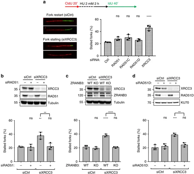
siRAD51D: – + – + siXRCC3 siCtrl RAD51C XRCC3 RAD51 RAD51D CtrlFig. 4 CX3 complex promotes reversed fork restart in U2OS cells. a DNAfiber analysis of U2OS cells transfected for 24–48 h (24 h for RAD51, 48 h for all
the other genes) with a control siRNA (Ctrl) or with siRNAs targetingRAD51, RAD51C, RAD51D, and XRCC3 to investigate replication fork restart upon HU.
Top: schematic CldU/IdU pulse-labeling protocol to evaluate fork restart upon HU treatment (2 mM, 2 h). Bottom-left: representative stalled and restarting
forks. Bottom-right: frequency of fork stalling for each genetic condition. Ina–d, the graph-bar depicts mean and SD from three independent experiments.
Statistical analysis: one-way ANOVA followed by Bonferroni test; ns not significant; ****p-value < 0.0001. b DNA fiber analysis of U2OS cells double
transfected with a control siRNA (Ctrl) or with a siRNA targetingXRCC3 (48 h) or RAD51 (24 h) to investigate replication fork restart upon HU treatments;
labeling protocol as ina. Top: levels of indicated proteins, assessed by western blot; Tubulin, loading control. In b and d multiple gels/blots were processed
in parallel, ensuring equal and comparable loading across gels. Bottom: frequency of fork stalling. ns not significant; **p-value = 0.0068 (1 versus 3) and
0.0087 (3 versus 4).c DNAfiber analysis of wild-type (WT) or ZRANB3 Knock-Out (KO) U2OS cells transfected with a control siRNA (Ctrl) or with
siRNAs targetingXRCC3 (48 h) to investigate replication fork restart upon HU treatments; labeling protocol as in a. Top: indicated protein levels, assessed
by western blot; Tubulin, loading control; asterisk, specific band. Bottom: frequency of fork stalling. ns not significant; ****p-value < 0.001. d DNA fiber
analysis of U2OS cells double transfected with a control siRNA (Ctrl), with siRNAs targetingXRCC3 (48 h) or RAD51D (60 h) to investigate replication fork
restart upon HU treatments; labeling protocol as ina. Top: indicated protein levels, assessed by western blot; KU70, loading control. Bottom: frequency of
Although both BRCA2 and XRCC3 were shown to play a role
downstream of fork remodeling, they display rather different, yet
possibly coordinated reversal-dependent functions. While BRCA2
defects lead to reversed fork degradation, but mild fork restart
defects
13,17,20, XRCC3 inactivation does not impair fork stability,
but severely impairs single-fork reactivation once the stalling agent
has been removed (Fig.
4), thus respectively handling a static (fork
protection) and dynamic (fork restart) replication stress response.
While the role of BRCA2 in fork protection is clearly linked to
RAD51 nucleofilament stabilization on the regressed arm
14,17, we
propose that XRCC3 assists fork restart by promoting
RAD51-mediated strand invasion of parental DNA by the regressed arm,
which may be important for at least a subset of fork restart events.
Consistently, the human CX3 complex is specifically observed to
promote RAD51 nucleofilament remodeling and stability, thereby
promoting strand invasion (Lumir Krejci, personal
communica-tion), similarly to what was previously reported for the unique
RAD51 paralogs complex in C. elegans
41. Alternatively, CX3 may
facilitate the recruitment of specialized factors, catalyzing fork
restoration via branch migration or controlled fork processing.
Overall, several different scenarios could be envisioned to explain
CX3 contribution to fork restart, and only a complete biochemical
reconstitution of this reaction in the future—including known fork
restart activities—will possibly clarify.
Importantly, BCDX2 inactivation, but not XRCC3
down-regulation, rescued chromosomal abnormalities upon fork stalling
in BRCA2-defective cells. These data are aligned with previous
findings with DNA translocase inactivation
22and reinforce the
concept of replication fork reversal as a double-edged sword in
genome maintenance, requiring tight biological control. While on
BRCA2
C
Abnormalities per metaphase
0 0.5 1.0 1.5 2.0 siBRCA2 siRNA: ns ns ns **** ns ns *** siCtrl BRCA2 Tubulin RAD51C RAD51D XRCC3 * siCtrl siBRCA2 35 35 35 55 250 RAD51 Fork protection ZRANB3 (SMARCAL1, HLTF) RPA Fork remodeling Fork restart B D X2 C Fork degradation Chromosomal instability X3
Fast restart (repriming?)
ns ** **
a
b
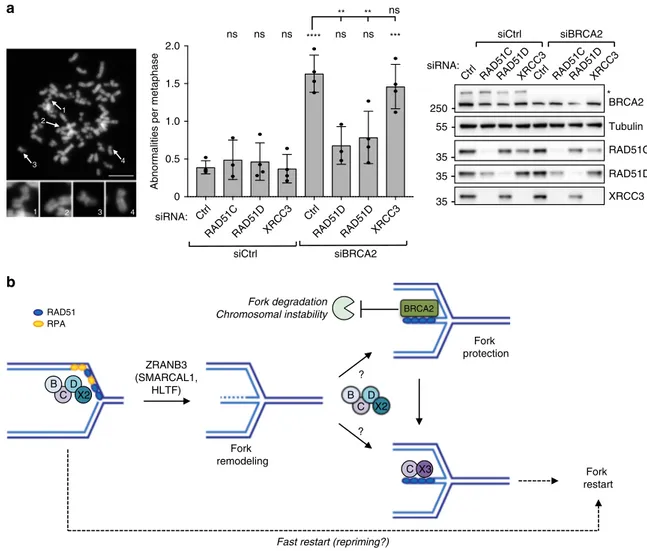
siRNA: 1 2 3 4 4 3 2 1 ? ? B D X2 C RAD51D XRCC3 RAD51D Ctrl RAD51D XRCC3 RAD51D RAD51C Ctrl XRCC3 RAD51D RAD51C Ctrl XRCC3 RAD51C CtrlFig. 5 BCDX2, but not CX3, promotes chromosomal instability in BRCA2-defective cells. a Metaphase spread analysis for detecting chromosomal
aberrations in U2OS cells double transfected with a control siRNA (Ctrl) or with siRNAs targetingRAD51C, RAD51D, and XRCC3 for 60 h and with BRCA2
siRNA for 48 h and treated with 4 mM HU for 5 hr. Left: representative metaphase spread image (scale-bar, 5μm); 1, 2, 3 representative breaks and 4
representative intact chromosome. Center: number of chromosomal abnormalities for each genetic condition. The graph-bar depicts mean and SD from at
least three independent experiments. Statistical analysis: one-way ANOVA followed by Bonferroni test; ns not significant; **p-value = 0.0012 (5 versus 6)
and 0.0020 (5 versus 7); ***p-value = 0.0001; ****p-value < 0.0001. Right: levels of indicated proteins, assessed by western blot; Tubulin, loading control;
asterisk, specific band. Multiple gels/blots were processed in parallel, ensuring equal and comparable loading across gels. Source data are provided in the
Source Datafile. b Proposed model for the sequential role of BCDX2 and CX3 paralog complexes in replication fork remodeling, protection and restart: the
BCDX2 complex helps RAD51 and ZRANB3 in driving fork remodeling. During fork stalling, regressed arms are protected from unscheduled nucleolytic
degradation by BRCA2-mediated stabilization of RAD51 nucleofilament. Preventing fork remodeling by BCDX2 inactivation suppresses genetic instability of
BRCA2-deficient cells upon sustained replicative stress. Similarly to RAD51, BCDX2 may be involved in fork protection (and possibly fork restart), but this
role may be genetically masked by its upstream role in promoting fork reversal. Once DNA synthesis can resume, the CX3 complex promotes reversed fork
restart, likely by engaging RAD51-bound regressed arms in efficient strand invasion events. In absence of fork remodeling, stalled forks undergo a fast, but
one hand fork reversal represents an important mechanism to
avoid replication stress-associated chromosomal breakage
5,8,58, it
can also drive clinically relevant pathogenic transactions in the
absence
of
functional
fork
protection
and
restart
mechanisms
10,13,20,22. Such data suggest that multiple defects in
the same remodeling pathway may occasionally combine, leading
to different outcomes in terms of genome stability. As
reversal-dependent degradation was shown to be a major molecular
determinant of chemosensitivity and -resistance in HR-defective
tumors
20, it will be essential to test the molecular phenotypes
upon cancer-associated mutations in RAD51 paralogs, and
thereby to possibly establish predictive parameters for
chemo-sensitivity in this subset of HR-defective tumors.
Methods
Cell culture and cell lines. Human U2OS and hTERT-RPE-1 cells were grown in Dulbecco’s modified Eagle’s medium (DMEM) supplemented with 10% fetal
bovine serum (FBS; GIBCO/Thermo Fisher Scientific) and 1%
penicillin-streptomycin antibiotics (100 U/mL penicillin and 100μg/mL streptomycin) under
standard cell culture conditions (humidified atmosphere, 6% CO2) at 37 °C. ZRANB3-proficient and Knock-Out U2OS cells were kindly provided by Dr. David Cortez.
RAD51 paralog CRISPSR-Cas9-based Knock-Out U2OS cells have been
generated and genetically characterized as recently reported46.
siRNA transfection and sequences. The QIBC-based siRNA screen was per-formed by reverse transfection of U2OS cells in CELLSTAR 96-well plates (Greiner Bio-One) for 48 h at a cell density of 5000 cells per well at the time of transfection
with Ambion Silencer Select siRNAs at afinal concentration of 5 nM using
HiPerFect reagent (Qiagen).
Individual siRNA transfections were performed using Lipofectamine RNAiMAX (ThermoFisher Scientific) at a concentration of 10 nM (Ambion Silencer Select) or 40 nM (Microsynth AG) according to manufacturer’s instruction and experiments performed between 48 and 72 h post transfection as indicated.
The oligonucleotides used for individual assays in this study are presented in Supplementary Table 2. Unless stated otherwise in the Figure legend siCtrl (as negative siRNA control), siRAD51C #2, siRAD51D #1, and XRCC3 #1 have been used.
Drugs and reagents. The following reagents were used to treat the cells for the
indicated time at the indicatedfinal concentrations before collection: Hydroxyurea
(HU; H8627, Sigma–Aldrich) was prepared in double-distilled H2O to obtain a
1 M stock and stored at−20 °C; Mirin (M9948, Sigma–Aldrich) was dissolved in
DMSO to yield a 50 mM stock and aliquots were stored at−80 °C; Camptothecin
(CPT; C9911, Sigma–Aldrich) was dissolved in DMSO to yield a 5 mM stock
(freshly made); Nocodazole (M1404, Sigma–Aldrich) was prepared in DMSO at the
final concentration of 1 mg ml−1, and aliquots were stored at−20 °C.
Immunoblotting. Cell were washed in cold PBS and lysed in NP-40 buffer (50 mM Tris-HCl [pH 7.5], 150 mM NaCl, 1% NP-40) supplemented with 1× protease inhibitor cocktail (cOmplete, Roche) and phosphatase inhibitors (20 mM NaF,
1 mM Na3VO4and 5 mM Na4P2O7). Cell extract were diluted with NuPAGE LDS
sample buffer (Thermo Fisher Scientific) containing DTT and heated at 60 °C for 10 min. Proteins, together with the PageRuler Plus Prestained Protein Ladder
(Thermo Fisher Scientific, 26620), were resolved on NuPAGE 4–12%, Bis-Tris or
3–8% Tris-Acetate gels (Thermo Fisher Scientific) using MOPS and Tris-Acetate SDS running buffers respectively, and transferred on nitrocellulose membranes. Membranes were blocked with TBS containing Tween-20 (0.1%) and 5% milk powder for 1 h at room temperature (RT), incubated with the indicated primary antibodies overnight at 4 °C and secondary antibodies for 1 h at RT. Protein were visualized using WesternBright ECL reagent (Advansta) and the Fusion Solo S imaging system (Vilber Lourmat). Uncropped and unprocessed scans of each blot
are provided in the Source Datafile.
The following primary antibodies were used: RAD51B mouse (sc-377192, Santa Cruz Biotechnology, 1:200), RAD51C rabbit (ab95069, Abcam, 1:2000), RAD51D rabbit (ab202063, Abcam, 1:500), XRCC2 mouse (sc-365854, Santa Cruz Biotechnology, 1:200), XRCC3 mouse (sc-271714, Santa Cruz Biotechnology, 1:200), BRCA2 mouse (OP-95, EMD Millipore, 1:500), RAD51 rabbit
(Bioacademia 70-002, 1:5000), ZRANB3 rabbit (23111-1-AP, Proteintech, 1:500),
KU70 mouse (ab202022, Abcam, 1:2000), andα-Tubulin mouse (T9026,
Sigma–Aldrich, 1:10000).
Immunostaining. Cells were grown on glass coverslips or 96-well plates, washed in PBS, pre-extracted for 5 min at 4 °C in CSK buffer (10 mM Hepes-KOH [pH 7.4], 300 mM sucrose, 50 mM NaCl, 3 mM MgCl2, 1 mM EGTA, 0.5% Triton X-100), fixed in 4% paraformaldehyde in PBS for 12 min at RT, washed twice in PBS,
permeabilized in PBS supplemented with 0.3% Triton X-100, washed twice in PBS and blocked with 3% BSA in PBS twice for 15 min. Rabbit polyclonal RAD51
antibody (Bioacademia 70-002, 1:2000) and secondary antibody (Alexa
fluor-ophores, Life Technologies) were diluted in 1.5% BSA in PBS and incubation were, respectively, performed for 2 or 1 h at RT. Coverslips were washed with PBS supplemented with 0.05% Tween-20 and incubated for 10 min with PBS containing
4′,6-Diamidino-2-Phenylindole Dihydrochloride (DAPI, 0.5 μg/mL) at RT to stain
DNA. Following three washing steps in PBS, coverslips were rinsed with distilled water, air-dried and mounted with ProLong Gold AntiFade (Thermo Fisher
Sci-entific). When indicated the Click-iT EdU Alexa Fluor Imaging Kit (Thermo Fisher
Scientific) was used for EdU detection according to manufacturer’s instruction. Quantitative image-based cytometry (QIBC). QIBC experiments were per-formed on an Olympus ScanR Screening System equipped with an inverted motorized Olympus IX83 microscope, a motorized stage, IR-laser hardware
autofocus, a fast emissionfilter wheel with single band emission filters, and a digital
monochrome Hamamatsu ORCA-FLASH 4.0 V2 sCMOS camera (2048 × 2048
pixel, pixel size 6.5 × 6.5μm, 12 bit dynamics), as described previously70,71. For
each condition, image information of large cohorts of cells (typically at least 500 cells for the UPLSAPO ×40 objective (NA 0.9) and at least 2000 cells for the UPLSAPO ×20 objective (NA 0.75)) was acquired under non-saturating condi-tions. Identical settings were applied to all samples within one experiment. Images were analyzed with the Olympus ScanR Image Analysis Software, a dynamic background correction was applied, nuclei segmentation was performed using an integrated intensity-based object detection module using the DAPI signal, and foci segmentation was performed using an integrated spot-detection module. All downstream analyses were focused on properly detected nuclei containing a 2C-4C DNA content as measured by total and mean DAPI intensities. Fluorescence
intensities were quantified and are depicted as arbitrary units. Color-coded
scat-terplots of asynchronous cell populations were generated with Spotfire data visualization software (TIBCO). Within one experiment, similar cell numbers were compared for the different conditions. For visualizing discrete data in scatterplots, mild jittering (random displacement of data points along the discrete data axes) was applied in order to demerge overlapping data points. For the siRNA screen,
genes were ranked according to a z-score (z= (x − μ)/σ with x being the mean
number of RAD51 foci per cell for each knockdown,μ being the mean number of
RAD51 foci per cell across all conditions, andσ being the standard deviation of the
mean number of RAD51 foci per cell across all conditions). Representative
scat-terplots and quantifications of independent experiments are shown. Representative
images, in which the individual color channels have been adjusted for brightness and contrast, accompany selected quantifications.
DNAfiber spreading analysis. Asynchronously growing cells were labeled with the
thymidine analogues 5-Chloro-2′-deoxyuridine (CldU, 30 μM), washed 3 times with
PBS, followed by 5-Iodo-2′-deoxyuridine (IdU, 250 μM) and treated with HU and
CPT as indicated58. The cells were quickly trypsinized and resuspended in ice-cold
PBS at 2.5 × 10⁵cells per ml. The labeled cells were diluted 1:1 with unlabeled cells,
and 3μl of cells were mixed with 7.5 μl of lysis buffer (200 mM Tris-HCl [pH 7.5],
50 mM EDTA, 0.5% (w/v) SDS) on a glass slide. After 9 min, the slides were tilted at 15°−45°, and the resulting DNA spreads were air-dried, fixed in 3:1 methanol/acetic
acid overnight at 4 °C. The DNAfibers were denatured with 2.5 M HCl for 1 h,
washed with PBS and blocked with 2% BSA in PBS supplemented with 0.1% Tween-20 for 40 min. The newly replicated CldU and IdU tracks were labeled (for 2.5 h in the dark, at RT) with anti-BrdU/CldU antibodies recognizing CldU (ab6326, Abcam, rat, 1:500) and BrdU/IdU (347580, Becton Dickinson, mouse, 1:100), respectively. After washing 5 × 3 min in PBS supplemented with 0.2% Tween-20, the following sec-ondary antibodies were used (incubated for 2 h in the dark, at RT): anti-mouse Alexa 488 (Molecular Probes, 1:300), anti-rat Cy3 (Jackson Immunoresearch, 1:150). After washing 5 × 3 min each in PBS supplemented with 0.2% Tween-20 the slides were air-dried completely and mounted with 20 uL/slide ProLong Gold AntiFade (Thermo Fisher Scientific). Images were acquired using an Olympus IX81 fluorescence microscope equipped with a CCD camera (Orca AG, Hamamatsu). CldU and IdU tract lengths were measured using the line tool in ImageJ64 software. In all the fork
restart experiments (Fig.4), defective fork restart (i.e., fork stalling) was defined as a
ratio (length of green vs red) <0.1.
Electron microscopic analysis of genomic DNA. Following the depletion of the protein of interest, asynchronous subconfluent cells were treated with 50 nM CPT for
1 h or 4 mM HU for 5 h. Where indicated, cells were pretreated with 50μM Mirin for
1 h. Cells were collected, resuspended in ice-cold PBS and crosslinked with 4,5′,
8-trimethylpsoralen (10μg/ml final concentration), followed by irradiation pulses with
UV 365 nm monochromatic light (UV Stratalinker 1800; Agilent Technologies). For
DNA extraction66, cells were lysed (1.28 M sucrose, 40 mM Tris-HCl [pH 7.5],
20 mM MgCl2, and 4% Triton X-100; Qiagen) and digested (800 mM guanidine–HCl,
30 mM Tris-HCl [pH 8.0], 30 mM EDTA [pH 8.0], 5% Tween-20, and 0.5% Triton X-100) at 50 °C for 2 h in presence of 1 mg/ml proteinase K. The DNA was purified using chloroform/isoamylalcohol (24:1) and precipitated in 0.7 volume of
iso-propanol. Finally, the DNA was washed with 70% EtOH and resuspended in 200μl
England Biolabs) were used to digest 6μg of mammalian genomic DNA for 5 h. RNase A (Sigma–Aldrich, R5503) to a final concentration of 250 ug/ml was added for the last 2 h of this incubation. The digested DNA was transferred into Microcon DNA
Fast Flow centrifugalfilters (Merck MRCF0R100) and washed two times with 200 ul
TE. Ultimately, the digested DNA was concentrated and recovered by inverting the filters. The Benzyldimethylalkylammonium chloride (BAC) method was used to spread the DNA on the water surface and then load it on carbon-coated 400-mesh nickel grids (G2400N, Plano Gmbh). Subsequently, DNA was coated with platinum using a High Vacuum Evaporator BAF060 (Leica). The grids were scanned using a
transmission electron microscope (Tecnai G2 Spirit; FEI; LaB6filament; high
ten-sion≤120 kV) and pictures were acquired with a side mount charge-coupled device
camera (2600 × 4000 pixels; Orius 1000; Gatan, Inc.) and processed with DigitalMi-crograph Version 1.83.842 (Gatan, Inc.). For each experimental condition at least70 replication fork molecules were analyzed in three different biological replicates by using ImageJ64 .
Chromosomal breakage and abnormalities by metaphase spreading. After transfection with the indicated siRNAs, cells were treated for 5 h with 4 mM HU. The compound was washed off three times with 1× PBS, upon which cells were released into fresh medium containing 200 ng/ml nocodazole for 16 h. Cells were harvested and swollen with 75 mM KCl for 20 min at 37 °C. Swollen mitotic cells were collected
andfixed with methanol:acetic acid (3:1). The fixing step was repeated two times.
Fixed cells were dropped onto prehydrated glass slides and air-dried overnight. The following day, slides were mounted with Vectashield medium containing DAPI. Microscopy was performed on a Leica DM6 B upright digital research microscope equipped with a DFC360 FX Leica camera. Images were analyzed using ImageJ64 and visible chromatid breaks/gaps were counted. For each experimental condition at least 50 metaphases were analyzed in three different biological replicates. Statistical analysis. For QIBC analysis, between 9 and 15 images per condition,
depending on the microscope objective used and the cell confluence were acquired
in an unbiased fashion from asynchronous cell populations grown on glass cov-erslips or multi-well plates. Typically, between 2000 and 5000 cells per condition were analyzed and representative single-cell data of cell cohorts of comparable size are shown as two-dimensional cell-cycle-resolved or one-dimensional scatterplots.
For DNAfiber length measurements, at least 150 fibers were scored for each
condition and every experiment was repeated at least twice. The results are depicted as median plus 10–90 percentile whisker plots and Kurskal–Wallis test was used for statistical analysis.
In all the other experiments, including DNAfiber analysis of fork restart, the
statistical significance for three different biological replicates was determined by
one-way ANOVA followed by Bonferroni test.
GraphPad Prism7 for MacOSX was used for all statistical analyses. Reporting summary. Further information on research design is available in the Nature Research Reporting Summary linked to this article.
Data availability
Source data are provided with this paper. All other data that support thefindings of this study are available from the authors upon reasonable request.
Received: 14 October 2019; Accepted: 16 June 2020;
References
1. Berti, M. & Vindigni, A. Replication stress: getting back on track. Nat. Struct.
Mol. Biol. 23, 103–109 (2016).
2. Forment, J. V. & O’Connor, M. J. Targeting the replication stress response in
cancer. Pharmacol. Ther. 188, 155–167 (2018).
3. Follonier, C., Oehler, J., Herrador, R. & Lopes, M. Friedreich’s
ataxia–associated GAA repeats induce replication-fork reversal and unusual
molecular junctions. Nat. Struct. Mol. Biol. 20, 486–494 (2013).
4. Neelsen, K. J., Zanini, I. M. Y., Herrador, R. & Lopes, M. Oncogenes induce
genotoxic stress by mitotic processing of unusual replication intermediates. J.
Cell Biol. 200, 699–708 (2013).
5. Zellweger, R. et al. Rad51-mediated replication fork reversal is a global
response to genotoxic treatments in human cells. J. Cell Biol. 208, 563–579 (2015).
6. Berti, M. et al. Human RECQ1 promotes restart of replication forks reversed by
DNA topoisomerase I inhibition. Nat. Struct. Mol. Biol. 20, 347–354 (2013).
7. Thangavel, S. et al. DNA2 drives processing and restart of reversed replication
forks in human cells. J. Cell Biol. 208, 545–562 (2015).
8. Neelsen, K. J. & Lopes, M. Replication fork reversal in eukaryotes: from dead
end to dynamic response. Nat. Rev. Mol. Cell Biol. 16, 207–220 (2015).
9. Cortez, D. Replication-coupled DNA repair. Mol. Cell 74, 866–876 (2019).
10. Berti, M., Cortez, D. & Lopes, M. The plasticity of DNA replication forks in response to clinically relevant genotoxic stress. Nat. Rev. Mol. Cell Biol.
10.1038–s41580–020–0257–5 (2020).
11. Betous, R. et al. SMARCAL1 catalyzes fork regression and Holliday junction migration to maintain genome stability during DNA replication. Genes Dev.
26, 151–162 (2012).
12. Kile, A. C. et al. HLTF’s ancient HIRAN domain binds 3′ DNA ends to drive replication fork reversal. Mol. Cell 58, 1090–1100 (2015).
13. Lemacon, D. et al. MRE11 and EXO1 nucleases degrade reversed forks and elicit MUS81-dependent fork rescue in BRCA2-deficient cells. Nat. Commun. 8, 860 (2017).
14. Mijic, S. et al. Replication fork reversal triggers fork degradation in BRCA2-defective cells. Nat. Commun. 8, 859 (2017).
15. Vujanovic, M. et al. Replication fork slowing and reversal upon genotoxic stress require PCNA polyubiquitination and ZRANB3 DNA translocase
activity. Mol. Cell 67, 882–890 (2017).
16. Bhat, K. P. & Cortez, D. RPA and RAD51: fork reversal, fork protection, and
genome stability. Nat. Struct. Mol. Biol. 25, 446–453 (2018).
17. Schlacher, K. et al. Double-strand break repair-independent role for BRCA2 in
blocking stalled replication fork degradation by MRE11. Cell 145, 529–542
(2011).
18. Schlacher, K., Wu, H. & Jasin, M. A distinct replication fork protection pathway connects Fanconi anemia tumor suppressors to RAD51-BRCA1/2. Cancer Cell 22, 106–116 (2012).
19. Wang, A. T. et al. A dominant mutation in human RAD51 reveals its function in DNA interstrand crosslink repair independent of homologous
recombination. Mol. Cell 59, 478–490 (2015).
20. Chaudhuri, A. R. et al. Replication fork stability confers chemoresistance in
BRCA-deficient cells. Nature 535, 382–387 (2016).
21. Przetocka, S. et al. CtIP-mediated fork protection synergizes with BRCA1 to suppress genomic instability upon DNA replication stress. Mol. Cell 72,
568–582.e6 (2018).
22. Taglialatela, A. et al. Restoration of replication fork stability in BRCA1- and
BRCA2-deficient cells by inactivation of SNF2-family fork remodelers. Mol.
Cell 68, 414–430.e8 (2017).
23. Kolinjivadi, A. M. et al. Smarcal1-mediated fork reversal triggers Mre11-dependent degradation of nascent DNA in the absence of Brca2 and stable Rad51 nucleofilaments. Mol. Cell 67, 867–881.e7 (2017).
24. Malacaria, E. et al. Rad52 prevents excessive replication fork reversal and protects from nascent strand degradation. Nat. Commun. 10, 1412–1419 (2019). 25. Sullivan, M. R. & Bernstein, K. A. RAD-ical new insights into RAD51
regulation. Genes (Basel) 9, 629 (2018).
26. Liu, T., Wan, L., Wu, Y., Chen, J. & Huang, J. hSWS1·SWSAP1 is an
evolutionarily conserved complex required for efficient homologous
recombination repair. J. Biol. Chem. 286, 41758–41766 (2011).
27. Abreu, C. M. et al. Shu complex SWS1-SWSAP1 promotes early steps in mouse meiotic recombination. Nat. Commun. 9, 3961 (2018).
28. Yonetani, Y. et al. Differential and collaborative actions of Rad51 paralog proteins in cellular response to DNA damage. Nucleic Acids Res. 33, 4544–4552 (2005).
29. Yokoyama, H. et al. Preferential binding to branched DNA strands and strand-annealing activity of the human Rad51B, Rad51C, Rad51D and Xrcc2 protein complex. Nucleic Acids Res. 32, 2556–2565 (2004).
30. Liu, N., Schild, D., Thelen, M. P. & Thompson, L. H. Involvement of Rad51C in two distinct protein complexes of Rad51 paralogs in human cells. Nucleic
Acids Res. 30, 1009–1015 (2002).
31. Masson, J. Y. et al. Identification and purification of two distinct complexes
containing thefive RAD51 paralogs. Genes Dev. 15, 3296–3307 (2001).
32. Miller, K. A. et al. RAD51C interacts with RAD51B and is central to a larger
protein complex in vivo exclusive of RAD51. J. Biol. Chem. 277, 8406–8411
(2002).
33. Takata, M. et al. Chromosome instability and defective recombinational repair
in knockout mutants of thefive Rad51 paralogs. Mol. Cell Biol. 21, 2858–2866
(2001).
34. Godthelp, B. C. et al. Mammalian Rad51C contributes to DNA cross-link resistance, sister chromatid cohesion and genomic stability. Nucleic Acids Res. 30, 2172–2182 (2002).
35. Bishop, D. K. et al. Xrcc3 is required for assembly of Rad51 complexes in vivo.
J. Biol. Chem. 273, 21482–21488 (1998).
36. Liu, N. et al. XRCC2 and XRCC3, new human Rad51-family members, promote chromosome stability and protect against DNA cross-links and other
damages. Mol. Cell 1, 783–793 (1998).
37. Pierce, A. J., Johnson, R. D., Thompson, L. H. & Jasin, M. XRCC3 promotes homology-directed repair of DNA damage in mammalian cells. Genes Dev. 13, 2633–2638 (1999).
38. French, C. A. et al. Role of mammalian RAD51L2 (RAD51C) in