HAL Id: hal-03097585
https://hal.archives-ouvertes.fr/hal-03097585
Submitted on 5 Jan 2021
HAL is a multi-disciplinary open access
archive for the deposit and dissemination of
sci-entific research documents, whether they are
pub-lished or not. The documents may come from
teaching and research institutions in France or
abroad, or from public or private research centers.
L’archive ouverte pluridisciplinaire HAL, est
destinée au dépôt et à la diffusion de documents
scientifiques de niveau recherche, publiés ou non,
émanant des établissements d’enseignement et de
recherche français ou étrangers, des laboratoires
publics ou privés.
Cullin 3, a cellular scripter of the non-proteolytic
ubiquitin code
Katerina Jerabkova, Izabela Sumara
To cite this version:
Katerina Jerabkova, Izabela Sumara.
Cullin 3, a cellular scripter of the non-proteolytic
ubiq-uitin code.
Seminars in Cell and Developmental Biology, Elsevier, 2019, 93, pp.100 - 110.
Contents lists available atScienceDirect
Seminars in Cell & Developmental Biology
journal homepage:www.elsevier.com/locate/semcdb
Review
Cullin 3, a cellular scripter of the non-proteolytic ubiquitin code
Katerina Jerabkova
a,b,c,d,e, Izabela Sumara
a,b,c,d,⁎aInstitut de Génétique et de Biologie Moléculaire et Cellulaire (IGBMC), Illkirch, France bCentre National de la Recherche Scientifique UMR 7104, Strasbourg, France cInstitut National de la Santé et de la Recherche Médicale U964, Strasbourg, France dUniversité de Strasbourg, Strasbourg, France
eInstitute of Molecular Genetics of the ASCR (IMG), Prague, Czech Republic
A R T I C L E I N F O Keywords: Cullin 3 Non-proteolytic signaling Ubiquitin code Substrates Cell division Development A B S T R A C T
Cullin-RING ubiquitin ligases (CRLs) represent the largest family of E3 ubiquitin ligases that control most if not all cellular processes. In CUL3-based CRLs, the substrate specificity is conferred by the interaction with one of around 183 existing BTB proteins, implying a broad spectrum of possible ubiquitylation signals and possible direct ubiquitylation substrates. Indeed, CUL3-based E3-ligases can catalyze various proteolytic and non-pro-teolytic ubiquitin signals regulating many physiological and pathophysiological states. Here, we discuss the recent studies focusing on the non-proteolytic CUL3-based signaling in mammalian cells, which emerge as im-portant pathways during cell division, embryonic development as well as other biological processes. Mechanistically, non-proteolytic ubiquitin signals generated by CUL3 E3-ligases often regulate substrates’ in-teractions with other downstream factors or their subcellular localization. Existing data also demonstrate an interplay with the proteolytic ubiquitylation catalyzed on the same substrates by different E3-ligases or by the same CUL3-BTB CRL3s on different substrates. In future, a deeper understanding of the upstream spatiotemporal regulatory mechanisms will help to dissect this fascinating CUL3 ubiquitin code.
1. Introduction
1.1. Diversity of the ubiquitin code
Since the discovery of ubiquitylation at the turn of years 70 s and 80 s [1], its important role has been implicated in almost all cellular processes in eukaryotes. Ubiquitylation relies on covalent attachment of ubiquitin to substrate proteins with help of E1, E2 and E3 enzymes and can be reversed by action of deubiquitylases (DUBs). Substrate proteins can be modified at their lysine (K) residues by single ubiquitin moiety (monoubiquitylation) or polymeric chains connected through either one of seven lysine residues or N-terminal methionine (M) in proximal ubiquitin (polyubiquitylation). In addition to homotypic ubiquitin polymers, recent studies support the important roles of mixed and branched polyubiquitin chains (heterotypic conjugates) composed of different linkages as well as multiubiquitylation, in which a single substrate is modified on several lysine residues (Fig. 1A). This plethora of possible ubiquitin modifications can be seen as “ubiquitin code” (Fig. 1A), which determines many different fates of modified substrates and is implicated in distinct cellular functions [2]. K48- and K11-linked ubiquitin chains usually target modified substrates for proteasomal
degradation [1,3]. Non-proteolytic functions of ubiquitylation were first discovered in 90 s implicating its role in regulating substrates’ in-tracellular localization, interactions and functions, such as for instance kinase activation [4–6]. For example, K63-linked ubiquitin chains regulate DNA repair, inflammation and endocytosis, M1-linked chains can activate nuclear factor kB (NF-kB) signaling and monoubiquityla-tion can control epigenetic pathways [7]. The complexity of ubiquitin code is further increased by the fact that ubiquitin can be modified by posttranslational modifications such as phosphorylation and acetyla-tion [8,9] and by the existence of chain editing or erasing DUBs as well as the effector proteins (ubiquitin receptors), which can specifically recognize and bind different ubiquitin signals [10], thereby targeting the modified substrates to downstream signaling components and cel-lular compartments. Since such a versatile ubiquitin code can ensure very robust and precise cellular signaling, it is not only important to understand the roles of different ubiquitin modifications on substrate proteins but also the enzymes conferring the substrate specificities, the E3 ubiquitin ligases.
https://doi.org/10.1016/j.semcdb.2018.12.007
Received 20 August 2018; Received in revised form 20 December 2018; Accepted 20 December 2018
⁎Corresponding author at: Institut de Génétique et de Biologie Moléculaire et Cellulaire (IGBMC), Illkirch, France.
1.2. Cullin 3
Cullin-RING ubiquitin ligases (CRLs) represent the largest family of E3 ubiquitin ligases. These multisubunit protein complexes are
assembled on the cullin scaffold proteins. Seven cullins CUL1, CUL2, CUL3, CUL4 A, CUL4B, CUL5 and CUL7 were reported to form different CRLs (CRL1, CRL2, CRL3, CRL4 A and B, CRL5 and CRL7, respectively) in human cells [11–13]. The C-terminal part of cullin proteins
Fig. 1. The ubiquitin code and Cul3-mediated non-proteolytic ubiquitin signaling.
(A) A schematic representation of possible ubiquity-lation modifications occurring in cells. Each type of ubiquitylation is depicted by a different color: mono-and multi-ubiquitylation, poly-ubiquitylation linked through any of seven Lysine (K) residues or N-term-inal Methionine (M1) as well as mixed and branched polyubiquitylation. Additional level of complexity is provided by posttranslational modifications on ubi-quitin molecules (not depicted in the scheme). Based on the steric positioning of the used residues, the polyubiquitin chains may adopt distinct conforma-tions (depicted in a schematic, inaccurate manner) ranging from more compact (K27, 29, 33, 48, bran-ched, mixed) to more linear (K6, K11, K63 and M1) topologies. (B) A schematic architecture of CUL3-based E3-ubiquitin ligase complexes (CRL3s). CUL3 (light gray) acts as a scaffold that interacts with the RBX1 (dark grey)/E2-Enzyme (yellow) module that transfers activated ubiquitin to the substrate (blue oval) and in its N-terminus with the BTB substrate adaptor protein providing substrate specificity sometimes with help of a substrate cofactors (or-ange). The ubiquitin-binding domain proteins (UBDs) (ubiquitin receptors, beige) may act downstream of the ubiquitylated substrates, while Deubiquitylating enzymes (DUBs, brown) reverse or edit the ubiquitin signals. (C) CUL3-BTB complexes can generate var-ious types of possible non-proteolytic signals on substrates (blue bold text) and thereby regulate de-picted fundamental biological processes.
associates with the RING-H2-domain proteins RBX1 or RBX2, which interact with the ubiquitin conjugating E2 enzymes and catalyze the transfer of ubiquitin to the substrates. Substrate specificity is mediated through the substrate-specific adaptor proteins that bind to the N-terminal part of cullins. In CUL3-based CRLs (CRL3s), the family of the BTB/POZ domain proteins (for “Bric-a-brac, Tramtrack and Broad complex/Pox virus and Zinc finger”, hereafter referred to simply as BTB) confers the substrate specificity [14–17]. In CRL3s, the BTB do-main interacts directly with the N-terminus of CUL3 (Fig. 1B). BTB proteins were first discovered in Drosophila [18] and are characterized by the presence of additional domains for protein-protein interactions (Kelch, Zinc-finger, MATH) that can directly recruit ubiquitylation substrates. Thus, a single adaptor molecule utilizing two protein in-teraction sites can link the CUL3 scaffold to the substrate, unlike in the CRL1s (with CUL1 as a scaffold), where separate linker and adaptor proteins build the substrate recognition module [11–13]. So far, around 183 BTB protein coding genes have been identified in human genome [19] from which 53 proteins have been shown to interact with CUL3 [20], suggesting a broad spectrum of possible ubiquitylation signals and substrates. Indeed, CUL3-based E3 ubiquitin ligases have been de-monstrated to catalyze various proteolytic and non-proteolytic ubi-quitin signals regulating different physiological and pathophysiological states [21,22]. In this review, we solely discuss the non-proteolytic CUL3-based signaling pathways, which emerge as important regulators of cell division, embryonic development but also many other important biological processes (Fig. 1C andTable 1).
2. Non-proteolytic CUL3 pathways during cell division and development
2.1. CUL3 and cell division
Cell division is a fundamental biological process that ensures precise partition of genetic material between two daughter cells during orga-nismal development. Defects in chromosome segregation are causally linked to different congenital diseases and to tumorigenesis. During prophase stage of eukaryotic mitotic division, the nuclear envelope is disassembled and chromosomes condense allowing the access of mi-crotubules that form the mitotic spindle. The duplicated centrosomes continue to separate to form symmetrical mitotic spindle, which makes
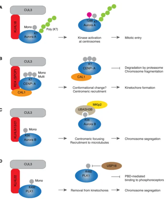
attachments to the kinetochores (kinetochore-microtubule (KT-MT) attachments) on all chromosomes during prometaphase. Only when all kinetochores are properly attached to microtubules and all chromo-somes are aligned at the metaphase plate, cells can separate their chromosomes. Following chromosome segregation during anaphase, the actinomyosin ring is formed and contracted to allow for the for-mation of the cleavage furrow and abscission during cytokinesis when two daughter cells are born. The coordinated action of protein kinases and phosphatases drives faithful mitotic progression in space and time [23]. Undoubtedly, the Anaphase Promoting Complex/Cyclosome (APC/C) is the main E3-ubiquitin ligase targeting the essential mitotic factors for proteasomal degradation thereby ensuring both direction-ality and irreversibility of cell division [24]. Interestingly, a number of proteolytic mitotic substrates of the APC/C are subject of non-proteo-lytic regulation by CUL3 during different mitotic stages. In human cells CUL3 localizes to the centrosomes before the onset of mitosis in late G2 and during prometaphase it associates with the mitotic spindle. The mitotic entry is dependent on the activation of cyclin-dependent kinase 1 (CDK1), which is directly controlled by mitotic cyclins and by the upstream phosphorylation signals involving, among others, polo-like kinase 1 (PLK1). Aurora A kinase localizes to the centrosomes leading to the activation of PLK1 resulting in CDK1 activation and mitotic entry [25–27]. CUL3 in a complex with KLHL18 adaptor protein is required for mitotic entry by activation of Aurora A at the centrosomes as monitored by threonine 288 phosphorylation located in the activation T loop of the kinase. CUL3-KLHL18 E3-ligase does not regulate localiza-tion of Aurora A to the centrosomes [28]. Unlike for the APC/C, which catalyzes ubiquitylation of Aurora A that targets this kinase for pro-teasomal degradation during mitotic exit [29], CUL3-KLHL18 drives non-proteolytic ubiquitylation (both mono and poly) of Aurora A (Fig. 2A). It would be interesting to understand what are the precise molecular mechanisms downstream of these modifications and if CUL3-mediated ubiquitylation of Aurora A stimulates interactions with any activatory proteins and/or ubiquitin receptors required for mitotic entry.
The centromere is the chromosomal region of kinetochore assembly, which directly attaches to the mitotic spindle. The histone H3 variant, CENP-A is an important marker for kinetochore specification and its recruitment to the centromere is necessary for the initiation of kine-tochore formation [30]. Assembly of the CENP-A-containing
Table 1
Substrates of CUL3-mediated non-proteolytic ubiquitylation. The table summarizes known non-proteolytic substrates of CUL3 CRLs. The roles of the indicated pathways are described in detail in the main text. The name of the substrate (in alphabetic order), ubiquitin site modification, type of ubiquitylation, adaptor protein and the specific function are depicted. “?” indicates unknown.
Substrate Site Type of Ub Adaptor protein Function
Aurora A ? Mono and poly KLHL18 Activation of Aurora A and mitotic entry
Aurora B K56 Mono KLHL9/
KLHL13/ KLHL21
Regulation of mitotic localization and chromosome segregation Caspase-8 K461,
other K Poly (K63 andK48-linked) ? Caspase-8 activation and aggregation, positive regulation of apoptosis
CENP-A ? Mono and multi RDX
CAL1
Centromeric levels of CENP-A and chromosome integrity Coronin7 K472 Poly (K33-linked) KLHL20 Localization of CRN7 to TGN and promoting post-Golgi trafficking D4.2,
D4.4
Lys Cys Ser/Thr
Poly KLHL12 Dopamine neurotransmission
EB1 K100 Mono KLHL21 Dynamics of cortical microtubules and FAs, cellular migration MacroH2 A1,
BMI1
? Mono,
Poly
SPOP Stable X chromosome inactivation
MCM3 K435 Poly KEAP1 MCM loading and regulation of DNA replication
PALB2 K20,
K25, K30 Mono KEAP1 Suppression of DNA damage repair by HR during G1
PLK1 K492 Mono KLHL22 Removal form kinetochores and faithful chromosome segregation SEC31 K647, K1217,
other K
Mono KLHL12 Assembly of large COPII coated vesicles and collagen secretion TCOF1, NOLC1 ? Mono KBTBD8 Ribosomes remodeling and neural crest specification
nucleosomes is independent of replication and relies on distinct loading factors. In Drosophila, loading of CENP-A on centromeres during mi-tosis depends on the factor called chromosome alignment defect 1 (CAL1), which is an ortholog of human AEG-1/MTDH/LYRIC. Regula-tion of CENP-A and CAL1 represent another interesting example for an interplay between proteolytic and non-proteolytic ubiquitylation (Fig. 2B). CUL3 associates with the adaptor protein Roadkill (RDX) (Drosophila ortholog of human Speckle-type POZ protein, SPOP), which leads to stabilization of CAL1 protein and the centromeric CENP-A le-vels. CAL1 functions as an additional linker protein that allows for mono- and multiubiquitylation of CENP-A perhaps by inducing a con-formational change or increasing the affinity to the chromatin of the CAL1/CENP-A complex [31]. Interestingly, in absence of RDX, CENP-A and CAL1 are subject of CUL3-indepentent degradation by proteasome, which leads to severe chromosome fragmentation and developmental defects (Fig. 2B). Thus, CUL3/RDX non-proteolytic ubiquitylation controls the amounts of CENP-A sufficient for centromeric main-tenance, while proteasomal degradation of CENP-A removes the excess of this protein [31].
Another direct substrate for CUL3-mediated non-proteolytic ubi-quitylation is Aurora B kinase, component of the chromosomal pas-senger complex (CPC). In addition to Aurora B, INCENP, Survivin and Borealin proteins are subunits of the CPC and all are co-dependent in
their mitotic localization and function [32]. The CPC is one of the key mitotic factors regulating a number of mitotic events including chro-mosome condensation, KT-MT attachment, activity of the Spindle As-sembly Checkpoint (SAC) and completion of cytokinesis. These mitotic CPC functions are insured by its dynamic localization during mitosis. Aurora B and entire CPC are found on chromosomes during prophase, concentrate at the centromeric region in prometaphase and metaphase and can be visualized in the midzone spindle region during anaphase [32]. CUL3-mediated monoubiquitylation of Aurora B is required for its centromeric focusing and spindle relocalization during anaphase [33]. Three different adaptor proteins KLHL9, KLH13 and KLHL21 can form a complex with CUL3 to monoubiquitylate Aurora B and to control mi-totic progression, chromosome segregation and completion of cyto-kinesis [33–36] (Fig. 2C). Identification of a downstream factor of Aurora B ubiquitylation, the ubiquitin receptor UBASH3B, unraveled important insights into spatiotemporal regulation of the CPC. UBASH3B can specifically interact with monoubiquitylated Aurora B at K56 using conserved UBA domain and with the motor protein MKlp2, which was previously implicated in relocalization of Aurora B to the anaphase midzone [37] (Fig. 2C). UBASH3B is both required and sufficient to transfer Aurora B to microtubules. Moreover, UBASH3B is able to trigger microtubule binding of INCENP, the component of CPC [37], which is important for the microtubule affinity of Aurora B [32].
Fig. 2. Non-proteolytic CUL3 pathways during cell division.
(A–D) CUL3 (gray) associates with the BTB substrate specific adaptors (red) sometimes with the help of cofactors (orange) to recruit specific substrate proteins (blue) involved in cell division (Aurora A, CENP-A, Aurora B and PLK1). The types of ubiquitin modifications and the modified lysine (K) residues (if known) are depicted. The cellular and biochemical ef-fects of substrates’ ubiquitylations are in-dicated (middle column) and their specific function in the biological process (right column). Pink circle depicts the activatory phosphorylation on Aurora A at Threonine (T) 288 (A), yellow depicts the microtubule motor protein MKlp2 that interacts with UBASH3B ubiquitin receptor (beige) to target Aurora B to microtubules (C), USP16 is DUB (brown) that can reverse monoubiquitylation of PLK1 (D). For details see the main text.
Depletion of UBASH3B leads to defects in centromeric focusing of Aurora B, inducing mitotic arrest, inhibition of chromosome segrega-tion and cellular death, while elevated levels of UBASH3B are sufficient to transfer Aurora B to microtubules even in the presence of high Cyclin B levels and prior to the onset of anaphase. Detailed microscopic ana-lysis demonstrated that Aurora B and MKlp2 can indeed associate with microtubules as they firmly attach to kinetochores before anaphase onset. Thus, UBASH3B can drive a switch-like mechanism of micro-tubule association of ubiquitylated Aurora B and CPC during mitosis [37]. This mechanism seems to be also important for fine tuning of mitotic checkpoint response (so called spindle assembly checkpoint, SAC), as high levels of UBASH3B (often observed in aggressive cancers) can silence SAC leading to premature and uncontrolled chromosome segregation and UBASH3B downregulation can potentiate SAC response
and inhibit proliferation of selected cancer cell lines [38]. Identification of this pathway may therefore open interesting therapeutic perspectives for future cancer therapies.
The role of monoubiquitylation in the regulation of dynamic loca-lization of substrates during mitosis can be also illustrated by the CUL3-PLK1 pathway. Substrate specific adaptor protein KLHL22 forms a complex with CUL3 and ubiquitylates lysine 492 (K492) located within the polo-box domain (PBD) of PLK1 [39,40] (Fig. 2D). PBD drives dy-namic localization of PLK1 during mitosis by phosphorylation-depen-dent association with various receptors including the kinetochore pro-tein BubR1 [41]. CUL3/KLHL22-mediated K492 ubiquitylation prevents binding of PLK1 to BubR1 and leads to removal of PLK1 from kinetochores at the metaphase to anaphase transition allowing for faithful chromosome segregation (Fig. 2D). Inhibition of this pathway
Fig. 3. Non-proteolytic CUL3 pathways during development.
(A–D) CUL3 (gray) associates with the BTB substrate specific adaptors (red) sometimes with the help of cofactors (orange) to recruit specific substrate proteins (blue) involved in development (BMI1, MacroH2 A1, SEC31, NOLC1 and TCOF1 and Caspase-8). The types of ubiquitin modifications and the modified lysine (K) residues (if known) are depicted. The cellular and biochemical effects of substrates’ ubiquitylations are indicated (middle column) and their specific function in the biological process (right column). Raise of Calcium (Ca2+) levels represents an activatory signal
for the CUL3-KLHL12 complex assembly with the cofactors PEF1 and ALG2 (orange) (B). p62 (beige) represents the ubiquitin receptor that interacts with ubiquitylated Caspase-8 (D). For details see the main text.
leads to accumulation of PLK1 at kinetochores, chromosome alignment defects, SAC potentiation and mitotic arrest resulting in apoptotic death [39,42,43]. It would be interesting to understand if kinetochore re-moval of K492-ubiquitylated PLK1 is helped by an action of a down-stream ubiquitin receptor analogous to UBASH3B. Nevertheless, the DUB USP16 was demonstrated to reverse the action of CUL3-KHL22 allowing for maintenance of kinetochore PLK1 levels prior to meta-phase and for establishment of initial KT-MT attachments and chro-mosome alignment [44] (Fig. 2D). Thus, USP16 and CUL3 ensure equilibrium of PLK1 ubiquitylation and faithful chromosome segrega-tion during mitosis [44,45]. Interestingly and similar to other substrates of CUL3, both Aurora B and PLK1 are also a subject of APC/C-mediated proteolytic ubiquitylation during mitotic exit [46].
2.2. CUL3 and development
Human development relies not only on faithful cell division but also on precise execution of differentiation programs and on cellular growth. During embryogenesis cells undergo fast rounds of divisions that give rise to various cell types, including pluripotent stem cells. The stem cells migrate and adopt specific fates to build all tissues of the growing embryo in a spatially and temporally coordinated manner. Gene ex-pression programs, epigenetic modifications and environmental factors determine distinct differentiation processes and defects in these path-ways can lead to severe congenital defects. In addition to its crucial involvement in cell division, CUL3-based complexes play a pivotal role during multiple stages of human development [47,48].
Involvement of CUL3 in early developmental stages is illustrated by its role in X chromosome inactivation in cells of female mammals. X chromosome carries around 1000 genes that are important for cell viability and proper development. Females carry two copies of X chromosome creating a possibility for toxic dosage of those genes. Therefore, a complex mechanism of induction of facultative hetero-chromatin on one X chromosome leads to its transcriptional silencing and keeps it in an inactive state. The non-coding RNA Xist (X-inactive specific transcript) labels the chromosome to be inactivated and recruits the complexes necessary for the silencing such as the polycomb group (PcG) proteins. The PcG consists of the initiation core complex (PRC2), which ubiquitylates histone H3 and this mark is further recognized by the maintaining complex (PRC1), which monoubiquitylates H2 A in embryonic and extraembryonic cells. Stably inactivated chromosome (Xi) carries several features of constitutive heterochromatin including histone hypoacetylation, DNA hypermethylation and enrichment of histone variant MacroH2 A. Interestingly, CUL3 together with the adaptor protein SPOP forms a complex with the member of the PRC1 complex BMI1 and separately with the histone variant MacroH2 A1 and ubiquitylates both of these substrates in human cells [49] (Fig. 3A). BMI1 is monoubiquitylated by CUL3, while MacroH2 A1 is both monoubiquitylated and polyubiquitylated, without inducing its pro-teasomal degradation (Fig. 3A). In female primary lung fibroblasts, MacroH2 A1 is strongly enriched at Xi site and colocalizes with BMI1 protein together with other components of PRC1 complex mostly during S-phase. Importantly, CUL3 or SPOP silencing abolishes locali-zation of MacroH2 A1 to Xi without affecting cell cycle progression. Additionally, silencing of CUL3, SPOP or MacroH2 A1, in the presence of DNA methylation and histone deacetylation inhibitors, leads to re-activation of Xi in female mouse fibroblasts (Fig. 3A). This suggests a crucial role for CUL3-mediated targeting of MacroH2 A1 to Xi and transcriptional silencing important for maintaining stable X chromo-some inactivation in synergy with other repressive imprints [49].
CUL3 also critically controls various differentiation programs in-cluding myogenesis, neurogenesis, chondriogenesis, osteogenesis and adipogenesis [47] and many of these pathways are based on non-pro-teolytic ubiquitin signaling. A study by Jin et al [50] identified a set of 31 BTB proteins that can interact with CUL3 in embryonic stem (ES) cells and several of them displayed a differential expression during
differentiation process. Similar to Nanog or other stem cell regulators [51], KLHL12, KBTBD8 and IBTK were highly expressed in mouse ES cells but downregulated during differentiation [50]. Indeed, during chondriogenesis, CUL3 paired with KLHL12 adaptor protein plays an important role in protein export by regulating the formation of mem-brane vesicles for secretion of proteins from transitional endoplasmic reticulum (tER, specialized regions of ER). Formation of these vesicles is carried out by Coat protein complex II (COPII), which consist of five core proteins: SAR1, SEC23, SEC24, SEC13 and SEC31 that are neces-sary for proper vesicle formation [52]. Size of these vesicles is critical for mediating export of small or large cargos. Interestingly, KLHL12 localizes to COPII vesicles and mediates monoubiquitylation of SEC31, which is crucial for formation of large vesicles allowing for collagen export to extracellular matrix (ECM) (Fig. 3B). Smaller cargos like Fi-bronectin and EGFR are unaffected by CUL3-KLHL12 depletion, sug-gesting that CUL3-mediated ubiquitylation is crucial for the assembly of large COPII coats, but dispensable for trafficking of smaller COPII ve-sicles. This mechanism is critical during early development when mouse ES cells undergo integrin-dependent division and thus proper ECM organization. Depletion of CUL3 or KLHL12 in mouse ES cells leads to cell clustering with aberrant adhesions and a delay in pro-liferation, which is reminiscent of depletion of integrin signaling pathways components [50]. The function of monoubiquitylated SEC31 does not appear to be strictly dependent on the position of specific ly-sine residues in cells somewhat questioning existence of an ubiquitin receptor for SEC31. Therefore, it would be interesting to understand mechanistic basis of COPII vesicle assembly acting downstream of SEC31. Excitingly, the upstream signals responsible for the activation of CUL3-KLHL12 during chondrocyte differentiation rely on a transient rise in cytosolic calcium levels allowing for the recruitment of calcium-binding proteins PEF1 and ALG2, which act as substrate specific co-adaptor factors for CUL3-KLHL12 E3 ligase and thereby enhanced SEC31 recognition and monoubiquitylation and collagen secretion [53] (Fig. 3B). This mechanism links CUL3-mediated ubiquitylation to cal-cium-regulated chondrocyte differentiation and craniofacial bone for-mation.
Another CUL3 adaptor protein highly expressed in ES cells, KBTBD8 plays an important role during development by promoting neural crest specification [50]. Neural crest is a group of cells originating from the neural tube that undergo extensive migration. Depending on the region they migrate to, they can give rise to a large number of differentiated cell types including neurons, adrenal gland and skeletal and connective tissue components of the head. CUL3-KBTBD8 complex is essential for expression of neural crest markers [50] (Fig. 3C). Depletion of CUL3 or KBTBD8 leads to severe loss of neural crest markers including FOXD3 and SOX10 and increase in central nervous system (CNS) precursors [54]. Importantly, CUL3-KBTBD8 in a complex with β-Arrestin as a cofactor, monoubiquitylates two paralogs NOLC1 and TCOF1. Mono-ubiquitylation stabilizes interaction of NOLC1 and TCOF1 and is ne-cessary for forming an interacting platform for RNA polymerase I re-quired for ribosomal remodeling (Fig. 3C). KBTBD8 depletion does not affect the total ribosome biogenesis, but it remodels the translational program of differentiating cells undergoing neural conversion. KBTBD8 specifically suppresses production of proteins from mRNAs coding for CNS precursors and does not affect translation of other mRNAs. Taken together, CUL3-KBTBD8 mediates remodeling of ribosomes that alters translation of specific mRNAs until cells have accomplished neural crest specification [54]. Mutation in TCOF1 and in RNA polymerase I cause the Treacher Collins Syndrome, a craniofacial developmental disorder, which is characterized by loss of cranial neural cells. Thus, the dis-covery of the role of CUL3-mediated ubiquitylation provides some im-portant insights into pathophysiology of this pediatric disease [54]. Interestingly, CUL3-KBTBD8-mediated monoubiquitylation requires prior multisite phosphorylation by CK2 kinase whose levels increase during embryogenesis at times of neural crest development [55]. Phosphorylation is a prerequisite for substrate recognition by SCF
E3-ligases [56], while APC/C often uses specialized motifs for substrate binding [24]. It seems therefore that CUL3-based complexes utilize different mechanisms including short motifs and oxidation [57,58], calcium levels [53] and phosphorylation [55] to confer substrate spe-cificity and spatiotemporal activation. In addition to CUL3-KBTBD8, CUL3-BTBD6 complex was shown to play an important role during late neuronal development and muscle formation although the direct CUL3-BTBD6 substrates have not been identified to date [59]. Differentiation programs of B and T cells can also be regulated by CUL3 in a complex with PLZF and BCL6 transcription factors that act as adaptor proteins recruiting CUL3 to the nucleus and regulating ubiquitylation pattern of chromatin factors [60,61], however the direct substrates of this E3-li-gase remain to be identified. CUL3 has been also implicated in the regulation of angiogenesis as illustrated by CUL3- BAZF-mediated
degradation of Notch signaling components, which leads to transcrip-tional regulation of genes promoting formation of new blood vessels [62]. However, CUL3 downregulation leads to more severe phenotype than BAZF depletion, suggesting an additional role of CUL3 in angio-genesis. Interestingly, CUL3 in a complex with another BTB adaptor ANKFY1 is required for Integrinβ1 localization to the plasma mem-brane, and thereby for cell adhesion and cellular spreading [63]. Better understanding of the mechanism of CUL3-mediated regulation of an-giogenesis, in particular identification of relevant direct substrates will likely help future development of more specific drugs to selectively inhibit angiogenic pathways during carcinogenesis.
Apoptosis is a complex process of programmed cellular death, which is important for embryonic development and adult tissue maintenance. Cell death can be activated from the inside of a cell (intrinsic pathway)
Fig. 4. Additional non-proteolytic CUL3 path-ways.
(A–E) CUL3 (gray) associates with the BTB substrate specific adaptors (red) sometimes with the help of cofactors (orange) to recruit specific substrate proteins (blue) involved in various
cellular processes, MCM3 in DNA replication, PALB2 in DNA repair, Coronin7 in protein trafficking, EB1 in cytoskeletal dynamics and migration and D4.2/D4.4 in neurotransmis-sion. The types of ubiquitin modifications and the modified lysine (K) residues (if known) are depicted. The cellular and biochemical effects of substrates’ ubiquitylations are indicated (middle column) and their specific function in the biological process (right column). USP11 is DUB (brown) that can reverse mono-ubiquitylation of PALB2 (B). EPS15 is the ubi-quitin receptor (beige) that interacts with K33-ubiquitylated Coronin7 (C). The precise mole-cular events triggered by D4.2/D4.4 poly-ubiquitylation (E) are currently unknown. For details see the main text.
or by outside stimulus as proapoptotic ligands (extrinsic pathway), which are then mediated by different downstream effectors. Extrinsic pathway is mediated through Caspase-8 and comprises of a cascade of events transmitting the ligand signal through death receptors (DR4, DR5), which trigger apoptosis by death-inducing signaling complex (DISC). Caspase-8 is first recruited to DISC, which mediates activation and self-processing of Caspase-8 releasing cleavage products to the cytoplasm for further activation of effector caspases. Proximity-induced dimerization stimulates activity of Caspase-8, which is potentiated by further aggregation critically regulated by CUL3. CUL3 interacts with Caspase-8 and catalyzes its polyubiquitylation in the C-terminal region, preferentially at K461 leading to Caspase-8 activation [64] (Fig. 3D). Both K63- and K48-linked polyubiquitylation of Caspase-8 are gener-ated by CUL3 and can lead to Caspase-8 activation. Biochemical ana-lysis of DISC-associated factors revealed presence of ubiquitin-binding polypeptide p62/Sequestosome-1, whose interaction is dependent on Caspase-8 modification by CUL3 and promotes Caspase-8 localization to ubiquitin-positive foci, further stimulating aggregation of Caspase-8 (Fig. 3D). This mechanism demonstrates an important role of CUL3 as a positive regulator of cell death through extrinsic apoptotic pathway [64]. It would be interesting to understand the identity of substrate specific adaptor or cofactor proteins involved in this pathway. 3. Additional non-proteolytic CUL3-based pathways
Besides their roles in cell division and development, CUL3-based complexes emerge as important regulators of non-proteolytic ubiquitin signaling involved of many other biological processes. These include DNA metabolism, intracellular transport, cytoskeletal remodeling, cell migration and neurotransmission. Future studies will likely demon-strate a direct or indirect involvement of these pathways in organismal development and/or link to human pathologies.
3.1. CUL3 during DNA synthesis and repair
Genome integrity relies on precise replication of genetic material during S phase whereby regulatory mechanisms ensure that DNA du-plication occurs only once during each cell division. This regulation is achieved by cell cycle dependent control of the replicative DNA heli-case, minichromosome maintenance (MCM) protein complex, an en-zyme responsible for DNA unwinding. Subunits of the hexameric MCM2-7 complex are extensively modified by posttranslational mod-ifications to ensure their tight regulation during cell cycle. The complex is loaded on chromosomes during G1 phase, activated in S phase and progressively unloaded upon replication fork termination [65]. Inter-estingly, biochemical analysis identified MCM3 as a Kelch-like ECH-associated protein 1 (KEAP1)-interacting factor and ubiquitylation substrate [66] (Fig. 4A). KEAP1 is a substrate specific adaptor of CUL3 complex that is involved in oxidative response through proteasomal targeting of antioxidant transcription factor NRF2 [67]. KEAP1 as-sociates with MDM3 during normal cell cycle and is loaded onto chromosomes during G1 together with MCM3 and unloaded in G2, after dissociation of MCM3 from chromatin [66]. Available data suggest that KEAP1 interacts with MCM3 when it is part of the entire MCM2-7 complex. CUL3-KEAP1 complex can ubiquitylate MCM3 on six lysine residues K229, K270, K283, K351, K435, K748 whereby K435 appears to be the main ubiquitin acceptor site (Fig. 4A). In contrast to some other established substrates of KEAP1, MCM3 is not targeted for pro-teasomal or lysosomal degradation and is not responsive to DNA-da-maging agents or to Reactive Oxygen Species (ROS) but may regulate MCM chromatin loading and/or its helicase activity [66] through some downstream MCM3 factors. Interestingly, in Xenopus laevis MCM7 subunit is polyubiquitylated specifically at the time of fork termination by yet unknown ligase. MCM7 is modified by K48 ubiquitin chain leading to unloading of MCM complex from chromatin, which can be reversed by treatment with the neddylation inhibitor MLN4924,
suggesting involvement of a CRL [68]. CUL3-KCTD5-MCM7 complex formation and CUL3-mediated MCM7 polyubiquitylation has been re-ported, yet the biological role of this modification remains unclear [69]. Integrity and stability of DNA are essential for cell viability and for correct transfer of genetic information to the daughter cells. To protect cellular genome from environmental, metabolic and replication da-mage, a complex DNA repair machinery has evolved [70]. Homologous recombination (HR) is one of the mechanisms of DNA repair, which occurs in late S and G2 phases and is suppressed during G1 phase of the cell cycle, in order to ensure that recombination occurs solely between two sister chromatids. Breast and ovarian tumor suppressors BRCA1, PALB2 and BRCA2 drive DNA repair of double strand breaks (DSB) by HR. BRCA1 interacts with PALB2 to recruit BRCA2 and RAD51 to the DSB site, an event which is abolished during G1 phase of cell cycle resulting in potent suppression of HR. Interestingly, the disruption of the interaction of BRCA1 with PALB2 is mediated by KEAP1 [71]. KEAP1 can recognize a short ETGE motif in PALB2 analogous to NRF2 [72]. CUL3-KEAP1 ubiquitylates N-terminus of PALB2 during G1 phase to suppress PALB2 binding to BRCA1, whereby K20, K25 and K30 sites are crucial for suppression of PALB2-BRCA1 interaction (Fig. 4B). In-terestingly, chemically induced monoubiquitylation-mimic mutant at K20 of PALB2 completely suppresses the interaction with BRCA1. PALB2 ubiquitylation can be reversed by USP11 deubiquitylase, re-storing formation of BRCA1-PALB2-BRCA2 complex (Fig. 4B). There is a high turnover of USP11 controlled by CUL4 during G1, which is also responsible for efficient USP11 degradation upon DNA-damage, thereby opposing CUL3-mediated regulation of BRCA1-PALB2-BRCA2 complex. This study demonstrates that suppression of HR in G1 is highly re-versible and plays a crucial role in genome integrity maintenance [71]. Taken together, based on these studies, KEAP1 emerges as an important CUL3 substrate adaptor in genome integrity maintenance, capable of supporting both proteolytic and non-proteolytic ubiquitylation under normal and stress conditions. Since KEAP1 mutations have been re-ported in various cancers [67], a deep understanding of underlying molecular mechanisms, in particular the factors or conditions that de-termine the type of substrate’s ubiquitylation by KEAP1, is urgently needed.
3.2. CUL3 in vesicle transport and in cytoskeletal remodeling
Cytoskeleton formed by microtubules and actin filaments creates a basis for intracellular protein transport, determines cellular shape and enables migration of cells. All these processes are pivotal for organismal development and critically depend on CUL3-based proteolytic as well as non-proteolytic ubiquitin signaling pathways [47].
Trans-Golgi network (TGN) is the major sorting complex where newly synthesized proteins are packed and transported in carriers to different compartments of the cell. CUL3-KLHL20 localizes to the TGN where it promotes biogenesis of TGN derived transport carriers [73]. It physically interacts with Coronin7 (Crn7) in 835–865 region of this protein. CUL3 mediates non-proteolytic, K33-linked polyubiquitylation of Crn7, which induces formation of TGN carriers by stimulating F-actin assembly at the Golgi. Ubiquitin binding protein EPS15 promotes Crn7 localization to Golgi (Fig. 4C). Indeed, downregulation of KLHL20, CUL3 or EPS15 or expression of the ubiquitylation mutant (K472R) of Crn7 prevents the assembly of the post-Golgi carrier tubules and in-hibits protein trafficking. Forced targeting of Crn7 to TGN is sufficient to facilitate post-Golgi trafficking, suggesting that the primary role of KLHL20-mediated ubiquitylation is to localize Crn7 to TGN (Fig. 4C). Thus, this study not only identifies an important role of CUL3 in protein trafficking but also unravels a novel signaling function of K33-linked polyubiquitin chains [73]. Interestingly, EPS15 levels can be controlled by CUL3 in a complex with the Speckle-type POZ protein-like (SPOPL) adaptor protein, which regulates endocytic trafficking and endosome maturation [74].
cellular migration. Focal adhesions (FA) are assembled at the cell front and disassembled at the rear and cortical microtubules are known to interact with FAs. Microtubule dynamics are controlled by opposing actions of microtubule stabilizing and destabilizing factors including a protein family that can specifically interact with the growing plus ends of microtubule polymers. For instance, end-binding 1 (EB1) protein can dynamically track microtubule tips by recognizing structural elements on growing microtubule ends. CUL3-KLHL21 monoubiquitylates EB1 thereby regulating microtubule dynamics at the cortex and FAs [75] (Fig. 4D). Interestingly, KLHL21 can transiently localize to the micro-tubule tips and FAs specifically at the leading edge of migrating cells. K100 located with the conserved Calponin Homology (CH) domain is the major ubiquitin acceptor site and its ubiquitylation may interfere with ability of EB1 to interact with microtubule ends (Fig. 4D). Non-ubiquitylable EB1 mutant does not support cellular migration and sta-bilizes ends of cortical microtubules and microtubules at the FAs, in-hibiting assembly and disassembly of FAs. Thus, the cross-talk of mi-crotubules reaching cortex and FAs is regulated by CUL3-KLHL21-mediated ubiquitylation of EB1 [75]. Since defects in cellular migration are linked to developmental disorders and to carcinogenesis, this study further confirms the important role of CUL3-mediated signaling. Moreover, it illustrates that highly dynamic and rapid cellular events can be dependent on ubiquitin attachment analogous to other post-translational modifications.
3.3. CUL3 in neurotransmission
In addition to its important roles in neural development, CUL3 also appears to control the function of the CNS. The neurotransmitter do-pamine is the primary ligand for dodo-pamine receptors, which are im-plicated in many neurological processes including cognition, learning, motor control, reward and endocrine signaling. Dopamine receptors are members of G-protein coupled receptors family that regulate adenylyl cyclase activity by binding to different G protein subunits. There are five types of dopamine receptors from two different families, where D4 receptor is particularly interesting due to its high polymorphism in a third intracellular (IC3) loop. A 16 amino acid sequence is repeated 2 to 11 times within IC3 loop, introducing different receptor variants de-signated as D4.2 to D4.11 with D4.2, D4.4 and D4.7 being most abundant. CUL3 adaptor KLHL12 specifically interacts with D4 receptor (D4.2, D4.4, D4.7), within the IC3 loop, but not with D1, D2L, D5 or M1-muskarinic orβ2-adrenergic receptors, and targets D4 for ubiqui-tylation [76] (Fig. 4E). Subsequent studies show that CUL3-KLHL12 interacts with both immature ER-associated D4 receptor and with ma-ture D4 receptors at plasma membrane, mediating non-proteolytic polyubiquitylation of both receptor types. D4 receptor ubiquitylation is not required for its localization to plasma membrane. Interestingly, D4 receptor interacts withβ-Arrestin, but despite the fact that KLHL12 is also able to bind toβ-Arrestin, this interaction does not depend on the ubiquitylation status of neither of those proteins and KLHL12 does not affect β-Arrestin ubiquitylation or localization [77] suggesting a pos-sibility that β-Arrestin acts as a cofactor similiar to CUL3-KBTBD6 complex (Figs. 3C,4E). Interestingly, a later study shows that D4 re-ceptors undergo unconventional ubiquitylation on non-lysine residues including cysteine, serine and threonine, suggesting that CUL3-KLHL12 complex can also catalyze non-lysine ubiquitylation [78]. Although CUL3-KLHL12 mediates specific ubiquitylation of D4.2 and D4.4, but not D4.7 dopamine receptor variants [78], the biological roles of these specific modifications in a context of agonist stimulation remain to be investigated.
4. General conclusions and future perspectives
Based on available studies, CUL3-based CRLs emerge as important regulators of fundamental cellular processes, many of which depend on non-proteolytic signaling and have a direct link to human pathology.
CUL3 is a very versatile writer of the non-proteolytic ubiquitin code. The non-proteolytic CUL3 signals range from mono- and multi-ubiquitylation to various types of polyubiquitin chains and can even occur on non-lysine residues. CUL3-mediated ubiquitylation can therefore regulate various fates of substrates. The most characterized outcomes of non-proteolytic substrates’ modifications by CUL3 are the changes in their intracellular localization (microtubules, kinetochores, chromatin, FAs) and in interactions with other factors or large protein complexes or assemblies. Future identification of the downstream modifiers, the effectors or the ubiquitin receptor proteins will certainly help a deep understanding of specific molecular fates of CUL3 sub-strates. Unfortunately, to date, only a handful of ubiquitin receptors and deubiquitylating enzymes was identified that can act in concert with CUL3 complexes. As exemplified by UBASH3B, the ubiquitin receptor for Aurora B or by the DUB USP16 that opposes the CUL3-KLHL22-mediated monoubiquitylation of PLK1, identification of such factors can not only deepen our molecular understanding of CUL3-based pathways but also provide some relevant insights for human patholo-gies. Another important question arises as to how CUL3 can switch the catalysis from the non-proteolytic to proteolytic signals. Binding of specific E2 enzymes to CUL3 complexes can very likely explain this ability but one could also envisage that the presence of different “de-grons” or motifs in the substrate proteins contributes to the strength of substrate binding and to catalysis of specific ubiquitin signals. For in-stance, proteolytic CUL3-KEAP1 substrate NRF2 contains two degrons, ETGE and DLG, while PALB2 containing one functional ETGE motif is not targeted for degradation by CUL3-KEAP1. It also appears that substrate binding by CUL3-BTB complexes is not restricted to motifs but can be also controlled by prior substrate phosphorylation somewhat analogous to CRL1 complexes or by changes in the intracellular calcium levels. This pioneering work lay a background for important future studies aiming at understanding the upstream molecular signals that control spatiotemporal activation of CUL3 E3-ligases that will further help to dissect the fascinating world of CUL3 non-proteolytic ubiquitin code.
Acknowledgements
We thank all members of Sumara group, Manuel Mendoza and Bill Keys for helpful discussions on the manuscript. We apologize to the many authors whose work could not be cited due to space restrictions. K.J. is supported by a fellowship from Gouvernement français et L’Institut français de Prague, a LabEx international PhD fellowship from IGBMC and a fellowship from the “Ligue Nationale Contre le Cancer”. This work was supported by the grant ANR-10-LABX-0030-INRT, a French State fund managed by the Agence Nationale de la Recherche under the frame program Investissements d’Avenir ANR-10-IDEX-0002-02. Research in I.S. laboratory is supported by IGBMC, CNRS, Fondation ARC pour la recherche sur le cancer, Institut National du Cancer (INCa), Ligue Nationale contre le Cancer, ANR and Sanofi-Aventis European Innovation Award (iAward).
References
[1] A. Hershko, A. Ciechanover, The ubiquitin system, Annu. Rev. Biochem. 67 (1998) 425–479,https://doi.org/10.1146/annurev.biochem.67.1.425.
[2] D. Komander, M. Rape, The ubiquitin code, Annu. Rev. Biochem. 81 (2012) 203–229,https://doi.org/10.1146/annurev-biochem-060310-170328.
[3] K.E. Wickliffe, A. Williamson, H.-J. Meyer, A. Kelly, M. Rape, K11-linked ubiquitin chains as novel regulators of cell division, Trends Cell Biol. 21 (2011) 656–663,
https://doi.org/10.1016/j.tcb.2011.08.008.
[4] Z.J. Chen, L. Parent, T. Maniatis, Site-specific phosphorylation of IkappaBalpha by a novel ubiquitination-dependent protein kinase activity, Cell 84 (1996) 853–862,
https://doi.org/10.1016/S0092-8674(00)81064-8.
[5] S. Janssens, A. Tinel, S. Lippens, J. Tschopp, PIDD mediates NF-kappaB activation in response to DNA damage, Cell 123 (2005) 1079–1092,https://doi.org/10.1016/ j.cell.2005.09.036.
[6] G.Y. Zhao, E. Sonoda, L.J. Barber, H. Oka, Y. Murakawa, K. Yamada, T. Ikura, X. Wang, M. Kobayashi, K. Yamamoto, S.J. Boulton, S. Takeda, A critical role for the
ubiquitin-conjugating enzyme Ubc13 in initiating homologous recombination, Mol. Cell 25 (2007) 663–675,https://doi.org/10.1016/j.molcel.2007.01.029. [7] Y. Kulathu, D. Komander, Atypical ubiquitylation – the unexplored world of
poly-ubiquitin beyond Lys48 and Lys63 linkages, Nat. Rev. Mol. Cell Biol. 13 (2012) 508–523,https://doi.org/10.1038/nrm3394.
[8] L. Herhaus, I. Dikic, Expanding the ubiquitin code through post-translational modification, EMBO Rep. 16 (2015) 1071–1083,https://doi.org/10.15252/embr. 201540891.
[9] K.N. Swatek, D. Komander, Ubiquitin modifications, Cell Res. 26 (2016) 399–422,
https://doi.org/10.1038/cr.2016.39.
[10] R. Yau, M. Rape, The increasing complexity of the ubiquitin code, Nat. Cell Biol. 18 (2016) 579–586,https://doi.org/10.1038/ncb3358.
[11] R.J. Deshaies, C.A.P. Joazeiro, RING domain E3 ubiquitin ligases, Annu. Rev. Biochem. 78 (2009) 399–434,https://doi.org/10.1146/annurev.biochem.78. 101807.093809.
[12] J.R. Lydeard, B.A. Schulman, J.W. Harper, Building and remodelling Cullin-RING E3 ubiquitin ligases, EMBO Rep. 14 (2013) 1050–1061,https://doi.org/10.1038/ embor.2013.173.
[13] E.S. Zimmerman, B.A. Schulman, N. Zheng, Structural assembly of cullin-RING ubiquitin ligase complexes, Curr. Opin. Struct. Biol. 20 (2010) 714–721,https:// doi.org/10.1016/j.sbi.2010.08.010.
[14] R. Geyer, S. Wee, S. Anderson, J. Yates, D.A. Wolf, BTB/POZ domain proteins are putative substrate adaptors for cullin 3 ubiquitin ligases, Mol. Cell 12 (2003) 783–790,https://doi.org/10.1016/S1097-2765(03)00341-1.
[15] L. Pintard, J.H. Willis, A. Willems, J.L. Johnson, M. Srayko, T. Kurz, S. Glaser, P.E. Mains, M. Tyers, B. Bowerman, M. Peter, The BTB protein MEL-26 is a sub-strate-specific adaptor of the CUL-3 ubiquitin-ligase, Nature 425 (2003) 311–316,
https://doi.org/10.1038/nature01959.
[16] L. Pintard, A. Willems, M. Peter, Cullin-based ubiquitin ligases: Cul3-BTB complexes join the family, EMBO J. 23 (2004) 1681–1687,https://doi.org/10.1038/sj.emboj. 7600186.
[17] L. Xu, Y. Wei, J. Reboul, P. Vaglio, T.H. Shin, M. Vidal, S.J. Elledge, J.W. Harper, BTB proteins are substrate-specific adaptors in an SCF-like modular ubiquitin ligase containing CUL-3, Nature 425 (2003) 316–321,https://doi.org/10.1038/ nature01985.
[18] S. Zollman, D. Godt, G.G. Prive, J.L. Couderc, F.A. Laski, The BTB domain, found primarily in zinc finger proteins, defines an evolutionarily conserved family that includes several developmentally regulated genes in Drosophila, Proc. Natl. Acad. Sci. U. S. A. 91 (1994) 10717–10721,https://doi.org/10.1073/pnas.91.22.10717. [19] P.J. Stogios, G.S. Downs, J.J. Jauhal, S.K. Nandra, G.G. Prive, Sequence and
structural analysis of BTB domain proteins, Genome Biol. 6 (2005),https://doi.org/ 10.1186/gb-2005-6-10-r82R82.
[20] E.J. Bennett, J. Rush, S.P. Gygi, J.W. Harper, Dynamics of cullin-RING ubiquitin ligase network revealed by systematic quantitative proteomics, Cell 143 (2010) 951–965,https://doi.org/10.1016/j.cell.2010.11.017.
[21] A.C. Andérica-Romero, I.G. González-Herrera, A. Santamaría, J. Pedraza-Chaverri, Cullin 3 as a novel target in diverse pathologies, Redox Biol. 1 (2013) 366–372,
https://doi.org/10.1016/j.redox.2013.07.003.
[22] P. Genschik, I. Sumara, E. Lechner, The emerging family of CULLIN3-RING ubi-quitin ligases (CRL3s): cellular functions and disease implications, EMBO J. 32 (2013) 2307–2320,https://doi.org/10.1038/emboj.2013.173.
[23] E.A. Nigg, Mitotic kinases as regulators of cell division and its checkpoints, Nat. Rev. Mol. Cell Biol. 2 (2001) 21–32,https://doi.org/10.1038/35048096. [24] J.M. Peters, The anaphase promoting complex/cyclosome: a machine designed to
destroy, Nat. Rev. Mol. Cell Biol. 7 (2006) 644–656,https://doi.org/10.1038/ nrm1988.
[25] T. Hirota, N. Kunitoku, T. Sasayama, T. Marumoto, D. Zhang, M. Nitta, K. Hatakeyama, H. Saya, Aurora-A and an interacting activator, the LIM protein Ajuba, are required for mitotic commitment in human cells, Cell 114 (2003) 585–598,https://doi.org/10.1016/S0092-8674(03)00642-1.
[26] L. Macurek, A. Lindqvist, D. Lim, M.A. Lampson, R. Klompmaker, R. Freire, C. Clouin, S.S. Taylor, M.B. Yaffe, R.H. Medema, Polo-like kinase-1 is activated by aurora A to promote checkpoint recovery, Nature 455 (2008) 119–123,https://doi. org/10.1038/nature07185.
[27] A. Seki, J.A. Coppinger, C.Y. Jang, J.R. Yates, G. Fang, Bora and the kinase Aurora a cooperatively activate the kinase Plk1 and control mitotic entry, Science 320 (2008) 1655–1658,https://doi.org/10.1126/science.1157425.
[28] S. Moghe, F. Jiang, Y. Miura, R.L. Cerny, M.-Y. Ming-Ying Tsai, M. Furukawa, The CUL3-KLHL18 ligase regulates mitotic entry and ubiquitylates Aurora-A, Biol. Open 1 (2012) 82–91,https://doi.org/10.1242/bio.2011018.
[29] R. Crane, A. Kloepfer, J.V. Ruderman, Requirements for the destruction of human Aurora-A, J. Cell. Sci. 117 (2004) 5975–5983,https://doi.org/10.1242/jcs.01418. [30] T. Hori, W.-H. Shang, K. Takeuchi, T. Fukagawa, The CCAN recruits CENP-A to the centromere and forms the structural core for kinetochore assembly, J. Cell Biol. 200 (2013) 45–60,https://doi.org/10.1083/jcb.201210106.
[31] D. Bade, A.-L. Pauleau, A. Wendler, S. Erhardt, The E3 ligase CUL3/RDX controls centromere maintenance by ubiquitylating and stabilizing CENP-A in a CAL1-de-pendent manner, Dev. Cell 28 (2014) 508–519,https://doi.org/10.1016/j.devcel. 2014.01.031.
[32] M. Carmena, M. Wheelock, H. Funabiki, W.C. Earnshaw, The chromosomal pas-senger complex (CPC): from easy rider to the godfather of mitosis, Nat. Rev. Mol. Cell Biol. 13 (2012) 789–803,https://doi.org/10.1038/nrm3474.
[33] I. Sumara, M. Quadroni, C. Frei, M.H. Olma, G. Sumara, R. Ricci, M. Peter, A Cul3-based E3 ligase removes Aurora B from mitotic chromosomes, regulating mitotic progression and completion of cytokinesis in human cells, Dev. Cell 12 (2007) 887–900,https://doi.org/10.1016/j.devcel.2007.03.019.
[34] S. Maerki, M.H. Olma, T. Staubli, P. Steigemann, D.W. Gerlich, M. Quadroni, I. Sumara, M. Peter, The Cul3-KLHL21 E3 ubiquitin ligase targets aurora B to midzone microtubules in anaphase and is required for cytokinesis, J. Cell Biol. 187 (2009) 791–800,https://doi.org/10.1083/jcb.200906117.
[35] S. Maerki, J. Beck, I. Sumara, M. Peter, Finding the midzone: the role of ubiquiti-nation for CPC localization during anaphase, Cell Cycle 9 (2010) 2921–2922,
https://doi.org/10.4161/cc.9.15.12740.
[36] I. Sumara, M. Peter, A Cul3-based E3 ligase regulates mitosis and is required to maintain the spindle assembly checkpoint in human cells, Cell Cycle 6 (2007),
https://doi.org/10.4161/cc.6.24.5068.
[37] K. Krupina, C. Kleiss, T. Metzger, S. Fournane, S. Schmucker, K. Hofmann, B. Fischer, N. Paul, I.M. Porter, W. Raffelsberger, O. Poch, J.R. Swedlow, L. Brino, I. Sumara, Ubiquitin receptor protein UBASH3B drives Aurora b recruitment to mitotic microtubules, Dev. Cell 36 (2016) 63–78,https://doi.org/10.1016/j.devcel. 2015.12.017.
[38] K. Krupina, C. Kleiss, S. Awal, I. Rodriguez-Hernandez, V. Sanz-Moreno, I. Sumara, UBASH3B-mediated silencing of the mitotic checkpoint: therapeutic perspectives in cancer, Mol. Cell. Oncol. 5 (2018) e1271494, ,https://doi.org/10.1080/23723556. 2016.1271494.
[39] J. Beck, S. Maerki, M. Posch, T. Metzger, A. Persaud, H. Scheel, K. Hofmann, D. Rotin, P. Pedrioli, J.R. Swedlow, M. Peter, I. Sumara, Ubiquitylation-dependent localization of PLK1 in mitosis, Nat. Cell Biol. 15 (2013) 430–439,https://doi.org/ 10.1038/ncb2695.
[40] T. Metzger, C. Kleiss, I. Sumara, CUL3 and protein kinases: insights from PLK1/ KLHL22 interaction, Cell Cycle 12 (2013),https://doi.org/10.4161/cc.25369. [41] S. Schmucker, I. Sumara, Molecular dynamics of PLK1 during mitosis, Mol. Cell.
Oncol. 1 (2014) e954507, ,https://doi.org/10.1080/23723548.2014.954507. [42] J. Beck, M. Peter, Regulating PLK1 dynamics by Cullin3/KLHL22-mediated
ubi-quitylation, cell cycle georget, Tex 12 (2013) 2528–2529,https://doi.org/10.4161/ cc.25839.
[43] C.A. McGourty, M. Rape, Cullin’ PLK1 from kinetochores, Nat. Cell Biol. 15 (2013) 347–348,https://doi.org/10.1038/ncb2722.
[44] X. Zhuo, X. Guo, X. Zhang, G. Jing, Y. Wang, Q. Chen, Q. Jiang, J. Liu, C. Zhang, Usp16 regulates kinetochore localization of Plk1 to promote proper chromosome alignment in mitosis, J. Cell Biol. 210 (2015) 727–735,https://doi.org/10.1083/ jcb.201502044.
[45] J. Liu, C. Zhang, The equilibrium of ubiquitination and deubiquitination at PLK1 regulates sister chromatid separation, Cell. Mol. Life Sci. CMLS 74 (2017) 2127–2134,https://doi.org/10.1007/s00018-017-2457-5.
[46] C. Lindon, J. Pines, Ordered proteolysis in anaphase inactivates Plk1 to contribute to proper mitotic exit in human cells, J. Cell Biol. 164 (2004) 233–241,https://doi. org/10.1083/jcb.200309035.
[47] W. Dubiel, D. Dubiel, D.A. Wolf, M. Naumann, Cullin 3-based ubiquitin ligases as master regulators of mammalian cell differentiation, Trends Biochem. Sci. 43 (2018) 95–107,https://doi.org/10.1016/j.tibs.2017.11.010.
[48]J.D. Singer, M. Gurian-West, B. Clurman, J.M. Roberts, Cullin-3 targets cyclin E for ubiquitination and controls S phase in mammalian cells, Genes Dev. 13 (1999) 2375–2387.
[49] I. Hernandez-Munoz, A.H. Lund, P. van der Stoop, E. Boutsma, I. Muijrers, E. Verhoeven, D.A. Nusinow, B. Panning, Y. Marahrens, M. van Lohuizen, Stable X chromosome inactivation involves the PRC1 Polycomb complex and requires his-tone MACROH2A1 and the CULLIN3/SPOP ubiquitin E3 ligase, Proc. Natl. Acad. Sci. U. S. A. 102 (2005) 7635–7640,https://doi.org/10.1073/pnas.0408918102. [50] L. Jin, K.B. Pahuja, K.E. Wickliffe, A. Gorur, C. Baumgartel, R. Schekman, M. Rape,
Ubiquitin-dependent regulation of COPII coat size and function, Nature 482 (2012) 495–500,https://doi.org/10.1038/nature10822.
[51] R.A. Young, Control of the embryonic stem cell state, Cell 144 (2011) 940–954,
https://doi.org/10.1016/j.cell.2011.01.032.
[52] D. Jensen, R. Schekman, COPII-mediated vesicle formation at a glance, J. Cell. Sci. 124 (2011) 1–4,https://doi.org/10.1242/jcs.069773.
[53] C.A. McGourty, D. Akopian, C. Walsh, A. Gorur, A. Werner, R. Schekman, D. Bautista, M. Rape, Regulation of the CUL3 ubiquitin ligase by a calcium-de-pendent co-adaptor, Cell 167 (2016) 525–538,https://doi.org/10.1016/j.cell.2016. 09.026e14.
[54] A. Werner, S. Iwasaki, C.A. McGourty, S. Medina-Ruiz, N. Teerikorpi, I. Fedrigo, N.T. Ingolia, M. Rape, Cell-fate determination by ubiquitin-dependent regulation of translation, Nature 525 (2015) 523–527,https://doi.org/10.1038/nature14978. [55] A. Werner, R. Baur, N. Teerikorpi, D.U. Kaya, M. Rape, Multisite dependency of an
E3 ligase controls monoubiquitylation-dependent cell fate decisions, ELife 7 (2018),
https://doi.org/10.7554/eLife.35407.
[56] J.R. Skaar, J.K. Pagan, M. Pagano, Mechanisms and function of substrate recruit-ment by F-box proteins, Nat. Rev. Mol. Cell Biol. 14 (2013) 369–381,https://doi. org/10.1038/nrm3582.
[57] M. Furukawa, Y. Xiong, BTB protein Keap1 targets antioxidant transcription factor Nrf2 for ubiquitination by the Cullin 3-Roc1 ligase, Mol. Cell. Biol. 25 (2005) 162–171,https://doi.org/10.1128/MCB.25.1.162-171.2005.
[58] K.I. Tong, A. Kobayashi, F. Katsuoka, M. Yamamoto, Two-site substrate recognition model for the Keap1-Nrf2 system: a hinge and latch mechanism, Biol. Chem. 387 (2006) 1311–1320,https://doi.org/10.1515/BC.2006.164.
[59] F.J. Bury, V. Moers, J. Yan, J. Souopgui, X.-J. Quan, N. De Geest, S. Kricha, B.A. Hassan, E.J. Bellefroid, Xenopus BTBD6 and its Drosophila homologue lute are required for neuronal development, Dev. Dyn. Off. Publ. Am. Assoc. Anat. 237 (2008) 3352–3360,https://doi.org/10.1002/dvdy.21748.
[60] R. Mathew, M.P. Seiler, S.T. Scanlon, A. Mao, M.G. Constantinides, C. Bertozzi-Villa, J.D. Singer, A. Bendelac, BTB-ZF factors recruit the E3 ligase cullin 3 to regulate lymphoid effector programs, Nature 491 (2012) 618–621,https://doi.org/
10.1038/nature11548.
[61] R. Mathew, A. Mao, A.H. Chiang, C. Bertozzi-Villa, J.J. Bunker, S.T. Scanlon, B.D. McDonald, M.G. Constantinides, K. Hollister, J.D. Singer, A.L. Dent, A.R. Dinner, A. Bendelac, A negative feedback loop mediated by the Bcl6-cullin 3 complex limits Tfh cell differentiation, J. Exp. Med. 211 (2014) 1137–1151,
https://doi.org/10.1084/jem.20132267.
[62] H. Ohnuki, H. Inoue, N. Takemori, H. Nakayama, T. Sakaue, S. Fukuda, D. Miwa, E. Nishiwaki, M. Hatano, T. Tokuhisa, Y. Endo, M. Nose, S. Higashiyama, BAZF, a novel component of cullin3-based E3 ligase complex, mediates VEGFR and Notch cross-signaling in angiogenesis, Blood 119 (2012) 2688–2698,https://doi.org/10. 1182/blood-2011-03-345306.
[63] M. Maekawa, K. Tanigawa, T. Sakaue, H. Hiyoshi, E. Kubota, T. Joh, Y. Watanabe, T. Taguchi, S. Higashiyama, Cullin-3 and its adaptor protein ANKFY1 determine the surface level of integrinβ1 in endothelial cells, Biol. Open 6 (2017) 1707–1719,
https://doi.org/10.1242/bio.029579.
[64] Z. Jin, Y. Li, R. Pitti, D. Lawrence, V.C. Pham, J.R. Lill, A. Ashkenazi, Cullin3-based polyubiquitination and p62-dependent aggregation of caspase-8 mediate extrinsic apoptosis signaling, Cell 137 (2009) 721–735,https://doi.org/10.1016/j.cell.2009. 03.015.
[65] T.D. Deegan, J.F.X. Diffley, MCM: one ring to rule them all, Curr. Opin. Struct. Biol. 37 (2016) 145–151,https://doi.org/10.1016/j.sbi.2016.01.014.
[66] K.M. Mulvaney, J.P. Matson, P.F. Siesser, T.Y. Tamir, D. Goldfarb, T.M. Jacobs, E.W. Cloer, J.S. Harrison, C. Vaziri, J.G. Cook, M.B. Major, Identification and characterization of MCM3 as a kelch-like ECH-associated protein 1 (KEAP1) sub-strate, J. Biol. Chem. 291 (2016) 23719–23733,https://doi.org/10.1074/jbc. M116.729418.
[67] K. Taguchi, H. Motohashi, M. Yamamoto, Molecular mechanisms of the Keap1-Nrf2 pathway in stress response and cancer evolution, Genes Cells 16 (2011) 123–140,
https://doi.org/10.1111/j.1365-2443.2010.01473.x.
[68] S.P. Moreno, R. Bailey, N. Campion, S. Herron, A. Gambus, Polyubiquitylation drives replisome disassembly at the termination of DNA replication, Science 346 (2014) 477–481,https://doi.org/10.1126/science.1253585.
[69] N. Rutz, R. Heilbronn, S. Weger, Interactions of cullin3/KCTD5 complexes with both cytoplasmic and nuclear proteins: evidence for a role in protein stabilization, Biochem. Biophys. Res. Commun. 464 (2015) 922–928,https://doi.org/10.1016/j.
bbrc.2015.07.069.
[70] A. Kalousi, E. Soutoglou, Nuclear compartmentalization of DNA repair, Curr. Opin. Genet. Dev. 37 (2016) 148–157,https://doi.org/10.1016/j.gde.2016.05.013. [71] A. Orthwein, S.M. Noordermeer, M.D. Wilson, S. Landry, R.I. Enchev, A. Sherker,
M. Munro, J. Pinder, J. Salsman, G. Dellaire, B. Xia, M. Peter, D. Durocher, A mechanism for the suppression of homologous recombination in G1 cells, Nature 528 (2015) 422–426,https://doi.org/10.1038/nature16142.
[72] J. Ma, H. Cai, T. Wu, B. Sobhian, Y. Huo, A. Alcivar, M. Mehta, K.L. Cheung, S. Ganesan, A.-N.T. Kong, D.D. Zhang, B. Xia, PALB2 interacts with KEAP1 to promote NRF2 nuclear accumulation and function, Mol. Cell. Biol. 32 (2012) 1506–1517,https://doi.org/10.1128/MCB.06271-11.
[73] W.-C. Yuan, Y.-R. Lee, S.-Y. Lin, L.-Y. Chang, Y.P. Tan, C. Hung, J.-C. Kuo, C.-H. Liu, M.-Y. Lin, M. Xu, Z.J. Chen, R.-C.-H. Chen, K33-linked polyubiquitination of coronin 7 by Cul3-KLHL20 ubiquitin E3 ligase regulates protein trafficking, Mol. Cell 54 (2014) 586–600,https://doi.org/10.1016/j.molcel.2014.03.035. [74] M. Gschweitl, A. Ulbricht, C.A. Barnes, R.I. Enchev, I. Stoffel-Studer, N.
Meyer-Schaller, J. Huotari, Y. Yamauchi, U.F. Greber, A. Helenius, M. Peter, A SPOPL/ Cullin-3 ubiquitin ligase complex regulates endocytic trafficking by targeting EPS15 at endosomes, ELife 5 (2016) e13841, ,https://doi.org/10.7554/eLife.13841. [75] T. Courtheoux, R.I. Enchev, F. Lampert, J. Gerez, J. Beck, P. Picotti, I. Sumara,
M. Peter, Cortical dynamics during cell motility are regulated by CRL3(KLHL21) E3 ubiquitin ligase, Nat. Commun. 7 (2016) 12810,https://doi.org/10.1038/ ncomms12810.
[76] P. Rondou, G. Haegeman, P. Vanhoenacker, K. Van Craenenbroeck, BTB Protein KLHL12 targets the dopamine D4 receptor for ubiquitination by a Cul3-based E3 ligase, J. Biol. Chem. 283 (2008) 11083–11096,https://doi.org/10.1074/jbc. M708473200.
[77] P. Rondou, K. Skieterska, A. Packeu, B. Lintermans, P. Vanhoenacker, G. Vauquelin, G. Haegeman, K. Van Craenenbroeck, KLHL12-mediated ubiquitination of the do-pamine D4 receptor does not target the receptor for degradation, Cell. Signal. 22 (2010) 900–913,https://doi.org/10.1016/j.cellsig.2010.01.014.
[78] K. Skieterska, P. Rondou, B. Lintermans, K. Van Craenenbroeck, KLHL12 promotes non-lysine ubiquitination of the dopamine receptors D4.2 and D4.4, but not of the ADHD-Associated D4.7 variant, PLoS One 10 (2015) e0145654, ,https://doi.org/ 10.1371/journal.pone.0145654.