Molecular Phylogeny and Taxonomy of Testate Amoebae
(Protist) and Host-Symbiont Evolutionary Relationships
within Mixotrophic Taxa
Thesis Submitted to
Faculty of Science, University of Neuchâtel in partial fulfillment of the requirements for the degree of
Doctor of Philosophy in Biological Science
By
Fatma Gomaa
Members of the jury:
Prof. Edward Mitchell: University of Neuchâtel, Thesis director, Switzerland Dr. Enrique Lara: University of Neuchâtel, Thesis co-director, Switzerland Dr. Christophe Praz: University of Neuchâtel, Switzerland
Prof. Jan Pawlowski: University of Geneva, Switzerland
Dr. Alexander Kudryavtsev: University of St-Petersburg, Russia Prof. Milcho Todorov: Bulgarian Academy of Sciences, Bulgaria
Accepted in 24/08/2012
University of Neuchâtel
U11
1e
UNIVERSITÉ DE NEUCHÂTEL Secrétariat-décanat de Faculté Rue Emile-Argand 11 2000 Neuchatel -Suisse Tél:+ 41 (0)32 718 2100 E-mail: secretariat.sciences@unine.chIMPRIMATUR POUR TH
ES
E
D
E DOCTORAT
La Faculté des sciences de l'Université de Neuchâtel
autorise l'impression de la présente thèse soutenue par
Madame Fatma Fathy Sayed GOMAA
Titre : Molecular Phylogeny and Taxonomy of Testate Amoebae
(Protist) and Host-Symbiont Evolutionary Relationships Within
Mixotrophic Taxa
sur le rapport des membres du jury:
• Prof. Edward Mitchell, directeur de thèse, UniNe • Dr Enrique Lara, UniNe
• Dr Christophe Praz, UniNe
• Prof. Jan Pawlowski, Université de Genève
• Dr Alexander Kudryavtsev, St. Petersburg State University, Russie • Prof. Milcho Todorov, Académie des Sciences de Bulgarie
Neuchâtel, le 30 avril'2013 Le Doyen, Prof. P. Kropf
“Travel through the earth and find how I originated creation”
Abstract
Résumé 8
11
1 Introduction 14
1. Phylogeny and diversity of eukaryotes, amoeboid protists and testate amoebae 15
1.1. The eukaryotes phylogeny and diversity 15
1.2. Phylogenetic position of amoeboid protists 17
1.3. What are the testate amoebae? 18
2. Evolution, phylogeny and physiology of testate amoebae 20
2.1. Fossil record & phylogeny 20
2.2. Physiology: the special case of mixotrophy 22
3. Testate amoebae in ecological and biogeographical research 24
3.1. Role of testate amoebae in nutrient cycling 24
3.2. Bioindicators and biomonitors 26
3.3. Testate amoebae as a model to study microbial biogeography 26
3.4. Current limitations to the use of testate amoebae in ecological research 27
4. Arcellinida (Lobosea) testate amoebae 28
4.1. Arcellinida taxonomy and diversity 28
4.2. Current problems and limitations in Arcellinida taxonomy 29
4.3. The molecular phylogeny of Arcellinida 31
5. Thesis Objectives 32
Bibliography 35
2
SSU rRNA Phylogeny of Arcellinida (Amoebozoa) Reveals that the Largest Arcellinid Genus, Difflugia Leclerc 1815, is not Monophyletic
48
Abstract 48
Introduction 48
Materials and methods 56
Results 49
Discussion 51
Acknowledgments 56
Bibliography 57
3 Amphitremida (Poche, 1913) is a New Major, Ubiquitous
Labyrinthulomycete Clade 60
Abstract 60
Introduction 60
Materials and methods 61
Results 62
Discussion 62
Acknowledgments 64
Bibliography
4
One alga to rule them all: Unrelated Mixotrophic Testate Amoebae (Amoebozoa, Rhizaria and Stramenopiles) Share the Same Symbiont (Trebouxiophyceae)
67
Abstract 68
Introduction 69
Materials and methods 73
Results 80
Discussion 85
Acknowledgments 89
Bibliography 89
5 Difflugia tuberspinifera (Amoebozoa: Arcellinida): A case of Fast
Morphological Evolution in Protists 98
Abstract 99
Introduction 100
Materials and methods 103
Results 106
Discussion 112
Acknowledgments 115
Bibliography 115
6 Discussion 120
1. Phylogeny and evolution of Arcellinida 121
1.1. New Arcellinida phylogeny based on SSU rRNA gene sequence 121
1.2. Problems and limitations in the current Arcellinida phylogenetic trees 122
1.3. What are the appropriate genetic molecular markers for Arcellinida phylogeny? 121
1.4. Evolution of Arcellinida 123
1.5. The role of adaptive phenotypic plasticity in Arcellinida evolution and adaption
to the new environments 124 1.6. Cryptic species in Arcellinida 126
2. Mixotrophic association as a driving force for evolution 127
3. Unveiling of novel testate amoebae clades and their implications 127
4. Future prospects
Bibliography
128 132
A Other project in which I have been involved during my PhD 137 Using DNA-Barcoding for sorting out protist species complexes A case study of
the Nebela-tincta-collaris-bohemica group (Amoebozoa, Arcellinida, Hyalospheniidae)
138
B Acknowledgments 155
Abstract
Molecular phylogenetic studies have considerably advanced our
understanding of the relationships among eukaryotes. In recent classification
schemes, amoeboid protists appeared scattered in more than 30 lineages within
Amoebozoa, Rhizaria, Stramenopiles, Opisthokonta, and Excavata. Amongst these,
some branches tended to develop a test or shell, often ornamented and
conspicuous, which has been used for more than 150 years as a diagnostic
character to describe more than 2000 species. Testate amoebae are characterized
by lobose or filose pseudopodia and one chamber shell that can be agglutinated,
proteinaceous, calcareous or siliceous. The acquisition of the shell happened several
times independently in the course of evolution. Furthermore, and in spite of the long
taxonomic tradition in testate amoebae research, the relationships between the
different taxa remained largely unresolved, some genera remaining still without
known phylogenetic affiliation.
In this thesis, we aimed at constructing a reliable phylogeny of the largest
testate amoebae order, the Arcellinida, using SSU rRNA gene sequences and
scanning electron microscopy analyses (chapters 2 and 5). Our results revealed
drastic contradictions with traditional taxonomy. Genus Difflugia, the largest
Arcellinid genus appeared not monophyletic, and divided in two major and distantly
related clades that grouped respectively the elongated/pyriform and the globular
species. Genus Netzelia was phylogenetically very closely related to the globular
Difflugia despite the inconsistencies in their shell structure.
In addition, Arcellinida tended to show an important morphological
distances. We also demonstrated that fast morphological evolution could also be
possible in this group. Difflugia tuberspinifera, an Asian endemic species had two
morphotypes (spiny and spineless) which shared highly similar SSU rRNA gene
sequences (99.8%) and identical introns and insertions, but could be nevertheless
discriminated on the base of their sequences. This result suggested a recent
morphological evolution, presumably due to some differing ecological factors that still
need to be clarified.
We determined also the phylogenetic position of two well known incertae
sedis genera of family Amphitremida, Amphitrema and Archerella (chapter 3), which
appeared surprisingly to be related to Labyrinthulomycetes (Stramenopiles), thus
forming a new clade of testate amoebae independent from others (i.e Amoebozoa,
Rhizaria). This study also illustrated that accurate taxonomy and phylogeny of
protists in general is of crucial important for understanding the evolution and diversity
of eukaryotes.
Testate amoebae have been also often found in association with some
photosynthetic organisms whose identity remained unknown. We identified the
symbionts of four different testate amoeba species using the chloroplastic gene rbcL
(ribulose-1, 5-diphosphate carboxylase/oxygenase large subunit) as a barcoding
gene. The majority of testate amoeba symbionts formed a consistent group with very
few sequence diversity that could be reasonably associated to a single species, in
spite of the fact that host species were taxonomically distantly related. Interestingly,
testate amoebae Chlorella symbionts were very closely related to Chlorella variabilis
and to Paramecium bursaria Chlorella symbionts. In the light of these results, we
proposed a general evolutionary scenario for association between heterotrophic
Overall, my thesis illustrated that the reliable phylogeny of testate amoebae
based on molecular and morphological approaches is not only essential prerequisite
for understanding their evolution, but it also will contribute in resolving debates
concerning their diversity and biogeography, and in general will increase their utility
as a model group of organisms for applied ecological research.
Keywords: Testate amoebae, Arcellinida, Difflugia, Amoebozoa, Rhizaria,
Stramenopiles, Protist, symbionts, Chlorella, Phylogeny, evolution, classification,
Résumé
Les recherches en phylogénie moléculaire on considérablement
avancé notre compréhension des relations entre eucaryotes. Les
classifications récentes placent les protistes amoeboides dans plus de 30
lignées au sein des Amoebozoa, Rhizaria, Stramenopiles, Opisthokonta, et
Excavata. Parmi celles-ci, certaines branches ont développé des thèques ou
coquilles, souvent ornementées et caractéristiques qui ont été utilisées depuis
plus de 150 ans comme caractère diagnostique pour décrire plus de 2000
espèces. Les thécamoebiens sont caractérisés par des pseudopodes lobés
ou filamenteux et une thèque à une chambre pouvant être agglutinée,
protéinique, calcaire ou siliceuse. L’acquisition de la thèque s’est faite
plusieurs fois de manière indépendante au cours de l’évolution. De plus, et
malgré la logue tradition de recherche en taxonomie sur les thécamoebiens,
les relations entre les différents taxons demeurent largement non-résolue,
l’affiliation phylogénétique de certains genres restant inconnue.
Le but de cette thèse était de construire une phylogénie fiable du plus
grand ordre d’amibes, les Arcellinida, en utilisant des séquences du gène
SSU rRNA et des analyses par microscopie électronique (chapitres 2 et 5).
Les résultats révèlent des contradictions drastiques avec la taxonomie
traditionnelle. Le genre Difflugia, le plus grand genre des Arcellinida, n’est pas
monophylétique et est divisé en deux clades bien distincts regroupant
respectivement les espèces allongées/pyriformes et les espèces globulaires.
Le genre Netzelia est phylogénétiquement proche des Difflugia globulaires
Par ailleurs, les Arcellinida démontrent un conservatisme
morphologique marqué ; les types morphologiques similaires correspondant
possiblement à des taxons génétiquement très distants. Nous démontrons la
possibilité d’une évolution morphologique rapide an sein de ce groupe.
Difflugia tuberspinifera, une espèce endémique d’Asie possède deux
morpho-types (avec et sans cornes) possédant des séquences similaire du gène SSU
rRNA gene (99.8%) et des introns et insertions identiques, mais mouvant
toutefois être discriminés sur la base de leur séquences. Ceci suggère une
évolution morphologique récente, possiblement liée à des facteurs
écologiques à déterminer.
Nous avons déterminé la position phylogénétique des deux genres
incertae sedis bien connus de la famille des Amphitrematidae, Amphitrema et Archerella (chapitre 3), qui de manière surprenante sont apparentés
Labyrinthulomycetes (Stramenopiles), formant ainsi un nouveau clade de
thécamoebiens indépendants des autres (c.à.d. Amoebozoa & Rhizaria).
Cette édute illustre également que la taxonomie et la phylogénie des protistes
en général est d’une importance cruciale pour comprendre l’évolution de la
diversité des eucaryotes.
Les thécamoebiens forment souvent des associations avec les
organismes photosynthétiques dont l’identité demeure toutefois inconnue.
Nous avons identifié les symbiontes de quatre thécamoebiens différents sur la
base du gène chloroplastique rbcL (ribulose-1, 5-diphosphate
carboxylase/oxygénase grande sub-unité) utilisé comme gène de barcoding.
La majorité des symbiontes de thécamoebiens ont pu être raisonnablement
taxonomiquement très distants. Fait intéressant, les Chlorelles symbiontes
des thécamoebiens étaient très proches de Chlorella variabilis ainsi que des
symbiontes de Paramecium bursaria. A la lumière de ces résultats, nous
proposons un scénario d’évolution de l’association entre hôtes hétérotrophes
et leur symbiontes photosynthétiques.
De manière générale, ma thèse illustre qu’une phylogénie fiable des
thécamobiens basée sur les approches morphologiques et moléculaires est
non-seulement un prérequis essentiel pour comprendre leur évolution, mais
contribuera aussi à résoudre des débats concernant leur diversité et leur
biogéographie, et en augmentera en général leur utilisé comme groupe
modèle d’organismes pour les recherches en écologie appliquée.
Mots-clés: Thécamoebiens, Arcellinida, Difflugia, Amoebozoa, Rhizaria,
Stramenopiles, protiste, symbiontes, Chlorella, phylogénie, évolution,
1
1. Phylogeny and diversity of eukaryotes, amoeboid protists and
testate amoebae
1.1 . The eukaryotes phylogeny and diversity
Eukaryotes are one of the three domains of life, along with Bacteria and Archaea
(Baldauf, 2008; Woese et al., 1990). Although most of our understanding of eukaryote
biology is related to the study of animals, land plants, and fungi, these three lineages
only represent a small part of the full extension of eukaryotic diversity (Patterson,
1999; Tekle et al., 2009, Katz et al., 2012). Indeed, the majority of eukaryotes are
unicellular organisms that are free living, photosynthetic, pathogenic or symbiotic, and
usually referred to as protists. Molecular phylogenetic studies demonstrated that
protists are the major constituents of the tree of eukaryotes and that they are present
among the major currently recognized supergroups in the tree of eukaryotes:
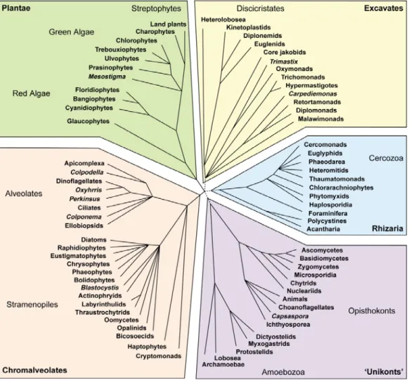
Opisthokonta, Amoebozoa, Plantae, Rhizaria, Excavata, Stramenopiles and Alveolata
(Baldauf, 2003; Keeling et al., 2005) (Figure 1.1). However, the phylogenetic
relationships among these major groups are still poorly resolved, and the stability of
some of them is still questionable. This is mainly due to the fact that the true diversity
of eukaryotes remains largely unknown and several groups remain unexplored by
molecular approaches (Bass and Cavalier-Smith, 2004; Parfrey et al., 2006).
Recent molecular phylogenetic studies unveiled novel major groups of eukaryotes
that provided a fundamental change in our understanding of eukaryotes diversity,
phylogeny and evolutionary history. A study by Burki et al. (2008) using a multigene
data set and over 65 species and 135 genes strongly suggested that Rhizaria,
stramenopiles and alveolates form a monophyletic megagroup (SAR assemblage)
supergroup, the Hacrobia, that comprises haptophytes, cryptophytes, katablepharids,
and telonemids, has recently been proposed (Okamoto et al., 2009). However, the
phylogenetic relationships within the Hacrobia is still questionable (Okamoto et al.,
2009).
Figure 1.1: A tree of eukaryotes by Keeling et al., (2005), showing the major supergroups, most of the groups are unicellular protists and algae. Note that Rhizaria
1.2. Phylogenetic position of amoeboid protists
The amoebae or amoeboid protists are considered one of the largest and most
diverse assemblages in the tree of eukaryotes (Pawlowski, 2008; Pawlowski and
Burki, 2009). Previously, all amoeboid protists were placed within Sarcodina. In the
current eukaryotic tree, Sarcodina is a polyphyletic group with Amoebozoa and
Rhizaria containing the highest number of species (Adl et al., 2005 ; Cavalier-Smith,
1998; Cavalier-Smith, 2002; Pawlowski and Burki, 2009). Other major eukaryotic
clades contain amoeboid protists. For instance, the naked amoeba forms
Vahlkampfia, Naegleria and Acrasis belong to Heterolobosea, characterized by the
presence of amoeboid stage, and were traditionally considered as Amoebozoa.
However, the SSU rRNA and protein sequence data confirmed their phylogenetic
position within excavates (Page and & Blanton, 1985; Simpson, 2003). Genus
Nuclearia (Nucleariids) are filose amoeba traditionally classified within Filosea (i.e.
Rhizaria) (Page, 1991). The SSU rRNA and multigene analyses confirmed that
Nucleariid amoebae branch within Opisthokonts at the base of fungi (Amaral-Zettler et
al., 2001; Steenkamp et al., 2006). It was shown that order Actinophryida
(ex-Heliozoa) branch within Stramenopiles (Nikolaev et al., 2004). Class
Synchromophyceae includes marine amoeboid algae that have a sessile amoeboid
stage and a non-sessile free floating life stage, while order Chrysamoebales includes
Chrysophytes that have amoeboid “Rhizopodial” vegetative cells during the greater
part of their life cycle. Ribosomal rRNA gene sequences placed both Chrysamoeba
and Synchroma within the Stramenopiles (Cavalier-Smith and Chao, 2006; Patil et al.,
2009). Members of family Amphitremidae are currently considered as incertae sedis
Other amoeboid eukaryotes still remain “homeless” or have not yet been
unambiguously assigned to any major lineage. This includes the Breviatea, a group of
free-living amoeboflagelates. Therefore, amoeboid eukaryotes are scattered through
all eukaryotic lineages except the plants (Pawlowski, 2008).
1.3. What are the testate amoebae?
Testate amoebae are free living single-celled eukaryotes, highly diverse and
abundant in a wide range of habitats such as wetlands, soil, mosses, freshwater, and
even marine environments. Testate amoebae are distinguishable from other shelled
amoeboid eukaryotes such as foraminifera, radiolarians and heliozoans by 1) their one
chambered shell that can be categorized into four main types: agglutinated (species
which include extraneous mineral particles in their shell, so-called “xenosomes”),
proteinaceous (species with flexible or rigid shell), calcareous or siliceous (secrete
their own regular siliceous platelets, so-called “idiosomes”, or produces shell of mixed
layers of both xenosomes and idiosomes (Meisterfeld, 2002a; Meisterfeld, 2002b;
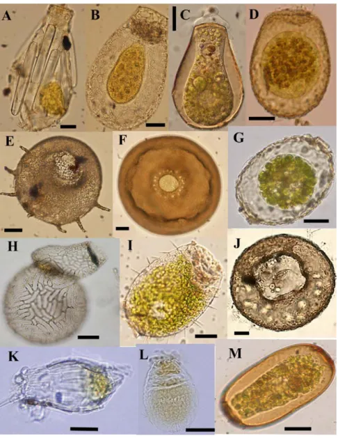
Wanner, 1999) (Figure 1.2 and 1.3), and 2) their pseudopodia that can be lobose or
Figure 1.2: Light micrographs of some testate amoebae illustrating their morphological variability. A) Difflugia bacillifera, B) Nebela marginata C) Hyalosphenia
papilio, D) Heleopera sphagni, E) Centropyxis aculeata, F) Arcella arenaria, G) Amphitrema wrightianum, H) Lesquereusia epistomium, I) Placocista spinosa, J) Centropyxis ecornis, K) Difflugia bacillariarum, L) Euglypha compressa, M) Archerella flavum. Scale bars indicate 20µm. All images by F. Gomaa except (H) E. Mitchell, and
2. Evolution, phylogeny and physiology of testate amoebae
2.1. Fossil record & phylogeny
Vase-shaped microfossils resembling extant arcellinid testate amoebae have
been found in marine deposits, and are considered among the oldest heterotrophic
eukaryotes with fossils, dating back to ca. 740 Mya in the Cryogenian period ((Porter
and Knoll, 2000; Porter et al., 2003). The age of these fossils has been corroborated
by a molecular clock study (Berney and Pawlowski, 2006). Molecular phylogenetic
studies based on ribosomal RNA and proteins gene sequences revealed that testate
amoebae are a polyphyletic assemblage of at least two major groups, the supergroup
Amoebozoa comprises Arcellinid testate amoebae ( Nikolaev et al., 2005; Gomaa et
al., 2012), and supergroup Rhizaria comprises Euglyphida, Chlamydophryidae
“Rhizaspididae”, Tectofilosida, and Gromiidae (Bhattacharya et al., 1995;
Cavalier-Smith, 1998; Nikolaev et al., 2003; Lara et al., 2007; Howe et al., 2011).
In addition to these groups, some taxa have not yet been placed in the tree of
eukaryotes. One of these is family Amphitremidae that are characterized by the
presence of a shell with two pseudostomes at the opposite ends of the shell. It
includes three genera, Amphitrema, Archerella and Paramphitrema. The first two
genera comprises organisms that possess filamentous anastomosing pseudopodia,
and most species harbor endosymbiotic zoochlorellae (Figure 1.2), while
Paramphitrema lives on marine and freshwater plants and algae, and is characterized
by linear pseudopodia. In addition, families Psammonobiotidae (Golemansky, 1979)
and Volutellidae (Sudzuki, 1979a), both characterized by filose pseudopodia and
organic and/or agglutinated shells. These organisms inhabit the supralittoral zone
often been found that eukaryotes with uncertain phylogenetic affinities can represent
key lineages, and information on these species can be very important for
understanding the eukaryotic evolution (Nikolaev et al., 2004; Yabuki et al., 2010 ).
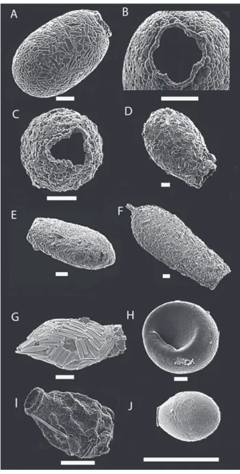
Figure1.3: Scanning electron micrograph of some testate amoebae illustrating the variability of the shell shape and composition. A) Centropyxis marsupiformis, B)
Apodera vas, C) Difflugia labiosa, D) Cyphoderia ampulla, E) Difflugia corona, F) Zivkovicia compressa, G) Euglypha brachiata H) Lesquereusia epistomium. Scale
bars indicate 20µm. Images: M. Todorov (A, C, E, F and H) and T. Heger (B, D and G).
2.2. Physiology: the special case of mixotrophy
Many heterotrophic species of protists are capable of building a symbiotic
relationship with phototrophic organisms. They can host either viable algal
endosymbionts or sequestered plastids (i.e. kleptoplastidy) from their algal prey that
remain functional for several days or even months, using by-products of their
photosynthesis (Johnson, 2011; Pillet et al., 2011). Such mixotrophic organisms
combine the advantages of both phototrophy and heterotrophy (Summerer et al.,
2007). Acquired phototrophy is generally more prevalent in nutrient-poor
environments, such as bogs, coral reefs and pelagic environment (Margulis and
Fester, 1992). The relationship between the host and their associated algal symbionts
is considered as a mutualistic relationship. The host usually supplies the algal cells
with nitrogen and CO2. In return, the algae supply the host with photosynthetic
products such as maltose and oxygen (Esteban et al., 2010; Summerer et al., 2007).
Generally, the mixotrophic individuals have higher growth rate in comparison to the
symbiotic-free individuals of the same species (Karakashian, 1963; Stabell et al.,
2002). In addition, it has been shown that the symbiotic algae have a photo-protective
role for their Paramecium bursaria host against UV damage besides their nutritional
benefits to their host (Summerer et al., 2009). Recently, the nature of algal symbiosis
in protists and invertebrates has attracted considerably scientific interest (Esteban et
al., 2010; Summerer et al., 2007; Hoshina and Imamura, 2008; Garcia-Cuetos et al.,
2005). However, most of our knowledge about the identity, diversity and host-symbiont
phylogenetic relationships concerns marine dinoflagellates of genus Symbiodinium,
the most prevalent symbiotic algae in Foraminifera, Radiolaria, Acantharea and coral
reefs (Garcia-Cuetos et al., 2005; Gast and Caron, 1996 and 2001; Santos et al.,
heterotrophic host and a phototrophic symbiont is found in lichens. In addition, the
ciliate Paramecium bursaria and its chlorophyte symbionts have become a model
system for studying host-symbiont specificity and evolutionary relationships (Hoshina
and Imamura, 2008; Summerer et al., 2008)
Several testate amoebae species are considered as obligate mixotrophs and
are always observed with their green-colored symbionts. These species are
distributed among three major eukaryotic clades (1) Hyalosphenia papilio and
Heleopera sphagni (Amoebozoa: Arcellinida) (Nikolaev et al., 2005), 2) Placocista spinosa (Rhizaria: Euglyphida) (Bhattacharya 1995, and(Cavalier-Smith, 1997), and 3)
Archerella flavum, Amphitrema wrightianum and Amphitrema stenostoma (incertae
sedis). Early experimental field study by Schönborn (1965) showed that Amphitrema
flavum (= Archerella) and Hyalosphenia papilio could not survive for long periods in
the dark. His study also demonstrated that Amphitrema flavum depend entirely on
their green symbiotic algae for their nutrition requirements, while H. papilio acquired
up to 40% of their nutrients from photosynthetic products released by algae and the
rest through phagotrophy (Schönborn, 1965). However, to date, the identity of their
symbionts and the nature of host-symbiont relationships remain unknown. We believe
that two factors have mainly hindered the early research on testate amoeba
mixotrophy. The first is the difficultly to establish and maintain cultures for both host
and the endosymbionts. The second is the impossibility of identifying species of green
algae (Chlorella-like) using only morphological criteria. Therefore, we performed the
first molecular phylogenetic study based on COI and rbcL gene sequences for both
host and their endosymbionts respectively. We identified the in hospite symbionts in
four mixotrophic testate amoebae species. We also assessed the symbiont diversity
Hyalosphenia papilio. Our study is based on samples collected from Sphagnum
peatlands (see chapter 4).
3. Testate amoebae in ecological and biogeographical research
3.1. Role of testate amoebae in nutrient cycling
Testate amoebae are mostly phagotrophic organisms, with a few exceptions of
mixotrophic (see chapter 4) and one phototrophic species (Paulinella chromatophora)
(Gilbert et al., 2000; Wilkinson, 2008; Yoon et al., 2006). The phagotrophic testate
amoeba species usually graze bacteria, fungi and algae, thus playing an important
role in soil nutrient recycling (Wilkinson, 2008). They can also be a food source for
other invertebrates such as nematodes, earthworms, collembolans, and predaceous
mites (Schroeter, 2001), thus re-entering the classical food chain (Bamforth and
Lousier, 1995; Wilkinson and Mitchell, 2010). Some testate amoebae species can
predate upon other protozoa and metazoan organisms such as nematodes and
rotifers (Yeates and Foissner, 1995; Gilbert et al., 2000). It was also shown that
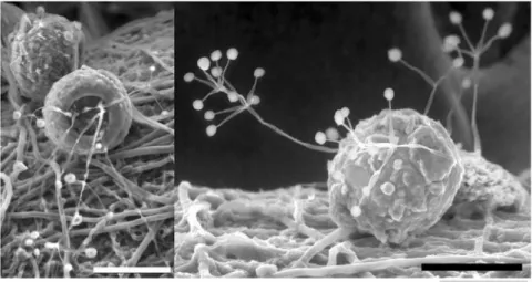
testate amoebae colonized with mutualistic fungi are good source for nutrients
particularly nitrogen components for plants in nutrient-poor soil (Figure 1.4) (Vohnîk et
al., 2009).
In addition, the testate amoebae with silica rich shells may also form an
important part of silica cycle and thus increasing the silica mineralization in soil (Aoki
et al., 2007; Wilkinson and Mitchell, 2010). Until recently our knowledge on testate
amoebae feeding habits was very limited due to the limitation in the methodologies
that used to identify both testate amoebae and their preys (Gilbert et al., 2000; Gilbert
et al., 2003). Jassey et al. (2012) succeeded to determine the feeding behavior of
some testate amoebae taxa using C and N stable isotope analysis. This study showed
identified preys along the Sphagnum shoots are probably the main factors influence
their feeding behavior (Jassey et al., 2012). However, much remains to be done in
order to better assess testate amoebae functional role in the microbial trophic network.
Figure 1.4: Phryganella acropodia colonized by fungal hyphae. Laboratory culture by Martin Vohník, Wilkinson (2008).
3.2. Bioindicators and biomonitors
Testate amoebae are considered as reliable bioindicators and biomonitors for
ecological and palaeoecological studies, in particular as proxies for hydrological
change, and therefore for paleoclimate reconstruction in peatlands (Charman and
Warner, 1997; Mitchell et al., 2000). Testate amoebae are also very sensitive to other
ecological gradients such as water table, dryness, pollutions and human activities (e.g.
deforestations or damming) (Booth, 2007; Payne et al., 2012). Thus making them
valuables biomonitors for current environmental health (Nguyen-Viet et al., 2008).
After death, their shells are very well preserved over time, therefore they are
considered as excellent microfossils useful for palaeoenvironmental reconstructions
(Charman et al., 2001; Mitchell et al., 2008).
3.3. Testate amoebae as a model to study microbial biogeography
Microorganisms have believed to be cosmopolitan because of their relativelysmall size, high dispersal rates, and high reproduction rates (Finlay, 2002; Finlay et
al., 2004). However, this classical cosmopolitan view or as it was proposed by
(Beijerinck)(1913) that “everything is everywhere, but, the environment selects’ ” has
changed during the late of the 20th century. Several studies illustrated that some
microbial organisms present a very characteristic morphology that cannot be confused
with any others, have restricted geographical distributions, and usually referred to it as
“flagship species” (Foissner, 2006; Smith et al., 2008). Some species of testate
amoebae are among the most striking examples of microorganisms that present
biogeographical patterns in their global distribution. For instance Apodera vas and
Alocodera cockayni and genus Certesella are restricted to the south of the Tropic of
thought to be endemic to Asia, such as Difflugia biwae (Kawamura, 1918) (in China
and Japan), Difflugia tuberspinifera (Yang et al., 2004) (in China), Difflugia mulanensis
(Yang et al., 2005) (in China), and Pentagonia zhangduensis (Qin et al., 2008) (in
China).
3.4. Current limitations to the use of testate amoebae in ecological
research
Testate amoebae are a problematic group of protists with respect to their
taxonomy and the identification and delimitation of species. This is mainly due to the
fact that their taxonomy is largely based on morphological characteristics, such as the
shell shape, size and composition, with only a limited contribution from molecular data
(Meisterfeld 2002a, b). As a result of this taxonomic confusion and limitations 1)
paleoecological studies generally underestimate their diversity and may group
together unrelated taxa. This may lead to errors in reconstructed environmental values
(see(Payne et al., 2011), 2) taxonomic uncertainties also undermine biogeographical
studies of testate amoebae (i.e. cosmopolitanism versus endemism) (see(Heger et al.,
2009; Mitchell and Meisterfeld, 2005), 3) their ecological and functional roles in the
environments remains unclear (Gilbert et al., 2000; Gilbert et al., 2003). It was recently
discovered that several morphospecies of testate amoebae hide cryptic diversity.
These cryptic species might possibly have either restricted or cosmopolitan
distributions, as well as different ecological roles in the ecosystem (see (Heger et al.,
2009; Heger et al., 2010; Heger et al., 2011; Kosakyan et al., 2012). Thus to improve
the utility of testate amoebae in different ecological research fields, their taxonomic
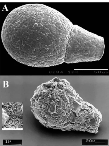
Figure 1.6: Scanning electron micrograph of A) Apodera vas and B) Lagenodifflugia
vas, illustrating that the relative morphological similarity among testate amoebae
species blurs the debate on their cosmopolitanism versus local endemism (Mitchell and Meisterfeld, 2005).
4. Arcellinida (Lobosea) testate amoebae
4.1. Arcellinida taxonomy and diversity
Molecular phylogenetic studies have established the phylogenetic position of
several testate amoeba taxa in the tree of eukaryotes (Bhattacharya et al., 1995;
Cavalier-Smith, 1997; Cavalier-Smith, 1998; Howe et al., 2011; Nikolaev et al., 2005),
groups and the proper assessment of their species diversity is lacking (Gomaa et al.,
2012; Howe et al., 2011; Kudryavtsev et al., 2009). This is mainly due to the lack of
gene sequences on some major taxa. Arcellinid testate amoebae are considered the
largest and the most diverse group of testate amoebae containing about three
quarters of all known species (Beyens and Meisterfeld, 2001; Meisterfeld, 2002a).
Meisterfeld divided arcellinids into three main orders; Arcellinina (characterised by a
membranous test; three families), Difflugiina (agglutinated test, twelve families) and
Phryganellina (pointed pseudopodia; two families) (Meisterfeld, 2002a). Although
about 1100 species have been described in Arcellinida based on morphological
criteria (Meisterfeld, 2002a), Adl et al., (2007) estimated that the true diversity of
Arcellinida could reach up to 10 000 species.
4.1. Current problems and limitations in Arcellinida taxonomy
The identification and classification of Arcellinida at genus and species level is
mainly based on shell dimensions, composition and shape; and the associated
morphological features with the shell such as the presence or absence of spines and
pores, or with the aperture such as the presence of collar, diaphragm, lobes, and teeth
(Beyens and Meisterfeld, 2001; Meisterfeld, 2002a; Wanner, 1999).
Several studies emphasized that the shell morphological features are
sometimes not sufficient to warrant species diagnoses particularly in the agglutinated
taxa like those in families Difflugiidae and Centropyxidae (Meisterfeld, 2002a; Ogden,
1983; Ogden and Hedley, 1980; Ogden and Meisterfeld, 1989). These species have
high diversity in shell composition due to the diverse type of the extraneous material
addition of these extraneous materials (Ogden, 1983). These morphological variations
can cause a considerable taxonomic confusion at species or even at genus level in
many taxa. Indeed, several authors described new species based on minor variations
on shell composition and shape. However, some of these newly described taxa are
actually not represent valid species and more likely fall within the natural
morphological variability within given species.
There are mounting evidences suggesting that the environmental factors such
as moisture, food source, temperature, the availability of extraneous material and pH
have a direct influence upon their morphology (Bobrov et al., 2004; Wanner, 1999).
Therefore, minor differences in the shell shape, composition could possibly be
response by amoeba towards the environmental factors and their combination and
probably are not genetically fixed characters.
This interpretation has been supported in several studies for example a
biometric data analyses on 32 natural populations of 24 species by Bobrov and Mazei
(2004) revealed a significant degree of morphological variability within local
population. A morphological, biometrical and ecological study on 2210 Centropyxis
individuals by Lahr et al., (2008) revealed a continuity in the morphospecies of C.
aculeata and C. discoides, and suggested that both species are actually the same
taxon that exhibit a highly morphological polymorphism in shell and aperture shape
and number of spines. A review on the morphological variability of testate amoebae by
Wanner (1999) emphasized that the effect of ecological factors on the shell
morphology should be taken into account, in order to estimate the range of genetic
4.2. The molecular phylogeny of Arcellinida
The application of DNA-based techniques to the study of Arcellinida
systematics is relatively recent. A first study by Nikolaev et al. (2005) placed
representatives of several Arcellinida genera together as a monophyletic group within
the eukaryotic superclass Amoebozoa (Nikolaev et al., 2005). Other molecular
studies, based on the SSU rRNA gene, were focused on the phylogeny of particular
groups within the Arcellinida, such as the Hyalospheniidae (Lara et al., 2008), and the
genera Spumochlamys (Kudryavtsev et al., 2009) and Arcella (Lahr et al., 2011; Tekle
et al., 2008). However, the phylogenetic tree of Arcellinida remains unresolved,
because very few species were characterized by molecular methods to date.
The main reasons explaining the limited number of Arcellinida sequences is probably due to the following:
1) The lack of suitable sets of PCR primers that can be generally used for the whole
arcellinid group and /or taxon-specific primers. Arcellinids are a very old taxa (Porter
and Knoll, 2000) and the genetic distances among taxa are very large, making the
design of PCR primers particularly challenging. In addition, contamination is a major
problem that scientist encounter when sequencing testate amoebae using general
eukaryotes primers set. There are two main source of such contamination A) these
microorganisms are free living and are generally isolated from a mixed pool of living
and non-living materials such as of soil, organic matters. Other protists some of them
quite small even pico-sized can easily be present (unnoticed) in the isolated specimen
(i.e. contamination with DNA derived from diverse living material). B) Arcellinida cells
epibionts or even contain undigested protist and metazoan prey. Therefore such
contamination can easily be amplified in the PCR reaction. Sometimes, these
co-amplified eukaryotes can even be closely related to Arcellinida, such as minute lobose
naked amoebae.
2) The majority of arcellinid taxa are difficult or even impossible to maintain in cultures.
3) The shell of many Arcellinida, such as members of genera Difflugia, Trigonopyxis,
Bullinularia, etc are opaque or dark in color and therefore, it is difficult to recognize if
the organism alive or dead.
5. Thesis Objectives
My PhD research focuses on the molecular phylogeny and taxonomy of testate
amoebae and host-symbiont evolutionary relationships within mixotrophic taxa. In this
thesis, I had three main objectives to work on, in order to achieve a better
understanding on phylogeny and evolution of testate amoebae:
The first aim is to revise and redefine the systematic of some arcellinid testate
amoebae taxa particularly those agglutinated species, based on both fine
morphological data using SEM and molecular data based on gene sequences mainly
the small subunit ribosomal RNA (SSU rRNA).
The second aim is to illustrate the phylogenetic position of some incertae sedis
testate amoebae taxa within the tree of eukaryotes.
The third aim is to investigate and evaluate the host-symbiont specificity and
evolutionary relationships in some mixotrophic testate amoebae taxa by obtaining
These objectives are addressed in four main chapters:
In chapter 2, we performed the first step toward resolving the Arcellinida phylogeny and taxonomy in general and in genus Difflugia in particular. We illustrated
that genus Difflugia (the largest arcellinid genus) is not monopyletic, by developing a
new set of SSU rRNA specific primers. We also stressed the major pitfalls that can be
encountered in the study of arcellinid phylogeny.
In chapter 3, we determined the phylogenetic position of two testate amoebae of unknown affiliation, Amphitrema wrightianum and Archerella flavum. These
organisms are well-known indicators in palaeoecological studies of Sphagnum
peatlands. Our molecular data based on SSU rRNA gene sequences showed that
they belong to labyrinthulomycete clade, a group of Stramenopiles that were hitherto
mostly known as marine osmotrophic organisms. In addition, we described a new
clade we named Amphitremida. The new clade is highly diverse genetically,
ecologically and physiologically.
In chapter 4, we explored the nature of the endosymbionts in four mixotrophic testate amoebae taxa (Hyalosphenia papilio, Heleopera sphagni, Placocista spinosa
and Archerella flavum); we also used Hyalosphenia papilio as a model to investigate
in details the host-symbiont specificity and evolutionary relationships. Our results
revealed a new lineage of symbiotic Chlorella that comprised, to date, only testate
amoebae endobiotic Chlorella.
In Chapter 5, we presented an example of fast morphological evolution within Arcellinida, in endemic Chinese Difflugia tuberspinifera the spiny and spineless
morphospecies. Our SSU rRNA and ITS gene sequences showed limited genetic
endemic D. tuberspinifera morphospecies have evolved recently from the same
ancestor (i.e. evolutionary very close related to each other) and the spines appeared
as a result of a strong selective pressure.
Bibliography
Adl, M.S., Leander, B.S., Simpson , A., Archibald, J.M., Anderson, O.R., Barta, J.R., Bass, D., Bowser, S.S., Brugerolle, G., Farmer, M.A., Karpov, S., Kolisko, M., Lane, C.E., Lodge, J., Lynn, D.H., Mann, D.G., Meisterfeld, R., Mendoza, L., Moestrup, Q., Mozley-Standridge, S.E., Smirnov, A.V., Spiegel, F.W, 2007. Diversity, nomenclature and taxonomy of protists. Systematic Biology 56(4), 684-689.
Adl, S., Simpson, A.G., Farmer, M.A., Andersen R.A., A.O.R., Barta, J.R., Bowser, S.S.,Brugerolle, G., Fensome, R.A., Fredericq, S., James, T.Y., Karpov, S., Kugrens, P., Krug, J., Lane, C.E., Lewis, L.A., Lodge, J., Lynn, D.H., Mann, D.G., McCourt, R.M., Mendoza, L.,Moestrup, O.,Mozley-Standridge, S.E., Nerad, T.A., Shearer, C.A.,Smirnov, A.V.,Spiegel, F.W. and Taylor, M.F., 2005. The new higher level classification of eukaryotes with emphasis on the taxonomy of protists. Journal of Eukaryotic Microbiolology 52, 399-451.
Amaral-Zettler, L.A., Nerad, C.J., O’Kelly, L., Sogin, A.M., 2001. The nucleariid amoebae: more protists at the animal-fungal boundary. Journal of Eukaryotic Microbiolology 48, 293-297.
Aoki, Y., Hoshino, M., Matsubara, T., 2007. Silica and testate amoebae in a soil under pine-oak forest. Geoderma 142, 29-35.
Baldauf, S., 2008. An overview of the phylogeny and diversity of eukaryotes. Journal of Systematics and Evolution 46 (3), 263-273.
Baldauf, S.L., 2003. The deep roots of eukaryotes. Science 300, 1703-1706.
Bamforth, S.S., Lousier, J.D., 1995. Protozoa in tropical litter decomposition. In: Reddy, M.V. (Ed.), Soil organisms and litter decomposition in the tropics. Oxford & IBH Publishing CO. PVT. LTD., New Delhi, Calcutta, pp. 59-73.
Bass, D., Cavalier-Smith, T., 2004. Phylum-specific environmental DNA analysis reveals remarkably high global biodiversity of Cercozoa (Protozoa). International Journal of Systematic and Environmental Microbiology 54, 2392-2404.
Beijerinck, M.W., 1913. De infusies en de ontdekking der backteriën. Jaarboek van de Koninklijke Akademie voor Wetenschappen. Müller, Amsterdam, the Netherlands. (Reprinted in Verzamelde geschriften van M.W. Beijerinck, vijfde deel, pp. 119-140. Delft, 1921).
Berney, C., Pawlowski, J., 2006. A molecular time-scale for eukaryote evolution recalibrated with the continuous microfossil record. Proceeding Royal Society London Series B-Biological Science 273, 1867-1872.
Beyens, L., Meisterfeld, R., 2001. Protozoa: Testate amoebae. Tracking environmental change using lake sediments. Volume 3: Terrestrial, algal, and siliceous indicators. Kluwer Academic Publischer, Dordrecht.
Bhattacharya, D., Helmchen, T., Melkonian, M., 1995. Molecular evolutionary analyses of nuclear-encoded small-subunit ribosomal-RNA identify an independent Rhizopod lineage containing the euglyphina and the chlorarachniophyta. Journal of Eukaryotic Microbiology 42, 65-69.
Bobrov, A.A., Andreev, A.A., Schirrmeister, L., Siegert, C., 2004. Testate amoebae (Protozoa : Testacealobosea and Testaceafilosea) as bioindicators in the Late Quaternary deposits of the Bykovsky Peninsula, Laptev Sea, Russia. Palaeogeography Palaeoclimatology Palaeoecology 209, 165-181.
Booth, R.K., 2007. Testate amoebae as proxies for mean annual water-table depth in
Sphagnum-dominated peatlands of North America. Journal of Quaternary Science 23,
43-57
Burki, F., Shalchian-Tabrizi K., Pawlowski, J., 2008. Phylogenomics reveals a new ‘megagroup’ including most photosynthetic eukaryotes. Biology Letters 4, 366-369. Cavalier-Smith, T., 1997. Amoeboflagellates and mitochondrial cristae in eukaryotic evolution; megasystematics of the new protozoan subkingdoms Eozoa and Neozoa. Arch Protistenkd 147, 237-258.
Cavalier-Smith, T., 1998. A revised six-kingdom system of life. Biological Reviews 73, 203-266.
Cavalier-Smith, T., 2002. The phagotrophic origin of eukaryotes and phylogenetic classification of Protozoa. International Journal of Systematic and Evolutionary Microbiology 52, 297–354.
Cavalier-Smith, T., Chao, E.E.Y., 2006. Phylogeny and megasystematics of phagotrophic heterokonts (Kingdom Chromista). Journal of Molecular Evolution 62, 388-420.
Charman, D.J., Caseldine, C., Baker, A., Gearey, B., Hatton, J., Proctor, C., 2001. Paleohydrological records from peat profiles and speleothems in Sutherland, northwest Scotland. Quaternary Research 55, 223-234.
Charman, D.J., Warner, B.G., 1997. The ecology of testate amoebae (Protozoa: Rhizopoda) in oceanic peatlands in newfoundland, Canada: Modelling hydrological relationships for palaeoenvironmental reconstruction. Ecoscience 4, 555-562.
Esteban, G.F., Fenchel, T., Finlay, B.J., 2010. Mixotrophy in ciliates. Protist 161(5), 621-641.
Finlay, B.J., 2002. Global dispersal of free-living microbial eukaryote species. Science 296, 1061-1063.
Finlay, B.J., Esteban, G.F., Fenchel, T., 2004. Protist diversity is different? Protist 155, 15-22.
Foissner, W., 2006. Biogeography and dispersal of micro-organisms: A review emphasizing protists. Acta Protozoologica 45, 111-136.
Garcia-Cuetos, L., Pochon, X., Pawlowski, J., 2005. Molecular evidence for host-symbiont specificity in soritid foraminifera. Protist 156(4), 399-412.
Gast, R.J., Caron, D.A., 1996. Molecular phylogeny of symbiotic dinoflagellates from Foraminifera and Radiolaria. Molecular Biology and Evolution 13, 1192-1197.
Gast, R.J., Caron, D.A., 2001. Photosymbiotic associations in planktonic foraminifera and radiolaria. Hydrobiologia 461, 1–7.
Gilbert, D., Amblard, C., Bourdier, G., Francez, A.-J., Mitchell, E.A.D., 2000. Le régime alimentaire des thécamoebiens. L’Année Biologique 39, 57-68.
Gilbert, D., Mitchell, E.A.D., Amblard, C., Bourdier, G., Francez, A.J., 2003. Population dynamics and food preferences of the testate amoeba Nebela tincta
major-bohemica-collaris complex (Protozoa) in a Sphagnum peatland. Acta Protozoologica 42, 99-104.
Golemansky, V., 1979. Thécamoebiens psammobiontes du supralittoral coréen de la mer Japonaise et description de deux nouvelles espèces - Rhumbleriella coreana n. sp. et Amphorellopsis conica n. sp. (Rhizopoda, Testacea). Acta Zoologica Bulgarica 12, 5-11.
Gomaa, F., Todorov, M., Heger, T.J., Mitchell, E.A.D, Lara, E., 2012. SSU rRNA phylogeny of Arcellinida (Amoebozoa) reveals that the largest Arcellinid genus,
Difflugia Leclerc 1815, is not monophyletic. Protist 163(3), 389-399.
Heger, T.J., Mitchell E.A.D., Ledeganck P., Vincke, S., Van de Vijver, B., Beyens, L., 2009. The curse of taxonomic uncertainty in biogeographical studies of free-living terrestrial protists: a case study of testate amoebae from Amsterdam Island. Journal of Biogeography 36, 1551-1560.
Heger, T.J., Mitchell, E.A.D., Golemansky, V., Todorov, M., Lara E, Leander, B.S., Pawlowski, J., 2010. Molecular phylogeny of euglyphid testate amoebae (Cercozoa: Euglyphida) suggests transitions between marine supralittoral and freshwater/ terrestrial environments are infrequent. Molecular Phylogenetics and Evolution 55, 113–122.
Heger, T.J., Pawlowski, J., Lara, E., Leander, B.S., Todorov, M., Golemansky, V., Mitchell, E.A.D, 2011. Comparing potential COI and SSU rDNA barcodes for assessing the diversity and phylogenetic relationships of cyphoderiid testate amoebae (Rhizaria: Euglyphida). Protist 162(1), 131-41.
Hoshina, R., Imamura, N., 2008. Multiple origins of the symbioses in Paramecium
bursaria. Protist 159(1), 53-63.
Howe, A.T., Bass, D., Scoble, J.M., Lewis, R., Vickerman, K., Arndt, H., & Cavalier-Smith, T., 2011. Novel cultured protists identify deep-branching environmental DNA clades of Cercozoa: New genera Tremula, Micrometopion, Minimassisteria, Nudifila,
Peregrinia. Protist 162 (2), 332-372.
Jassey, V.E., Shimano, S., Dupuy, C., Toussaint, M.L., Gilbert, D., 2012. Characterizing the feeding habits of the testate amoebae Hyalosphenia papilio and
Nebela tincta along a narrow "Fen-Bog" gradient using digestive vacuole content and
(13)C and (15)N isotopic analyses. Protist 163(3), 451-464.
Johnson, M.D., 2011. The acquisition of phototrophy: Adaptive strategies of hosting endosymbionts and organelles. Photosynthetic Research 107(1), 117-132.
Karakashian, S., 1963. Growth of Paramecium bursaria as influenced by the presence of algal symbionts. Physiological Zoology 36 1, 52-68.
Katz, L.A., Grant, J.R., Parfrey, L.W., Burleigh, J.G., 2012. Turning the crown upside down: Gene tree parsimony roots the eukaryotic tree of life Systematic Biology 61(4),653-60.
Kawamura, T., 1918. Japanese Freshwater Biology. Syoukabou, Tokyo, Japan (in Japanese).
Keeling, P.J., Burger, G., Durnford, D.G., Lang, B.F., Lee, R.W., Pearlman, R.E., Roger, A.J., Gray, M.W., 2005. The tree of eukaryotes. Trends in Ecology and Evolution 20, 671-676.
Kosakyan, A., Heger, T.J., Leander, B.S., Todorov, M., Mitchell, E.A.D., Lara, E., 2012. COI barcoding of Nebelid testate amoebae (Amoebozoa: Arcellinida): Extensive cryptic diversity and redefinition of family Hyalospheniidae Schultze. Protist 163, 415– 434.
Kudryavtsev, A., Pawlowski, J., Hausmann, K., 2009. Description and phylogenetic relationships of Spumochlamys perforata n. sp and Spumochlamys bryora n. sp (Amoebozoa,Arcellinida). Journal of Eukaryotic Microbiology 56, 495–503.
Lahr, D.J.G., Bergmann, P.J., Lopes, S.G.B.C., 2008. Taxonomic identity in microbial eukaryotes: A practical approach using the testate amoeba Centropyxis to resolve conflicts between old and new taxonomic descriptions. Journal of Eukaryotic Microbiology 55, 409-416.
Lahr, D.J.G., Nguyen, T.B., Barbero, E., Katz, L.A., 2011. Evolution of the actin gene family in testate lobose amoebae (Arcellinida) is characterized by two distinct clades of paralogs and recent independent expansions. Molecular Biology and Evolution 28, 223–236.
Lara, E., Heger, T.J., Ekelund, F., Lamentowicz, M., Mitchell, E.A.D., 2008. Ribosomal RNA genes challenge the monophyly of the Hyalospheniidae (Amoebozoa: Arcellinida). Protist 159, 165-176.
Lara, E., Heger, T.J., Mitchell, E.A.D., Meisterfeld, R., Ekelund, F., 2007. SSU rRNA reveals a sequential increase in shell complexity among the Euglyphid testate amoebae (Rhizaria: Euglyphida). Protist 158, 229-237.
Margulis, L., Fester, R., 1992. Symbiosis as a source of evolutionary innovation: speciation and morphogenesis. Cambridge, MA, USA: MIT Press.
Meisterfeld, R., 2002a. Order Arcellinida Kent, 1880. In Lee JJ, Leedale GF, Bradbury P (eds) The illustrated guide to the protozoa. vol 2, Second edition, Society of Protozoologists, Lawrence, Kansas, USA, pp 827–860.
Meisterfeld, R., 2002b. Testate amoebae with filopodia. The illustrated guide to the protozoa (ed. by J.J. Lee, G.F. Leedale and P. Bradbury), Society of protozoologists, Lawrence, Kansas, USA,pp 1054-1084.
Mitchell, E.A.D., Borcard, D., Buttler, A.J., Grosvernier, P., Gilbert, D., Gobat, J.M., 2000. Horizontal distribution patterns of testate amoebae (Protozoa) in a Sphagnum
magellanicum carpet. Microbial Ecology 39, 290-300.
Mitchell, E.A.D., Charman, D.J., Warner, B.G., 2008. Testate amoebae analysis in ecological and paleoecological studies of wetlands: past, present and future. Biodiversity and Conservation 17, 2115-2137.
Mitchell, E.A.D., Meisterfeld, R., 2005. Taxonomic confusion blurs the debate on cosmopolitanism versus local endemism of free-living protists. Protist 156, 263-267.
Nguyen-Viet, H., Bernard, N., Mitchell, E.A.D., Cortet, J., Badot, P.-M., Gilbert, D., 2008. Effect of lead pollution on testate amoebae communities living in Sphagnum fallax: an experimental study. Ecotoxicology and Environmental Safety 69, 130-138.
Nikolaev, S.I., Berney, C., Fahrni, J., Mylnikov, A.P., Aleshin, V.V., Petrov, N.B., Pawlowski, J., 2003. Gymnophrys cometa and Lecythium sp are core Cercozoa: evolutionary implications. Acta Protozoologica 42, 183-190.
Nikolaev, S.I., Berney C., Fahrni J., Bolivar I., Polet S., Mylnikov A.P., Aleshin V.V., Petrov, N., Pawlowski, J., 2004. The twilight of Heliozoa and rise of Rhizaria: an emerging supergroup of amoeboid eukaryotes. Proceeding of the National Academy of Science USA 101 (21), 8066-8071.
Nikolaev, S.I., Mitchell, E.A.D., Petrov, N.B., Berney, C., Fahrni, J., Pawlowski, J., 2005. The testate lobose amoebae (order Arcellinida Kent, 1880) finally find their home within Amoebozoa. Protist 156, 191-202.
Ogden, C.G., 1983. Observations on the systematics of the genus Difflugia in Britain (Rhizopoda, Protozoa). Bulletin of the Natural History Museum. Zoology series 44, 1-73.
Ogden, C.G., Hedley, R.H., 1980. An Atlas of Freshwater Testate Amoebae. Oxford University Press, London, 222p.
Ogden, C.G., Meisterfeld, R., 1989. The taxonomy and systematics of some species of Cucurbitella, Difflugia and Netzelia (Protozoa: Rhizopoda); with an evaluation of diagnostic characters. European Journal of Protistology 25, 109-128.
Okamoto, N., Chantangsi, C., Horák, A., Leander, B., Keeling, P., 2009. Molecular phylogeny and description of the novel Katablepharid Roombia truncata gen. et sp. nov., and establishment of the Hacrobia taxon nov. PLoS ONE 4 (9), 7080-7090.
Page, F.C., 1991. Nackte Rhizopoda und Heliozoea, Gustav Fischer Stuttgart. p. 170. Page, F.C., & Blanton, L., 1985. The Heterolobosea (Sarcodina: Rhizopoda), a new class uniting the Schizopyrenida and the Acrasidae (Acrasida). Protistologica 21, 121-132.
Parfrey, L.W., Barbero, E., Lasser, E., Dunthorn, M.S., Bhattacharya, D., Patterson, D.J., and Katz, L.A., 2006. Evaluating support for the current classification of eukaryotic diversity. PLOS Genetics 2, 2062-2073.
Patil, V., Bråte, J., Shalchian-Tabrizi K, KS., J., 2009. Revisiting the phylogenetic position of Synchroma grande. Journal of Eukaryotic Microbiology 56(4),394-6.
Patterson, D.J., 1999. The diversity of eukaryotes. American Naturalist 154, 96-124.
Pawlowski, J., 2008. The twilight of Sarcodina: a molecular perspective on the polyphyletic origin of amoeboid protists. Protistogy 5, 281-302.
Pawlowski, J., Burki, F., 2009. Untangling the phylogeny of amoeboid protists. Journal of Eukaryotic Microbiology 56(1),16-25
Payne, R., Lamentowicz, M., Mitchell, E.A.D., 2011. The perils of taxonomic inconsistency in quantitative palaeoecology: experiments with testate amoeba data. Boreas 40,15–27.
Payne, R., Mitchell, E.A.D., Nguyen-Viet, H., Gilbert, D., 2012. Can pollution bias peatland paleoclimate reconstruction? Quaternary Research 78 (2), 170-173
Pillet, L., de Vargas, C., Pawlowski, J., 2011. Molecular identification of sequestered diatom chloroplasts and kleptoplastidy in Foraminifera. Protist 162 (3), 394-404.
Porter, S.M., Knoll, A.H., 2000. Testate amoebae in the Neoproterozoic Era: Evidence from vase- shaped microfossils in the Chuar Group, Grand Canyon. Paleobiology 26, 360-385.
Porter, S.M., Meisterfeld, R., Knoll, A.H., 2003. Vase-shaped microfossils from the Neoproterozoic Chuar Group, Grand Canyon: A Classification guided by Modern Testate Amoebae. Journal of Paleontology 77, 409-429.
Qin, Y.M., Xie, S.C., Swindles, G.T., Gu, Y.S., Zhou, X.G., 2008. Pentagonia zhangduensis nov. spec., (Lobosea, Arcellinida), a new freshwater species from China. European Journal of Protistology 44, 287–290.
Santos, S.R., Shearer, T.L., Hannes, A.R., Coffroth, M.A., 2004. Fine-scale diversity and specificity in the most prevalent lineage of symbiotic dinoflagellates (Symbiodinium, Dinophyceae) of the Caribbean. Molecular Ecology 13 (2), 459-69.
Schönborn, W., 1965. Untersuchungen über die Zoochlorellen-Symbiose der Hochmoor-Testaceen. Limnologica (Berlin) 3, 173-176.
Schroeter, D. (2001). Structure and function of the decomposer food webs of forests along a European North-South-transect with special focus on Testate Amoebae (Protozoa). PhD-thesis, Department of Animal Ecology, University Giessen, p.172.
Simpson, A.G.B., 2003. Cytoskeletal organization, phylogenetic affinities and systematics in the contentious taxon Excavata (Eukaryota). International Journal of Systematic and Evolutionary Microbiology 53, 1759-1777.
Smith, H., Bobrov, A., Lara, E., 2008. Diversity and biogeography of testate amoebae. Biodiversity and Conservation 17 (2), 329-343.
Smith, H.G., Wilkinson, D.M., 2007. Not all free-living microorganisms have cosmopolitan distributions - the case of Nebela (Apodera) vas Certes (Protozoa : Amoebozoa : Arcellinida). Journal of Biogeography 34, 1822-1831.
Stabell, T., Andersen, T., Klaveness, D., 2002. Ecological significance of endosymbionts in a mixotrophic ciliate—an experimental test of a simple model of growth coordination between host and symbiont. Journal of Planktonic Research 24(9), 889-899.
Steenkamp, E.T., Wright, J., Baldauf, S.L., 2006. The protistan origins of animals and fungi. Molecular Biology and Evolution 23, 93-106.
Sudzuki, M., 1979a. Marine Testacea of Japan. Sesoko marine science laboratory, technical report 6, 51-61.
Sudzuki, M., 1979b. Tentative key to marine interstitial testacea from Japan. Japanese Journal of Protozology 12, 10-12.
Summerer, M., Sonntag, B., Hoertnagl, P., and Sommaruga, R., 2009. Symbiotic ciliates receive protection against UV damage from their algae: A test with
Paramecium bursaria and Chlorella Protist 160, 233-243.
Summerer, M., Sonntag, B., Sommaruga, R., 2007. An experimental test of the symbiosis specificity between the ciliate Paramecium bursaria and strains of the unicellular green alga Chlorella. Environmental Microbiology 9(8), 2117-22.
Summerer, M., Sonntag, B., Sommaruga, R., 2008. Ciliate–symbiont specificity of freshwater endosymbiotic Chlorella. Journal of Phycology 44, 77–84.
Tekle, Y.I., Grant, J., Anderson, O.R., Nerad, T.A., Cole, J.C., Patterson, D.J., Katz, L.A., 2008. Phylogenetic placement of diverse amoebae inferred from multigene analyses and assessment of clade stability within ‘Amoebozoa’ upon removal of varying rate classes of SSU-rDNA Molecular Phylogenetics and Evolution 47, 339– 352.
Tekle, Y.I., Parfrey, L.W., Katz., L.A., 2009. Molecular data are transforming hypotheses on the origin and diversification of eukaryotes. BioScience 59, 471–481.
Vohník, M., Burdíková, Z., Albrechtová, J., Vosátka, M., 2009. Testate amoebae (Arcellinida & Euglyphida) vs. Ericoid mycorrhizal and DSE fungi: A possible novel interaction in the mycorrhizosphere of ericaceous plants? Microbial Ecology 75 (1), 203-214.
Wanner, M., 1999. A review on the variability of testate amoebae: Methodological approaches, environmental influences and taxonomical implications. Acta Protozoologica 38, 15-29.
Wilkinson, D.A., 2008. Testate amoebae and nutrient cycling: peering into the black box of soil ecology. Trends in Ecology & Evolution 23, 596-598.
Wilkinson, D.M., Mitchell, E.A.D., 2010. Testate amoebae and nutrient cycling; with particular reference to soils. Geomicrobiology 27, 520-533.
Woese, C.R., O., K., M.L., W., 1990. Towards a natural system of organisms: proposal for the domains Archaea, Bacteria, and Eucarya. Proceeding of the National Academy of Science USA 87(12), 4576–4579.
Yabuki, A., Inagaki, Y., Ishida, K., 2010 Palpitomonas bilix gen. et sp. nov.: A novel deep-branching heterotroph possibly related to Archaeplastida or Hacrobia. Protist 161(4), 523-38.
Yang, J., Beyens, L., Shen, Y.F., Feng, W.S., 2004. Redescription of Difflugia
tuberspinifera Hu, Shen, Gu et Gong, 1997 (Protozoa: Rhizopoda: Arcellinida:
Difflugiidae) from China. Acta Protozoologica 43, 281–289.
Yang, J., Meisterfeld, R., Zhang, W.J., Shen, Y.F., 2005. Difflugia mulanensis nov. spec., a freshwater testate amoeba from Lake Mulan, China. European Journal of Protistology 41, 269– 276.
Yeates, G. W., Foissner, W.,1995. Testate amoebas as predators of nematodes. Biology and Fertility of Soils 20(1), 1-7.
Yoon, H.S., Reyes-Prieto, A., Melkonian, M., Bhattacharya, D., 2006. Minimal plastid genome evolution in the Paulinella endosymbiont. Current Biology 16, R670-R672.
2
SSU rRNA Phylogeny of Arcellinida
(Amoebozoa) Reveals
that the Largest Arcellinid Genus,
Difflugia
Leclerc 1815,
is not Monophyletic
SSU
rRNA
Phylogeny
of
Arcellinida
(Amoebozoa) Reveals
that
the
Largest
Arcellinid
Genus, Difflugia
Leclerc
1815,
is
not
Monophyletic
FatmaGomaaa,b,1, MilchoTodorovc, ThierryJ.Hegera,d, EdwardA.D.Mitchella,and
EnriqueLaraa,1
aLaboratoryofSoilBiology,UniversityofNeuchâtel,RueEmile-Argand11,2000Neuchâtel,Switzerland bAinShamsUniversity,FacultyofScience,ZoologyDepartment,Cairo,Egypt
cInstituteofBiodiversityandEcosystemResearch,BulgarianAcademyofSciences,2GagarinSt.,
1113Sofia,Bulgaria
dDepartmentsofBotanyandZoology,UniversityofBritishColumbia,Vancouver,BC,Canada
Thesystematicsoflobosetestateamoebae(Arcellinida),adiversegroupofshelledfree-living unicel-lulareukaryotes,isstillmostlybasedonmorphologicalcriteriasuchasshellshapeandcomposition. Fewmolecularphylogeneticstudieshavebeenperformedontheseorganismstodate,andtheir phy-logeny suffers from typical under-sampling artefacts, resulting in a still mostly unresolved tree. In orderto clarifythe phylogeneticrelationships amongarcellinid testateamoebaeat the inter-generic andinter-specificlevel,andtoevaluatethevalidityofthecriteriausedfortaxonomy,weamplifiedand sequencedthe SSUrRNAgeneof ninetaxa-Difflugiabacillariarum,D.hiraethogii,D.acuminata,D. lanceolata,D.achlora,Bullinulariagracilis,Netzeliaoviformis,PhysochilagriseolaandCryptodifflugia oviformis.Ourresults,combinedwithexistingdatademonstratethefollowing:1)Mostarcellinidsare dividedintotwomajorclades,2)thegenusDifflugiaisnotmonophyletic,andthegeneraNetzeliaand
Arcellaarecloselyrelated,and3)CryptodifflugiabranchesatthebaseoftheArcellinidaclade.These resultscontradictthetraditionaltaxonomybasedonshellcomposition,andemphasizetheimportance ofgeneralshellshapeinthetaxonomyofarcellinidtestateamoebae.
Keywords:Arcellinida;phylogeny;Amoebozoa;SSUrRNA;Difflugia. Introduction
Testate lobose amoebae (Order: Arcellinida Kent, 1880) are abundant in soils, mosses, and fresh-water and are more rarely found in marine environments. They are considered as reliable bioindicators and biomonitors of environmental
1Correspondingauthors;fax+41327183001
e-mail fatma.gomaa@unine.ch(F.Gomaa), enrique.lara@unine.ch(E.Lara).
gradients, changes or pollution in terrestrial, (Mitchell et al. 2008), moss (Nguyen-Viet et al. 2008)andlimnetichabitats(Schönborn1973;Wall etal.2010).Astheirshellsarewellpreservedover timeinlakesedimentsandpeat,theyarecommonly used forquantitativepalaeoecological reconstruc-tion(Charman2001).Yet,anaccuratetaxonomyis aprerequisiteto theefficientuseof anyorganism for bioindication purposes (Birks 2003). Arcellinid systematics is presentlybasedalmost exclusively on themorphology and composition of theirshell
