HAL Id: tel-01878356
https://tel.archives-ouvertes.fr/tel-01878356
Submitted on 21 Sep 2018
HAL is a multi-disciplinary open access
archive for the deposit and dissemination of sci-entific research documents, whether they are pub-lished or not. The documents may come from teaching and research institutions in France or abroad, or from public or private research centers.
L’archive ouverte pluridisciplinaire HAL, est destinée au dépôt et à la diffusion de documents scientifiques de niveau recherche, publiés ou non, émanant des établissements d’enseignement et de recherche français ou étrangers, des laboratoires publics ou privés.
Regulation of adult muscle stem cell quiescence by
Notch signalling
Meryem Baghdadi
To cite this version:
Meryem Baghdadi. Regulation of adult muscle stem cell quiescence by Notch signalling. Development Biology. Université Pierre et Marie Curie - Paris VI, 2017. English. �NNT : 2017PA066461�. �tel-01878356�
Complexité du vivant - ED515
Equipe Cellules Souches et Développement, CNRS URA 3738 Institut Pasteur, 25 rue du Docteur Roux, 75015 Paris
Régulation de la quiescence des cellules souches du
muscle squelettique par la voie Notch
Regulation of adult muscle stem cell quiescence by
Notch signalling
Par Meryem Baghdadi
Thèse de doctorat de Cellules souches et Médecine Régénérative
Dirigée par Shahragim Tajbakhsh
Présentée et soutenue publiquement le 19 Septembre 2017 Devant un jury composé de :
M. le Pr. Thierry Jaffredo Président du jury Mme la Dr. Jyotsna Dhawan Rapportrice M. le Pr. Freddy Radtke Rapporteur Mme. la Dr. Silvia Fre Examinatrice Mme la Pr. Maria Dominguez Examinatrice M. le Pr. Shahragim Tajbakhsh Directeur de thèse
We will either find a way, or make one Hannibal
Acknowledgements
First and foremost, I would like to express my sincere gratitude to all the jury members for accepting to evaluate my thesis and for they precious time especially at this time of the year. I would like to thank Pr. Freddy Radtke and Dr. Jyotsna Dhawan for evaluating my manuscripts, their precious comments and for coming all the way from Switzerland and India respectively to attend my defence.
Also, Pr. Thierry Jaffredo that first evaluated me as a Master 2 student in 2013 and for accepting to be the president of the PhD jury today. I hope you are satisfied by the evolution of the master student I was.
My sincere thanks also go to Dr. Maria Dominguez for accepting to evaluate my thesis as a micro-RNA expert, and for travelling from Spain to be present at my defence.
And finally, I would like to thank Dr. Silvia Fre for accepting to be in my PhD committee before being part of my PhD jury today. Thank you so much for all the advices and support for these past four years. You were more than a tutor to me.
“Mentor: noun, experienced and trusted adviser”. I was lucky enough to have two of them!
Firstly, I would like to thank my PhD supervisor Shahragim for “rescuing” me when I quit my first Masters lab. Thank you Shahragim, for your trust during these past 4 years, and for the freedom you gave me. Freedom to try, to fail, try again and eventually succeed. Thank you for the freedom to disagree with you and be myself. I have learnt so much with you, scientifically speaking but also in life: “You are never dead until you are dead”, right? You always have been very demanding but that was really stimulating and challenging. Your support and trust were a driving force all along this tough road. Thank you again for all the opportunities and cards you gave me for the future, I will do my best to use them wisely. Secondly, I want to sincerely thank my “unofficial” supervisor/other mentor and friend Philippos, for his trust and support. Thank you for involving me in the Collagen project: we did such a nice work together! I cut my teeth working with you at the bench and you contributed to the scientist I am today. Thank you, for caring about me when I work too much, and for pushing me when I lose motivation. I will never thank you enough for the trust you put in me; it gave me the fuel to keep going.
I hope that both of you, Shah and Pmour are proud of the scientist I became and of the work that we have done together.
Je souhaiterais remercier tous les membres, passés et présents, du Tajbakhsh Lab; je vous souhaite à tous beaucoup de réussite. A ceux qui ont cru en moi qui ont quitté le labo, j’espère vous revoir bientôt et partager encore avec vous.
Je souhaiterais remercier David Castel pour m’avoir légué la suite de son projet microRNA, j’espère que tu es satisfait de ce que j’en ai fait.
Je remercie également Barbara et Gérard Dumas qui m’ont appris tous les basiques du muscle, et du labo lorsque je suis arrivée et bien après.
Merci Eglantine pour ton aide lors de l’écriture de ma première revue, j’ai autant appris de la phylogénie que de la gestion de la frustration. Merci Brendan pour tout ce que tu m’as appris en biologie moléculaire et toutes les discussions que nous avons eu; j’espère ne pas avoir été trop nulle comme élève. Zuza, merci pour tes attentions quotidiennes et ton aide spontanée. In addition to these very talented postdoc, I also want to deeply thank the fireball Daniela, for your involvement in the miR project, your energy and ideas. Sylvain, même si ça n’a pas toujours été facile entre nous, je te remercie pour ton aide technique quand tu le pouvais. Merci à toi Marc pour ton énergie, ton aide constante et ta bonne humeur (après la banane de 10h). Francesca, things have not always been easy but you always tried to help me the way you could! Thank you.
Je vous souhaite à tous beaucoup de succès et la réussite à laquelle vous aspirez.
Je remercie ceux qui sont devenus plus que des collègues ; Glenda, ma superwoman Juanita! Merci d’avoir partagé avec moi cette force que tu as et que j’admire tant. Merci de me comprendre autant et de m’avoir soutenu au labo et en dehors. Merci à au brillant technicien, Gilles pour son aide technique. Ta bienveillance et ta gentillesse me redonnent le sourire et m’apaise quand tout va mal. J’aimerai te mettre dans une Pokeball et t’emmener avec moi.
Merci Clémire pour le travail fabuleux que tu fais dans la joie et la bonne humeur. J’adore venir dans ton jardin secret ou règne musique et bonnes ondes.
Merci à Sophie et Sandrine du CIH pour votre aide technique et aussi pour m’avoir allumer le FACS tant de fois! Grâce à vous j’ai gagné énormément de temps et beaucoup rit !
Je remercie certains membres de l’équipe Chrétien : David B, David H, Pierre, Patricia et Franck pour leur aide technique et les vannes à chaque fois que je passe au slide-scanner ! Je remercie aussi tous les membres du département et plus particulièrement les membres de Miria team, pour les discussions stimulantes et l’aide lors de manip’ ponctuelles.
Merci Chloé pour ton soutien infaillible lors de ce parcours que l’on a fait l’une à côté de l’autre.
Merci Léo pour ton aide technique et pour ton soutien quand je « craque ».
Merci du fond du cœur à ma dimère d’enzyme, Céline, de venir d’aussi loin pour moi ! Tout comme Marielle, qui m’a appris à utiliser une pipette en 3e année et qui est
aujourd’hui une post-doc brillante. J’espère que tu es fière du moi maintenant !
Elodie, Quynh-Lan, Emmanuelle et Marie, vous êtes encore mieux que les sœurs dont j’ai pu rêver. Merci pour votre amitié/amour qui me rappelle que je ne suis jamais seule. Merci de me sortir de mon labo et de mon lit ! Je vous adore.
Merci Joao pour ton soutien et ta force. J’ai pu avancer grâce à toi et à la confiance que tu m’infuses. Je suis tellement fière de partager ma vie avec un scientifique si brillant et homme qui m’a toujours poussé à respecter mes valeurs. Tu m’as appris l’acceptation et l’amour de soi malgré le monde extérieur. J’ai hâte de te retrouver.
Merci à mon père d’adoption, Gabriel pour son soutien depuis toujours.
Et pour terminer, tout cela n’aurait jamais été possible sans ton soutien inconditionnel Maman. Merci d’être toujours là pour moi, dans les bons et mauvais moments. J’espère un jour n’être pour mes enfants que la moitié de la mère que tu as été pour moi. Que Dieu te garde auprès de moi.
Table of Contents
Abstract ... 1
Résumé ... 3
INTRODUCTION ... 5
Chapter 1. ... 7
Skeletal muscle and its resident stem cells ... 7
1. Skeletal muscle structure and function ... 9
1.1. Skeletal muscle as a contractile unit ...9
1.2. Muscle regeneration ...10
2. Satellite cells as adult skeletal muscle stem cells ... 13
2.1. A brief history ...13
2.2. Molecular regulation of muscle stem cell emergence ...14
2.3. Heterogeneity in the muscle stem cell population ...17
3. Functions of muscle stem cells ... 19
3.1. Adult myogenesis ...19
3.1.1. Satellite cell activation and differentiation ... 21
3.1.2. Satellite cell self-renewal ... 22
Chapter 2. ... 25
Stem cell niche is essential for quiescence ... 25
1. Stem cell quiescence ... 27
1.1. Identification of quiescent stem cells ...27
1.2. Ex vivo induction of quiescence ...28
1.3. Molecular signature of quiescence ...28
1.3.1. Epigenetic control ... 28
1.3.2. Cell cycle regulators ... 29
2. Molecular signature of MuSCs ...29
2.1.1. Calcitonin receptor ... 30
2.1.2. Teneurin-4 or Odz4 ... 32
2. The stem cell niche ... 33
2.1. Extracellular matrix: powerful modulator of cell behaviour ...34
2.2. ECM-cell interaction ...36
2.3. Biophysical properties of ECM ...36
2.4. Collagens constitute a major component of the ECM ...38
2.4.1. Insights from Collagen V ... 38
3. The MuSCs niche ... 39
3.1. Extracellular matrix and associated factors ...40
Chapter 3. ... 43
Post-transcriptional regulation of myogenesis: a role for
microRNAs ... 43
1. The discovery of microRNAs ... 45
2. MicroRNAs: Genomics, biogenesis, mechanism and function .... 45
2.1. Biogenesis of microRNAs ...45
2.2. MicroRNAs arise from distinct genomic loci ...47
2.3. MicroRNA prediction tools ...48
3. MicroRNAs in cell and tissue regulation ... 49
4. Regulation of myogenesis by microRNAs ... 50
5. Inhibition of microRNAs using “Antagomirs” ... 53
Chapter 4. ... 55
Notch signalling is a pleiotropic regulator of stem cells ... 55
1. An introduction to the world of Notch ... 57
2. Notch receptors, ligands and the cascade ... 57
3. Notch targets genes and their regulation ... 61
4. Notch signalling in the regulation of stem cell fate ... 62
5. Notch signalling in skeletal muscle and satellite cells ... 63
RESULTS ... 69
Part I: ... 71
Notch-induced Collagen V maintains muscle stem cells by
reciprocal activation of the Calcitonin Receptor ... 71
Part II: ... 111
The Notch-induced microRNA-708 maintains quiescence and
regulates migratory behavior of adult muscle stem cells ... 111
CONCLUSIONS AND PERSPECTIVES ... 153
1. Context of this thesis project ... 155
2. Notch signalling regulates ECM niche components ... 156
3. Notch signalling positions MuSCs in their niche ... 158
4. Potential regulation of Notch signalling by microRNAs ... 161
ANNEX 1: Review ... 163
Regulation and phylogeny of muscle regeneration ... 163
ANNEX 2: Resource paper ... 175
Comparison of multiple transcriptomes using a new analytical
pipeline Sherpa exposes unified and divergent features of
quiescent and activated skeletal muscle stem cells ... 175
ANNEX 3: ... 213
Small-RNA sequencing identifies dynamic microRNA
deregulation during muscle lineage progression ... 213
Abstract
Adult skeletal muscles can regenerate after repeated trauma, yet our understanding of how adult muscle satellite (stem) cells (MuSCs) restore muscle integrity and homeostasis after regeneration is limited. In the adult mouse, MuSCs are quiescent and located between the basal lamina and the myofibre. After injury, they re-enter the cell cycle, proliferate, differentiate and fuse to restore the damaged fibre. A subpopulation of myogenic cells then self-renews and replenishes the stem cell pool for future repair. The paired/homeodomain transcription factor Pax7 is expressed all skeletal muscle stem and progenitor cells and various genetically modified mice have exploited this locus for isolation and analysis of MuSCs. When MuSCs are removed from their niche, they rapidly express the commitment marker Myod and proliferate. The basal lamina that ensheaths MuSCs is rich in collagens, non-collagenous glycoproteins and proteoglycans. Whether these and other extracellular matrix (ECM) proteins constitute functional components of MuSCs niche remains unclear. Moreover, although signalling pathways that maintain MuSCs quiescence have been identified, how these regulate stem cell properties and niche composition remains largely unknown. Sustained, high activity of the Notch signalling pathway is critical for the maintenance of MuSCs in a quiescence state. Of interest, whole-genome ChIP for direct Notch/Rbpj transcriptional targets identified specific micro-RNAs and collagen genes in satellite cells. Using genetic tools to conditionally activate or abrogate Notch signalling, we demonstrate that the expression of these target genes is controlled by the Notch pathway in vitro and in vivo. Further, we propose that Collagen V and miR708 can contribute cell-autonomously to the generation of the MuSC niche via a Notch signalling-regulated mechanism.
Résumé
Le muscle squelettique adulte est capable de se régénérer à plusieurs reprises après blessure grâce à sa population de cellules souches résidentes : les cellules satellites. Cependant, les mécanismes impliquant les cellules satellite dans la recouvrement de l’homéostasie et de l’intégrité musculaire ne sont toujours pas clairs. Chez l’adulte, les cellules satellites sont quiescentes et localisées dans une niche entre la lame basale et la fibre musculaire. Après blessure, elles entrent à nouveau dans le cycle cellulaire, prolifèrent, se différencient et fusent afin de restaurer les fibres endommagées. Le pair-homeo domaine facteur de transcription Pax7 marque les cellules souches périnatales et postnatales et permet l’isolation de ces cellules à l’état souche et activé. Lorsque la niche des cellules satellite est altérée elles expriment rapidement le marqueur d’activation Myod puis prolifèrent. La lame basale des cellules souches est riche en collagène, glycoprotéines qui ne font pas partie de la famille des collèges et de protéoglycan. Cependant, le mécanisme de fonction de ces protéines de la matrice extracellulaire (MEC) dans le maintien de la cellule satellite dans sa niche est toujours inconnu. De plus, l’interaction entre la MEC et des voies de signalisation cellulaire essentielles au maintien des cellules souches quiescentes sont toujours un mystère. Nous avons identifiés la voie Notch comme effecteur indispensable à la quiescence des cellules satellites. Un ChIP screening dans des cellules musculaires nous a permit d’identifier des micro-RNAs et collagènes spécifiques comme des gènes cibles de la voie Notch. L’utilisation d’outils génétiques permettant de moduler l’activité de la voie Notch démontrent que ces micro-RNAs et collagènes sont régulés transcriptionnellement par la voie Notch in vitro et in vivo. Nous proposons que le Collagène de type V et miR-708, induits par Notch, peuvent autoréguler la niche des cellules souches.
Chapter 1.
1. Skeletal muscle structure and function
1.1. Skeletal muscle as a contractile unit
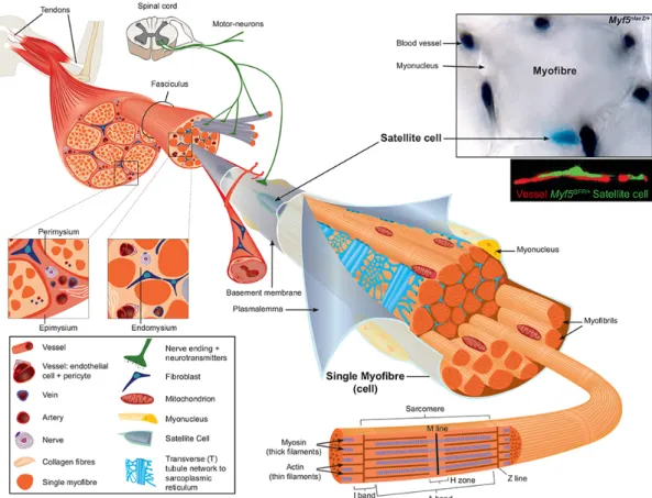
Skeletal muscle is one of the largest tissues in mammals. It allows voluntary movement and plays a key role in regulating metabolism and homeostasis of the organism. Throughout evolution, skeletal muscle is essentially defined by the succession of motor units which consists of a motoneuron and all of the muscle fibres innervated by that motoneuron (Figure 1). Myofibres are multinucleated cells and compose the cellular units of mature skeletal muscles. The structure of myofibres is strikingly repetitive at all sites in the organism, and the basic principles that govern the development of myofibres are conserved from Drosophila to humans. This structure is illustrated by the linear and repetitive arrangement of sarcomeres composed by an actin and myosin network together with associated proteins that enable muscle contraction (Figure 1). Different fibre types have been described, and these can be classified as slow-oxidative, fast-oxidative-glycolytic, and fast- glycolytic (Peter et al., 1972). The inherent contractile speed of each fibre-type cluster is determined essentially by the myosin motor protein isoform that is expressed predominantly. For example, the slow-oxidative unit expresses primarily a slow myosin heavy chain (MyHC) gene designated as slow or type I. The fast-oxidative unit expresses a combination of the fast type IIa and IIx MyHC genes, whereas the fast-glycolytic unit expresses both the fast IIb and IIx MyHC genes (Larsson et al., 1991). The accessibility of the hind limb Tibialis anterior muscle (below the knee), a mix of slow and fast fibres, has made it one the major sites for experimentation in studies on muscle homeostasis and regeneration. Finally, skeletal muscle allows the study of plasticity at the tissue and cellular level in different conditions such as overload (exercise), sarcopenia (muscle loss), ageing, and disease (myopathies).
The resident stem cells of skeletal muscle, historically called satellite cells, are located between the basement membrane containing a basal lamina, and the plasmalemma of the muscle fibre (Mauro, 1961) (Figure 1). Importantly, ≈90% of Muscle stem cells (MuSCs) are located in tight proximity with vessels (within 21μm) (Christov et al., 2007) (Figure 1), suggesting a communication between the vasculature and the MuSCs.
1.2. Muscle regeneration
The remarkable regenerative ability of skeletal muscle was shown several decades ago in rats that had received weekly injections of bupivacaine (anaesthetic drug that blocks sodium channels (see, (Gayraud-Morel et al., 2009)) for 6 months, and did not show reduction or exhaustion of muscle fibres repair capacity (Sadeh et al., 1985). Similarly in mouse, after 50 bupivacaine injections into the TA muscle mice regenerated their muscle without loss of myofibres or gain of fibrotic areas (Luz
Figure 1. Scheme of skeletal muscle and associated structures. Skeletal muscles in
general are attached at each end to the bone via tendons. Three connective tissue layers can be distinguished in skeletal muscle. The epimysium is the deep facia component that encloses the entire muscle and it is contiguous with the tendon and endosteum (facia surrounding bone). The perimysium encloses individual muscle fibers into fascicules (bundles). The endomysium is located between fibers and it encloses individual muscle fibers. Within the muscle cell (myofibre) the major intracellular source of calcium needed for muscle contraction is the sarcoplasmic reticulum, which connects to the transverse (T) tubules, and these surround the sarcomeres. Satellite cells are located between the basement membrane and the plasmalemma of the myofibre. Note the close proximity of the vessel, stained with India ink on the muscle section, and satellite cell from adult Myf5nlacZ/+ mouse stained with X-gal (upper image), or immunostained with
et al., 2002). In human, skeletal muscle injuries resulting from direct trauma (contusions), partial tears, fatigue, following surgical procedures or myopathies are common and present a challenge in traumatology, as therapy and recuperation are not well supported. The most commonly used acute murine injury models involve intramuscular injection of myotoxins (cardiotoxin and notexin), BaCl2, and mechanical injury (freeze, needle or crush injuries) (Gayraud-Morel et al., 2009; Hardy et al., 2016) (see also Annex 1). For the purpose of our study, we will focus on the injury following the injection of myotoxins. Cardiotoxin (CTX, protein kinase C inhibitor) and Notexin (NTX, phospholipaseA2) are isolated from snake venom, and they trigger an increase in Ca2+
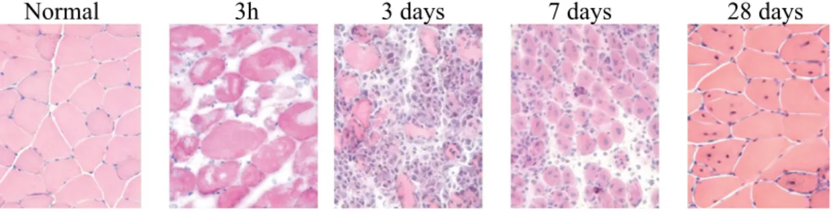
influx followed by fibre depolarization and consequently myofibre hypercontraction and necrosis (Gayraud-Morel et al., 2009; Hardy et al., 2016). After trauma, skeletal muscle regeneration follows three distinguishable and overlapping phases (Figure 2). The first phase of degeneration following severe injury is characterized by necrosis and significant inflammation (0 to 5 days post-injury (dpi)). After clearance of cellular debris, new fibres form and they transiently express embryonic and neonatal Myosin Heavy Chain (MyHC) from 3-14 dpi. The remodelling phase is characterized by hyperplasia and hypertrophy regulated in part by the IGF-1/Akt and TGFβ /Smad pathways. IGF-1 affects the balance between protein synthesis and protein degradation thus inducing muscle hypertrophy, whereas TGFβ negatively controls muscle growth (Schiaffino et al., 2013).
Figure 2. Regeneration of Tibialis anterior (TA) muscle after myotoxin injury. Three
hours after injury with the snake venom notexin, severe necrosis is apparent. After 3 days, most of the necrotic fibres are cleared by immune infiltrate and empty spaces are colonized by new myoblasts derived from satellite cells after activation and proliferation. Seven days post-injury, myoblasts continue to proliferate and fuse to restore fibre homeostasis (central nuclei). By 28 days, muscle regeneration appears to be complete histologically with the presence of centrally located myonuclei, a hallmark of regeneration. (Gayraud-Morel et al., 2009)
Although satellite cells play a crucial role in restoring myofibres following injury, it is clear that other cells types impact on the regeneration process (Figure 3) (see Annex 1). For example, fibro-adipogenic progenitors (FAPs) reside in the muscle interstitium and they play a significant myogenic and trophic role in muscle physiology during regeneration (Fiore et al., 2016; Joe et al., 2010; Lemos et al., 2015; Uezumi et al., 2010). Similarly, macrophages play a critical role during the initial stages following tissue damage as they are required for phagocytosis and cytokines release. The first wave of macrophages (peak at 3dpi) promotes myoblast proliferation via the secretion of pro-inflammatory molecules such as TNFα (Tumor Necrosis Factor α), INFα (Interferon α) and IL6 (Interleukin 6) (Lu et al., 2011a). Subsequently, macrophages undergo a phenotypical and functional switch toward an anti-inflammatory fate characterized by the production of IL4 and IL10, for example (Arnold et al., 2007). As mentioned previously, this anti-inflammatory response stimulates FAPs, mesoangioblasts, and also directly myoblasts to promote differentiation and fusion (Chazaud et al., 2003; Saclier et al., 2013). In addition, pericytes, located peripheral to the endothelium of microvessels, are known to be involved in blood vessel growth, remodelling, homeostasis, and permeability (Armulik et al., 2011) (Figure 3). The integrity of vessels is essential for muscle repair and homeostasis and it has been proposed that microvascular insufficiency could be responsible for the local inflammation and necrosis observed in both dystrophin-deficient mouse and human (Cazzato, 1968). Moreover, pericytes in skeletal muscles are constituents of the satellite cell niche where they secrete molecules such as IGF1 (insulin growth factor-1) or ANGPT1 (angiopoetin-1) to modulate postnatal myofibres growth and satellite cell entry in quiescence, respectively (Kostallari et al., 2015).
2. Satellite cells as adult skeletal muscle stem cells
2.1. A brief history
The regenerative potential of muscle was first shown in the 1860s, but almost a century elapsed before the satellite cell was discovered. Using electron microscopy, Alexander Mauro observed a group of mononucleated cells located at the periphery of the adult skeletal muscle fibres from the Tibialis anticus of the Xenopus and rat (Mauro, 1961). These cells were named satellite cells due to their localisation on the periphery of the myofibres (Figure 4).
The absence of satellite cells in cardiac muscle prompted him to speculate a role for these cells as skeletal-muscle specific precursor cells: "satellite cells are merely
Figure 3. Synoptic view of the different cell populations involved in muscle repair.
Although the generation of new fibres is dependent on MuSCs, other cell types such as macrophages, monocytes, mesenchymal stromal cells (including FAPs, mesoangioblasts and PICs), pericytes and fibroblasts are also critical for the regeneration process. (Baghdadi and Tajbakhsh, Annex 1).
Figure 4. Electron micrograph of a typical myonucleus (A) and satellite cells (B) in mouse.
Muscle satellite cell (S) is inside the basal lamina (arrowheads) and outside the sarcolemma (arrows) with an independent cytoplasm. In contrast, a myonucleus (M) is located inside the sarcolemma of the muscle fibre. Bar: 1μm. (Sinha-Hikim et al., 2003)
dormant myoblasts that failed to fuse with other myoblasts and are ready to recapitulate the embryonic development of skeletal muscle fibre when the main multinucleate cell is damaged" (Mauro, 1961). Interestingly, the position of this cell adjacent to the myofibre appears to be highly conserved in evolution, and similar satellite cells have been observed in multiple species, from the arthropods to mammals (see Baghdadi and Tajbakhsh, Annex 1). Electron microscopy also revealed other morphological characteristics of satellite cells: large nuclear-to-cytoplasmic ratio, few organelles, small nucleus, and condensed interphase chromatin.
The role of satellite cells in regeneration was first assessed after crush injury to the small web muscles of the East African fruit bat, Eidolon helvum (Church and Noronha, 1965). This study reported that satellite cells disappear from the highly injured area at the same time as the emergence of mitotic myoblasts, then reappear on myotubes after repair. Authors provided evidence that satellite cells were skeletal muscle “reserve cells”, capable of generating new fibres upon injury and replenishing the initial pool of cells. Additional [3
H]-Thymidine tracing experiments combined with electron microscopy demonstrated that satellite cells are mitotically quiescent in adult muscle contribute to myofibre nuclei upon injury (Moss and Leblond, 1970; Reznik, 1969). The same studies also demonstrated that satellite cells give rise to proliferating myoblasts (myogenic progenitors cells), which were previously shown to form multinucleated myotubes in vitro (Konigsberg, 1963; Snow, 1977; Yaffe, 1969). Moreover, in vivo [3
H]-Thymidine donor satellite cells specific labelling after free grafting of the muscle showed the presence of labelled nuclei on the periphery of regenerated myofibres in the host (Gutmann et al., 1976).
2.2. Molecular regulation of muscle stem cell emergence
During early development, muscle stem/progenitor cells migrate underneath the dorsal part of the somites called the dermomyotome (DM) and differentiate into mononucleated myocytes to form the myotome. In response to key transcription factors, committed myocytes align and fuse to generate small multinucleated myofibres during primary myogenesis in the embryo (from E11-E14.5), then myofibres containing a few hundred myonuclei during secondary myogenesis (from
continued myoblast fusion, or hyperplasia, is followed by muscle hypertrophy (Sambasivan and Tajbakhsh, 2007; Tajbakhsh, 2009; White et al., 2010) (Figure 5).
The developmental origin of satellite cells was first shown in a chick-quail chimera study: satellite cells of quail origin were found after replacement of chick somitic mesoderm by one from quail. In addition, electroporation of the central dermomyotome (the dorsal somite) in the trunk with a molecular marker showed that marked cells gave rise to Pax7+ satellite cells after hatching, thereby establishing the dermomyotome origin of satellite cells, in chick (Armand et al., 1983; Gros et al., 2005). Further evidences that satellite cells also originate from Pax3/7+ cells coming from the somites have been reported in the mouse (Kassar-Duchossoy et al., 2005; Relaix et al., 2005).
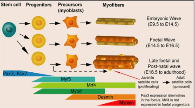
Emerging satellite cells are found underneath a basement membrane from about 2 days before birth in mice and they further proliferate until the mid-perinatal stage (Kassar-Duchossoy et al., 2005). The majority of quiescent MuSCs are established from about 2-4 weeks after birth (Tajbakhsh, 2009; White et al., 2010). During prenatal and postnatal myogenesis, stem cell self-renewal and commitment are governed by a gene regulatory network that includes the paired⁄homeodomain transcription factors Pax3 and Pax7, and basic helix-loop-helix (bHLH) myogenic regulatory factors (MRFs), Myf5, Mrf4, Myod and Myogenin (Figure 5). Pax3 plays a critical role in establishing MuSCs during embryonic development (except in cranial-derived muscles) and Pax7 during late foetal and perinatal growth. Indeed, Pax3:Pax7 double mutant mice exhibit severe hypoplasia due to a loss of stem and progenitor cells from mid embryonic stages, and these Pax genes appear to regulate apoptosis (Relaix et al., 2006; Relaix et al., 2005; Sambasivan et al., 2009). During perinatal growth, Pax7 null mice are deficient in the number of MuSCs and fail to regenerate muscle after injury in adult mice (Lepper et al., 2009; Oustanina et al., 2004; Seale et al., 2000; von Maltzahn et al., 2013).
Experiments using simple or double knockout mice have shown the temporal and functional roles of these different factors during myogenesis. Myf5, Mrf4 and Myod assign myogenic cell fate of muscle progenitor cells to give rise to myoblasts
(Kassar-Duchossoy et al., 2004; Rudnicki et al., 1993; Tajbakhsh et al., 1996) whereas Myogenin plays a crucial role in myoblast differentiation prenatally (Hasty et al., 1993; Nabeshima et al., 1993) but not postnatally as the conditional mutation of Myogenin in the adult has a relatively mild phenotype (Knapp et al., 2006; Meadows et al., 2008; Venuti et al., 1995). In the adult, Myod deficient mice that survive have increased precursor cell numbers accompanied by a delay in regeneration (Megeney et al., 1996; White et al., 2000); whereas Myf5 null mice display a slight delay in repair (Gayraud-Morel et al., 2007). These studies suggested that Myf5, Mrf4 and Myod could in some cases compensate for each other's function. Whereas Mrf4 plays a role in embryonic progenitors, Myf5 and Myod continue to regulate muscle progenitor cell fate throughout foetal and postnatal life. Interestingly, additional transcription factors have been shown to interact with MYOD to regulate myogenesis. For instance, ChiP-seq data demonstrated that KLF5 (Kruppel-like factor, member of a subfamily of zinc-finger transcription factors) (Hayashi et al., 2016) as well as RUNX1 (Umansky et al., 2015) binding to Myod-regulated enhancers is necessary to activate a set of myogenic differentiation genes.
The MRFs form heterodimers with members of the E-protein bHLH family (E2A, E2-2 and HEB) and bind to a consensus E-box sequence (CANNTG) to activate muscle-specific gene expression. Although there are millions of consensus E-boxes in the genome that can bind of the myogenic bHLH factors, the productivity of this occupancy and the specifity of binding is determined by flanking nucleotides in the E-box, thereby effectively reducing the number of sites that are functional (Cao et al., 2010).
It is likely that MRFs combined with other transcription factors fine-tune the myogenesis process and it would be important to further explore the set of co-activators/repressors required for each step of muscle repair.
2.3. Heterogeneity in the muscle stem cell population
Compelling evidence from several studies has demonstrated that the satellite cell population is heterogeneous regarding their gene set of expression, proliferation rate, differentiation potential, stemness and even survival.
One remarkable example is demonstrated by the heterogeneity in satellite cells derived from skeletal muscle arising from different developmental origins: head (non-segmented paraxial mesoderm) versus limb (somites) that showed distinct molecular signatures. Cranial mesoderm derived muscles (except extraoculars) are Tbx1-dependent, whereas somite-derived muscles are Pax3-dependent (Sambasivan et al., 2011a). Furthermore, Alx4, Pitx1/2 are specifically expressed in the cranial mesoderm-derived extraoccular muscles (EOM) (Sambasivan et al., 2009). In addition, EOM-derived satellite cells showed greater ex vivo growth, self-renewal capacities and in vivo transplantation efficiency (Stuelsatz et al., 2015).
Similarly, single fibre transplantation experiments suggested that heterogeneity exists in muscles with the same developmental origin, but different anatomical location: MuSCs isolated from EDL (Extensor digitorium longus) or soleus muscles have superior engraftment potential compared to MuSCs from TA (Tibialis anterior) (Collins et al., 2005). Given that the MuSCs were grafted with their adjacent fibre in those experiments, this result could also be explained by the heterogeneity in the stem cell niche rather than cell-autonomous properties of the satellite cells.
Figure 5. Expression of MRFs during lineage progression of myogenesis.
Pax3 and Pax7 expressions decline in the foetus. Myf5, Myod and Mrf4 expressing instruct to the progenitors cell a myogenic program. Desmin is an intermediate filament protein express in the muscle and Myosin is a component of the contractile apparatus. Around E16.5 Pax7+ cells appear in satellite cell position (see also Fig. 6). (Sambasivan and Tajbakhsh, 2007)
Strikingly, even within a single muscle cell population, heterogeneity has been reported. Continuous in vivo labelling with the thymidine analogue BrdU (5′-bromo-2′-deoxyuridine) in 4weeks-old rats revealed two populations: about ≈80% of satellite cells readily marked over the first 5 days and a slow cycling minority of cells not fully saturated upon 2 weeks of treatment. This second population named “reserve cells” was proposed to maintain quiescence during muscle growth/homeostasis and enter cell-cycle only upon trauma (Schultz, 1996). Furthermore, freshly isolated single myofibres from Myf5nlacZ
and Myf5Cre
;R26RYFP
mice showed ≈13% of MuSCs that never express Myf5 (Pax7+
/β-gal—
; Pax7+ /YFP—
, respectively), suggesting a more stem-like fate (Kuang et al., 2007). This Myf5— population is capable of asymmetric cell division and replenish the stem cell pool upon engraftment, whereas the Myf5+ undergo differentiation. These results suggest a hierarchical organisation of quiescent MuSCs: with a more stem population that will give rise to the more committed cells upon activation while self-renew to repopulate the quiescent niche. However, this phenotype is less pronounced with another Myf5Cre
allele, and eventually all satellite cells experience Myf5 expression, therefore it is unclear how the genetically modified mice reflect stem-like behaviour over time (Sambasivan et al., 2013). Indeed, the presence/absence of labelling relies on the efficiency of the Cre-recombinase that has been shown to not faithfully represent Myf5 expression in every condition, a phenomenon that has been reported also for other tissues (Comai et al., 2014).
To address some of these issues, a Tg:Pax7-nGFP mouse has been used to fractionate the satellite cell population in both quiescent and injured muscles based on the nGFP intensity. Interestingly, fractionation of the Pax7-nGFP population by FACS into Pax7High
(Top 10%) and Pax7Low
(Bottom 10%) revealed that the Pax7High
population displays more stem-like features such as lower metabolic activity, longer time to enter cell cycle compared to Pax7Low
that express more activation/differentiation genes (e.g: Myod, Myogenin, see below section 3.1.2), and higher expression of stem cell markers. Notably, Pax7High
cells were considered to be in a more dormant cell state (deeper quiescence), however serial transplantation of these subpopulations did not show dramatic differences in contribution to the niche (Rocheteau et al., 2012).
Recent technological advancements in single cell RNAseq, methylome analysis and mass cytometry now permit investigations of cellular heterogeneity within specific cell populations (Angermueller et al., 2016; Grun et al., 2016; Spitzer and Nolan, 2016). For example, analysis of single cells by multiparameter sequencing-based analysis, specifically RNAseq and bisulfite based methylome analysis, allows the investigation of epigenetic, genomic and transcriptional heterogeneities. Although powerful, some limitations include sequence depth and coverage of the genome. On the other hand, CyTOF based mass cytometry is based on a combination of markers conjugated to metal isotopes, and this led to the identification and classification of subpopulations of myogenic cells following muscle injury (Porpiglia et al., 2017). These emerging technologies can be used to assess the relative potential and role of a whole population at the single cell level and promise to give further insights into understanding MuSC heterogeneities.
3. Functions of muscle stem cells
3.1. Adult myogenesis
The absolute requirement for MuSCs was shown by genetic elimination of satellite cells postnatally using an inducible diphtheria toxin system that leads to an arrest in translation and subsequent cell death. This resulted in failed regeneration and replacement of the damaged muscle tissue with inflammatory and adipogenic cells (Lepper et al., 2011; Murphy et al., 2011; Sambasivan et al., 2011b). Nevertheless, some outstanding questions remain regarding the potential role of other interstitial cells in muscle repair (see Baghdadi and Tajbakhsh, Annex 1).
Examination of β-galactosidase activity in Myf5nlacZ mice indicated that the Myf5 locus is active in 90% of quiescent satellite cells, which suggests that most satellite cells are committed to the myogenic lineage (Beauchamp et al., 2000). Satellite cell physiology and progression throughout the myogenic program are tightly controlled by a hierarchy of transcription factors (Yablonka-Reuveni and Rivera, 1994) (Figure 6). At homeostasis, MuSCs remain quiescent and reside in G0-phase within their sublaminal niche contiguous to the myofibre (Schultz et al., 1978). It is thought that all adult quiescent satellite cells express the transcription factor Pax7 (Seale et al.,
2000); its paralogue Pax3 is also expressed in a subset of satellite cells of certain muscles (Relaix et al., 2006). While Pax3 plays a critical role during embryonic myogenesis, most satellite cells, however, downregulate Pax3 before birth (Kassar-Duchossoy et al., 2005). As mentioned above, in myogenesis, Pax7 and Pax3 play overlapping but non-redundant roles. These functional differences can be explained by differential binding affinities for paired versus homeobox motifs, suggesting differences in DNA binding and chromatin status affinities (Soleimani et al., 2012). Upon injury, MuSCs activate, re-enter the cell cycle and undergo cellular division to give rise to myoblasts, a highly proliferative transient amplifying cell population (Figure 6). In the adult, MRFs are also responsible for both myogenic lineage specification as well as for the regulation differentiation. Although MYF5, but not MYOD protein is expressed in satellite cells, Myod and Myf5 genes are both rapidly upregulated upon activation (Cooper et al., 1999; Gayraud-Morel et al., 2012). Finally, terminal differentiation is initiated by the downregulation of Pax7 (Olguin and Olwin, 2004) and the upregulation of Myogenin and Mrf4 to generate elongated myocytes that will further fuse into myotubes (Cornelison et al., 2000; Cornelison and Wold, 1997) (Figure 6). Essentially, a subpopulation of activated satellite cells, exit the cell cycle and return to quiescence in order to maintain the stem cell pool for future regeneration (Figure 6).
Figure 6. Muscle regeneration following different forms of injury. Following
mild or severe injury, quiescent muscle stem cells (MuSCs) activate, differentiate and fuse to repair the damaged fibre. The myogenic process is tightly regulated by the action of key transcription factors and regulators. (Baghdadi and Tajbakhsh, Annex 1)
3.1.1. Satellite cell activation and differentiation
Immediately following muscle injury, Myod expression is rapidly upregulated and MYOD protein is already detectable within satellite cells as early as 12 h after injury, before the first cell division that takes place from about 20h (Rocheteau et al., 2012; Smith et al., 1994). This early expression of Myod is proposed to be associated with a subpopulation of committed satellite cells, which are poised to differentiate without proliferation (Rantanen et al., 1995). In contrast, the majority of satellite cells express either Myod or Myf5 by 24h following injury and subsequently co-express both factors (Cornelison and Wold, 1997; Gayraud-Morel et al., 2012; Zammit et al., 2002) (Figure 6). Interestingly, ectopic expression of Myod in NIH-3T3 and C3H10T1/2 fibroblasts is sufficient to activate the complete myogenic program in these cells (Hollenberg et al., 1993); thus expression of Myod is an important determinant of myogenic commitment and differentiation, and its absence promotes proliferation and delayed differentiation (Myod—/—
)(Sabourin et al., 1999). During satellite cell activation, Pax7 and Pax3 target genes to promote proliferation and commitment to the myogenic lineage, while repressing genes that induce terminal myogenic differentiation (Soleimani et al., 2012). For example, PAX7 and PAX3 induce the expression of Myf5 by direct binding to distal enhancer elements and Myod by binding to the proximal promoter (Bajard et al., 2006; Hu et al., 2008). Moreover, p38 kinase (p38γ) also negatively regulates the transcriptional potential of Myod by phosphorylation, which leads to a repressive Myod complex occupying the Myogenin promoter (Gillespie et al., 2009). This observation is supported by the premature expression of Myogenin and reduced proliferation of myoblasts in p38-decificent muscle (Gillespie et al., 2009).
Terminal differentiation is initiated by the expression of Myogenin and later Mrf4 (Smith et al., 1994; Yablonka-Reuveni and Rivera, 1994) (Figure 6). ChIP-on-chip experiments (Bergstrom et al., 2002; Cao et al., 2006) and ChIP-Seq analysis (Cao et al., 2010) revealed MYOD and MYOGENIN specific target genes. These studies suggested a hierarchical organization involved in satellite cell activation and differentiation with regard to MRFs. MYOD directly activates Myogenin and Mef2 transcription factors, a large portion of downstream targets are muscle-specific
structural and contractile genes, such as those encoding actins, myosins, and troponins, essential for proper myofibres function.
p38α/β kinase stimulates the binding of MYOD and MEF2s to the promoters of muscle-specific genes, leading to the recruitment of chromatin remodelling complexes promoting myogenesis (Cox et al., 2003; Wu et al., 2000).
Besides MRFs and their regulators, other post-transcriptional factors have been shown to be involved in myogenic differentiation such as micro-RNAs (see Chapter 3).
3.1.2. Satellite cell self-renewal
The self-renewing capability of MuSCs has been demonstrated by series of transplantation experiments and clearly showed their remarkable ability to sustain the capacity for muscle repair. For example, transplantation of a single myofibre and its resident MuSCs (7-22/fibre) into irradiated muscles of immunodeficient dystrophic mice (nude; mdx) showed that MuSCs can give rise to over 100 new myofibres, expand and support further rounds of muscle regeneration (Collins et al., 2005). Similarly, purification of MuSCs followed by transplantation showed that they both contribute to muscle repair of nude; mdx mice and colonize the stem cell niche (Montarras et al., 2005). The self-renewing capability of satellite cells was further shown by serial transplantations of isolated Pax7-nGFP cells in pre-injured immunocompromised mice (Rocheteau et al., 2012); GFP+ cells were collected up to seven rounds of transplantations. Finally, single cell transplant experiments demonstrated that a single freshly isolated MuSC is capable to give rise to progeny cells and to self-renew upon injury (Sacco et al., 2008).
To study self-renewal ex vivo, two models are generally used: 1) floating isolated single myofibres where MuSCs will proliferate in clusters formed by activated, differentiated and self-renewed cells within 72h in the absence of cell fusion (Figure 7); 2) reserve cell model, where cells plated at high density will form myotubes and this is accompanied by the emergence of non-proliferative single cells (Pax7+
) adjacent to the myotubes (Figure 7).
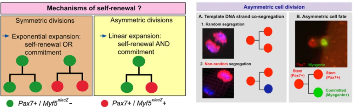
Stem cells can divide, commit to differentiation and self-renew in two fashions: asymmetrically (one daughter stem cell and one daughter committed cell) or symmetrically (two identical daughter cells, either renewed or committed). The balance between asymmetric versus symmetric division depends on several intrinsic and extrinsic cues, however how this is regulated, during growth and regeneration remains largely unknown (Collins et al., 2005; Motohashi and Asakura, 2014; Yennek et al., 2014). Asymmetric cell divisions have been reported in myogenic cells in several studies by following the differential distribution of transcription factors (Pax7, Myod, Myogenin), non-random DNA segregation (NRDS) of old and new DNA strands using nucleotide analogues, reporter gene expression, and dystrophin/Par complex (Kuang et al., 2007; Rocheteau et al., 2012; Shinin et al., 2006; Yennek et al., 2014).
For example, when myogenic cells were isolated on myofibres, asymmetric divisions were reported to occur when the mitotic spindle is perpendicular to the myofibre axis with the satellite stem cell (Pax7+
/Myf5—
) in close contact with the basal lamina and the committed cell (Pax7+
/Myf5+
) adjacent to the myofibre plasma membrane (Kuang et al., 2007). Furthermore, Wnt7a, through its receptor Frizzled-7, was reported to be upregulated in Pax7+
/Myf5+
cells, and it induced polarized expression of Vangl2, an Figure 7. Ex vivo study of satellite cell self-renewal. Left: Schematic
representation of single myofibre satellite cell renewal model. Within 72h after isolation, a single MuSC will give rise to a cluster composed by self-renewed cells Pax7+ (green), differentiated cells Myod+/Pax7— (red) (Zammit et al., 2006). Right: Culture of murine cells showing differentiated MyHC+ myotubes (red) and tightly associated Pax7+ satellite cells (green; arrows) that returned to quiescence: reserve cells (Abou-Khalil et al., 2013).
effector of the planar cell polarity pathway, which was required for Wnt7a-mediated satellite cell expansion (Le Grand, Jones, Seale, Scime, & Rudnicki, 2009).
In other studies, NRDS was reported in satellite cells ex vivo and in vivo (Yennek and Tajbakhsh, 2013). Semiconservative replication of DNA can result in random or non-random segregation of older template and nascent DNA strands in daughter cells during cell division. Pulse-chase DNA labelling experiments using thymidine analogues (BrdU, EdU (5′-ethynyl-2′-deoxyuridine)) in injured muscle showed that up to 80% of the Pax7High
activated population (by extrapolation, 8% of total GFP population) performs non-random or template DNA segregation (NRDS or TDSS) (Rocheteau et al., 2012; Shinin et al., 2006; Yennek et al., 2014). Interestingly, NRDS was directly associated with cell fates: the more stem cell Pax7+
/Myogenin— retains the old strand while the committed cell Pax7—
/Myogenin+
inherits the newly synthesized strand (Figure 8) (Conboy et al., 2007; Rocheteau et al., 2012; Yennek et al., 2014).
Figure 8. Proposed models for satellite cell self-renewal via asymmetric cell division.
Left: Satellite cell self-renewal can be achieved by either symmetric or asymmetric cell division. Symmetric divisions can amplify the stem cell pool, or generate differentiated cells whereas asymmetric divisions result in maintenance of one stem cell and the generation of one differentiated daughter cell (Sambasivan and Tajbakhsh, 2007). Right: (A) Random DNA segregation where daughter cells inherit old and new DNA strands (1) or only one cell is labelled with BrdU indicating non-random DNA segregation (2). (B) Asymmetric division and cell fate: the division of one Pax7+ cell gives rise to one stem (red) and one committed (green) daughter cell. (Yennek and Tajbakhsh, 2013)
Chapter 2.
1. Stem cell quiescence
Cellular quiescence is a reversible, non-proliferative G0-arrested state characterized by the ability to re-enter the cell cycle and generate progenitor cells in response stimuli, such as trauma. The quiescence state was extensively studied in the budding yeast Sacchromyces cerevisiae as a mode of survival that can be induced by nutrient deprivation (Herman, 2002). Similar conditions were noted in mammalian cells in vitro (Zetterberg and Larsson, 1985). The presence of quiescent adult stem cells in multiple tissues and organs highlights the essential role of this cell state.
1.1. Identification of quiescent stem cells
Due to the low numbers of quiescent stem cells (QSCs) in a given tissue, our understanding of this cell state has been limited to the absence of markers associated with proliferation and differentiation. For example, nucleotide analogues (3
H-TdR, BrdU, EdU), endogenous markers of proliferation (PCNA, a DNA polymerase accessory protein expressed in S-phase), Ki67 (ribosomal RNA transcription associated protein), MCM-2 (protein involved in replication origins, S-phase) and phospho-Histone3 (M-phase specific) can be detected by autoradiography or immunofluorescence (Conboy et al., 2007; Shinin et al., 2006). More recently, histone tagged proteins (H2B-GFP/YFP) have been used as their association with DNA is replication-dependent thereby allowing live imaging by videomicroscopy (Foudi et al., 2009; Tumbar et al., 2004). QSCs have also been identified based on label retention. Label retention is based on the premise that a dividing cell will dilute away an incorporated label (e.g. nucleotide analogue, H2B-GFP), whereas a QSC, or slow-cycling cell, will retain the label for longer periods of time. The presence or absence of label-retaining cells (LRCs) has been for a long time the only tool to determine if a population of stem cells was quiescent; however, it has become increasingly clear that this approach is not sufficient. In high-turnover tissues such as the small intestinal epithelium, lineage-tracing experiments allow the distinction of at least two populations with stem cell potential: the long-retaining reserve cells (+4) and the proliferating stem cells (Lgr5+) (Buczacki et al., 2013). Similarly, the skin houses a first proliferative stem population at the basal layer of the epidermis and a quiescent population in the bulge of the hair follicle (HFSC) (Ito et al., 2005). Interestingly, in both cases, the active stem population was proposed to be involved
in tissue homeostasis whereas the quiescent, LRCs appear to be mobilized upon injury (Li and Clevers, 2010).
1.2. Ex vivo induction of quiescence
Cellular quiescence can be mimicked in vitro by modulating cell culture conditions such as the nutrient concentration or adherence cues. The loss of adherence has been shown to induce both mouse and human myoblasts back to quiescence by culture in suspension in a methylcellulose gel (Milasincic et al., 1996; Sellathurai et al., 2013). Similarly, culture on soft substrate induces the loss of contractile property and can trigger a quiescent-like state (Gilbert et al., 2010). Although fibroblasts respond well to the deprivation of nutrients/mitogens, myoblasts tend to differentiate rather than go back to quiescence (Arora et al., 2017; Rumman et al., 2015).
1.3. Molecular signature of quiescence
1.3.1. Epigenetic control
Recent epigenetic studies showed that during development, chromatin configuration becomes more and more restrictive as cells commit and differentiate into specific lineages. One key determinant of gene expression is the landscape of histone modifications often associated with gene activation or repression. For example, actively transcribed genes are commonly marked by trimethylation of histone 3 lysine 4 (H3K4me3) around their transcription start sites (TSSs) and H3K36me3 in the gene body, whereas Polycomb group (PcG) complex-mediated H3K27me3 is associated with transcriptional repression (Jenuwein and Allis, 2001). Some chromatin regions, referred to as bivalent domains are marked by both H3K4me3 and H3K27me3. They are frequently located in close proximity to TSS and have been shown to mark master regulators of cell lineage, maintaining ES cell in this poised state mentioned above (Bernstein et al., 2007; Li et al., 2012).
Regarding MuSCs, histone profiles in quiescent versus activated (2, 3, and 5dpi) satellite cells has been performed by mass-spectrometry-based proteomics and highlighted a time-dependent shift towards a heterochromatic state during activation (Schworer et al., 2016). Complementary to this study, chromatin
immunoprecipitation sequencing (ChIP-seq) combined with transcriptomic analysis in quiescent and activated satellite cells also showed a switch from permissive state in quiescence to a more repressed state in activation (Liu et al., 2013a). Quiescence to activation transition is marked by the retention of H3K4me3 and a dramatic increase of H3K27me3 mark at the TSSs. Finally, the fine-tuned epigenetic regulation of establishment and/or maintenance of the reversible quiescent state has been recently demonstrated in MuSCs, where the H3K9 methyl-transferase PRDM2 binds to thousands of promoters mostly marked by the repressive H3K9me2 mark such as the G0-arrest inducing gene Ccna2 (Cheedipudi et al., 2015).
1.3.2. Cell cycle regulators
Cyclin-dependent kinase inhibitors (CKIs) such as p21, p27 inhibit CDK2, and CDK4 respectively are expressed in QSCs to block cell cycle progression (Sherr and Roberts, 1999). The genetic loss of p21 or p27 induces exhaustion of HSCs due to a high proliferative capacity (Zou et al., 2011). Similarly, MuSCs deficient for p21 (p21 KO) increase their proliferation rate but fail to undergo differentiation (Hawke et al., 2003); meaning that different CKIs are involved in the exit from the cell cycle triggered by differentiation (Mohan and Asakura, 2017).
Rb family proteins (Rb, p130 and p107) are guardians of the G1/S transition and inhibit cell cycle progression by controlling S-phase transcription factors (Weinberg, 1995). HSCs deficient for Rb proteins have an enhanced proliferation and fail to replenish the stem cell pool in the bone marrow after transplantation (Viatour et al., 2008). Rb proteins are highly expressed in quiescent MuSCs, and their genetic inactivation induce accelerated cell cycle entry, loss of myogenic differentiation and ultimately cell death (Hosoyama et al., 2011). Interestingly, p300 has been shown to suppress myogenic differentiation genes; thus Rb proteins block cell cycle progression and differentiation of MuSCs (Carnac et al., 2000).
2. Molecular signature of MuSCs
Transcriptomic analysis comparing quiescent and activated satellite cells have been done by several labs (Farina et al., 2012; Fukada et al., 2007; Garcia-Prat et al., 2016; Liu et al., 2013a; Lukjanenko et al., 2016; Pallafacchina et al., 2010). Although many quiescence specific genes are found in all data sets, the variations in the
experimental procedures raise questions regarding reproducibility. For instance, for in vivo satellite cell activation, several techniques were used to induce the injuries including BaCl2, or myotoxins. Cell extraction protocols also varied among the different studies: i) using transgenic mice expressing a reporter gene that marks satellite cells and ii) using a combination of antibodies targeting surface cell antigens specific to satellites cells (see Annex 2). In an attempt to normalize these differences, we developed a standardized pipeline for comparing quiescent versus activation data sets. An initial analysis of 11 samples revealed a quiescent transcriptional signature that includes already known genes such as Calcitonin receptor, Teneurin-4 and Collagen genes (type 5 and 6) (see Annex 2; manuscript in preparation).
Furthermore, histone landscape analysis coupled with microarray in quiescent versus activated satellite cells showed that genes expressed at high levels in quiescence were marked only by H3K4me3 (Liu et al., 2013a). This list of genes included a large number of known quiescent-specific genes such as Pax7, Cd34, Odz4 and Calcitonin receptor (Calcr), and Notch target genes Hey1, Hey2, and HeyL. Notably, this list of genes was dominated by genes encoding glycoproteins. Given that glycoproteins are integral membrane proteins that often play an important role in cell and cell-matrix interactions (Moremen et al., 2012), these glycoproteins that expressed at high levels in QSCs may be important mediators of interactions within the niche (see Section 2 below). In the context of our work, we focus on two quiescent-specific genes: Calcitonin receptor and Teneurin-4.
2.1.1. Calcitonin receptor
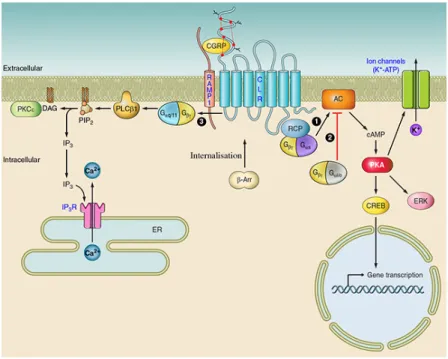
The calcitonin receptor (Calcr) belongs to the secretin-like family of is a G-protein- coupled seven transmembrane protein (GPCR) arising from a 70kb gene composed of 12 encoding exons. In human and rodents, alternative splicing gives rise to two Calcr isoforms: Calcr-C1α and Calcr-C1β. As the Calcr is widely expressed, it has been proposed that its tissue-specific expression is regulated by the single transmembrane co-receptor of the RAMPs: RAMP1-3 (receptor activity modifying protein) (Russell et al., 2014). Upon glycosylation, the heterodimerization of both CALCR and one of the RAMP peptides is required for the mature protein to be
1998). It is still unclear how the dimerization of RAMPs with the CALCR is regulated, especially in cell types that coexpress several RAMP isoforms (Figure 9).
Figure 9. Intracellular mediated signalling of calcitonin receptor. Binding of CGRP
ligand to the CALCR/RAMP receptor can activate multiple signalling pathways. (1) The activation of adenylate cyclase (AC) by Gαs G-protein subunit, triggers the elevation of intracellular cAMP, thereby activating protein kinase A (PKA), resulting in the phosphorylation of multiple downstream targets. These targets may include potassium-sensitive ATP channels (KATP channels), extracellular signal-related kinases (ERKs), or transcription factors, such as cAMP response element-binding protein (CREB). (3) Reports in osteoblasts have also shown evidence of Gαq/11-mediated signalling, involving activation of PLC-1, cleaving phosphatidylinositol 4,5-bisphosphate (PIP2) to form inositol trisphosphate (IP3) and diacylglycerol (DAG). IP3 binds to the IP3 receptor (IP3R) on the endoplasmic reticulum (ER), causing calcium release and thus raising cytoplasmic concentrations. DAG may activate PKCε, which in turn phosphorylates proteins further downstream. Upon activation, GPCR forms a complex with β-arrestins (β-Arr), that undergoes dynamin/clathrin dependent endocytosis for further lysosomal degradation or endosome recycling (Walker et al., 2010). Adapted from (Russell et al., 2014)
To date, the only known ligand of CALCR is the polypeptide hormone calcitonin (CT), synthetized by the thyroid gland and known to regulate serum calcium levels. The main targets of CT are the osteoclasts where it inhibits bone resorption via interaction with CALCR. Although it has other roles in the blood, kidney, CNS, respiratory system, gastrointestinal system and sperm, whether its function is mediated by CT is unclear (Russell et al., 2014). Upon activation, CALCR triggers a downstream pathway involving Gsα protein described in Figure 9.
Quiescent MuSCs specifically express the C1α isoform and all three RAMP isoforms. Calcr is downregulated during activation and is absent in activated cells (2, 5 and 7d post-injury), then it is re-expressed by 14dpi when the majority of satellite cells return to quiescence (Yamaguchi et al., 2015). Interestingly, the specific ablation of Calcr in satellite cells (Pax7CreERT2
; Calcrflox
) induces an exit of satellite cells from the quiescence niche followed by apoptosis, resulting in partial a loss of the stem cell poll (Yamaguchi et al., 2015). Furthermore, in vitro activation of CALCR with the synthetic peptide Elcatonin induces the cAMP-PKA pathway to inhibit the expression of cyclin-related genes (like Ccnd1, Ccna2, and Skp2) resulting in the active maintenance of the G0-quiescent state (Yamaguchi et al., 2015).
2.1.2. Teneurin-4 or Odz4
Odz is the vertebrate homologue of the Drosophila odd Oz pair-rule gene and encodes a large type II transmembrane protein family: teneurins (Tenm). In vertebrates, there are four Odz/Tenm numbered 1-4 mainly expressed in the CNS (Tucker et al., 2007). Although Odz4 function has been studied in chick embryo neuron patterning (Kenzelmann-Broz et al., 2010) and mouse oligodendrocyte differentiation (Suzuki et al., 2012), the role of the teneurins and their mechanisms of action remain largely unknown. When the intracellular domain of teneurins are targeted by immunostaining on cells in vitro, they localize to the nucleus whereas the extracellular domain remains at the membrane, suggesting that they might be cleaved and act as transcription factors similar to Notch (see Chapter 4)(Bagutti et al., 2003). However, whether ODZ/TENM binds to DNA and activates transcription of specific genes has yet to be demonstrated.
Odz4 and Odz3 are both present in satellite cells, however only Odz4 expression shows a clear restriction to quiescent satellite cells, and its expression reappears between 5-7 days post-injury (Fukada et al., 2007). Odz4 contains 33 exons that can give rise to 12 different coding proteins by alternative splicing. Interestingly, in the study reporting the role of Odz4 in oligodendrocyte differentiation, the authors also indicated that focal adhesion kinase (FAK), a key regulator of cell adhesion, is activated downstream of Odz4 (Suzuki et al., 2012); therefore, in quiescent MuSCs, Odz4 might control cell adhesion and/or differentiation.
The only study involving Odz4 in muscle used a transgenic mouse originally designed to study the role of a recombinant FLAG-tagged perlecan (heparin sulfate proteoglycan) specifically in cartilage under the control of Col2a1 promoter (Suzuki et al., 2012). Homozygous null mice developed severe tremors in the hindlimbs and paralysis due to hypomyelination in the CNS, hereafter named “furue” (japanese term for tremor): FurueTg(Hspg2)2Yy. Because this phenotype was likely caused by the transgene insertion, FISH (fluorescent in situ hybridization) and screening of a bacterial artificial chromosome library prepared from Furue mice allowed the identification of a transgene insertion into intron 5 of Odz4, located on chromosome 7 (Suzuki et al., 2012). The analysis of Furue mice showed hypoplasia in perinatal and adult animals in addition to a decrease in MuSCs number, subsequently inducing a delay in regeneration upon injury (Ishii et al., 2015). Moreover, upon injury, Odz4-deficient satellite cells atypically maintained high proliferation capacities and the activation marker MYOD 7dpi (Ishii et al., 2015). However, the constitutive repression of Odz4 raises questions about the specificity of its action in the satellite cell population as muscle growth and repair involve the collaboration of diverse cell regulators. Furthermore, the innervation of muscle is critical for its proper development and regeneration, thus the hypomyelination of the CNS showed by Suzuki and collegues has high probability to affect muscle function in general as nervous input is altered (Suzuki et al., 2012).
Finally, in the mutant embryos Pax3Cre/+; Rbpjflox/flox; Myod—/—a decrease of Odz4 expression was observed in isolated myoblasts suggesting that Odz4 and Notch functions might be correlated (Brohl et al., 2012). Accordingly, we showed that Odz4 is a Notch pathway target genes (see Results, part II).
2. The stem cell niche
The concept of the “niche” proposed to represent the specific microenvironment that maintains and instructs stem cells (Schofield, 1978). Extensive studies that investigated Drosophila and Caenorhabditis elegans (C. elegans) adult SC niches in vivo have confirmed the critical role of the niche in modulating stem cell behaviour (Byrd and Kimble, 2009; de Cuevas and Matunis,