RESEARCH OUTPUTS / RÉSULTATS DE RECHERCHE
Author(s) - Auteur(s) :
Publication date - Date de publication :
Permanent link - Permalien :
Rights / License - Licence de droit d’auteur :
Dépôt Institutionnel - Portail de la Recherche
researchportal.unamur.be
University of Namur
Proton irradiation orchestrates macrophage reprogramming through NFB signaling
Genard, Géraldine; Wera, Anne Catherine; Huart, Camille; Le Calve, Benjamin; Penninckx,
Sébastien; Fattaccioli, Antoine; Tabarrant, Tijani; Demazy, Catherine; Ninane, Noëlle;
Heuskin, Anne Catherine; Lucas, Stéphane; Michiels, Carine
Published in:
Cell Death and Disease DOI:
10.1038/s41419-018-0757-9 Publication date:
2018
Document Version
Publisher's PDF, also known as Version of record
Link to publication
Citation for pulished version (HARVARD):
Genard, G, Wera, AC, Huart, C, Le Calve, B, Penninckx, S, Fattaccioli, A, Tabarrant, T, Demazy, C, Ninane, N, Heuskin, AC, Lucas, S & Michiels, C 2018, 'Proton irradiation orchestrates macrophage reprogramming through NFB signaling', Cell Death and Disease, vol. 9, no. 7, 728. https://doi.org/10.1038/s41419-018-0757-9
General rights
Copyright and moral rights for the publications made accessible in the public portal are retained by the authors and/or other copyright owners and it is a condition of accessing publications that users recognise and abide by the legal requirements associated with these rights. • Users may download and print one copy of any publication from the public portal for the purpose of private study or research. • You may not further distribute the material or use it for any profit-making activity or commercial gain
• You may freely distribute the URL identifying the publication in the public portal ? Take down policy
If you believe that this document breaches copyright please contact us providing details, and we will remove access to the work immediately and investigate your claim.
A R T I C L E
O p e n A c c e s s
Proton irradiation orchestrates
macrophage reprogramming through
NF
κB signaling
Géraldine Genard
1, Anne-Catherine Wera
1, Camille Huart
1, Benjamin Le Calve
1, Sébastien Penninckx
2,
Antoine Fattaccioli
1, Tijani Tabarrant
2, Catherine Demazy
1, Noëlle Ninane
1, Anne-Catherine Heuskin
2,
Stéphane Lucas
2and Carine Michiels
1Abstract
Tumor-associated macrophages (TAMs) represent potential targets for anticancer treatments as these cells play critical roles in tumor progression and frequently antagonize the response to treatments. TAMs are usually associated to an M2-like phenotype, characterized by anti-inflammatory and protumoral properties. This phenotype contrasts with the M1-like macrophages, which exhibits proinflammatory, phagocytic, and antitumoral functions. As macrophages hold a high plasticity, strategies to orchestrate the reprogramming of M2-like TAMs towards a M1 antitumor phenotype offer potential therapeutic benefits. One of the most used anticancer treatments is the conventional X-ray radiotherapy (RT), but this therapy failed to reprogram TAMs towards an M1 phenotype. While protontherapy is more and more used in clinic to circumvent the side effects of conventional RT, the effects of proton irradiation on macrophages have not been investigated yet. Here we showed that M1 macrophages (THP-1 cell line) were more resistant to proton irradiation than unpolarized (M0) and M2 macrophages, which correlated with differential DNA damage detection. Moreover, proton irradiation-induced macrophage reprogramming from M2 to a mixed M1/M2 phenotype. This
reprogramming required the nuclear translocation of NFκB p65 subunit as the inhibition of IκBα phosphorylation
completely reverted the macrophage re-education. Altogether, the results suggest that proton irradiation promotes
NFκB-mediated macrophage polarization towards M1 and opens new perspectives for macrophage targeting with
charged particle therapy.
Introduction
The immune system takes part in both cancer elim-ination and tumor development, especially through its
activation and then its adaption to cancer cells1. This
process is called the cancer immunoediting and it per-fectly illustrates the ambivalent function of the immune system in cancer. In addition, the effectiveness of treat-ments such as X-ray orγ-ray radiotherapy (RT) is partially
conditioned by the presence of immune cells into the tumor. For example, ablative radiation (20 Gy) on local
tumor significantly reduced the tumor volume in
wild-type (WT) but not in nude (T-cell-deficient) mice bearing
a melanoma (B16 melanoma cells)2. In line with this
observation, local RT produces an abscopal response, which is an antitumor effect on distant unirradiated tumors (metastases), by triggering systemic immune
responses3. Another example is the systemic immune
activation in mice previously treated by local RT and immune checkpoint inhibitors that prevented the tumor
growth in mice rechallenged with the same tumor4.
However, the number of infiltrating tumor-associated macrophages (TAMs) is known to limit radiotherapy © The Author(s) 2018
Open Access This article is licensed under a Creative Commons Attribution 4.0 International License, which permits use, sharing, adaptation, distribution and reproduction in any medium or format, as long as you give appropriate credit to the original author(s) and the source, provide a link to the Creative Commons license, and indicate if
changes were made. The images or other third party material in this article are included in the article’s Creative Commons license, unless indicated otherwise in a credit line to the material. If
material is not included in the article’s Creative Commons license and your intended use is not permitted by statutory regulation or exceeds the permitted use, you will need to obtain
permission directly from the copyright holder. To view a copy of this license, visithttp://creativecommons.org/licenses/by/4.0/.
Correspondence: Carine Michiels ([email protected])
1
Cellular Biology Research Unit (URBC)—NARILIS, University of Namur, Namur, Belgium
2
Laboratory of Analysis by Nuclear Reaction (LARN / PMR)—NARILIS, University of Namur, Namur, Belgium
Edited by H.-U. Simon
1234567890() :,; 1234567890( ):,; 1234567890() :,; 1234567890( ):,;
efficiency and is directly correlated to a poor prognosis5 . Inside the tumor, TAMs represent up to 50% of the host infiltrating cells6, 7
, meaning that these cells are highly recruited into the tumor site and play critical roles in tumor development. It has been shown that RT promotes the recruitment of macrophages into the tumor, favoring
tumor relapse after treatment8. Indeed, TAM depletion or
inhibition of monocyte recruitment into the tumor site combined to RT induce tumor regression in mouse can-cer models and increase the survival of cancan-cer patients9, 10
. Another attractive and effective strategy is the re-education of TAMs towards an antitumoral phenotype.
Macrophages display a remarkable plasticity allowing them to fulfill a multiple range of functions. These multitask skills rely on two opposite phenotypes, M1 versus M2. These two subpopulations are classified as two extremes of a linear scale between which exists a
multitude of intermediate states11. Proinflammatory M1
macrophages, also called classically activated macro-phages, exhibit enhanced pathogen phagocytosis,
pro-mote inflammation, and activate immune system. On
the opposite, anti-inflammatory M2 macrophages,
referred to as alternatively activated macrophages, contribute to tissue repair, matrix remodeling, and angiogenesis, and also repress the immune system. It is well accepted that M1 macrophages exert antitumor functions while M2 macrophages show protumoral activity and, unfortunately, are usually the most
repre-sentative TAM population into the tumor12. M2-like
TAMs display their harmful actions by promoting genetic instability, stem cell nurturing, angiogenesis,
metastasis spreading, and local immunosuppression8.
The polarization of macrophages towards an M1 or an M2 phenotype is driven by the activity of diverse tran-scription factors and miRNAs. Among trantran-scription
factors, NFκB plays a central role to influence the
inflammatory macrophage status. While the active
het-erodimer NFκB (p50–p65) promotes the transcription
of proinflammatory genes, such as TNFα, IL-6, and
IL1β, the inactive homodimer NFκB (p50–p50) prevents
the transcription of proinflammatory genes and confers
the anti-inflammatory status to M2 macrophages13.
The plasticity of TAMs and their ability to be repro-grammed, especially from M2 to M1 phenotype, make them an attractive target for anticancer therapies.
Con-ventional radiotherapy (X-rays or γ-rays) initiated the
polarization of differentiated but unpolarized (M0) mac-rophages towards M1 when exposed to moderate doses
(1–10 Gy) (for a review, see ref. 14
). In addition, the
combination of CD8+ T-cell transfer and γ-ray used at
moderate doses (2 Gy) re-educated TAMs towards an M1
phenotype in a pancreatic tumor mice model10. Other
studies also reported TAM reprogramming after low dose
of whole-body irradiation15, 16. However, no study has
established the reprogramming of M2 into M1 macro-phages with local conventional RT only. This suggests that RT alone is not sufficient to reverse macrophage polarization.
Over the last decades, efforts were aimed to improve the delivery of conventional RT using image guidance. Despite these improvements, side effects associated to this treatment have remained severe. To spare surrounding healthy tissues, protontherapy presents an increasing interest, thanks to the charged nature of the particles and its depth dose profile. In more details, the one-shot energy release at the end of the charged particle track allows the improvement of dose conformation. This is added to the fact that the track of charged particles can be easily
deviated by a magneticfield to precisely target the tumor.
As for X-ray irradiation, the deposited energy by charged particle beam promotes the ionization of DNA through reactive oxide species (ROS) production. In addition to these indirect DNA damage, charged particles also directly interact with DNA, resulting in more complex
DNA damage17. Our work demonstrated the ability of
protontherapy to induce macrophage reprogramming. By using THP-1-derived M0, M1 and M2 macrophages, we evidenced that proton irradiation, but not X-ray irradia-tion, induced a partial switch from M2 to M1 macro-phages. This macrophage reprogramming is orchestrated, at least in part, by an NFκB activation.
Results
To address these goals, THP-1 cells were differentiated (M0) and polarized (M1 or M2) in irradiation chambers
(Fig. 1). These special devices were placed at the end of
the accelerator-produced proton beam and the effects on macrophages were then analyzed.
M1 macrophages are more resistant to moderate doses of proton irradiation than M0 and M2 macrophages
In order to evaluate the cell viability after proton
irradiation, ethidium bromide−acridine orange staining
was performed (Fig. 2a) and the number of dead cells
(orange) and viable cells (green) was counted (Fig.2b).
The cell viability slightly decreased 8 h (Fig. 2b) after
moderate proton irradiation doses (0–10 Gy), as
indi-cated by a survival of 82% for M0 macrophages, 82% for M1 macrophages, and 78% for M2 macrophages when irradiated with a dose of 10 Gy. Proton irradiation at 10 Gy further lowered the viability of M0 and M2
macro-phages respectively to 42 and 50% after 16 h (Fig. 2b).
Surprisingly, the viability of M1 macrophages was only slightly affected (92%) 16 h after proton irradiation at doses as high as 10 Gy. These results suggest an early radioresistance of the M1 phenotype to moderate pro-ton irradiation doses, compared to the two other phenotypes.
M1 radioresistance correlates with more intenseγH2AX
and 53BP1 labeling
In order to further examine the influence of proton
irradiation on macrophages, phosphorylated H2AX (Ser
139) (γH2AX), a sensitive marker for DNA double-strand
breaks (DSBs), was evaluated by immunofluorescence
labeling (Fig. 3a). Quantifications of γH2AX labeling
(Fig.3b) 15 min after irradiation indicated a similar profile for the three macrophage phenotypes when irradiated at
different doses (3, 5, and 10 Gy). However, the
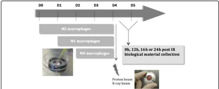
Fig. 1 Schematic outline of the irradiation procedure for macrophages. THP-1 monocytes were differentiated into macrophages (M0) with 150 nM PMA in cloning cylinder, placed at the center of the irradiation chamber. Macrophages were polarized in M1 phenotype with 10 pg/ml LPS and 20 ng/ml IFN-γ during 24 h incubation or were polarized in M2 phenotype with 20 ng/ml IL-4 and IL-13 during 48 h incubation. THP-1 monocytes were differentiated on the appropriate day, in order to obtain the three phenotypes on day 4. Following the experiment that was performed, the biological material was collected 8, 12, 16, or 24 h after proton or X-ray irradiation
B
M0 0 2 4 6 8 10 0 20 40 60 80 100 Dose (Gy) Vi ab ili ty (% ) * *** 16h 8h *** * M1 0 2 4 6 8 10 0 20 40 60 80 100 Dose (Gy) V ia bilit y (% ) 16h 8h M2 0 2 4 6 8 10 0 20 40 60 80 100 Dose (Gy) V ia bili ty (% ) *** * *** *** 16h 8h ** *** ***A
Fig. 2 Early radioresistance of M1 macrophages after moderate doses of proton irradiation. M0, M1, and M2 macrophages were irradiated with different doses of protons. Viability was assessed by ethidium bromide−acridin orange at different times postirradiation. a Representative ethidium bromide−acridine orange staining images of M1 and M2 macrophages 16 h after proton irradiation (0 Gy and 10 Gy). Lived cells appeared in green while dead cells are stained in orange. b Quantification of viability (%) in M0, M1, and M2 macrophages, 8 or 16 h after proton irradiation. N= 3 for each dose (mean ± SD). One-way ANOVA analyses followed by Dunnett’s multiple comparisons post-tests were performed on data; *p≤ 0.05; **p < 0.01; ***p < 0.001
quantifications of γH2AX labeling (Fig.3c) over the time
post irradiation (0–360 min) indicated a similar profile for M0 and M2 macrophages while M1 phenotype exhibited
a higher level of phosphorylated H2AX. In more details,
γH2AX labeling increased 5 min after proton irradiation
(3 Gy) in M0 and M2 phenotypes and was mostly
decreased 2 h after irradiation. In contrast, the γH2AX
labeling was increased and plateaued as long as 6 h after irradiation in M1 macrophages, indicating an increasing and prolonged detection of DSBs in this phenotype. In
order to confirm these data, 53BP1 intensity was also
assessed by immunofluorescence labeling (Fig.3d). 53BP1
is known for its role in DNA repair machinery. Similarly
to γH2AX labeling, the quantification of 53BP1 intensity
revealed a sustained labeling over the time in M1 mac-rophages, while 53BP1 intensity was decreased 1 h after
proton irradiation in M0 and M2 macrophages (Fig.3e).
As chromatin conformation could influence the detection and the repair of DSBs, we evaluated the heterochromatin content by MNAse I assay in the three different pheno-types. The radioresistance of M1 macrophages was not linked to a higher level of DNA condensation before irradiation since there was no difference in the hetero-chromatin content in the three phenotypes (Fig. S1). In
addition to sustainedγH2AX, the analysis of ROS content
by flow cytometry revealed that M1 macrophages better
managed H2O2treatment (Fig. S2). Taken together, these
results indicated that the radioresistance of M1 macro-phages to proton irradiation could be related to higher DSB detection and/or to better efficiency of DNA repair machinery in this phenotype, and also correlates with a better elimination of ROS.
Proton irradiation induces macrophage reprogramming
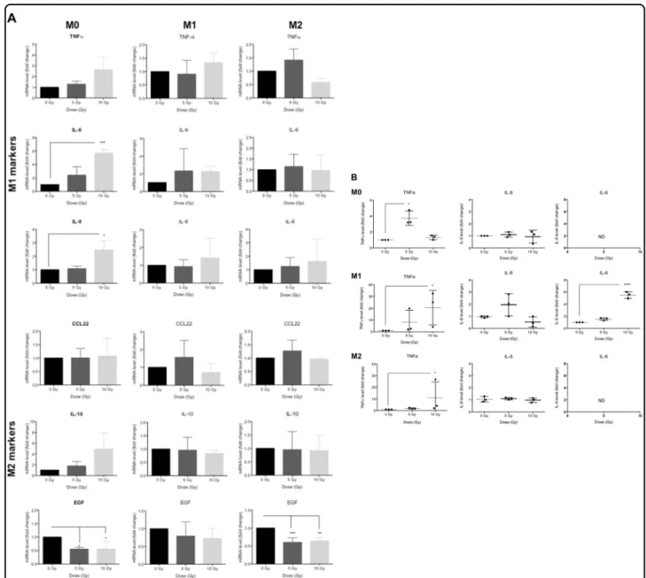
Proton irradiation of M0, M1, and M2 macrophages with moderate doses promotes the reprogramming of M0 and
M2 macrophages towards an M1 phenotype (Fig. 4).
mRNA levels of several M1 markers (IL-6 and IL-8)
increased significantly in M0 macrophages (Fig. 4a, left
panel) exposed to 10 Gy. At the same time, we observed a significant reduction of the mRNA expression of EGF, a specific marker of M2 phenotype. Consistent with these results, the secretion of TNFα was also significantly higher (Fig.4b, left panel) in this macrophage phenotype after 5 Gy of proton irradiation. Taken together, these results indi-cated a polarization of M0 macrophages towards a M1 phenotype after moderate doses of proton irradiation. The
irradiation of M1 macrophages (Fig.4a, middle panel) did
not exhibit any change in mRNA expression for M1 (TNFα,
IL-6 and IL-8) and M2 (CCL22, IL-10 and EGF) markers.
The secretion of IL-6 and TNFα was then quantified after
moderate doses of irradiation (Fig.4b, middle panel). The results revealed a strengthening of the M1 phenotype in M1 macrophages. The proton irradiation of M2 macro-phages (Fig.4a, right panel) led to a significant decrease in
EGF mRNA expression at 5 and 10 Gy while the expression of other M2 markers remained unchanged. The mRNA level of M1 markers was not affected by proton irradiation. However, the irradiation of M2 macrophages generated a significant increase in TNFα secretion (Fig.4b, right panel). As a whole, proton irradiation (5 and 10 Gy) initiated the polarization of M2 macrophages towards the M1 pheno-type, thus generating an intermediate phenotype. Similarly to other studies, X-ray irradiation did not succeed to induce a reprogramming of M2 macrophages towards an M1 phenotype in our model (Fig. S3). The mRNA expression of M1 and M2 markers was not affected by X-ray irradiation in M0 and M2 macrophages, while the mRNA level of EGF was reduced in irradiated M1 macrophages. In addition, the secretion of TNFα was elevated in irradiated M1 macro-phages, consistent with a strengthening of the M1 pheno-type in M1 macrophages.
Proton irradiation orchestrates NFκB p65 nuclear translocation in macrophages
To investigate the mechanism underlying macrophage reprogramming after moderate doses of proton
irradia-tion, the nuclear translocation of NFκB p65 subunit was
(seefigure on previous page)
Fig. 3 M1 resistance to proton irradiation correlates with a higherγH2AX and 53BP1 labeling. M0, M1, and M2 macrophages were irradiated
with different doses of protons. The evaluation of DNA damage following proton irradiation was performed by phosphorylated H2AX (γH2AX) or
53BP1 labeling. a Representative immunofluorescence labeling of γH2AX for M0, M1, and M2 macrophages 15 min after proton irradiation.γH2AX
labeling appears in green and nuclei are stained in blue (To-pro). b Quantification of the mean γH2AX intensity per nucleus 15 min after proton
irradiation. Results are expressed in mean pixel intensity value and are normalized to the nonirradiated condition (fold change). Quantifications were performed on minimumfive images per condition; representative experiment (N = 3, mean ± SD). c Mean γH2AX intensity after proton irradiation (3
Gy), several times postirradiation. Each point corresponds to the meanγH2AX intensity at time t (Tt) normalized to time 0 (T0). Quantifications were
performed on minimum ten images per condition; representative experiment, N= 3 (mean ± SD). One-way ANOVA analyses were performed on data, followed by Dunnett’s post-tests; *p ≤ 0.05; **p < 0.01; ***p < 0.001. d Representative immunofluorescence labeling of 53BP1 for M0, M1, and M2 macrophages 15 min after proton irradiation. 53BP1 labeling appears in green and nuclei are stained in blue (To-pro). e Quantification of the 53BP1 intensity per nucleus after proton irradiation (3 Gy), several times postirradiation. Each point corresponds to the mean 53BP1 intensity at time t (Tt), normalized to time 0 (T0). Quantifications were performed on five images per condition; N = 1 (mean ± SD). One-way ANOVA analyses were performed on data, followed by Dunnett’s post-tests; *p ≤ 0.05; **p < 0.01; ***p < 0.001
evaluated after the irradiation. This translocation was analyzed in M0, M1, and M2 macrophages at different times postirradiation (0, 5, 15, 30, 60, 90, 120, and 360 min) (data not shown) and was observed in the three
phenotypes 2 h after the irradiation (Fig. 5a). The
quan-tification of highly positive cells for NFκB p65 suggested a
nuclear translocation of NFκB p65 in M0, M1, and M2
macrophages after proton irradiation (5 and 10 Gy) (Fig.5b).
NFκB inhibition reverts macrophage reprogramming induced by proton irradiation
To study the role of NFκB in proton beam-mediated macrophage reprogramming, we assessed the mRNA level of M1 and M2 markers in M0 and M2 macrophages 12 h after Bay 11-7082 (IKK inhibitor) treatment combined
with proton irradiation (Fig. 6). Based on preliminary
results, we chose to treat macrophages during 12 h with 5 µM of Bay 11-7082 inhibitor. Indeed, for longer
Fig. 4 Proton irradiation induces macrophage reprogramming. M0, M1, and M2 macrophages were irradiated with different doses of protons (0, 5, and 10 Gy). a 24 h after proton irradiation, mRNA levels of M1 (TNFα, IL-6, IL-8) and M2 (CCL22, IL-10, EGF) markers were assessed by RT-qPCR (N= 3, mean ± SD). One-way ANOVA analyses followed by Dunnett’s multiple comparison tests were performed to evaluate the significance (*p≤ 0.05; **p < 0.01; ***p < 0.001). b 24 h after proton irradiation, TNFα, IL-8, and IL-6 secretion was evaluated by ELISA. Results are expressed in pg/ng of proteins and are normalized to the nonirradiated condition (fold change) for each macrophage phenotype; ND was used for not detected (N= 3, mean ± SD). One-way ANOVA analyses followed by Kruskal−Wallis multiple comparison tests were performed on data (*p ≤ 0.05; **p < 0.01; ***p < 0.001)
incubation time and for higher concentrations, we observed a cell death higher than 20% (data not shown).
In accordance with the results from Fig. 4a, the proton
irradiation (10 Gy) of M0 macrophages displayed a
non-significant increase in M1 marker mRNA levels (TNFα,
IL-6, and IL-8) 12 h after irradiation while the mRNA
expression of M2 markers was not influenced by the
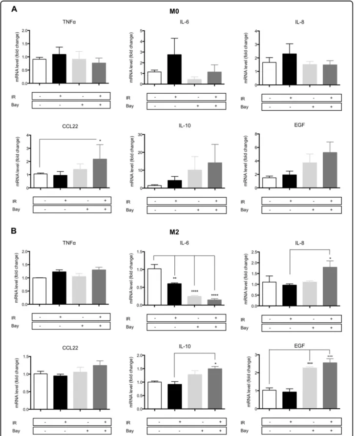
irradiation (Fig.6a). The combination of the IKK inhibitor with the proton irradiation completely inhibited the programming of M0 into M1 phenotype and induced a nonsignificant increase in M2 marker expression (CCL22, IL-10, and EGF). Although Bay 11-8072 treatment alone drove the expression of IL-10 and EGF, the combination of the inhibitor with proton irradiation induced a much higher expression of these genes. The same experiment
has been performed for M2 macrophages (Fig.6b). As it
was aforementioned, the irradiation of M2 macrophages had no effect on the M1 marker mRNA levels after 24 h
(Fig.4a) and we detected no change in the TNFα and IL-8
mRNA expression after 12 h (Fig. 6b). However, a
decrease in IL-6 expression was noticed in the same condition. When proton irradiation was combined to
NFκB inhibition, diverse effects were observed on the
expression of M1 markers. No effect was observed on
TNFα expression, while the expression of IL-8
surpris-ingly increased when proton irradiation was combined to Bay 11-7082 treatment. As the activation of NFκB reg-ulates the expression of IL-8, it is surprising to observe an elevation of the IL-8 mRNA level in M2 macrophages irradiated in the presence of Bay 11-7082. However, the transcription of IL-8 is also regulated by other transcrip-tion factors, such as the activator protein 1 (AP-1), that
could be also activated when NFκB is inhibited in
proton-irradiated M2 macrophages18. On the other hand, IL-6
expression was strongly decreased by the IKK inhibitor: its expression decreased by four times with Bay 11-7082
B M0 0 5 10 0 5 10 15 20 25 Dose (Gy) C el ls w ith n ucl ea r NF !B p 65 ( % ) M1 0 5 10 0 5 10 15 20 25 Dose (Gy) C el ls w ith n ucl ea r NF !B p 65 ( % ) M2 0 5 10 0 5 10 15 20 25 Dose (Gy) C el ls w ith n ucl ea r NF !B p 65 ( % ) * A
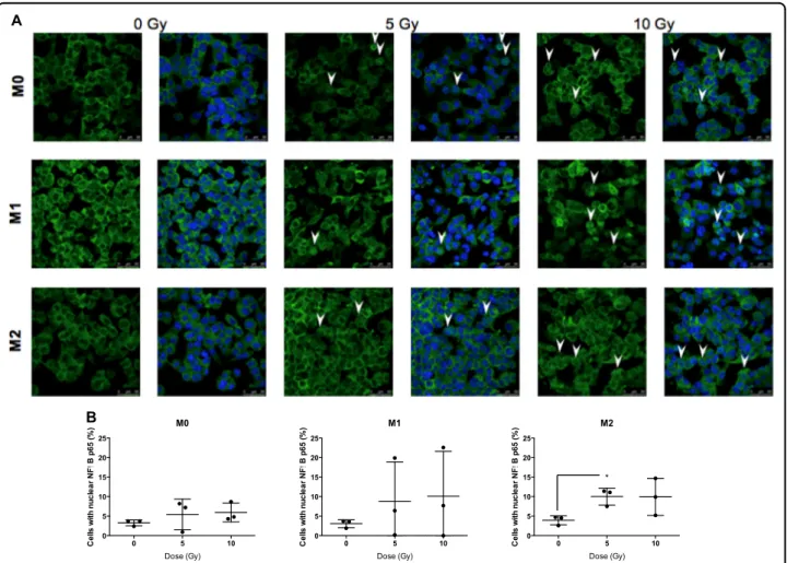
Fig. 5 Proton irradiation induces nuclear translocation of p65 (NFκB). M0, M1, and M2 macrophages were irradiated by different doses of protons. 2 h after the irradiation, the nuclear translocation of p65 was evaluated by NFκB p65 immunofluorescence labeling. NFκB p65 is stained in green and nucleus appears in blue. a Nuclear translocation of NFκB p65 is indicated (arrow) on representative immunofluorescence labeling images for M0, M1, and M2 macrophages after irradiation. b Quantification of the mean NFκB p65 intensity per nucleus 2 h after proton irradiation. Results are expressed in percentage of cells with nuclear NFκB p65. Quantifications were performed on minimum five images per condition (N = 3, mean ± SD). An unpaired t test was performed on data (*p≤ 0.05; **p < 0.01; ***p < 0.001)
Fig. 6 NFκB inhibition reverts proton irradiation-induced macrophage reprogramming. M0 and M2 macrophages were irradiated (IR) with protons (10 Gy) with or without NFκB inhibitor (Bay 11-7082—5 μM). The inhibitor was added 1 h before irradiation for the following 12 h. Macrophage polarization was evaluated by RT-qPCR 12 h after proton irradiation by the analysis of M1 (TNFα, IL-6, IL-8) and M2 (CCL22, IL-10, EGF) marker mRNA level in (a) M0 macrophages (N= 3, mean ± SD) and b M2 macrophages (N = 4, mean ± SD). Two-way ANOVA analyses followed by Tukey’s multiple comparison tests were performed on data; *p ≤ 0.05; **p < 0.01; ***p < 0.001
alone and by five times with the combination. For M2
markers, proton irradiation alone did not alter
the expression of these markers after 24 h (Fig.4a). In the same line, 10 Gy of irradiation did not change the
expression of M2 markers after 12 h (Fig. 6b). However,
the combination of NFκB inhibitor to proton irradiation
induced a higher expression of M2 markers. The elevation of M2 marker expression was stronger in M2 macro-phages exposed to the combined treatment than for macrophages treated with the inhibitor alone. In
conclu-sion, the NFκB inhibitor combined to proton irradiation
completely prevented the programming of M0 macro-phages towards an M1 phenotype. On the contrary, it promoted the programming of M0 macrophages towards an M2 phenotype. In addition, M2 macrophages reinforce their M2 phenotype when exposed to proton irradiation in combination with Bay 11-7082.
Discussion
As M2-like TAMs play an important role in tumor promotion and treatment failure, the reprogramming of
these cells has been shown to be an efficient way to
promote tumor regression. It is already known that macrophages are more radioresistant compared to other cell types19. Indeed, the fusion of macrophages with breast cancer cell line (MCF7) resulted in hybrids developing the abilities to resist high radiation doses and to repair DNA
damage faster than the parental MCF7 cells20. In the
present study, we demonstrated the radioresistance of M1 macrophages compared to M0 and M2 phenotypes. Sev-eral evidences highlighted the radioresistance of M1 macrophages in the literature. For example, BALB/c mice
naturally exhibiting a TH1/M1 (TH1 lymphocytes and M1
macrophages) response were more radioresistant than
C57BL/6 mice naturally exhibiting a TH2/M2 response16.
TNFα play key roles in macrophage radioresistance since
an intact TNFα signaling is needed for radioresistance.
Indeed, the deletion of tumor necrosis factor receptor 1 or 2 (TFNR1 or TNFR2) in mice rendered macrophages radiosensitive21. In contrast to our observations, a recent in vitro study indicated that unpolarized (M0) and M1 macrophages were more sensitive to X-ray irradiation than M2 macrophages. The divergent results could be explained by the use of higher LPS concentration (100 ng/ml instead of 10 pg/ml in the present study) for the polar-ization process before the irradiation22.
The resistance of M1 macrophages to proton irradiation
correlated with a higher level of phosphorylated H2AX,
possibly indicating either more DSBs, or a higher activity of DNA damage repair. Another explanation could rely on the ability of macrophages to detoxify irradiation-induced radicals formed upon H2O radiolysis. It is well established
that the radioresistance of macrophages is conferred by a high production of anti-oxidative molecules, such as
manganese superoxide dismutase (MnSOD). Higher
MnSOD expression is associated to a resistance against damaging effects from ROS and reactive nitrogen species23.
Indeed, the scavenging of ROS by N-acetyl-L-tryptophan
glucopyranoside (NATG) in J774A.1 macrophages provided a protection against radiation-dependent apoptosis24. Fur-thermore, another in vitro study revealed that mouse peri-toneal macrophages polarized towards the M1 phenotype with methionine displayed increased SOD activity and
decreased ROS production25. In our experiments, the
ana-lysis of ROS management revealed a more efficient
elim-ination of ROS in M1 macrophages when challenged with
H2O2compared to M0 and M2 macrophages (Fig. S2). In
conclusion, the radioresistance of M1 macrophages to proton irradiation is related to a higher DNA damage detection. Several causes like a larger antioxidant pool, a more active DNA repair machinery and the differential promoter methylation of genes involved in DNA repair should be taken in considerations for future studies.
Macrophage re-education represents a promising
approach to reverse the fate of tumor and generates
tumor regression. To our knowledge, this is the first
observation of in vitro macrophage reprogramming with particle therapy. Previous studies with conventional
radiotherapy (X-rays or γ-rays) revealed an efficient
pro-gramming of unpolarized macrophages (M0) towards the M1 phenotype but not M2 reprogramming towards M1-like phenotype (for a review, see ref.14). For example, the irradiation of monocyte-derived macrophages with frac-tionated doses (2 Gy 5 × /week) induced the polarization towards M1-like macrophages as evidenced by a higher expression of proinflammatory genes and a
down-regulation of anti-inflammatory genes26
. Another study also revealed the programming of unpolarized macro-phages towards a proinflammatory profile after moderate dose of γ-irradiation (2 or 4 Gy)27. In the last study, the authors also reported an increased expression of IL-6, IL-8,
and TNFα in γ-irradiated (4 Gy) human
monocyte-derived macrophages. While the use of X-ray irradiation induced an M1 polarization of unpolarized human monocyte-derived macrophages, Raw 264.7 cells,
PMA-differentiated THP-1 cells and peritoneal macrophages14,
our results indicated that X-ray irradiation failed to pro-gram PMA-differentiated THP-1 macrophages in our experimental settings. In our experiments, a resting time period of 24 h allowed a decrease in NFκB gene cluster expression, upregulated during PMA-induced
differ-entiation28. This may explain the discrepancy with
pre-viously reported results. The release of TNFα by M0, M1, and M2 macrophages after proton irradiation confirms the reprogramming of macrophages to an M1-like
phe-notype. Indeed, TNFα is a potent anti-M2 polarization
factor and strongly correlates with an M1-like
was shown for the three phenotypes upon irradiation. EGF is well known to be involved in both angiogenesis
and cell migration, two roles fulfilled by M2
macrophages30.
The active heterodimer NFκB p50–p65 is predominant
for M1 activation, leading to a proinflammatory profile
while the induction of the inactive homodimer NFκB
p50–p50 inhibits the expression of proinflammatory
genes in M2 macrophages12,13. Our results demonstrated
the implication of NFκB p65 subunit in macrophage
reprogramming after moderate doses of proton irradia-tion. In addition, the combination of proton irradiation to an IKK complex inhibitor (Bay 11-7082) completely aborted the re-education towards a M1 phenotype in M0 and M2 macrophages. In general, NFκB is activated in irradiated cancer cells when doses are comprised in the range of 7−10 Gy31–33
. Similarly to our study, the X-ray
irradiation of monocyte-derived macrophages also
induced the upregulation of total and phosphorylated p65, from 1 to 6 h after irradiation at 2 Gy and 10 Gy. This was
correlated to the reduced expression of anti-inflammatory
genes26. Furthermore, the whole-body irradiation of RT5
insulinoma bearing mice increased NFκB p65
phosphor-ylation in tumors, partially explaining the TAM repro-gramming after γ-irradiation15. Proton irradiation is also more likely to induce more severe DNA damage and
higher ROS production compared to photons34. These
damages activate the DNA repair system, including ATM (ataxia-telangiectasia mutated) kinase, notably responsible
for the translocation of NFκB p65 into the nucleus35
. Hence, ROS production and double DNA strand breaks are strongly linked to NFκB p65 activation14,36
. In addi-tion, other studies have demonstrated the activation of NFκB p65 in cancer cells after high LET charged particle irradiation. Indeed, heavy ions with a LET of
100–300 keV/μm showed up to nine times higher
potential to activate the NFκB pathway compared to
X-rays37while carbon ions with a LET under 73 keV/μm
induced an NFκB p65 activation twice as high as the one
induced by conventional radiotherapy38. For future
stu-dies, the use of high LET radiation should be considered, as it can potentiate the activation of NFκB in macrophages and induce apoptosis in cancer cells. However, further investigations are needed to clarify if charged particle therapy may be used for all cancer types. Indeed, con-stitutive activation of NFκB is associated to tumor growth for several human cancer cells such as breast cancer, colon cancer, prostate cancer, and lymphoid cancer. In cancer cells, NFκB plays key roles in cell survival, notably by regulating the expression of genes involved in cell survival and cell cycle38. For conventional radiotherapy,
the use of NFκB inhibitors increased the radiosensitivity
of many cancer cells39. However, no study has been
per-formed yet to assess the balance between NFκB-mediated
survival and apoptosis in high LET radiation context. Also, the activation of the immune system by particle therapy could overcome the activation of NFκB in cancer cells. These questions reveal a need for further investi-gations regarding the use of charged particle therapy in cancer.
Our study provides direct evidences that there could be a selection of M1 macrophages in tumors with proton irradiation, since this phenotype is more radioresistant. Furthermore, we showed that 10 Gy of proton irradiation is able to program M0 macrophages towards M1 pheno-type, to enhance the M1 phenotype in M1 macrophages and to initiate an M1 phenotype in M2 macrophages. This re-education is due, at least in part, to NFκB p65 activa-tion. Therefore, our data open new perspectives for macrophage clinical targeting with charged particle ther-apy. In the future, these results need to be confirmed by in vivo experiments, as other cell types are present in the tumor microenvironment and could influence the response to proton irradiation. It has to be noted that macrophage reprogramming after proton irradiation may
not be exclusively associated to NFκB activation. Indeed,
the interaction between several pathways (NFκB, MAPK,
and IRF/STAT pathways) may be involved in this process and needs to be further investigated. Moreover,
metabo-lism reprogramming has been demonstrated to influence
macrophage polarization40, 41 and should be taken into
account in the further studies.
Materials and methods
Differentiation of monocytes to macrophages and macrophage polarization
Human monocytic cell line (THP-1–ATCC TIB-202) were grown in Roswell Park Memorial Institute (RPMI 1640, Gibco #21875034) culture medium containing 10% of heat inactivated fetal bovine serum (Gibco) and supplemented with 10 mM Hepes (Gibco, #15630-056),
2 mM pyruvate (Gibco, #11360-039), 2.5 g/L D-glucose
(Merck) and 50 pM β-mercaptoethanol (Gibco,
#31350-010). As the proton accelerator used in this study pro-duces a horizontal beam, cells were plated in suitable irradiation chambers, composed of two stainless steel
rings with the central holes covered by 3μm Mylar foils
(Goodfellow). THP-1 monocytes were seeded at the center of irradiation chambers, on the Mylar foil, and differentiated into macrophages with 150 nM phorbol 12-myristate 13-acetate (PMA, Sigma P8139). For this step, a cloning cylinder (6.4 mm diameter size, Sigma
C3983-50EA) was placed at the center of the chamber andfilled
with Cell Tak (VWR #354240) for coating. Differentiated THP-1 cells tend to form clusters when seeded on Mylar foil and Cell tak allowed a homogenous monolayer. The cloning cylinders were rinsed with milli Q water and were filled with 190 μl of medium containing 50,000 THP-1
monocytes, penicillin streptomycin 10,000 U/ml (Fisher, #15140122) and PMA for 24 h incubation. The differ-entiation medium was replaced with RPMI medium for a further 24 h incubation. Macrophages were polarized in M1 phenotype with 10 pg/ml lipopolysaccharides (LPS,
Sigma; #8630) and 20 ng/ml interferon γ (IFN-γ, R&D
Systems, #285-IF) during 24 h incubation or were polar-ized in M2 phenotype with 20 ng/ml interleukin 4 (IL-4, R&D Systems, #204-IL) and 20 ng/ml interleukin 13 (IL-13, R&D Systems, #213-ILB) during 48 h incubation as
described in ref. 42. The gene expression of M1 (IL-1β,
TNFα, CXCL10, and IL-6) and M2 (CCL18, CCL22,
CD206, and IL-10) markers were analyzed to verify the macrophage phenotype of M0, M1, and M2 macrophages in the irradiation chambers (Fig. 4S).
Proton irradiation
Four days before irradiation, THP-1 monocytes were differentiated in the irradiation chambers to generate M2 macrophages. To obtain M1 or M0 macrophages, cells were seeded 3 or 2 days respectively before irradiation. Just before proton irradiation, culture medium was
changed by CO2independent medium (Gibco #18045054)
supplemented with 2 mM L-glutamine and 3.75 g/L of
D-glucose. The irradiation chambers were placed at the
end of a 2 MV Tandem accelerator (High Voltage Engi-neering Europa) available at the University of Namur. The experimental set-up and irradiation procedure are
described elsewhere43, 44. Briefly, an H+ homogenous
1 cm2broad beam went through a 1-μm-thick Si3N4 exit
foil. The chambers were placed vertically at 3 mm from the exit window of the accelerator. The linear energy transfer (LET) of protons was set to 25 keV/μm and the
dose ratefixed to 2 Gy/min. After irradiation, the
cham-bers were replaced in the CO2 incubator until the
experiments were performed.
One of the constraints of this set-up is the need to have cells that adhere to the Mylar foil that constitutes the bottom of the irradiation chamber. We cannot use usual
plastic flaks/plates since the proton beam generated by
our particle accelerator does not have enough energy to go through this material. Most cell lines do not adhere to this material and/or form clusters that do not allow a homogeneous irradiation of all cells. Hence, this work has been performed using THP1-derived macrophages only.
Determination of cell viability
Because of the Cell Tak, the cells could not be detached from the Mylar foil. Ethidium bromide (1 mg/ml Sigma E8751)−acridin orange (0.3 mg/ml, Sigma A6014) (EBAO) was used at 1:100 dilution in PBS to analyze viability. The cells were stained with EBAO for 5 min, 8 h, or 16 h after irradiation. Cells were then rinsed with PBS and were observed with Olympus stream microscope
beyond UV light. Live cells are stained in green while dead cells are stained in orange. Quantifications were
per-formed with Aphelion Lab® software on approximately
1000 cells.
Immunofluorescence labeling
Cells werefixed for 10 min with paraformaldehyde 4%
in PBS, and then permeabilized with triton 0.1% for 5 min.
The cells were rinsed three times with PBS–BSA 2%
(bovine serum albumin) and incubated overnight at 4 °C with primary antibody in the irradiation chambers.
Pri-mary antibodies were 1:800 (γH2AX, BioKe 2577), 1:400
(NFκB p65, Cell Signaling D14E12) or 1:1500 (53BP1, Novus NB100-304) diluted in PBS–BSA 2%. Cells were rinsed three times with PBS–BSA 2% and then incubated for 1 h with secondary antibody 1:1000 diluted (Alexa Fluor 488-conjugated anti-rabbit IgG antibody; Molecular Probes, #A11034) at room temperature. After washing the cells three times with PBS, nuclei were stained with TOPRO-3 (1:80 diluted in RNase solution). Cells were then washed three times with PBS and the Mylar foil was cut around the cell drop and was deposited on a micro-scope slide, cells on top. A coverslip was mounted using Mowiol (Sigma) above the cells and the cells were observed with a confocal microscope (SP5, Leica). A constant photomultiplier gain value and a constant laser
power were used to take the pictures. Quantifications for
γH2AX intensity or 53BP1 foci were performed using
Aphelion Lab® software on approximately 1000 cells. For
γH2AX, the quantification was performed as followed:
mean of (Number of green pixels × intensity of each
pixel) / nucleus. Usually, the quantification of γH2AX
labeling is performed by countingγH2AX foci. However,
as high doses were used in this study, large numbers of clustered foci were observed and it was impossible to discriminate foci at these doses. Therefore, we choose to
quantify the mean intensity of γH2AX per nucleus.
Apoptotic cells were not taken into account for the
quantification. Quantification for 53BP1 foci was
per-formed similarly. Quantification for NFκB p65 was
per-formed as followed: number of cells with higher pixel
numbers than mean+ 2 standard deviations,
representa-tive of cells with high translocation of NFκB p65.
Relative quantification of mRNA levels
Total RNA was extracted from cells in irradiation chambers with RNeasy micro kit and DNAse protocol (QIAGEN # 74004). Reverse transcription was performed
on 1μg using Transcriptor first strand cDNA synthesis kit
(Roche #4309155). The quantitative Real Time PCR was
performed using Viia 7 (Thermo Fisher Scientific) with
SYBRGreen PCR Master Mix (Applied Biosystems, #4309155) and primers (IDT, 300 nM). 40S ribosomal protein S9 (RPS9) was selected as the housekeeping gene
for normalization, based on its constant expression within all samples. The primers used for RT-qPCR are sum-marized in supplementary information (table S1).
Cytokine quantification
Secreted cytokine (IL-6, CXCL-8/IL-8, and TNFα)
analysis was assessed using ELISA kit, according to the
manufacturer’s recommendations (Quantikine, R&D
Systems D6050, D8000C and DTA00C). Culture media
were diluted in CO2independent medium 5 and 50 times
for TNFα and IL-8, respectively, while the culture
med-ium was not diluted for IL-6. For all samples, concentra-tions were normalized by total protein (μg proteins/ml) determined by Folin method.
IKK inhibition
Bay 11-7082 inhibitor (3-((4-methylphenyl)sulfonyl)-(2E)-propenenitrile; Selleckchem S2913) selectively and irreversibly inhibits the phosphorylation of IκB-α and
then prevents the NFκB activation. This inhibitor was
added at 5μM in CO2 independent medium 1 h before
irradiation and the cells were incubated for 12 h after
irra-diation in the presence of the inhibitor. TNFα (20 ng/ml,
R&D Systems #210-TA-020) was used as a positive
con-trol to verify the nuclear translocation of NFκB p65. In
order to validate the inhibition of p65 nuclear
transloca-tion in our model, cells were incubated with TNFα and
Bay 11-7082 (Fig. 5S).
Statistical analysis
Statistical analyses were performed using Graph Pad Prism software. Data are reported as mean ± 1SD of N independent experiments. A p value of <0.05 was con-sidered as significant.
Acknowledgements
The authors thank Denis Mottet (Ph.D.) for advices on the MNAse assay. We would like to acknowledge Romain Tonneau for the computational method. We are thankful to the technological platforms SIAM and Morph-Im (University of Namur). G.G. is a recipient of a Télévie grant (National Fund for Scientific Research (FNRS)). A.-C.W. and B.L.C. benefit from a Postdoctoral Move-in-Louvain-Marie Curie grant. C.H. is a recipient of an FSR (fonds special de la recherche) grant from the University of Namur. S.P. is supported by FNRS (Ph.D. grant).
Authors’ contributions
G.G. carried out the experiments and wrote the manuscript. A.-C.W., C.H., and S. P. helped for the experiments. T.T., A.F., C.D., and N.N. contributed to the acquisition of data. B.L.C. and A.-C.W. reviewed the manuscript. C.M., S.L., and A.-C.H. conceived of the study, participated in its design and coordination, and revised the manuscript. All authors read and approved thefinal manuscript.
Conflict of interest
The authors declare that they have no conflict of interest.
Publisher's note
Springer Nature remains neutral with regard to jurisdictional claims in published maps and institutional affiliations.
Supplementary Information accompanies this paper at (https://doi.org/ 10.1038/s41419-018-0757-9).
Received: 1 February 2018 Revised: 27 May 2018 Accepted: 29 May 2018
References
1. Mittal, D., Gubin, M. M., Schreiber, R. D. & Smyth, M. J. New insights into cancer immunoediting and its three component phases—elimination, equilibrium and escape. Curr. Opin. Immunol. 27, 16–25 (2014).
2. Lee, Y. et al. Therapeutic effects of ablative radiation on local tumor require CD8+T cells: changing strategies for cancer treatment. Blood 114, 589–595 (2009).
3. Weichselbaum, R. R., Liang, H., Deng, L. & Fu, Y. X. Radiotherapy and immu-notherapy: a beneficial liaison? Nat. Rev. Clin. Oncol. 14, 365–379 (2017). 4. Dovedi, S. J. et al. Acquired resistance to fractionated radiotherapy can be
overcome by concurrent PD-L1 blockade. Cancer Res. 74, 5458–5468 (2014). 5. Lewis, C. E. & Pollard, J. W. Distinct role of macrophages in different tumor
microenvironments. Cancer Res. 66, 605–612 (2006).
6. van Ravenswaay Claasen, H. H., Kluin, P. M. & Fleuren, G. J. Tumor infiltrating cells in human cancer. On the possible role of CD16+macrophages in anti-tumor cytotoxicity. Lab Invest. 67, 166–174 (1992).
7. Gentles, A. J. et al. The prognostic landscape of genes and infiltrating immune cells across human cancers. Nat. Med. 21, 938–945 (2015).
8. Mantovani, A., Marchesi, F., Malesci, A., Laghi, L. & Allavena, P. Tumour-associated macrophages as treatment targets in oncology. Nat. Rev. Clin. Oncol. 14, 399–416 (2017).
9. Pienta, K. J. et al. Phase 2 study of carlumab (CNTO 888), a human monoclonal antibody against CC-chemokine ligand 2 (CCL2), in metastatic castration-resistant prostate cancer. Invest. New Drugs 31, 760–768 (2013).
10. Klug, F. et al. Low-dose irradiation programs macrophage differentiation to an iNOS(+)/M1 phenotype that orchestrates effective T cell immunotherapy. Cancer Cell 24, 589–602 (2013).
11. Mosser, D. M. & Edwards, J. P. Exploring the full spectrum of macrophage activation. Nat. Rev. Immunol. 8, 958–969 (2008).
12. Biswas, S. K. & Mantovani, A. Macrophage plasticity and interaction with lymphocyte subsets: cancer as a paradigm. Nat. Immunol. 11, 889–896 (2010).
13. Wang, N., Liang, H. & Zen, K. Molecular mechanisms that influence the macrophage m1-m2 polarization balance. Front. Immunol. 5, 614 (2014). 14. Genard, G., Lucas, S. & Michiels, C. Reprogramming of tumor-associated
macrophages with anticancer therapies: radiotherapy versus chemo- and immunotherapies. Front. Immunol. 8, 828 (2017).
15. Prakash, H. et al. Low doses of gamma irradiation potentially modifies immunosuppressive tumor microenvironment by retuning tumor-associated macrophages: lesson from insulinoma. Carcinogenesis 37, 301–313 (2016). 16. Nowosielska, E. M., Cheda, A., Wrembel-Wargocka, J. & Janiak, M. K. Effect of
low doses of low-let radiation on the innate anti-tumor reactions in radio-resistant and radiosensitive mice. Dose Response 10, 500–515 (2012). 17. Ilicic, K., Combs, S. E. & Schmid, T. E. New insights in the relative radiobiological
effectiveness of proton irradiation. Radiat. Oncol. 13, 6 (2018).
18. Roebuck, K. A. Regulation of interleukin-8 gene expression. J. Interferon Cyto-kine Res. 19, 429–438 (1999).
19. Tsai, C. S. et al. Macrophages from irradiated tumors express higher levels of iNOS, arginase-I and COX-2, and promote tumor growth. Int J. Radiat. Oncol. Biol. Phys. 68, 499–507 (2007).
20. Lindstrom, A., Midtbo, K., Arnesson, L. G., Garvin, S. & Shabo, I. Fusion between M2-macrophages and cancer cells results in a subpopulation of radioresistant cells with enhanced DNA-repair capacity. Oncotarget 8, 51370–51386 (2017). 21. Meng, Y. et al. Blockade of tumor necrosis factor alpha signaling in tumor-associated macrophages as a radiosensitizing strategy. Cancer Res. 70, 1534–1543 (2010).
22. Leblond, M. M. et al. M2 macrophages are more resistant than M1 macro-phages following radiation therapy in the context of glioblastoma. Oncotarget 8, 72597–72612 (2017).
23. Hachiya, M., Shimizu, S., Osawa, Y. & Akashi, M. Endogenous production of tumour necrosis factor is required for manganese superoxide dismutase expression by irradiation in the human monocytic cell line THP-1. Biochem J. 328(Pt 2), 615–623 (1997).
24. Malhotra, P., Adhikari, M., Singh, S. K. & Kumar, R. N-acetyl tryptophan gluco-pyranoside (NATG) provides radioprotection to murine macrophage J774A.1 cells. Free Radic. Res. 49, 1488–1498 (2015).
25. Dos Santos, L. M. et al. Methionine and methionine sulfoxide treatment induces M1/classical macrophage polarization and modulates oxidative stress and purinergic signaling parameters. Mol. Cell. Biochem. 424, 69–78 (2017).
26. Teresa Pinto, A. et al. Ionizing radiation modulates human macrophages towards a pro-inflammatory phenotype preserving their invasive and pro-angiogenic capacities. Sci. Rep. 6, 18765 (2016).
27. Wu, Q. et al. NOX2-dependent ATM kinase activation dictates pro-inflammatory macrophage phenotype and improves effectiveness to radia-tion therapy. Cell Death Differ. 24, 1632–1644 (2017).
28. Chanput, W., Mes, J. J. & Wichers, H. J. THP-1 cell line: an in vitro cell model for immune modulation approach. Int. Immunopharmacol. 23, 37–45 (2014). 29. Murray, P. J. Macrophage polarization. Annu. Rev. Physiol. 79, 541–566 (2017). 30. Sasaki, T., Hiroki, K. & Yamashita, Y. The role of epidermal growth factor receptor in cancer metastasis and microenvironment. Biomed. Res. Int. 2013, 546318 (2013).
31. Hellweg, C. E. The Nuclear Factor kappaB pathway: a link to the immune system in the radiation response. Cancer Lett. 368, 275–289 (2015). 32. Schaue, D., Kachikwu, E. L. & McBride, W. H. Cytokines in radiobiological
responses: a review. Radiat. Res. 178, 505–523 (2012).
33. Formenti, S. C. & Demaria, S. Combining radiotherapy and cancer immu-notherapy: a paradigm shift. J. Natl. Cancer Inst. 105, 256–265 (2013).
34. Alan Mitteer, R. et al. Proton beam radiation induces DNA damage and cell apoptosis in glioma stem cells through reactive oxygen species. Sci. Rep. 5, 13961 (2015).
35. Hayden, M. S. & Ghosh, S. Shared principles in NF-kappaB signaling. Cell 132, 344–362 (2008).
36. Huang, T. T., Wuerzberger-Davis, S. M., Wu, Z. H. & Miyamoto, S. Sequential modification of NEMO/IKKgamma by SUMO-1 and ubiquitin mediates NF-kappaB activation by genotoxic stress. Cell 115, 565–576 (2003).
37. Hellweg, C. E. et al. Activation of the nuclear factor kappaB pathway by heavy ion beams of different linear energy transfer. Int J. Radiat. Biol. 87, 954–963 (2011).
38. Hellweg, C. E. et al. Carbon-ion-induced activation of the NF-kappaB pathway. Radiat. Res. 175, 424–431 (2011).
39. Criswell, T., Leskov, K., Miyamoto, S., Luo, G. & Boothman, D. A. Transcription factors activated in mammalian cells after clinically relevant doses of ionizing radiation. Oncogene 22, 5813–5827 (2003).
40. Liu, P. S. et al. alpha-ketoglutarate orchestrates macrophage activation through metabolic and epigenetic reprogramming. Nat. Immunol. 18, 985–994 (2017). 41. Kelly, B. & O’Neill, L. A. Metabolic reprogramming in macrophages and
den-dritic cells in innate immunity. Cell Res. 25, 771–784 (2015).
42. Genin, M., Clement, F., Fattaccioli, A., Raes, M. & Michiels, C. M1 and M2 macrophages derived from THP-1 cells differentially modulate the response of cancer cells to etoposide. BMC Cancer 15, 577 (2015).
43. Riquier, H. et al. Effects of alpha particle and proton beam irradiation as putative cross-talk between A549 cancer cells and the endothelial cells in a co-culture system. Cancers (Basel) 7, 481–502 (2015).
44. Wéra, A.-C., R, H., Heuskin, A.-C., Michiels, C. & Lucas, S. In vitro irradiation station for broad beam radiobiological experiments. Nucl. Instrum. Methods Phys. Res. B 269, 3120–3124 (2011).