HAL Id: tel-03231370
https://tel.archives-ouvertes.fr/tel-03231370v2
Submitted on 22 May 2021
HAL is a multi-disciplinary open access
archive for the deposit and dissemination of sci-entific research documents, whether they are pub-lished or not. The documents may come from teaching and research institutions in France or abroad, or from public or private research centers.
L’archive ouverte pluridisciplinaire HAL, est destinée au dépôt et à la diffusion de documents scientifiques de niveau recherche, publiés ou non, émanant des établissements d’enseignement et de recherche français ou étrangers, des laboratoires publics ou privés.
Supramolecular biological protein assemblies study using
solid-state NMR
Nadia El Mammeri
To cite this version:
Nadia El Mammeri. Supramolecular biological protein assemblies study using solid-state NMR. Other. Université de Bordeaux, 2020. English. �NNT : 2020BORD0217�. �tel-03231370v2�
THÈSE PRÉSENTÉE
POUR OBTENIR LE GRADE DE
DOCTEUR DE
L’UNIVERSITÉ DE BORDEAUX
ÉCOLE DOCTORALE DES SCIENCES CHIMIQUES
SPÉCIALITÉ CHIMIE PHYSIQUE
Par Nadia EL MAMMERI
Étude d'assemblages protéiques biologiques supramoléculaires
par RMN du solide
Sous la direction de : Dr. Antoine LOQUET
Soutenue le 04/12/2020
Membres du jury :
M. DUFOURC, Erick J. Directeur de Recherche, CNRS, Bordeaux Président Mme BÖCKMANN, Anja Directrice de Recherche, CNRS, Lyon Rapporteur Mme FRANCETIC, Olivera Directrice de Recherche, Institut Pasteur, Paris Rapporteur M. SAUPE, Sven Directeur de Recherche, CNRS, Bordeaux Examinateur M. MACKERETH, Cameron Directeur de Recherche, INSERM, Bordeaux Examinateur M. KOVACS, Ákos T. Professeur, DTU, Danemark Examinateur M. GALAN, Jorge E. Professeur, Université Yale, USA Examinateur M. LOQUET, Antoine Directeur de Recherche, CNRS, Bordeaux Directeur de thèse
To my parents
And Reda, Sophia, Amine,
Titre : Étude d'assemblages protéiques biologiques
supramoléculaires par RMN du solide
Résumé : Ma thèse a pour buts l'élucidation fonctionnelle et l'étude structurale de
nano-machines biologiques en utilisant principalement la RMN du solide : (i) La protéine SesB trouvée chez Nectria haematococca, dont l'assemblage en fibrilles amyloïdes, serait impliquée dans les mécanismes de signalisation de mort cellulaire programmée. En utilisant la RMN du solide avec rotation à l’angle magique, un nouveau modèle structural de cet amyloïde a été établi pour les fibrilles amyloïdes SesB de Nectria haematococca. (ii) Des protéines appelées TasA, TapA et CalY dont l'assemblage en fibrilles amyloïdes est impliqué dans la formation et l'intégrité des biofilms bactériens chez Bacillus subtilis et Bacillus cereus. Des différences structurales ont été observées entre ces deux espèces. Bacillus subtilis et quelques mutants (mutations de gènes impliqués dans la formation de biofilms) ont été étudiés en utilisant la RMN du solide sur cellules entières pour comprendre l'impact de la délétion de ces composants sur la paroi cellulaire globale et la composition de la matrice. (iii) HET-s, l'amyloïde fonctionnelle fongique aujourd’hui très étudié, est utilisé ici comme système modèle dans une stratégie visant à utiliser les avantages combinés de la DNP, de la rotation très rapide à l’angle magique et de la dilution de spin dans le contexte d'études de biologie structurale. Il a été prouvé qu'une utilisation pertinente de ces techniques constitue une stratégie potentiellement robuste pour la caractérisation structurale des assemblages supramoléculaires pour augmenter la sensibilité des analyses par RMN. (iv) Des polymères protéiques inhabituels, en forme de ruban, appelés R-bodies (« refractile bodies » de type 51), que l'on trouve chez de nombreuses espèces bactériennes, comme Caedibacter et Pseudomonas. Une attribution complète des signaux RMN des R-bodies a été réalisée en utilisant la RMN du solide avec rotation à l’angle magique à très grande vitesse. Un modèle structurel des monomères est établi et tient compte de leur changement conformationnel en fonction du pH, ainsi que de leur capacité à relarguer des biomolécules.
Mots clés : assemblages supramoléculaires, biologie structurale, RMN du solide, machine
Title: Supramolecular biological protein assemblies study using
solid-state NMR
Abstract: I have worked on elucidating and studying biological nanomachines using mainly
solid-state NMR (SSNMR): (i) The protein SesB found in Nectria haematococca whose assembly into amyloid fibrils is thought to be involved in programmed-cell death signalling mechanisms. Using magic-angle spinning SSNMR, a novel structural amyloid fold model for
Nectria haematococca amyloid fibrils was established. (ii) Proteins called TasA, TapA, and
CalY found in Bacillus subtilis and Bacillus cereus whose assembly into amyloid fibrils is involved in biofilm formation and integrity. Bacillus subtilis and its mutants have been studied using whole-cell SSNMR to understand the impact of biofilm matrix components deletion in the overall cell wall, and matrix composition. The protein TasA found in Bacillus subtilis has been observed to perturb liposomes as membrane models. (iii) HET-s, the well documented fungal functional amyloid, used here as a model system in a scheme to use the combined advantages of DNP, fast MAS, and spin dilution in the context of structural biology studies. A relevant use of DNP in combination with fast MAS and specific labelling has been proven to be a potential robust strategy for structural characterization of supramolecular assemblies. (iv) Unusual, ribbon-like protein polymers called R-bodies (Type 51 refractile bodies) found in many bacterial species, such as Caedibacter and Pseudomonas. A complete resonance assignment of R- bodies was achieved using very fast MAS SSNMR. A structural model of the monomers was established and accounts for their interesting pH-dependent switch, as well as the ability to deliver biomolecules.
Keywords: Supramolecular assemblies, structural biology, solid-state NMR, bacterial machine
UNITE DE RECHERCHE
ACKNOWLEDGMENTS / REMERCIEMENTS
First, I would like to express my most sincere gratitude to Antoine Loquet for allowing me to conduct this research under his auspices. I am especially grateful for his trust, advice and the independence he allowed me throughout the years. I take the time to also gratefully thank Erick Dufourc, who supported me in all stages of my PhD research and after. He always gave me constant encouragement, and support despite his busy schedule.
I am deeply grateful to all the members of the jury. I thank Anja Böckmann, and Olivera Francetic for agreeing to read the manuscript and evaluate my thesis, and Erick J. Dufourc, Sven Saupe, Cameron Mackereth, Ákos Kovacs and Jorge E. Galán for accepting to participate in the defense of this thesis.
I would like to acknowledge Professor Jorge E. Galán who welcomed me in his team in Yale University for five months for a fruitful and enriching collaboration. This experience has been a major part of my personal and professional development. I wish to thank the Fulbright Commission Franco-américaine organization for sponsoring my stay with Professor Jorge E. Galán at Yale University, New Haven, USA.
My thesis work was made possible through numerous collaborations with brilliant researchers. I would like to thank all participants of the following work with whom we had fascinating scientific discussions. I am honoured to have worked with Prof. Jorge E. Galán, Dr. Maria Lara-Tejero, Dr. Guido Pintacuda, Dr. Anne Lesage, Dr. Diego Romero, Professor Justin Kollman, Dr. Sven Saupe, Dr. Brice Kauffmann, Dr. Sophie Lecomte, and Prof. Erick J. Dufourc.
I express my most sincere appreciation to Axelle Grélard, Estelle Morvan, Mélanie Berbon, Antoine Dutour, and Mathilde Bertoni who helped me to learn biochemical and biophysical techniques in my laboratory. They have been a constant support in the past three years. I warmly thank Birgit Habenstein for her support and encouragements. It was a real pleasure to work with Axelle Raymond, and I thank her for her help in the biological lab. I am glad to have met so many interesting and pleasant people in the three years that I have spent in the Loquet team. I have learned from all of you. Thank you!
Gaëlle, Arpita, Mathilde, we’ll always have teatime. Gaëlle, Jaya, we’ll always have Paris.
Pony, thanks for your time, support, and conversation. After five years, we’ve finally made it.
Finally, to my family, my parents, Sophia, Reda, Amine, Anissa, Salma, Amel, Lina, Adam and Robert Porifera, it goes without saying, thank you for everything. No need to read any further. I’m even surprised you got this far!
Table of contents
LIST OF PUBLICATIONS ... 13
DISCLAIMER... 13
INTRODUCTION... 15
1.1.SSNMR AS A TOOL FOR STRUCTURAL BIOLOGY ... 15
1.1.1. Biological assemblies... 15
1.1.2. The interesting case of amyloid fibrils ... 16
1.2.BIOCHEMICAL METHODOLOGY ... 19
1.2.1. Sample preparation ... 19
1.2.2. Assembly and polymorphism... 20
1.2.3. Isotope labeling ... 21
1.3.BIOPHYSICAL METHODOLOGY ... 23
1.3.1. SSNMR experiments for biomolecular assemblies ... 23
1.3.2. Data analysis ... 24
1.3.3. Restraint detection and identification ... 25
1.3.4. 1H-detection for structural investigations ... 28
1.4.STRUCTURE CALCULATION ... 29
1.4.1. Manual structure calculation ... 30
1.4.2. Automated structure calculation ... 30
1.5.THESIS OBJECTIVES ... 31
CHAPTER 1: HIGH-RESOLUTION 1H-DETECTED STUDY OF BIOMOLECULES BY DYNAMIC NUCLEAR POLARIZATION ENHANCED VERY FAST MAS NMR ... 33
1.1.CONTEXT ... 33
1.2.METHODOLOGY ... 35
1.2.1. Isotopically enriched protein expression ... 35
1.2.2. Protein purification and assembly ... 36
1.2.3. NMR spectroscopy and DNP ... 36
1.3.RESULTS AND DISCUSSION ... 37
1.3.1. Model system ... 37
1.3.2. Polarizing source concentration optimization ... 37
1.3.3. Effect of MAS rate on DNP enhancement ... 39
1.3.4. Two-dimensional SSNMR ... 39
1.3.5. Proof of concept ... 40
1.3.6. Structural analysis ... 41
1.3.7. Biological relevance ... 43
1.4.CONCLUSION ... 43
CHAPTER 2: A NOVEL FUNGAL FUNCTIONAL AMYLOID FAMILY INVOLVED IN PROGRAMMED CELL-DEATH IN NECTRIA HAEMATOCOCCA ... 45
2.1.CONTEXT ... 45
2.2.METHODOLOGY ... 48
2.2.1. Bacterial strains and culture conditions ... 48
2.2.2. Cell growth and protein expression ... 48
2.2.3. Cell growth and protein expression in D2O ... 49
2.2.5. X-ray diffraction... 50
2.2.6. Solid-state NMR ... 50
2.2.7. Protein preparation for electron microscopy ... 51
2.2.8. Negative-staining electron microscopy... 51
2.3.RESULTS ... 51
2.3.1. SesA, SesB and the sigma motif region of HET-eN form fibrils in vitro ... 51
2.3.2. The sigma motif region of SesA, SesB and HET-eN form prions ... 53
2.3.3. Recombinant SesB sigma motif fibrils display amyloid features ... 53
2.3.4. The sigma motif is a ~50 amino acid-long ordered segment ... 54
2.3.5. SesB fibrils constitute a novel amyloid core, different from other functional amyloids ... 57
2.3.6. The sigma motif fold is conserved in the partner protein SesA ... 60
2.4.DISCUSSION ... 60
2.5.CONCLUSION AND PERSPECTIVES ... 61
CHAPTER 3: MOLECULAR ARCHITECTURE OF BACTERIAL AMYLOIDS IN BACILLUS BIOFILMS ... 63
3.1.NONSTANDARD ABBREVIATIONS ... 64
3.2.ABSTRACT ... 64
3.3.KEYWORDS ... 64
3.4.INTRODUCTION... 64
3.5.MATERIALS AND METHODS ... 67
3.5.1. Bacterial strains and culture conditions ... 67
3.5.2. Plasmid construction ... 67
3.5.3. Cell growth and protein expression ... 68
3.5.4. Protein purification ... 68
3.5.5. Biofilm formation and extracellular complementation assays ... 69
3.5.6. Assembly of filaments ... 69
3.5.7. ThT measurements ... 70
3.5.8. Dynamic light scattering experiments ... 70
3.5.9. Co-assembly SSNMR experiments ... 70
3.5.10. Proteinase K digestion assay ... 70
3.5.11. X-ray diffraction... 71
3.5.12. Solution NMR ... 71
3.5.13. Solid-state NMR ... 72
3.5.14. Secondary structure analysis of fibrils by solid-state NMR... 72
3.5.15. Attenuated Total Reflection Fourier Transform Infrared Spectroscopy (ATR-FTIR) ... 72
3.5.16. Electron microscopy and immunoelectron microscopy ... 73
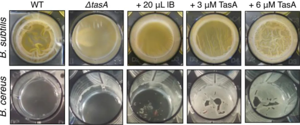
3.6.RESULTS ... 73
3.6.1. E. coli expression of TasA and functionality ... 73
3.6.2. B. cereus TasA and CalY maintain an unfolded conformation in solution ... 75
3.6.3. B. subtilis and B. cereus TasA functional amyloids show various macroscopic morphologies... 77
3.6.4. B. subtilis and B. cereus TasA functional amyloids display a cross-β architecture ... 78
3.6.5. B. subtilis and B. cereus TasA functional amyloid filaments reveal different assembly kinetics ... 79
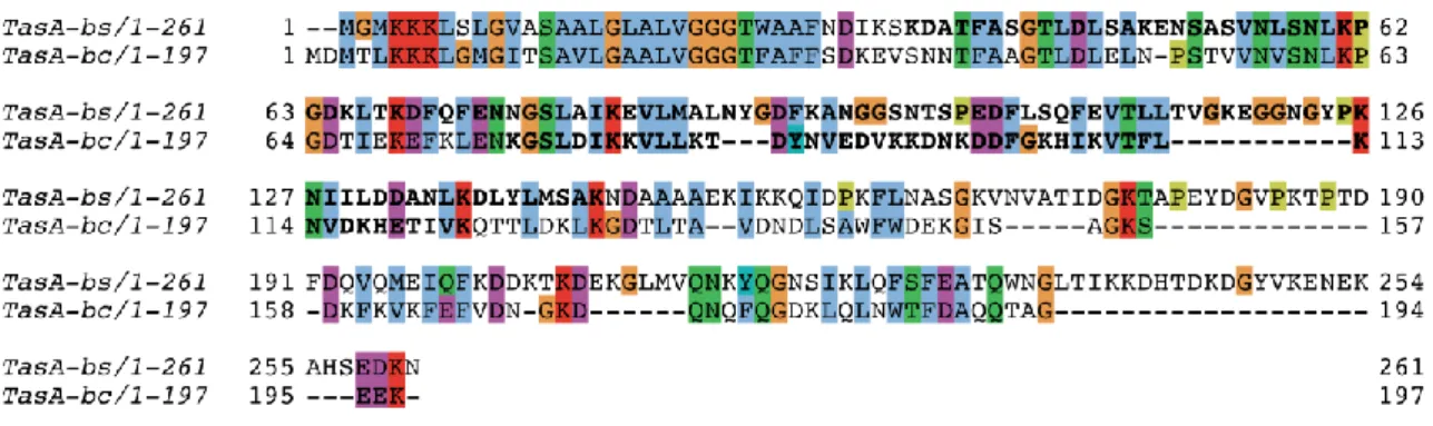
3.6.6. B. subtilis and B. cereus TasA functional amyloids share the same structural fold but different local structural polymorphism ... 81
3.6.7. Comparison of secondary structure propensity of TasA functional amyloids
between B. subtilis and B. cereus... 84
3.6.8. Accessory proteins TapA and CalY self-assemble and adopt the same structural fold as observed for TasA filaments ... 88
3.6.9. In vitro co-assembly between TasA and its accessory protein does not perturb the global filament architecture but catalyze its fibrillization ... 91
3.7.DISCUSSION ... 93
3.8.SUPPLEMENTARY DATA ... 97
CHAPTER 4: BACILLUS SUBTILIS BIOFILM MOLECULAR ORGANIZATION IS MODULATED BY THE EXPRESSION OF TASA AND EXTRACELLULAR POLYMERIC SUBSTANCES ... 105
4.1.NONSTANDARD ABBREVIATIONS ... 105
4.2.CONTEXT ... 105
4.3.METHODOLOGY ... 108
4.3.1. Bacterial strains and culture conditions ... 108
4.3.2. Solid-state NMR ... 108
4.4.RESULTS ... 108
4.4.1. Polysaccharides in the extracellular matrix and the cell wall are flexible ... 108
4.4.2. WT Bacillus subtilis biofilm content analysed by SSNMR ... 110
4.4.3. Biofilms of eps-, tasa-, tapa-deleted mutants are dynamically heterogeneous ... 115
4.4.4. eps-, and tasa-, tapa-deleted double mutants biofilms display drastic changes in bio-content ... 117
4.5.DISCUSSION ... 120
4.6.CONCLUSION ... 122
CHAPTER 5: THE INNER ROD PROTEINS OF THE TYPE III SECRETION SYSTEM IN SALMONELLA AND PSEUDOMONAS STRAINS... 125
5.1.CONTEXT ... 125
5.2.METHODOLOGY ... 127
5.2.1. Bacterial material ... 127
5.2.2. Cell growth and protein expression ... 128
5.2.3. PrgJ/PscI purification ... 128
5.2.4. spaPQR purification ... 129
5.2.5. Isolation of the needle complex substructures ... 129
5.2.6. Solid-state NMR ... 130
5.2.7. Secondary structure analysis by solid-state NMR ... 130
5.2.8. Negative-staining electron microscopy... 131
5.3.RESULTS AND DISCUSSION ... 131
5.3.1. PscI from Pseudomonas ... 131
5.3.2. PrgJ from Salmonella ... 134
5.4.DISCUSSION ... 139
5.5.CONCLUSION AND PERSPECTIVES... 140
CHAPTER 6: STRUCTURAL INSIGHTS INTO THE ‘KILLER TRAIT’ CONVEYED BY SUPRAMOLECULAR REFRACTILE BODIES WITH SOLID-STATE NMR ... 141
6.1.NONSTANDARD ABBREVIATIONS ... 141
6.2.CONTEXT ... 142
6.3.METHODOLOGY ... 144
6.3.2. Cell growth and protein expression ... 144
6.3.3. R bodies extraction ... 144
6.3.4. 31P NMR ... 145
6.3.5. Solid-state NMR ... 145
6.3.6. Secondary structure analysis by solid-state NMR ... 146
6.3.7. Negative-staining electron microscopy... 146
6.4.RESULTS ... 147
6.4.1. E. coli expression of R bodies and pH-dependent macroscopic morphologies ... 147
6.4.2. Very-fast MAS SSNMR characterization of R bodies at pH 7 ... 148
6.4.3. Monomeric RebA and RebB are composed of helical structural elements ... 152
6.4.4. R bodies contain non-protein biomolecules ... 154
6.4.5. Structural basis of the pH-dependent macro-morphological switch ... 157
6.4.6. R bodies can store and release biomolecules and thus act as a delivery nano-machine ... 160
6.5.CONCLUSION ... 162
TABLE OF FIGURES ... 165
CONCLUSIONS AND PERSPECTIVES ... 175
List of Publications
(1) Jan Stanek, Loren B Andreas, Kristaps Jaudzems, Diane Cala, Daniela Lalli, Andrea Bertarello, Tobias Schubeis, Inara Akopjana, Svetlana Kotelovica, Kaspars Tars, Andrea Pica, Serena Leone, Delia Picone, Zhi‐ Qiang Xu, Nicholas E Dixon, Denis Martinez, Melanie Berbon, Nadia El Mammeri*, Abdelmajid Noubhani, Sven Saupe, Birgit Habenstein, Antoine Loquet, Guido Pintacuda. (2016) NMR spectroscopic assignment of backbone and side‐chain protons in fully protonated proteins: microcrystals, sedimented assemblies, and amyloid fibrils. Angewandte Chemie International Edition, 55: 15504- 15509.
(2) Antoine Loquet, Nadia El Mammeri*, Jan Stanek, Mélanie Berbon, Benjamin Bardiaux, Guido Pintacuda, Birgit Habenstein. (2018) 3D structure determination of amyloid fibrils using solid-state NMR spectroscopy. Methods, 138: 26-38.
(3) Birgit Habenstein, Nadia El Mammeri*, James Tolchard, Gaëlle Lamon, Arpita Tawani, Mélanie Berbon, Antoine Loquet. (2019) Structures of type III secretion system needle filaments. Springer, Berlin, Heidelberg book chapter, 1- 23.
(4) El Mammeri N.*, Hierrezuelo J., Tolchard J., Cámara‐Almirón J., Caro‐Astorga J., Álvarez‐Mena A., Dutour A., Berbon M., Shenoy J., Morvan E., Grélard A., Kauffmann B., Lecomte S., de Vicente A., Habenstein B., Romero D. and Loquet A. (2019), Molecular architecture of bacterial amyloids in Bacillus biofilms. The FASEB Journal, 33: 12146- 12163. (5) J. Shenoy, N. El Mammeri*, A. Dutour, M. Berbon, A. Saad, A. Lends, E. Morvan, A. Grélard, S. Lecomte, B. Kauffmann, F.‐X. Theillet, B. Habenstein, A. Loquet. (2019) Structural dissection of amyloid aggregates of TDP‐43 and its C‐terminal fragments TDP‐35 and TDP‐16. FEBS J, 287: 2449-2467.
(6) A. Daskalov, D. Martinez, V. Coustou, N. El Mammeri*, M. Berbon, L.B. Andreas, B. Bardiaux, J. Stanek, A. Noubhani, B. Kauffmann, J.S. Wall, G. Pintacuda, S.J. Saupe, B. Habenstein, A. Loquet. (2020) Structural and molecular basis of cross- seeding barriers in amyloids. BioRxiv
Disclaimer
The text and figures of the Introduction section and Chapter 3 have been taken or adapted from published articles. (2) Loquet et al., 3D structure determination of amyloid fibrils using solid-state NMR spectroscopy. Methods, 2018; (4) El Mammeri et al., Molecular architecture of bacterial amyloids in Bacillus biofilms. The FASEB Journal, 2019.
For clarity purposes, figures are numbered independently in each chapter and comprise the chapter section number.
Introduction
1.1.SSNMR AS A TOOL FOR STRUCTURAL BIOLOGY ... 15
1.1.1. Biological assemblies... 15
1.1.2. The interesting case of amyloid fibrils ... 16
1.2.BIOCHEMICAL METHODOLOGY ... 19
1.2.1. Sample preparation ... 19
1.2.2. Assembly and polymorphism... 20
1.2.3. Isotope labeling ... 21
1.3.BIOPHYSICAL METHODOLOGY ... 23
1.3.1. SSNMR experiments for biomolecular assemblies ... 23
1.3.2. Data analysis ... 24
1.3.3. Restraint detection and identification ... 25
1.3.4. 1H-detection for structural investigations ... 28
1.4.STRUCTURE CALCULATION ... 29
1.4.1. Manual structure calculation ... 30
1.4.2. Automated structure calculation ... 30
1.5.THESIS OBJECTIVES ... 31
1.1. SSNMR as a tool for structural biology
1.1.1. Biological assemblies
Supramolecular assemblies of proteins, nucleic acids, lipids, sugars, etc, are very common in most biological cell functions. Solid-state NMR spectroscopy (SSNMR) has been playing a key role in understanding and studying biological samples that are insoluble, non-crystalline, or even intrinsically disordered (1-5). In the past decades a wide range of biological systems has been put through SSNMR scrutiny: viral capsids (6), filaments (7), amyloid fibrils (8), membrane proteins in native environments (9-15), large supramolecular assemblies (16), etc. With crucial milestones including the molecular conformation of A fibrils from Robert Tycko’s work (17-20), transthyretin fibril structure from Bob Griffin's laboratory (21, 22), or the structure of the functional amyloid HET-s by Beat Meier (23), SSNMR has now become a major actor in structural biology. SSNMR offers a unique awareness of heterogeneous and “imperfect” biological samples (2) thus, in combination with cryo-electron microscopy (cryo-EM), giving access to a wide scope of biological events ranging from highly ordered assemblies (SSNMR: up to about ~25 kDa; cryo-EM: even higher) to highly polymorphic entities. In addition to the structural information, SSNMR also allows the study of interactions of molecules of different sizes and natures, dynamics, local and overall order, local motions, etc (24-26). SSNMR is often used in synergy with X-ray crystallography, solution NMR, and cryo- or conventional EM in biological studies (27). To date, 135 protein/peptide 3D structures
determined by SSNMR have been deposited at the PDB (Protein Data Bank), including different types of protein complexes such as membrane-associated systems, macromolecular assemblies (filaments, fibrils, capsids), or microcrystalline proteins.
SSNMR methodologies require the use of isotopic 13C/15N enrichment of proteins through labelled precursors introduced in the cell cultures prior to protein expression (28-35). Indeed, at natural abundance, cells contain native matter made of 1H at 99.98%, 13C at 1.11%, and 15N at 0.36%, these isotopes being active in NMR. In such conditions, NMR experiments which depend on specific nuclear properties of carbon and nitrogen atoms might be difficult to implement. Various labelling schemes have been developed in the past decades ranging from uniformly labelled samples to highly site-specific 13C/15N enrichment (see section 1.2). Another type of SSNMR application is the study of entire cells, bacteria, fungi, etc, called whole-cell NMR (36). In such studies, isotopic enrichment is applied on the entire cells. Many studies report the detailed analysis of bacterial or plant cell wall components, inclusion bodies, live cells, etc (37-43). With the use of isotopic labelling, multidimensional spectroscopy, and
13C, 1H, 15N, 31P detection, SSNMR groups have reported detailed accounts of the
carbohydrate, protein, lipid, and other molecules’ composition within complex organisms.
1.1.2. The interesting case of amyloid fibrils
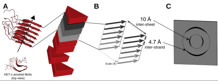
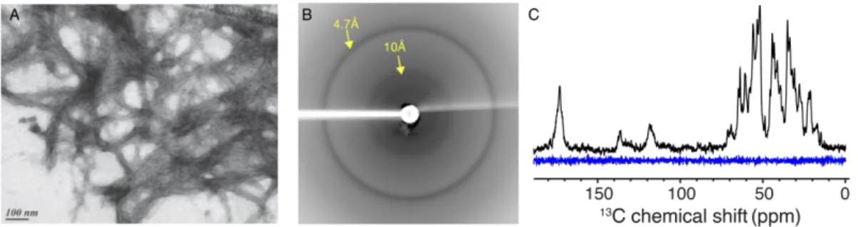
Amyloid-forming proteins have long been a target of choice in the field of SSNMR as their intrinsic polymorphism had long hindered their study at atomic resolution by other biophysical methods (44-51). The term amyloid is by definition suitable for any protein-based supramolecular assembly with birefringence features and the propensity to bind the Congo red dye, displaying non-branched fibrils of approximately 10 nm in diameter (52). The characterization of such unique entities has then been extended to the presence of the so-called cross-β arrangement consisting in stacked β-strands perpendicular to the fibril axis that arrange as intermolecular β-sheets along the fibril axis (Figure 0-1A). This structural hallmark, presently considered to be a much more robust portrayal of the amyloid family, is adequately pictured through typical X-ray diffraction patterns (53, 54), in which are inferred some structural characteristics, as typical inter-strand and inter-sheet distances of 4.7 Å and 10 Å, respectively (Figure 0-1B-C).
Figure 0-1: Structural features of amyloid fibrils. (A) General morphology and monomer stacking of an amyloid fibril with HET-s fibrils (55) here used as an illustration with a singular two-layer β-solenoid amyloid fold; (B) representation of the so-called cross-β quaternary structure in which β-sheets are stacked along the fibril axis with a 4.7 Å spacing distance as inter-strand interactions, and 10 Å as inter-sheet contacts ; (C) schematic representation of a typical X-ray diffraction pattern, highly representative of the mentioned structural aspects associated with amyloid proteins.
Interestingly, the physician Rudolph Virchow first introduced the word amyloid in 1854, while describing brain-related cellulose-like materials, called corpora amylacea (56). It was later demonstrated that amyloid entities were in fact a novel class of proteins, which displays the ability to undergo conformational change and adopt the cross-β architecture, thus forming amyloid fibrils. Such observations paved the way for ground-breaking discoveries in a multitude of research fields. This considerable attention fundamentally arises from their distinct association with a series of deadly (neuro-)degenerative disorders, called amyloidosis, such as Alzheimer’s, Parkinson’s diseases and type 2 diabetes (49, 57-62).
However, more recently it has become evident that the amyloid family can actually be split into two sub-classes, regarding their pathological or beneficial effects. These amyloids, executing native cellular functions, have been identified in a range of organisms from bacteria to mammals and are termed "functional amyloids" (63-69). Biofilm formation, hormone storage, or cellular signal transduction are some of many examples illustrating the presence of amyloid-like systems involved in biological processes (70-77).
Such duality of effects, non-native aggregation and functional assembly, reveals the complexity of the amyloid fold. Several reports suggest that virtually any protein, when set in the proper conditions/environment, could adopt an amyloid-like β-strand-rich conformation, thus advocating the amyloid fold as a universal global free-energy minimum for polypeptide chains (78-80). Nonetheless, the implication of functional amyloids in selective biological
B
4.7 Å inter-strand 10 Å inter-sheetC
HET-s amyloid fibrils (top view)
A
processes such as cellular signal transduction (71, 74-76) indicate that the recruitment strategies of amyloid monomers can be very specific and not necessarily related to misfolding events.
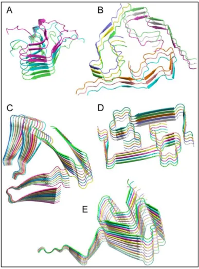
The non-crystalline and insoluble character of amyloid fibrils poses many obstacles for the structural investigations of these systems at the atomic level, based on well-established techniques such as X-ray crystallography or solution NMR spectroscopy. Additionally, a certain level of structural heterogeneity on the mesoscopic level, often encountered in amyloid fibrils, has hampered the use of cryo-EM until recently (81, 82). SSNMR is a method of choice to achieve keen characterization of supramolecular assemblies in general (4, 5, 25, 83-86), and more specifically of amyloid fibrillar assemblies at high resolution (87-90). This is illustrated by the first structure determination of prion amyloid fibrils that Meier and co-workers achieved in 2008 (55), or by the discovery of the 3D fold of amyloid fibrils of α-synuclein (91) and A (92-96), solved by SSNMR techniques. SSNMR capacity to probe the local order of “imperfect” inhomogeneous biological assemblies stands behind its efficiency over amyloid fibrils (97). Most high-resolution structures of amyloid fibrils have been elucidated using SSNMR as the main method: to date, 35 amyloid protein/peptide structures or structural models have been deposited in the PDB. Figure 0-2 displays some of the most relevant structures of amyloid fibrils elucidated using SSNMR.
Figure 0-2: Selection of amyloid fibrils atomic structures determined by solid-state NMR. (A) HET-s(218-289) prion fibrils, automated calculation with CYANA (PDB 2KJ3) (23); (B) Aβ1-40 fibrils in Alzheimer’s disease brain tissue, manual calculation with XPLOR-NIH (PDB 2M4J) (94); (C) Aβ1-40 D23N "Iowa" mutant fibrils, manual calculation with Rosetta (PDB 2MPZ) (98); (D) Aβ1-40 E22Δ "Osaka" mutant fibrils, automated calculation with CYANA (PDB 2MVX) (99); (E) Human α-synuclein fibrils, manual calculation with XPLOR-NIH (PDB 2N0A) (91). Figure was generated with the PyMOL Molecular Graphics System (Schrödinger, LLC).
1.2. Biochemical methodology
1.2.1. Sample preparation
Both chemical synthesis and recombinant protein production in Escherichia coli are used for structure determination of supramolecular assemblies. For whole-cell NMR, the introduction of labelled isotopes can be achieved by providing 13C/2H/15N sources in the culture medium. Most small protein/peptide samples (up to about 30-50 residues) are prepared based on well-known Fmoc-protection solid-phase chemical synthesis methods (100, 101), and HPLC-based purification. Higher molecular-weight proteins, however, are usually produced by E. coli-based recombinant expression followed by purification steps, although expression in the native organism has also been used. The heterologous expression has the advantage that isotope labeling (2H, 13C, 15N) can be easily implemented by introducing the labeled precursors in the bacterial culture medium. Furthermore, an arsenal of different selective labeling strategies
based on heterologous expression has been developed and successfully applied to determine structures. In the case of amyloid proteins, during bacterial protein production, monomers generally accumulate in inclusion bodies and need to be extracted (e.g., using 8 M urea, or 6 M guanidinium chloride), followed by an enrichment and purification process, which could employ one or several affinity- and/or size-exclusion- and/or hydrophobic-chromatography procedures. Note that the optimal protein production conditions may vary between the production in rich, unlabeled and in poor bacterial expression medium that can be supplemented with labeled or unlabeled 2H, 13C, 15N sources. It is therefore beneficial in terms
of cost and time effort to optimize the production in unlabeled poor medium before using isotope-labeled sources.
1.2.2. Assembly and polymorphism
For biological systems that require an in vitro post-purification assembly, prior to actual NMR data recording and analysis, the polymerization process represents a significant step in the sample preparation because the conditions during the assembly play a major role in driving the protein subunits to adopt one specific homogeneous molecular structure, a heterogeneous variant or several structural polymorphs in the fibrillar sample. Incubation temperature and duration, pH, protein concentration, nature of the buffer, presence/quantity of additional compounds (e.g., metal ions, chaotropic agents, chelating agents, redox reagents, antibacterial molecules, etc.), protein state upon rotor-packing (i.e. hydrated or lyophilized sample), are some of the numerous conditions that need to be thoroughly optimized with both SSNMR and transmission electron microscopy as control tools. The optimization steps are crucial since conformational and structural differences in the fibril arrangement can be observed following only slight changes in sample preparation. Indeed, obtaining highly homogeneous samples is a compelling task in the field of SSNMR and the polymorph selection is often guided by the assembly conditions. The sample heterogeneity and polymorphism observed for amyloid fibrils, for example, is under constant discussion (47, 87, 102), and polymorphism as well as fold instability might carry biological significance. Different structural polymorphs in disease-related amyloids lead to strains that might cause different clinical and pathological phenotypes (103-106) and that can vary in cellular toxicity (107-109). SSNMR spectroscopy serves as a probe of the molecular order at the atomic level, as it can detect slight differences in local conformational states in the assembly. Indeed, the level of sample homogeneity is directly
reflected in the SSNMR line width and has thus a significant impact on the structural analysis (88-90, 97).
1.2.3. Isotope labeling
SSNMR approaches require 15N and 13C isotopic enrichment of biological samples to
enhance the signal abundance and allow site-specific studies. Protocols for supramolecular assemblies preparation for SSNMR resemble those employed in solution NMR studies, but differ in that the final sample needs to be assembled from the monomeric subunits or kept assembled (4). The development of isotopic labelling strategies has greatly enhanced the power of SSNMR in structural studies as the labelling schemes allow to selectively retrieve the desired information (i.e. a specific position of labelled 13C and 15N nuclei within the amyloid proteins), to reduce dipolar truncation by spin dilution (distribution of labelled 13C sites based on metabolic pathways of the used precursor), and to alleviate spectral congestion (110-113).
13C-15N enrichment can either derive from chemically labelled precursors during
solid-phase chemical synthesis, for which the limitations in labelling combination are of chemical nature, or from the carbon sources that are used during recombinant expression. During recombinant protein production, metabolic pathways of the host introduce the 13C-labeling to specific positions in the amino acids (111). The same considerations apply when isotopically enriching whole cells. Numerous selective 13C-labeling strategies have been exploited for SSNMR, such as using [U-13C]-, [1-13C]-, [2-13C]-, [1,3-13C2]- glucose or glycerol derivatives.
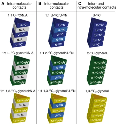
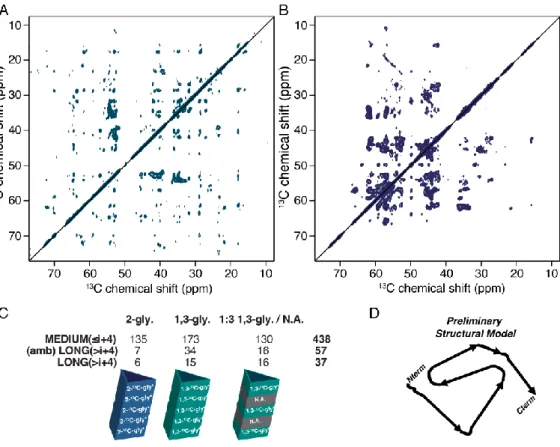
Figure 3 displays some of these labelling systems that can be used either for intra-molecular
or inter-molecular interaction determination (Figure 3 - illustrated with the use of a fibril for clarity purposes). Another production and labelling strategy is the use of cell-free techniques, which is an in vitro tool that mimics a bacterial system and is used to produce proteins while keeping a relevant control over 13C/15N enrichment (114, 115).
Figure 0-3: Labelling schemes utilized for amyloid structure determination using SSNMR (NB: * is added when
15N-labelling can be added if 15N-detected SSNMR experiments are required). (A) any mix between 13C-labelled and unlabelled (natural abundance, N.A.) proteins will distinctly prevent inter-monomer 13C-13C correlations, thus allowing the unambiguous discrimination between inter- and intra-molecular interactions; (B) any mix between 13C-labelled and 15N-labelled proteins is extremely helpful in determining the fibril stacking registry (i.e. in or out); (C) both inter- and intra-monomer interactions can be detected, while specific 13C-glycerol derivatives allow spin dilution.
1H-detected SSNMR is an attractive alternative to 13C-detected methods, which could
in perspective alleviate the need for isotopic enrichment. However, 1H-detected SSNMR methods at moderate to fast MAS (Magic Angle Spinning; approx. 20-60 kHz; see section 1.3) need to be coupled not only to 13C- and 15N-labelling to increase the separation of resonances
in multidimensional spectra, but also to different levels of deuteration to ensure narrow linewidths in the 1H dimension and sensitivity of detection (i.e., 1H dipolar coupling broadens
NMR signals at low MAS). For example, extreme levels of proton dilution in side-chains are obtained by the expression in D2O/H2O mixtures (e.g., the ratio of 9:1) and the use of [2H/13
C-]glucose as the sole carbon source, followed by a purification in D2O (116, 117). Alternatively,
an excess of D2O can be used for the expression, and a mixture of D2O/H2O can be used during
the purification/assembly step to partially reintroduce only exchangeable amide protons in contact with the aqueous buffer (118, 119). However, a high dilution of protons severely limits the probability of observing 1H-1H contacts (120), therefore either a full back-exchange (in
sites) has been proposed to record amide-amide (121-123), methyl-amide and methyl-methyl (124-128) interproton distance restraints. However, the amount of potentially available structural information is also significantly reduced, which can impair the high-resolution structure determination. This approach can be used with automation for small model proteins (126), but not on a larger molecular scale if not supplemented by other long-range restraints (129, 130). Also, incomplete exchange of amide 1H can severely hamper the spectral analysis by 1H-detected SSNMR methods, particularly if a refolding protocol for the protein is lacking
(119, 131-133). With the advent of fast spinning MAS probes (> 60 kHz), intermediate approaches were proposed, yielding a significantly lower deuteration level in side chains (and in particular, a full occupancy at H sites), such as “inverse fractional deuteration” (132) where
2H,13C-glucose and 100% H
2O is used for expression, and either D2O or H2O at the purification
step. Recently, Meier and co-workers employed partial (about 75%) deuteration for 1 H-detection of amyloid fibrils of HET-s(218-289) at 60 kHz MAS, where a H2O:D2O ratio of 1:3
was used for expression, and either pure D2O or H2O was used for washing the resulting fibrils
(134). While this approach favours resolution of 1HN and 1H resonances over occupancy of
1H sites, the non-uniform 2H-induced shifts reduce the resolution in the 13C dimension or, for
a given sample, cause systematic inconsistencies of 13C and 13C shifts in different spectra (or in different regions of the same spectrum) (134). A number of studies have recently demonstrated that in the ultrafast MAS regime (100-111 kHz) a large set of 1H-1H restraints can be obtained for fully-protonated microcrystalline proteins, viral assemblies or membrane-embedded proteins, thus alleviating all issues related to deuteration (133, 135-138).A detailed investigation on 1H linewidths showed that at 111 kHz MAS a minor resolution gain is expected even with a high level of deuteration for microcrystalline proteins (139), while partial deuteration remains beneficial for more dynamic systems such as membrane proteins (140) or fibrils.
The distinction of inter- and intramolecular contacts in 1H-detected SSNMR remains a
challenge for sensitivity reasons, and until now is possible with a (uniform) mixed labelling with 15N or 13C, or by deuterating one part of the assembly (141).
1.3. Biophysical methodology
1.3.1. SSNMR experiments for biomolecular assemblies
SSNMR has emerged as the method of choice for investigation of insoluble non-crystalline biopolymers at atomic resolution. The method is mainly based on two tools to
circumvent issues such as line broadening and low signal to noise ratios associated with 13C and 15N. (i) Proteins in solid state move very slowly, so that the spectral lines are broadened considerably compared to solution NMR spectra on soluble proteins, for which the isotropic motion averages out several anisotropic NMR interactions. The use of MAS allows for the recovery of spectral resolution. Indeed, the sample is spun at the magic angle (i.e., 54.7°; this angle averages out several anisotropic interactions that are orientation-dependent) relative to the magnetic field. Therefore, many line-broadening interactions, such as CSA, quadrupolar and dipolar interactions, are partially averaged out by the MAS, leading to significant line-narrowing and an increase in resolution and signal to noise ratio. (ii) Cross-polarization (CP) is a well-established SSNMR experiment used to increase sensitivity of 13C and 15N detection. Indeed, SSNMR usually uses 13C and 15N nuclei, with low gyromagnetic ratios, which require long experimental times to provide suitable signal. CP allows magnetization to be transferred from 1H (high gyromagnetic ratio) to heteroatoms (e.g. 13C and 15N) that are dipolar coupled to enhance the signal and decrease the experimental time. To date, mostly 13C-based structure elucidation strategies have been employed on supramolecular assemblies, but 1H-detected fast MAS are increasingly reported for this kind of systems.
1.3.2. Data analysis
1D, 2D, and 3D SSNMR spectra of supramolecular assemblies are usually firstly recorded based on 13C-13C, 13C-15N, 13C-15N-13C, 15N-13C-13C or (1H-)15N-1H correlations in the aim of assigning different resonances to the protein atoms, as chemical shift assignments are a prerequisite for extracting structural information using NMR spectroscopy. The level of complexity during the resonance assignment process typically depends on the diversity of the amino acid composition, the presence or absence of repetitive sequence motifs, the extent of undetectable residues (due to static or dynamic disorder), structural homogeneity in the sample, available SSNMR technology and methodology and also on the availability of other biophysical data. Assigned SSNMR resonances (mainly 13Cα, 13Cβ) allow the secondary
structure determination of all detected residues, based on the use of secondary chemical shifts (142).
When aiming at robust and high-resolution analysis, spectral crowdedness can hamper peak observation, and thus the 3D structural information collection, which is achieved by measuring inter- and intra-molecular distance restraints, obtained using polarization transfers between two atoms (usually 1H, 13C, or 15N), located below ~2-10 Å in space. Spin dilution
achieved through the above-mentioned labelling strategies (i.e. 13C-glucose and 13 C-glycerol-derived precursors) offers higher spectral resolution by reducing scalar one-bond 13C-13C couplings. In addition, the 13C spin dilution improves the efficiency of polarization transfer between through-space coupled 13C-13C pairs, by decreasing the phenomenon of dipolar truncation (143-145). [1-13C]-, [2-13C]-, [1,3-13C
2]- glucose (or glycerol) labelling strategies
allow the almost complete removal of one-bond dipolar, and J-couplings, thus considerably improving the spectral resolution, by a factor of ~2 or larger (85, 112, 146, 147). Indeed, the reduced number of 13C-labeled sites lessens the spectral overlap and permits the sequential
assignment of short- and medium-range correlations. Many labelling combinations have been developed over the years, in order to improve the data quality, and to target specific structural details. Both intra- and inter-molecular restraints are gathered prior to model calculations as described below.
1.3.3. Restraint detection and identification
For 13C/15N based restraint identification, several labelling and monomer-interaction combinations can be useful (Figure 3): (i) mix between 13C-labeled (or 13C-15N) and natural-abundance proteins for intra-molecular contacts (146) only (from 1:1 to 1:4 molar ratios) (Figure 3A); (ii) mix between two different labelling schemes (usually contains 50% of U-15N with either 50% of [U-13C]-glucose (148), [2-13C]-, or [1,3-13C2]-glucose (147) or glycerol
(149)) for inter-molecular contacts only (usually 1:1 molar ratio) (Figure 3B); (iii) [U-13 C]-glucose, [2-13C]-, or [1,3-13C2]-glycerol residue labelling for all types of through-space contact
detection (Figure 3C), based on 2D and 3D spectroscopy (150, 151).
The specific 1:1 [1-13C]-glucose / [2-13C]-glucose mix has been employed, for the measurement of definite intermolecular long-range distances (150, 152). For example, using a 1:1 U-15N/U-13C mixed labelling scheme can reveal the amyloid fibril register (i.e., -strand packing), by comparing an NCA experiment (usually displaying the backbone correlation
15N/13Cα of a residue) of a U-13C-15N sample and the one of the 1:1 U-15N/U-13C mix. Indeed,
an in-register stacking will show perfect overlap of the two experiments, and the 13Cα of a residue i will be in direct contact with the backbone 15N labelling of the same residue on the adjacent monomer i 1, thus showing the same 15N/13Cα correlations as the one observed in a uniformly 13C-15N labelled sample. Generally, most long-range through-space correlations will appear on the [2-13C]-glycerol and [1,3-13C2]-glycerol samples, and the same labelling scheme
discriminate inter- from intra-molecular residue contacts (Figure 3C); the applied molar ratio between labelled and unlabelled monomers should reflect a compromise, to prevent both 13
C-13C inter-molecular correlations and data with too low signal (e.g. having only ~50% of the
sites 13C-labeled in the supramolecular assemblies will reduce the signal to noise ratio by ~4). The 13C-13C correlations, displayed as peaks on the spectra, are usually considered as
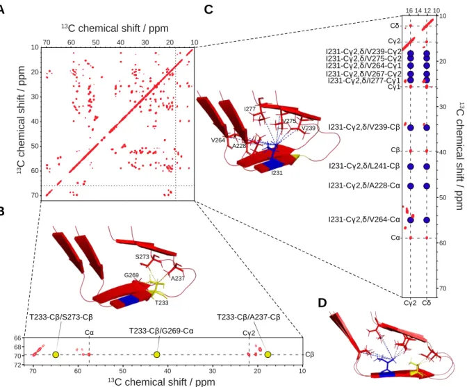
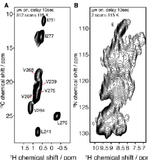
four different types of contacts: (i) intra-residual 13C-13C correlations, which account for 13 C-labeled sites of the same residue; (ii) short-range correlations, which comprise inter-residual interactions (between a residue i and the residues i+1) (also referred to as “sequential correlations”); (iii) medium-range correlations include the correlations from i to i+2 to i+4; (iv) long-range 13C-13C correlations contain i to >i+4 contacts, crucial for determining long-range through-space restraints further used during structure calculations. Indeed, in optimal conditions (e.g., high spectral resolution, high signal to noise ratios, no polymorphism, etc.) a complete and thorough analysis of the SSNMR data should provide a list of 13C-13C or 1H-1H long-range correlations subdivided into four groups: unambiguous inter-molecular, ambiguous inter-molecular, unambiguous intra-molecular, and ambiguous intra-molecular distances; both the quantity and quality of restraints are to be considered. Figure 4 illustrates such data set with the example of HET-s fibrils (55), in which reported distances involving T233 and I231 are displayed.
Figure 0-4: Illustration of long range through-space distance determination, based on HET-s(218-289) amyloid core (55). (A) 2D-13C-13C PDSD spectrum (50ms mixing time) of HET-s(218-289) uniformly labelled (13C/15N) recorded on a 800MHz spectrometer (proton frequency). (B) Excerpts of the PDSD spectrum shown in A, displaying only threonine correlations. T233 resonances are highlighted with dashes (C, C, C2). Reported distances regarding T233 are displayed with yellow dots and illustrated on the HET-s(218-289) structure (PDB ID: 2RNM). (C) Excerpts of the PDSD spectrum shown in A, (ppm window: 10-17.5). I231 resonances are highlighted with dashes (C, C, C1, C2, C). Reported distances regarding I231 are displayed with blue dots and illustrated in HET-s(218-389) structure (PDB ID: 2RNM). For clarity purposes, overlapping peaks were disregarded. (D) HET-s structure (55), as shown in B-C, from a lateral point of view. All mentioned distances were determined based on either PDSD long-mixing time experiments (with [2-13C]- or [1,3-13C
2]-glycerol labelling schemes), or CHHC spectra (with a fully labelled HET-s amyloid core sample).
Interestingly, the 3D structure model construction of supramolecular assemblies often requires hybrid approaches, which employ data acquired using other techniques, such as X-ray diffraction, or scanning transmission electron microscopy (STEM), which give information about the mass-per-unit-lengths parameters of filamentous objects (5, 92, 153, 154). From such bio-objects, a data set, comprising knowledge about the molecular atomic structure from SSNMR experiment and fibril morphology with STEM analysis, will be the basis for the complex model calculation and energy minimization processes.
1 3C c h e m ic a l s h if t / p p m 13C chemical shift / ppm T233-Cβ/S273-Cβ T233-Cβ/G269-Cα T233-Cβ/A237-Cβ Cα Cγ2 Cβ Cα Cγ1 Cγ2 Cδ Cγ2 Cδ I231-Cγ2,δ/V264-Cγ1 I231-Cγ2,δ/V275-Cγ2 I231-Cγ2,δ/V239-Cγ2 I231-Cγ2,δ/V239-Cβ I231-Cγ2,δ/L241-Cβ I231-Cγ2,δ/A228-Cα I231-Cγ2,δ/I277-Cγ1 I231-Cγ2,δ/V264-Cα I231-Cγ2,δ/V267-Cγ2 T233 A237 S273 G269 I231 A228 V264 V239 V275 I277 13C chemical shift / ppm 1 3 C c h e m ic a l s h ift / p p m 70 60 50 40 30 20 10 70 60 50 40 30 20 10 16 14 12 10 70 60 50 40 30 20 10 70 60 50 40 30 20 10 72 70 68 66 A B C D Cβ
1.3.4. 1H-detection for structural investigations
1H detection boosts sensitivity and allows the direct observation of 1H-1H proximities. 1H-1H
proximities represent natural restraints for structure calculation, easy to interpret, and in contrast to 13C-13C interactions, are a direct indication of physical close contacts, especially
relevant across -strands or within hydrophobic patches.
The capabilities of 1H-detected techniques with respect to a full structure determination in the
solid state have been demonstrated in a number of microcrystalline systems (121, 122, 124-126, 155). MAS rates in the 40-60 kHz range were employed, aided by extensive sample deuteration and selective reintroduction of protons (see section 2.1), allowing a significant reduction in the sample amount required, as well as faster spectral acquisition and unbiased automated data analysis. The approach has recently been extended toward the determination of a membrane-embedded target, in combination with 13C-13C constraints (123, 128). Notably, this approach considerably accelerates resonance assignment, which represent the first but crucial phase of an NMR structure determination and provides a quick evidence of the secondary structure adopted by the protein. In the field of amyloid proteins and in a pioneering study, Reif and coworkers demonstrated high-resolution 1H-detected spectra for Alzheimer disease -amyloid peptide A (1-40), using 20 kHz MAS and a deuterated sample back-reprotonated at the amide sites at 25-50% (119). There, 2D-1H,15N fingerprint correlation spectra were used as a clear and rapid readout of sample homogeneity, enabling the optimization of fiber preparation through multiple seeding cycles. Recently, Meier and coworkers showed that 1H,15N,13C inter-residue correlations can be used to transfer available 13C and 15N assignments to -protons in a partially (above 75%) deuterated sample of HET-s(218-289) at 60 kHz MAS (134).
The use of MAS rates beyond 100 kHz allows to extend the approach and removes the requirement for proton dilution by deuteration, opening the way to the sensitive detection of amide and aliphatic protons in fully protonated proteins. In particular, a set of experiment was proposed for sequence-specific backbone and aliphatic side-chain resonance assignments. Only 500 g of sample and a few days of data acquisition were used in this study (135). Overall, this opens the perspective of determining tertiary and quaternary structures of all supramolecular assemblies by directly probing side-chain-to-side-chain and/or backbone-to-side-chain 1H-1H proximities (133).
1.4. Structure calculation
Despite their topological peculiarities, high-resolution 3D structures can be determined from SSNMR data using the same general procedure as for solution NMR, i.e.: (i) conversion of assigned NMR observables into structurally meaningful restraints, (ii) calculation of hundreds of conformers by restrained molecular-dynamics simulation and (iii) selection of best fitting conformers as final structure bundle. Detected cross-peak signals in relevant hetero- or homo-nuclear spectra are first converted into restraints restricting the distance between the different assigned nuclei. In SSNMR studies, distance restraints often take the form of an upper-bound distance that can be either estimated from peak intensity and known reference distances or from L-shape curves obtained from multiple structure calculation trials with increasing upper-limits (23). It is worth noting that the success of structure calculation strongly relies on a critical number of long-range (between residues separated by more than 5 residues in sequence) and inter-molecular restraints (between different subunits).
The restraint set for structure calculation can be supplemented by dihedral angle restraints on φ/Ψ backbone angles, predicted from backbone secondary chemical shifts using tools like TALOS+ (156). On the basis on the collected restraints, several conformers (usually hundreds) are then calculated using dedicated software, such as CYANA (157), UNIO (158), ARIA (159), XPLOR-NIH (160) or Rosetta (161) that perform restrained molecular modeling.
?-? ?-? ?-? A-B A-A Manual calculation Automated calculation Initial fold Final structure
+
Symmetry restraintsAxial stacking Interfacial symmetry Identity restraints
+
Hydrogen-bond restraints+
Backbone torsion-angle restraints! " N Cα C' N H O Random conformation
Figure 0-5: General procedure for 3D structure calculation of amyloid fibrils structures from SSNMR. First, distance restraints derived from unambiguously assigned cross-peaks or peaks with low spectral ambiguity are used for structural calculation to establish the initial fold of the fibrils (Manual calculation). Next, the restraint set is supplemented with all identified cross-peaks (generally with high spectral ambiguity) that will be assigned automatically and yielding a high precision bundle of conformers as the final structure (Automated calculation). Owing to the highly ordered nature of amyloid fibrils, symmetry restraints are employed to ensure a symmetric arrangement of subunits during calculation (identity restraints, axial stacking and interfacial symmetry, if applicable). Additionally, backbone dihedral angle restraints, derived for secondary chemical shifts, and distance restraints enforcing hydrogen-bonds are implemented in order to impose a regular conformation and proper stacking of stands.
1.4.1. Manual structure calculation
Direct application of automated NMR cross-peak assignment and structure calculation tools used routinely for solution NMR data is pragmatically limited for amyloid fibrils due to not only the lower spectral resolution of SSNMR but also the difficulty to disentangle intra- and inter-molecular restraints, the latter potentially originating from axial (along the fibril axis) or lateral (between protofibrils) contacts. It is thus common practice to first perform structure calculation using solely SSNMR restraints that could be assigned unambiguously or with very limited spectra ambiguity that can be identified manually by a trained operator (ca. a few dozens of long-range and a handful of inter-molecular restraints at least). Such manual
structure calculation (Figure 5) generally determines an initial fold with reasonable precision
that will serve to identify new compatible restraints for additional rounds of manual calculation. At this stage, it is important to carefully replicate restraints for all equivalent subunits to respect the symmetric nature of the fibrils.
1.4.2. Automated structure calculation
Alternatively, one can proceed with automated structure calculation (Figure 5) using all remaining cross-peaks for which unambiguous assignment cannot be safely obtained manually. For instance, the CYANA software can assist in automatically collecting assignment possibilities by matching cross-peaks positions with the list of assigned chemical shifts according to user defined tolerance windows. These tolerances for assignment of cross-signals should be reflective of the linewidth in the different dimensions of the originating spectrum. CYANA then carries out an iterative protocol by alternating calculation of conformer bundles and selection of the most probable assignments compatible with the structures, hence reducing human bias in assigning cross-peaks.
1.5. Thesis objectives
The following thesis manuscript is divided into six independent chapters based on the different projects that my PhD work encompasses.
Chapter 1 relays a methodological approach to use DNP (Dynamic Nuclear Polarization) in
the context of structural biology with the combined advantages of fast MAS (up to 40 kHz MAS) and selective labelling. We performed such methodological development in collaboration with Dr Guido Pintacuda and Dr Anne Lesage from Lyon ENS in an attempt to set up a proof-of-concept of 1H detection of proteins under DNP conditions and further the knowledge of DNP applications to biological samples.
Chapter 2 encompasses the study of a novel functional amyloid family involved in
programmed cell-death of Nectria haematococca, highly reminiscent of the HET-s paradigm in Podospora anserina. HET-s is a model protein for functional amyloids and prions as it stands as a fascinating amyloid protein whose assembly is thought to be part of a signalling cascade. Dr. Sven Saupe, from the University of Bordeaux, identified a three-gene system encoding for three proteins called SesA, SesB, and het-eN, which share a short segment in their N- or C-terminal ends with high sequence homology. The purpose of the study is to characterize the activity in vivo and the structural features in vitro, in close collaboration with Dr. Sven Saupe. Identifying a new functional amyloid is of great interest in the mystery of functional vs pathological amyloids. An acute understanding of how functional amyloid differ from pathological misfolded proteins is still lacking. We used SSNMR to investigate the structural fold of SesB amyloid motif and found that SesB fold seems to constitute a new functional amyloid family.
Chapter 3 mentions our study of another functional amyloid entity called TasA. TasA is
involved in Bacillus subtilis and Bacillus cereus biofilm matrix composition. Biofilms are surface-attached bacterial communities in which bacteria an extracellular matrix (ECM) is secreted. The ECM is composed of exopolysaccharides (EPS), proteins, DNA, etc. This work has been done in collaboration with Dr. Diego Romero, from the University of Malaga. The goal of this study is to analyse yet another functional amyloid. Several reports have already proposed TasA to form amyloid fibrils within the extracellular matrix of the biofilm thus being responsible for the biofilm integrity and protect bacteria from external and mechanical stresses. In addition to TasA, the proteins TapA and CalY, the partner proteins in Bacillus subtilis, and
Bacillus cereus, respectively. This broad functional versatility and their remarkable ability to
form robust filamentous self-assemblies contribute to general interesting and wonder surrounding functional amyloids in the bacterial kingdom. This chapter combines a
biochemical/biophysical approach to a microbiological one as we report the differences displayed by the species Bacillus subtilis and the pathological Bacillus cereus.
Chapter 4 resumes such focus on biofilm formation as we investigate the impact of
biofilm-related mutations, in eps, tasA, and tapA, on the overall biofilm matrix composition and on the general dynamical features of the biofilm. This chapter introduces the use of SSNMR multidimensional techniques as a powerful tool to obtain the composition of cell wall molecules in intact Bacillus biofilms. Changes in molecular composition of the cell wall has been studied for three different mutants and compared to wild type biofilms to decipher the impact of mutations on the cell wall composition.
Chapter 5 focuses on the inner rod proteins of the type three secretion system of
Gram-negative bacteria. Type 3 secretion systems (T3SSs) are in the centre of Gram-Gram-negative bacterial ability to directly deliver effector proteins from their cytoplasm to that of eukaryotic cells via a protein-based needle complex, through a most conserved battery of homologous genes required to assemble a nanomachine, also called the “injectisome”. This appendage passes through the bacterial barrier composed of plasma membranes, peptidoglycan layer and the extracellular space. Such work has been done in collaboration with Prof. Jorge Galán from Yale University. Following the elucidation of the needle complex, we set to characterize the inner rod, which constitutes the basis of the needle by connecting it to the basal body. We wish to study two proteins, PscI in Pseudomonas, and PrgJ in Salmonella, which form the inner rod. In the context of this study, I was able to work with Prof. Jorge Galán for five months. Chapter 5 thus displayed the work we have done which focuses on the in vitro characterization of the inner rod proteins, as we discuss the structural and sequence conservation of the inner rod proteins.
Chapter 6 relates the work we have done on a complex and mysterious bacterial
supramolecular assembly called refractile bodies (a.k.a. R bodies). R bodies (in this study, type 51 refractile bodies) are unusual, ribbon-like protein polymers found in many bacterial species, such as Caedibacter and Pseudomonas. In 1938, Tracy M. Sonneborn observed that some
Paracemium aurelia strains were able to kill other sensitive strains of the same species. This
ability was initially linked to an extranuclear factor, called kappa, which was associated with the production and release of a toxin in the medium. R bodies are large protein biopolymers that look like ribbons and have the ability to roll and unroll in a pH-dependent fashion. Such work is done in collaboration with Dr. Justin Kollman, from Seattle University, USA. No structural has been uncovered so far, and we set to characterize such unusual structural
Chapter 1: High-resolution
1H-detected study of biomolecules by
Dynamic Nuclear Polarization Enhanced very fast MAS NMR
1.1.CONTEXT ... 33 1.2.METHODOLOGY ... 35
1.2.1. Isotopically enriched protein expression ... 35 1.2.2. Protein purification and assembly ... 36 1.2.3. NMR spectroscopy and DNP ... 36
1.3.RESULTS AND DISCUSSION ... 37
1.3.1. Model system ... 37 1.3.2. Polarizing source concentration optimization ... 37 1.3.3. Effect of MAS rate on DNP enhancement ... 39 1.3.4. Two-dimensional SSNMR ... 39 1.3.5. Proof of concept ... 40 1.3.6. Structural analysis ... 41 1.3.7. Biological relevance ... 43 1.4.CONCLUSION ... 43 1.1. Context
As SSNMR methods are increasingly employed to characterize non-crystalline and insoluble biomaterials (84, 162-170), recent and on-going advances regarding both sensitivity improvement (171-176) and proton-detected approaches (136, 177-186) are expected to boost MAS SSNMR tremendous role in understanding supramolecular assemblies (1, 2, 187). For more than half a century, SSNMR has been favorably used in the study of molecular structure, organization and dynamics of non-crystalline and insoluble samples, from chemistry to biology. Consequently, improvements in NMR sensitivity are continually sought to aid in the observation of dilute moieties and to accelerate data acquisition.
An interesting challenge of NMR-based methods in general is the signal sensitivity (171, 173, 188, 189). For decades, the fields of non-biological materials have used Dynamic Nuclear Polarization (DNP) (190-193) for improving such issue and reduce the samples concentrations as well as experimental times. DNP is based on the hyperpolarization of nuclei by transferring large electron polarization to the surrounding nuclei (172-174, 188, 194-198). Such transfer is achieved through continuous microwave (MW) radiations that saturate the electron transition. This phenomenon requires the use of cryogenic temperature (below 200 K) and organic radicals as electron sources. High power and frequency gyrotrons are used and so far, NMR
signals have been enhanced by a factor of ~10-200 for biological samples in numerous studies. The use of such remarkable technology still requires the optimisation of several parameters: MW strength, temperature, the type of polarization agent used, overall radical concentration and insertion into the sample, MAS frequency, magnetic field strength, etc (172).
Dynamic nuclear polarization (DNP) has revolutionized SSNMR for material sciences (174, 196, 199-206), wherein microwave (µw) irradiation of the sample drives electron-nuclear spin polarization to enhance the observable NMR signal which, for solid samples, with a theoretical enhancement limit of ~660 for protons (192). Here, we report an approach combining fast MAS, 1H-detected SSNMR, and DNP coupled with site-specific labelling in the analysis of a
biological system. Altogether with severe spin dilution, three-dimensional proton detected SSNMR spectroscopy becomes possible for macromolecular protein assemblies while allowing improvements in sensitivity. The detection of SSNMR signals encoding for proton-proton proximities is demonstrated and paves the way for future structure elucidation of protein assemblies otherwise intractable by conventional biophysical techniques.
The use of DNP on proteins and biological assemblies (172, 197, 207) has been in constant increase through the past two decades as new SSNMR methodologies and instrumentations have been developed (208-212), and the chemical design of more efficient polarizing agents (213-218). Many biological objects have been studied with the help of DNP-enhanced SSNMR, including large complexes (219-226), fibrils (227-232), microcrystals (176), viral particles (233-235), membrane proteins (16, 236-241), entire organisms (194, 242-246), cellular cell walls (38, 247, 248), or lipidic systems (198, 249-253).
DNP-based SSNMR is now at a critical stage where proton-detection has yet to be implemented in such experiments, as signal enhancement combined with high sensitivity nuclei such as protons, is expected to be extremely useful. Similar to the race for proton detection “traditional” SSNMR has witnessed in the past two decades, DNP-enhanced SSNMR is now experiencing the same experimental trends. Although the current state of SSNMR advances gives access to more than 100 kHz magic-angle spinning, no DNP-equipped SSNMR setup is commercially available with the ultra-fast MAS technology. The current theoretical understanding of cross-effect (CE) DNP and empirical DNP observations of increasing MAS speed also raise questions regarding the potential efficiency of hyperpolarization with ultra-fast MAS (197, 254, 255). The very-fast MAS SSNMR probes (~60 kHz) with cryogenic cooling which have been constructed and paired with the obligatory DNP apparatus (a gyrotron microwave source and waveguide) therefore remain pertinent to SSNMR proton detection (256-258). Inherently lower