SCIENTIFIC ARTICLE
Normal values of Wiberg
’s lateral center-edge angle
and Lequesne
’s acetabular index–a coxometric update
Clément M. L. Werner&Leonhard E. Ramseier&
Thomas Ruckstuhl&Jeff Stromberg&
Carol E. Copeland&Clifford H. Turen&
Kaspar Rufibach&Samy Bouaicha
Received: 26 February 2012 / Revised: 16 April 2012 / Accepted: 17 April 2012 / Published online: 15 May 2012 # ISS 2012
Abstract
Background The historical pathological cut-off values for Wiberg’s lateral center-edge (LCE) angle and Lequesne’s
acetabular index (AI) are below 20° and above 12° for the LCE and AI, respectively. The aim of this study was to reassess these two angles more than 50 years after their introduction using a standardized conventional radiological measurement method, considering changing social habits and their associated physiological changes.
Methods A total of 1,226 anteroposterior radiographs of the pelvis (2,452 hips) were obtained according to a strict stan-dardized radiographic technique allowing reliable measure-ments of the LCE angle and the AI.
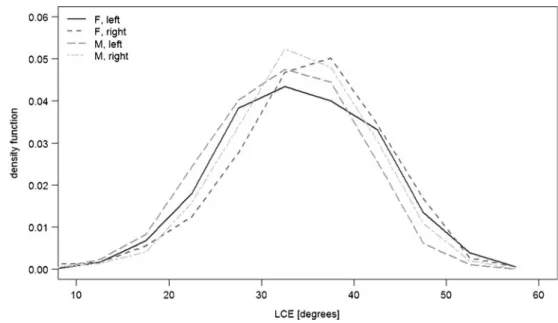
Results Distributions of the LCE and AI were pronouncedly Gaussian, with mean values of 33.6° for the LCE and 4.4° for the AI. The 2.5th and 97.5th empirical percentiles were 18.1 and 48.0° for the LCE and−6.9 and 14.9° for the AI. These intervals contained 95 % of the data in our large sample. Small but statistically significant differences between the sexes and right and left hips have been demonstrated. Correlation be-tween age and coxometric indices was low.
Conclusion The above findings do not conflict with the historical benchmarks. Statistical differences between sexes and between right and left hips were not clinically relevant. No conclusion can be drawn about coxometric indices and clinical manifestations of hip dysplasia.
Keywords Hip . Acetabulum . Lateral center-edge angle . Acetabular index . Pelvic radiographs
Introduction
The two radiographic indices primarily used to quantify the lateral coverage of the femoral head by osseous acetabular structures are Wiberg’s lateral center-edge angle (LCE) and the Lequesne’s acetabular index (AI), introduced in 1939 and 1963, respectively [1, 2]. Since then, orthopaedic
C. M. L. Werner (*)
:
S. BouaichaDivision of Trauma Surgery, University Hospital Zurich, Raemistrasse 100,
8091 Zürich, Switzerland e-mail: [email protected] S. Bouaicha
e-mail: [email protected]
C. M. L. Werner
:
J. Stromberg:
C. E. Copeland:
C. H. Turen R Adams Cowley Shock Trauma Center,University of Maryland Medical Systems, 22 South Greene Street,
Baltimore, MD 21201, USA J. Stromberg e-mail: [email protected] C. E. Copeland e-mail: [email protected] C. H. Turen e-mail: [email protected] L. E. Ramseier
:
T. RuckstuhlDepartment of Orthopaedics, Balgrist University Hospital Zurich, Forchstrasse 340, 8008 Zürich, Switzerland L. E. Ramseier e-mail: [email protected] T. Ruckstuhl e-mail: [email protected] K. Rufibach
Biostatistics Unit, Institute for Social and Preventive Medicine, University of Zurich,
Hirschengraben 84, 8001 Zürich, Switzerland
e-mail: [email protected] DOI 10.1007/s00256-012-1420-7
surgeons all over the world have included these values in their diagnostic and therapeutic considerations, in the context of dysplastic hip assessment and preoperative planning of cor-rective osteotomies. Both the LCE and AI are measured on anteroposterior radiographs of the pelvis. The anatomic land-marks for the LCE are the lateral edge of the acetabulum, the center of the femoral head, and a perpendicular line to the horizontal axis of the pelvis. The AI is measured between a line parallel to the horizontal pelvic plane through the most medial extent of the“sourcil” and a line tangent to its most lateral extent. Values below 20° for the LCE and above 12° for the AI were considered pathological by the two authors.
After introduction of new surgical procedures for re-orientation of the acetabulum in dysplastic hips, such as the Bernese periacetabular osteotomy [3], determination of LCE and AI has regained attention as an accurate assess-ment of the acetabulum. As a result, it is mandatory that it be measured pre-, intra- and postoperatively.
Over the last few decades, social environments have changed and, in general, people are heavier, taller, and older than when these indices were proposed 50 years ago. Few data exist about the validity of these coxometric values in the contemporary generation [4]. Additionally, there is evi-dence that pelvic tilt or rotation significantly influences measurement outcomes [5].
However, few studies, with relatively small sample sizes [6], exist that quantify Wiberg’s and Lequesne’s angles
using a reliable, standardized measurement procedure of the anteroposterior pelvic radiographs as previously de-scribed [7].
Our study was therefore driven by questions as to wheth-er the LCE and AI measured by using a reliable, standard-ized measurement method [7] in a large sample size population would be the same as those determined by Wiberg and Lequesne. In addition, we wondered about any differences between“normal values” of those cited in the past century and today’s population.
Materials and methods Patients
Anteroposterior pelvic radiographs of 1,635 trauma patients (3,270 hips) admitted to the R Adams Cowley Trauma Center, Baltimore, MD, USA, were extracted from the electronic radiological database. Of these patients, 1,226 (2,452 hips) met the inclusion criteria of having conventional radiographs of the pelvis obtained in a correct standardized radiographic tech-nique [7]. The eligible patients included 871 males and 355 females (71/29 %). Ages ranged from 14 to 97 years, with a median age of 38.9 years.
Validation of radiographs
Rotation in the axial and sagittal plane of anteroposterior pelvic radiographs may significantly change outcome meas-urements of coxometric indices [5,6]. In order to minimize this error, the criteria of Siebenrock et al. [7] were applied to determine those cases to be further assessed. Only AP pelvic radiographs that demonstrated alignment of the tip of the coccyx with the middle of the symphysis and a distance between the sacrococcygeal joint and the symphysis less than 32 mm in men and less than 47 mm in women were included. False rotation in the frontal plane could be simply corrected electronically with the picture-archiving communication sys-tem (PACS) imaging program, and therefore such radiographs were not excluded. All radiographic measurements were per-formed by orthopaedic residents, fellows, and staff members (C.M.L.W, T.R., J.S., C.E.C, and C.H.T.).
Measurements on radiographs
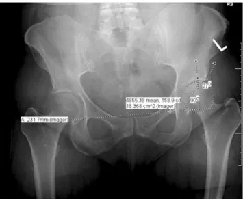
The LCE was measured according to the following steps. First, the horizontal plane of the pelvis was determined by drawing a horizontal line along the inferior boundaries of the two teardrops. Second, a perpendicular line crossing the center of the femoral head, defined by the midpoint of a congruent circle, was drawn up to the acetabular roof. The intersection of this vertical line with the acetabular roof represented the starting point for further measuring. Finally, a line from the center of the femoral head was drawn out to the most lateral extent of the acetabulum. The angle between the perpendicular line and the oblique line, touching the lateral extent of the acetabulum, is Wiberg’s angle (Fig.1). Lequesne’s AI was measured by first drawing a line paral-lel to the horizontal plane of the pelvis as described above (teardrop to teardrop), touching the medial border of the sclerotic weight-bearing portion of the acetabular roof, the so-called sourcil. A line originating from the medial extent of the sourcil was drawn out to the lateral extent of the weight-bearing portion of the acetabulum, neglecting any osteophyte formation. The angle between these two lines crossing at the medial border of the sourcil is Lequesne’s AI (Fig.2).
All values were obtained using digital measurement tools of the PACS imaging program.
Statistical analysis
Gaussianity of angle data is graphically illustrated using frequency polygons. Normal ranges were defined as the interval from the 2.5th to the 97.5th empirical percentile, i.e., the interval that contains 95 % of the data. Standard t-test confidence intervals were computed when assessing quantities with one measurement per patient. For quantities that involved measurements of both sides of a patient,
meaning dependent measurements, we computed confi-dence intervals using standard errors derived from a gener-alized least squares (GLS) model, which assumes a compound symmetry correlation structure. Similarly, groups were compared using the t-test if independent observations were analyzed; if dependent observations were present, the t-test was used to estimate the coefficient in a GLS model including intercept and the grouping variable under consid-eration. Pearson’s correlation coefficient was used to assess the extent of linear association between continuous varia-bles. Confidence intervals for proportions were computed using Wilson’s method. No correction for multiple testing was done in this analysis. All confidence intervals were computed using a confidence level of 95 %, and all tests were computed at a significance level of alpha00.05. All
analyses were performed using R. The add-on package “nlme” was used to compute the GLS models [8,9].
Source of funding
This study was conducted without any external funding.
Results
Mean overall Wiberg’s LCE angle was 33.6° (95 % CI: 33.2–34.0°) and Lequesne’s AI was 4.4° (95 % CI: 4.1−4.7°). The measurements extended from a minimum of 2.6° to a maximum of 57.1° for the LCE angle and from −18.8° to 33° for the AI. The values exhibited a Gaussian distribution (see Figs. 3 and 4) for both as well as for each sex and side. The reference range for LCE was calculated as 18.1° to 48.0° and for AI from−6.9° to 14.9°. Considering 20° for LCE angle as inferior cut-off level as proposed by Wiberg, 95.0 % (95 % CI: 93.7–96.1) of all left hips and 96.6 % (95 % CI: 95.4–97.5) of all right hips in our study population were characterized as“normal.” When 12° was defined as the superior cut-off for Lequesne’s AI, 94.0 % (95 % CI: 92.5–95.2) of all left hips and 92.4 % (95 % CI: 90.8–93.8) of all right hips could be identified as “normal.” LCE and AI measurements showed small but significant differences between male and female population (p<0.05), where LCE values were higher in females and AI values were higher in male individuals. Also differences could be found between right and left hips, where right hips showed higher values than left hips for both LCE and AI. We did not find large correlations between coxometric indi-ces and age (LCE/age: r00.19 and AI/age: r0−0.07). As
Fig. 1 Digital measurement technique of Wiberg’s lateral center-edge angle
Fig. 2 Digital measurement technique of Lequesne’s acetab-ular index
expected a negative correlation was found between LCE and AI (r0−0.61). For details see also Table1.
Discussion
In 1939, Wiberg’s monograph “Studies on dysplastic acetabula and congenital subluxation of the hip joint” mentioned for the first time a new method of measuring the lateral coverage of the femoral head. “In order to form an opinion on the roentgenologic features of a normal acetabulum,” he examined 50 males and 50 females aged 20–35 years who had functionally “blame-less” hips. Aware of methodological weakness, he obtained radiographs of the 200 hip joints and measured
the angle between a line parallel to the longitudinal axis of the body crossing the center of the femoral head and a line tangential to the lateral edge of the acetabular roof. He found that the majority of both men and women showed angles between 26 and 35°; Wiberg considered angles between 20 and 25° as uncertain and below 20° as pathological [1].
In 1963, Lequesne described the“horizontal toit externe” (THE) angle, better known in the English literature as the acetabular index (AI). This angle is a diagnostic tool to determine the orientation of the acetabular roof in the coro-nal plane and therefore describes the superolateral coverage of the femoral head in an anteroposterior radiograph of the adult pelvis. The measurement is consistent with the work published in 1925 by Hilgenreiner to determine the
Fig. 3 Normal distribution of the Wiberg’s lateral center-edge angle in males, females, right and left hips
Fig. 4 Normal distribution of the Lequesne’s acetabular index in males, females, right and left hips
obliquity of the immature acetabular roof, “Pfannendach-winkel” (German for “socket roof angle”) [10]. The mea-surement results from an adjacent line to the weight-bearing portion of the acetabulum with its sclerotic, arcuated aspect (“sourcil cotyloidien”) and a horizontal line drawn out from the most medial point of the weight-bearing area. Normal values were determined in his 1954 doctoral thesis by mea-suring 200 anteroposterior radiographs of patients with oste-oarthritis of the hip, and angles below 10° were considered normal, from 10 to 12° as borderline and above 12° as path-ological [2]. In the current literature the AI often is ascribed to Tönnis who re-launched this measurement procedure in 1976 when he introduced the hip value [11].
The methodological approaches of both Wiberg and Lequesne lacked a reproducible, standardized radio-graphic technique and were derived based on a small number of individuals. While these benchmarks have been used for decades in the diagnosis of abnormal hips, the widespread application of their “normal” val-ues is worthy of consideration.
Although historic benchmarks of Wiberg and Lequesne were published without comprehensive and reproducible methodology, our findings in today’s general population do not conflict with traditional cut-offs. In contrast, the historical thresholds between normal and pathological hips are close, differing only a few degrees from our“normal” range (<20° vs. 18.1° for LCE and >12° vs. 14.9° for AI). Considering the historical cut-offs for dysplastic hips, only 3–8 % of our study population could be identified as path-ological for LCE or AI.
No reasonable explanation is evident for the findings that LCE and AI values are significantly different between sexes and right to left hips. Taking into account that the differ-ences do not exceed 0.9 and 1.2° for AI and LCE between sexes and 0.5 and 1.5° for AI and LCE between the right
and left hips, these significant findings have no clinical relevance.
In contrast to the changing orientation of the acetabulum with progression of age during childhood [12], no correla-tion of LCE and AI values with age is found in the adult hip. The anticipated inverse correlation (−0.61) of the two angles was verified in our study.
Even though our results show narrow ranges of dis-tribution of the values and statistically significant com-parisons, only indirect statements can be made about the cut-off levels between normal and pathological radio-graphic indices. Another limitation is the possibility of selection bias as the study population consisted of trau-ma victims instead of “normal” volunteers. The large number of patients included in our study as well as the presumption that trauma patients reflect the general population may support this as a reasonable sample choice. Due to the large sample size, no inter- or intra-observer reliability was measured.
The clinical relevance of the data presented is that they represent current and accurate measurement references of normal coxometric indices, which may be considered as baseline values in acetabular re-orientation surgery such as periacetabular osteotomy.
Conclusion
Our study demonstrates that in today’s general population Wiberg’s LCE angle and Lequesne’s AI measured by stan-dardized method show a parametric distribution with mean values of 33.6° for the LCE and 4.4° for the AI. Normal ranges including 95 % of observations ranged from 18.1 to 48.0° for the LCE and from−6.9 to 14.9° for the AI. These findings do not conflict with the historical benchmarks.
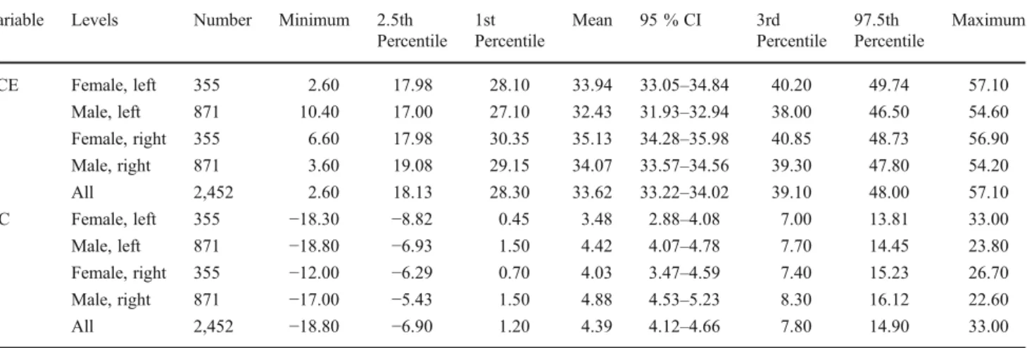
Table 1 Descriptive statistics of Wiberg’s lateral center-edge (LCE) angle and Lequesne’s acetabular index (AI) angles per sex and side Variable Levels Number Minimum 2.5th
Percentile 1st Percentile Mean 95 % CI 3rd Percentile 97.5th Percentile Maximum
LCE Female, left 355 2.60 17.98 28.10 33.94 33.05–34.84 40.20 49.74 57.10 Male, left 871 10.40 17.00 27.10 32.43 31.93–32.94 38.00 46.50 54.60 Female, right 355 6.60 17.98 30.35 35.13 34.28–35.98 40.85 48.73 56.90 Male, right 871 3.60 19.08 29.15 34.07 33.57–34.56 39.30 47.80 54.20 All 2,452 2.60 18.13 28.30 33.62 33.22–34.02 39.10 48.00 57.10 AC Female, left 355 −18.30 −8.82 0.45 3.48 2.88–4.08 7.00 13.81 33.00 Male, left 871 −18.80 −6.93 1.50 4.42 4.07–4.78 7.70 14.45 23.80 Female, right 355 −12.00 −6.29 0.70 4.03 3.47–4.59 7.40 15.23 26.70 Male, right 871 −17.00 −5.43 1.50 4.88 4.53–5.23 8.30 16.12 22.60 All 2,452 −18.80 −6.90 1.20 4.39 4.12–4.66 7.80 14.90 33.00
Statistical differences between sexes and between right and left hips are not of clinical relevance. No conclusion can be made about coxometric indices and clinical manifestation of hip dysplasia.
References
1. Wiberg G. Studies on dysplastic acetabula and congenital sublux-ation of the hip joint. Acta Chir Scand. 1939:5–135.
2. Lequesne M. Coxometry. Measurement of the basic angles of the adult radiographic hip by a combined protractor. Rev Rhum Mal Osteoartic. 1963;30:479–85.
3. Ganz R, Klaue K, Vinh TS, Mast JW. A new periacetabular osteotomy for the treatment of hip dysplasias. Technique and preliminary results. Clin Orthop Relat Res. 1988:26–36. 4. Ozcelik A, Omeroglu H, Inan U, Ozyurt B, Seber S. Normal values
of several acetabular angles on hip radiographs obtained from individuals living in the Eskisehir region. Acta Orthop Traumatol Turc. 2002;36:100–5.
5. Tannast M, Zheng G, Anderegg C, et al. Tilt and rotation correc-tion of acetabular version on pelvic radiographs. Clin Orthop Relat Res. 2005;438:182–90.
6. Fowkes LA, Petridou E, Zagorski C, Karuppiah A, Toms AP. Defining a reference range of acetabular inclination and center-edge angle of the hip in asymptomatic individuals. Skeletal Radiol. 2011;40:1427–34.
7. Siebenrock KA, Kalbermatten DF, Ganz R. Effect of pelvic tilt on acetabular retroversion: a study of pelves from cadavers. Clin Orthop Relat Res. 2003:241–8.
8. Pinheiro J, Bates D, DebRoy S, Sarkar D, and the R Core Team. nlme: linear and nonlinear mixed effects models. 1–90.
9. RDevelopmentCoreTeam. R: language and environment for statis-tical computing. 2010.
10. Hilgenreiner H. Zur Frühdiagnose und Frühbehandlung der ange-borenen Hüftgelenkverrenkung. Med Klinik 1925:1385–9. 11. Tonnis D. Normal values of the hip joint for the evaluation of
X-rays in children and adults. Clin Orthop Relat Res. 1976:39–47.
12. Tonnis D, Brunken D. Differentiation of normal and pathological acetabular roof angle in the diagnosis of hip dysplasia. Evaluation of 2294 acetabular roof angles of hip joints in children. Arch Orthop Unfallchir. 1968;64:197–228.