1 Title Page
Chondrocyte secretome: a source of novel insights
and exploratory biomarkers of osteoarthritis
Christelle Sanchez, PhD
1,2christelle.sanchez@ulg.ac.be
Anne-Christine Bay-Jensen, PhD
2,3acbj@Nordicbioscience.com
Thomas Pap, MD
2,4thomas.pap@uni-muenster.de
Mona Dvir-Ginzberg, PhD
2,5 monad@ekmd.huji.ac.ilHelen
Quasnichka PhD
2,7h.quasnichka@surrey.ac.uk
Richard Barrett-Jolley, DPhil (Oxon)
2,6rbj@liverpool.ac.uk
Ali Mobasheri, DPhil (Oxon)
2,7,8,9.10a.mobasheri@surrey.ac.uk
Yves Henrotin, MT, PT, PhD
1,2yhenrotin@ulg.ac.be
Affiliations:
1. Bone and Cartilage Research Unit, Arthropôle Liège, University of Liège, CHU Sart-Tilman, Belgium 2. The D-BOARD European Consortium for Biomarker Discovery
3. Department of Rheumatology, Biomarkers and Research, Nordic Bioscience, Herlev Hovedgade 207, 2730 Herlev, Denmark
4. Institute of Experimental Musculoskeletal Medicine, University Hospital Munster, Domagkstrasse 3, D-48149 Munster, Germany
5. Institute of Dental Sciences, Faculty of Dental Medicine, Hebrew University of Jerusalem, P. O. Box 12272, Jerusalem 91120, Israel
6. Department of Musculoskeletal Biology, Institute of Ageing and Chronic Disease, Faculty of Health & Life Sciences, University of Liverpool, Liverpool, United Kingdom
7. Department of Veterinary Pre-Clinical Sciences, School of Veterinary Medicine, University of Surrey, Guildford, GU2 7AL, United Kingdom
8. Faculty of Health and Medical Sciences, Duke of Kent Building, University of Surrey, Guildford, Surrey, GU2 7XH, United Kingdom
9. Arthritis Research UK Centre for Sport, Exercise and Osteoarthritis, Queen’s Medical Centre, Nottingham, NG7 2UH, United Kingdom
10. Center of Excellence in Genomic Medicine Research (CEGMR), King Fahd Medical Research Center (KFMRC), Faculty of Applied Medical Sciences, King Abdulaziz University, Jeddah, 21589, Kingdom of Saudi Arabia
Corresponding author: Dr Christelle Sanchez, Bone and Cartilage Research Unit, Arthropôle
Liège, University of Liège, CHU Sart-Tilman, 4000 Liège, Belgium. Email:
2
ABSTRACT
The extracellular matrix (ECM) of articular cartilage is comprised of complex networks of proteins and glycoproteins, all of which are expressed by its resident cell, the chondrocyte. Cartilage is a unique tissue given its complexity and ability to resist repeated load and deformation. The
mechanisms by which articular cartilage maintains its integrity throughout our lifetime is not fully understood, however there are numerous regulatory pathways known to govern ECM turnover in response to mechanical stimuli. To further our understanding of this field, we envision that proteomic analysis of the secretome will provide information on how the chondrocyte remodels the surrounding ECM in response to load, in addition to providing information on the metabolic state of the cell. In this review, we attempt to summarize the recent mass spectrometry-based proteomic discoveries in healthy and diseased cartilage and chondrocytes, to facilitate the discovery of novel biomarkers linked to degenerative pathologies, such as osteoarthritis (OA).
KEYWORDS
Cartilage, Chondrocyte, Osteoarthritis, Biomarker, Proteomics, Secretome, Extracellular
matrix (ECM)
RUNNING TITLE
3
INTRODUCTION
To gain a deeper understanding of the mechanisms that drive osteoarthritis (OA), it is important to appreciate the underlying biology of healthy and diseased joint tissues. Of interest is the pathophysiology of articular cartilage, and the processes that govern synthesis and organisation of extracellular matrix (ECM) components secreted by chondrocytes into the pericellular milieu. The chondrocyte is the unique resident cell of articular cartilage and thus solely responsible for ECM composition and regulation. Chondrocyte metabolism is influenced by its micro-environment, and in return influences ECM composition, organization and ultimately the mechanical resilience of cartilage [1-3]. As such, chondrocytes play a key role in ECM remodelling in physiological and pathological conditions [4].
It is commonly established that healthy articular chondrocytes change into different phenotypes as OA develops and progresses:
(i) A catabolic phenotype develops, associated with an increase in proteolytic enzymes and reactive oxygen/nitrogen species, in response to mechanical stress and inflammatory cytokines, tumour necrosis factor (TNF)-and interleukin (IL)-1, leading to further ECM degradation.
(ii) An anabolic phenotype emerges that is associated with regeneration of the ECM, including increased collagen type II and proteoglycan expression regulated by growth factors
(transforming growth factor (TGF)-, bone morphogenetic protein (BMP)s and insulin growth factor (IGF)-I), expressed either by the surrounding joint tissue or by the chondrocytes themselves.
(iii) A hypertrophic phenotype develops, manifesting in expression of type X collagen and induction of apoptosis, ultimately resulting in osteophyte formation.
4 (iv) A fibroblastic-like phenotype with an increased number of dedifferentiated chondrocyte and
expression of type I collagen.
(v) Lastly, a chondroblastic phenotype emerges with expression of foetal type IIA collagen, type III collagen and early/late differentiation markers[5].
The specific phenotype that any individual chondrocyte exhibits is dependent on the zone in which the chondrocyte is situated and the stage of OA progression. In the upper zone, cellular proliferation and hypertrophy are observed, whereas the mid and deep zones display increased expression of type II collagen [6]. As OA progresses, cartilage is lost and chondrocytes undergo senescence, due to a combination of replicative exhaustion and oxidative stress [7].
Eventually, chondrocytes will undergo apoptosis, and the articular cartilage will be destroyed. The ultimate goal of mass spectrometry-based proteomics strategies is the identification of a specific tissue-derived secretome that is unique to the diseased chondrocyte and surrounding ECM and is able to distinguish between healthy and diseased cartilage.
Because protein expression is dependent on environmental conditions, the secretome is highly dynamic in composition and turnover. In 2010, Agrawal et al. suggested defining the secretome as “the global group of secreted proteins into the extracellular space by a cell, tissue, organ, or organism at any given time and conditions through known and unknown secretory mechanisms involving constitutive and regulated secretory organelles”[8]. In this narrative review, we only considered the chondrocyte (or cartilage) as the source of secreted proteins, which have been uncovered by state-of-the-art mass spectrometry-based proteomics techniques.
Proteomic techniques include different methods, all relying on the separation of proteins and their further analysis using either gel-based (Two-dimensional electrophoresis) or gel-free methods. Protein separation methods are coupled to a mass spectrometer for identification of sequence by mass spectrometry [9, 10]. In differential analysis, the peptides may be marked with stable isotope at various stages of the analysis process, or Label-free methods could be performed. Several modes of analysis are
5 available in mass spectrometry[10]. They differ markedly by the ionization source of the sample. The main sources used in proteomic analysis are matrix-assisted laser desorption/ionization (MALDI) and surface-enhancer laser desorption/ionization (SELDI) [10, 11]. These techniques allow a soft ionization of molecules without excessive fragmentation [9, 11].
More than the total tissue protein extract, it is expected that well-defined protein fractions such as the secretome could be a source of novel OA biomarkers, with the potential to predict disease severity and monitor progression.
MATERIALS AND METHODS
A literature search was performed in Pubmed/Medline and Scopus, identifying articles published between January 2004 and March 2016. Keywords used in 'Any fields' were; (chondrocyte OR cartilage) AND secretome (19 relevant papers out of 38 found), or (chondrocyte OR cartilage) AND (proteomic OR mass spectrometry) (65 articles relevant papers out of 290 found). Only results from mass spectrometry-based studies were included in this article. The review has considered all species, even if the majority of studies have been performed on human source of chondrocytes or cartilage. Only research articles published in English were included. Supplementary files of all papers were analysed and included in this review.
CHONDROCYTE SECRETOME
Recent mass spectrometry-based proteomic studies have identified several proteins which form the chondrocyte secretome. In this part of the review, we focused on proteins identified either directly in the secretome of cartilage explants [12-20] or chondrocytes cultures [18, 21-31]), both of which are listed in Table 1. We further complete this list with proteins recently identified by proteomic analysis performed directly on fresh chondrocytes or cartilage tissue[32-36] (sometimes de-cellularized [37]) whereby different locations in the joint were compared, or healthy joint tissues were compared with
6 OA tissues. The characteristics of all these studies are summarized in Table 2. Proteins have been classified in different sections: ECM proteins, cytokines and growth factors, enzymes and
miscellaneous.
ECM proteins
As expected, the most abundant ECM proteins produced by chondrocytes, and detected by proteomic analysis, are collagens and proteoglycans, Table 1. 13 collagens are found in the chondrocytes
secretome, of which type II, VI and XII are the most abundant[35]. Collagen type XII is also known to interact with other cartilage elements such as cartilage oligomeric matrix protein (COMP), decorin and fibromodulin [38]. Collagen type II and VI levels are increased in the secretome of chondrocytes taken from the medial condyle of patients with early OA (Mankin score 0-3), compared with samples taken from patients with severe OA (Mankin score 5-10) [25].
Beside the collagens, other ECM proteins found in high abundance in the chondrocyte secretome include; aggrecan, HPLN-1 (proteoglycan link protein), biglycan, COMP, fibronectin, prolargin (Proline-arginine-rich end leucine-rich repeat protein (PRELP)), matrilin-3, cartilage acidic protein-1 (CRTAC1 or ASPIC), latent-transforming growth factor beta-binding protein-1 (LTBP1), extracellular matrix protein-1 (ECM-1), tenascin, lubricin and chitinase-3-like protein 1 (CHI3L1), also known as YKL-40). CHI3L1 was found at lower levels in cartilage explants treated with IL-1β compared to controls[12]. CHI3L1, is a biomarker of OA found in synovial fluid and serum[39], and plays a role in tissue remodelling and inflammation. The concentration of CHI3L1 in OA synovial fluid positively correlates with levels of matrix metalloproteinase (MMP)-1, MMP-3, IL-6 and IL-17.
IL-6 and IL-17 enhanced CHI3L1 production in human primary chondrocyte cultures[40] and CHI3L1 serum concentration positively correlated with osteophyte size in OA patients[41]. This protein is more abundant in knee compared to hip cartilage [37] and in OA compared to healthy cartilage [27] and is considered to be an early OA biomarker in the Stenberg’ proteomic study, which compared early and severe OA secretomes [25].
7 In comparison with normal cartilage explants, CRTAC1 is found elevated in the OA cartilage
secretome [13]. This protein is considered a cartilage specific protein, appearing early during
chondrocyte differentiation of mesenchymal stem cells (MSCs) [26]. Expression of CRTAC1 allows discrimination between human chondrocytes and osteoblasts, or MSCs, in monolayer cell culture [26, 42]. This protein is therefore considered a good marker of chondrocytic differentiation of MSCs.
LTBP1 also seems to be an important ECM protein secreted early in the differentiation of MSCs to chondrocytes [26]. Beside its structural role in the ECM, this protein is involved in the storage and the activation of TGF-β1, and therefore may play an important role in facilitating cartilage homeostasis.
ECM-1 is a protein involved in endochondral bone formation serving as a negative regulator of bone mineralization [43]. It is able to enhance the proliferation of endothelial cells during angiogenesis[44] and inhibit MMP-9 proteolytic activity[45]. Louridos et al observed that the level of ECM-1 secreted by cells originating from damaged OA cartilage is 3-fold higher than that of healthy cartilage [13].
Periostin secretion from explants increases with cartilage degradation [13], and was only found in cultured OA chondrocytes [35]. Expression of periostin was previously shown to be elevated in OA cartilage compared to normal, and located in the pericellular ECM close to damaged areas of cartilage [46, 47]. Periostin is able to increase expression of IL-6, IL-8, MMP-1,-3,-13 and a disintegrin and metalloproteinase with thrombospondin motifs (ADAMTS)-4 by chondrocytes in vitro[46, 47]. Fibronectin and fibromodulin are secreted at comparable levels by OA and normal chondrocytes, but fragmentation of these ECM proteins is found to differ depending on whether the cell is healthy or diseased [36, 48].
Cytokines and growth factors
Classical cytokines and chemokines like IL-6, IL-8, CCL8-20, CXCL1-3-5-8, CSF-1 are detected by tandem mass spectrometry only after an IL-1β stimulation of chondrocytes, cultured either in explant or in monolayer [12, 14, 19, 22, 27].
8 The IGFBP family is widely represented in chondrocyte secretome by IGFBP-2,-3,-4,-5,-6 and -7 [12, 21, 22, 24-27]. Expression of IGFBP-3,-4,-6 and -7 is increased in early OA [25]. These proteins serve as a carrier protein for IGF-1 and modulate the availability of IGF-1 to bind its receptor. Furthermore, IGFBP3 is known to induce chondrocyte apoptosis during OA[49]. In contrast IGFBP5 that has a positive role in cartilage anabolism, and inhibits enzymatic degradation; in fact IGFBP3 promotes cartilage extracellular matrix formation in vivo in the DMM OA rat model [50].
Among growth factors detected in the chondrocyte secretome, gremlin-1, chondromodulin and pleiotrophin are known to be involved in hypertrophic differentiation regulation. Gremlin-1 is a BMP antagonist presenting reduced secretion from OA chondrocytes compared to normal [14][35], and plays an important role in bone development by inhibiting bone formation [51]. Gremlin-1 is therefore, potentially, a potent inhibitor of chondrocyte hypertrophy in cartilage. Chondromodulin is also important for chondrocytes stabilization, acting by inhibiting hypertrophic differentiation, and is shown to be secreted only by superficially located chondrocytes [34]. Pleiotrophin is a secreted heparin-binding peptide expressed in mesodermal and neuroectodermal cells during development, but rarely in adult tissues. Pleiotrophin is abundant in foetal and juvenile cartilage, but not in mature. Furthermore, pleiotrophin is re-expressed in chondrocytes in early OA, and is involved in the
clustering and proliferation of chondrocytes observed in the early stages of OA [52]. Pleiotrophin is an inducer of hypertrophy during chondrogenic differentiation of MSCs [53] and is also a potent pro-angiogenesis factor.
Proteolytic enzymes and their regulators
Proteomic analysis of the chondrocyte secretome shows that the most abundant family of enzymes secreted is the MMPs, along with their endogenous inhibitors, the TIMPs. MMP-1 and 3, and TIMP-1, are particularly abundant. TIMP-3 appears to be more abundant in normal hip cartilage than knee, but MMP-1 is contrarily abundant in normal knee cartilage [37]. According to these studies, MMP-13 was only identified following IL-1β-stimulation [15], or is at very low levels without stimulation [14]. With the exception of MMP-2 [13], MMP protein levels do not vary substantially between normal and
9 OA chondrocytes, but all increase with IL-1β treatment. TIMP-1 and -2 levels are decreased in OA medial condyles [25], and TIMP-1 is increased with TGF-β1 stimulation [24].
The other family of metalloproteinases expected to be found in cartilage is the ADAMTSs. Although ADAMTSs were found in some proteomic secretome studies [12, 22, 25], they were not identified in all studies; most likely due to varying extraction methods which can eliminate ADAMTSs, along with the glycosaminoglycan (GAG) attachments of proteoglycans [15]. ADAMTS's may not be easily identified in mass spectrometry-based proteomic studies also due their relatively low abundance. Indeed, saturation of detectors with high abundance ions, type of protease used in mass spectrometry based studies (mainly trypsin) which may lead to non-identification of ionized peptides as they are too small, too large or contain amino acid sequences less likely to be 'seen' following ion identification. Positive studies, which identified ADAMTSs in the secretome, were all performed using OA
chondrocytes or OA cartilage explant culture [12, 22, 25]. A recent study published by Svala, showed that peptides cleaved from aggrecan, following IL-1β stimulation in chondrocytes, were generated by MMPs, but not by ADAMTSs [14]. This finding confirms the importance of MMPs in cartilage ECM breakdown mediated by IL-1β. However, the fragmentation patterns, and differential distribution between cartilage and synovial fluid, are consistent with the existence of at least two proteolytic pathways for aggrecan degradation in human OA, generating both 342FFGV- and 374ARGS-fragments [54]. Both the MMP-generated N-terminus of 342FFGV- fragment and the aggrecanase 374ARGS- fragment are found in the OA synovial fluid, but only the 342FFGV is found in cartilage tissue itself [54].
A further intriguing proteomic study was conducted to elucidate the target of MMPs and ADAMTSs in articular cartilage [20, 55]. Human articular cartilage[55] or crude equine cartilage proteoglycan extract[20] was digested by the addition of exogenous metalloproteases, including MMP2, 3, 8, 9, -12, and -13 and the aggrecanases ADAMTS-4 and ADAMTS-5. Digestion products were identified by proteomic methods, and complete sequences of generated peptides were determined. A wide variety of peptides, originating from collagen types I, II, and III, biglycan, prolargin, fibromodulin, fibronectin,
10 decorin, COMP, cartilage intermediate-layer protein, megakaryocyte-stimulating factor, clusterin, mimecan, aggrecan, and lumican, were obtained [20, 55]. MMP-2 was the most active protease used in the study, and the aggrecanases were the least active in generating peptides from cartilage digestion. The aggrecanases showed a preference for cleaving proteoglycan-containing proteins. However, all of the proteases cleaved many types of cartilage ECM proteins [55]. Interestingly, IL-1β treatment generated many COMP neopeptides [14, 20]. Some biglycan and COMP neopeptides were identified as being generated by ADAMTS-4 or MMP-3 and even increased by IL-1β concerning COMP [20] are found increased in the horse OA synovial fluid compared to normal one, i.e. 191CIEMGGNPL for biglycan and 149CEACPPGYSGPTHEGVGM166 and 87AQCAPGSCFPGVACTQ102 for COMP[56].
Proteomics studies also highlight a serine protease, HTRA1, as one of the most abundant secreted
protein by chondrocytes.
This enzyme cleaves aggrecan, within the interglobular domain, in human cartilage [57]. Levels of HTRA1-generated aggrecan fragments, containing the VQTV(356)neoepitope, were significantly elevated in OA cartilage compared with cartilage from healthy joints; implicating HTRA1 as a critical protease involved in proteoglycan turnover and cartilage degradation during degenerative joint disease [57]. Cleavage of aggrecan by HTRA1 was strongly enhanced by HTRA1 agonists such as CPII, a C-terminal hexapeptide derived from the C-propeptide of procollagen IIα1 [57]. HTRA1 is preferentially localized in the deep layer of cartilage [34] and increased during chondrocyte differentiation of MSCs [26], and by TGF-β1 stimulation [24]. HTRA1 is reduced 4-fold in OA vs normal chondrocytes [35], with no difference detected between OA and normal
decellularized cartilage [37].
Procollagen C-endopeptidase enhancer (PCOLCE)-1, and 2, are proteins which enhance procollagen C-proteinase activity, commonly identified in many proteomic studies. Furthermore, the C-terminal processed domain of PCPE (CT-PCPE) may have metalloproteinase inhibitory activity. PCOLCE1 is increased during chondrocyte differentiation of MSCs [26] and in OA cartilage [37], but is decreased with IL-1β [14, 15] and with TGF-β1 [24].
11 Other enzymes that could be important in the regulation of collagen metabolism are the procollagen-lysine,2-oxoglutarate 5-dioxygenases 1 and 2. Expression of these enzymes is increased upon IL-1β stimulation [22, 58], and they could be responsible for an over glycosylation of collagen fibrils, observed in OA, which decreases fibril flexibility [58].
Another set of enzymes found to be greatly increased in the OA chondrocyte secretome are the lysosomal enzymes, including cathepsins, phospholipases, peroxiredoxins, ovochymase-1 [36, 37]. In fact, cathepsin activity based probes have detected increased cathepsin B activity in blood and
synovial fluid of early OA patients, while cathepsin S was associated with RA [59].
Other enzyme families detected in chondrocyte secretome comprise of SERPINs, superoxide
dismutases (SODs), triosephosphate isomerase and lysozyme C enzymes. These are modulated by IL-1β, but are not significantly modified with the disease, except SERPINA3. SERPINA3, also known as α1-antichymotrypsin, is mainly located in the superficial layer of the articular cartilage [34] and is decreased during OA [35].
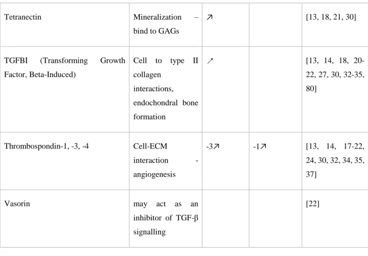
Miscellaneous Secretome Proteins
The chondrocyte secretome contains many proteins involved in cellular regulatory pathways, cell-cell or cell-ECM interactions, including chaperons, alarmins, apolipoproteins and chondrocalcin.
Chondrocalcin is the C-terminal of type II collagen, associated with the calcification of cartilage ECM.
Gelsolin, clusterin and transforming growth factor-beta-induced protein ig-h3 (TGF appeared in nearly all secretome studies and are one of the most highly secreted proteins by chondrocytes [14-19, 22, 24, 26, 30, 34, 35, 37]. Gelsolin is increased in OA compared to normal chondrocytes[35, 37]. Clusterin inhibits protein aggregation and apoptosis. This protein is detected more in healthy than osteoarthritic cartilage[37]. TGFBI is known to be involved in the interaction of the cell with type II collagen, and plays an important role in endochondral bone formation. Indeed, this factor is an
12 inhibitor of mineralization. It was found to be highly secreted by OA chondrocytes, compared to normal chondrocytes[13], and present at a greater abundance in hip OA than healthy cartilage[33].
Osteonectin is involved in ECM-cytokine interactions. Similar to osteopontin, osteonectin is highly secreted in deep layer chondrocytes[34], and found to be increased in the secretome of medial condylar early OA cartilage[16, 25, 27]. TGF-β1 decreases the production of osteonectin[24].
Finally, thrombospondin-1,-3,-4 are all involved in cell-ECM interaction and angiogenesis, and are also secreted by chondrocytes [13, 14, 17, 19, 21, 24, 32, 34, 35, 37]. They are more abundant in the superficial layer of cartilage[34]. Thrombospondin-3 is increased in OA compared to normal
chondrocyte secretomes[13], and thrombospondin-1 is increased with IL-1β treatment[19].
DISCUSSION
The “omics” approach is a general exploratory approach used to investigate alterations in an
enormous number of genes, transcripts, proteins, lipids and metabolites, in healthy versus
diseased tissues. The challenge of using this approach is to identify those candidates that are
specifically involved in the disease process. The most commonly used omics approaches
include genomics, proteomics, lipidomics, metabolomics and transcriptomics. Such “omics”
technologies applied to serum or urine samples have uncovered numerous new biomarkers,
which are ubiquitous molecules in most cases. Therefore, it would be beneficial to refine
“omics” technologies specifically to joint tissues (cartilage, bone, meniscus, synovial
membrane) and compare “omics” profiles of the various articular tissues taken at different
stages of evolution and correlate those amongst the various diseased tissues for the joint. In
particular, the secretome of chondrocytes is of great interest because this approach provides a
range of biomarkers reflecting the metabolic changes occurring in the main tissue affected by
OA. Further, by investigating the chondrocyte secretome we increase the chance of
13
this paper, we have reviewed and summarized data produced by proteomic analysis of
cartilage or chondrocyte culture supernatants. Interestingly, some proteins have been found to
increase in the secretome of OA explants or OA chondrocytes, while few were found
decreased in comparison with normal secretome. Among these, some have been found in
synovial fluid, serum or urine and proposed as potential biomarkers. For example, type II
collagen[60-64], aggrecan[63, 65, 66], lumican[67, 68], COMP[64, 66], gelsolin[67, 69],
fibulin-3[70], mimecan[69], periostin[13, 71, 72], SERPINs[67, 70, 73] are found in these
fluids in their native form, but also post-transcriptionally modified and/or fragmented. Some
of these proteins or protein fragments have demonstrated to be burden of disease, prognostic,
and efficacy of intervention or diagnostic soluble biomarkers, according the Burden of
Disease, Investigative, Prognostic, Efficacy of Intervention and Diagnostic (BIPEDS) criteria.
Some peptides generated during type II collagen degradation have been particularly well
investigated, because type II collagen is the most specific protein of cartilage. Further, their
concentration appears to be modified in joint disease. As previously mentioned, many
aggrecan neoepitopes are found in cartilage tissue, but those generated by aggrecanases are
largely released from cartilage and found in blood circulation, while the majority of
fragments generated by MMPs remain entrapped in the tissue[54].
Although the proteomic approach is a good approach to study secretome, there are some
limitations associated with this method. Quantitative assessment of proteins secretome using
these proteomic techniques remain hazardous, because the method promotes the
quantification of some proteins relative to their solubility, binding to other components, their
size, length, amino acid sequence and also their post-translational modifications. Small
14
are needed to assure the identification of the protein, they are probably excluded from the
final observation.
In addition, there are some missing links in the secretome study. For example, there is to date
few studies really dedicated and designed to investigate the dynamic kinetics of cartilage
metabolism during OA disease. Most of the studies compare healthy and end-stage OA
chondrocyte secretome, with the aim to discover new proteins secreted by the chondrocytes.
For example, CRTAC1, HTRA1, PCOLCE, LTBP1, ECM-1, gremlin-1, clusterin, have been
identified using this approach and now need more investigations to decipher their role in
chondrocyte metabolism during OA pathogenesis.
The abundance of diverse models used in these proteomics studies also make them difficult to
interpret. In monolayer, chondrocytes certainly have a different secretome than in their native
3D environment[31]. In explant culture, the majority of the newly synthesized proteins
remains entrapped in the ECM and only degraded products are released into the
supernatant[18]. Mechanical stimulation of the explant should be performed to mimic the
physiological flow and help the protein to be released from tissue.
Posttranslational modifications like glycosylation, glycation, nitration are seldom
investigated. A metabolomics approach would allow to investigate these changes and to
complete knowledge coming from proteomic.
Many supposed cytoplasmic proteins are found in the chondrocyte secretome using proteomic
technics. This finding seems surprising but can be explained by the secretion of ECM-derived
vesicles by chondrocytes. Articular cartilage vesicles (ACVs) are 50–150 nm
membrane-bound extracellular organelles found in normal articular cartilage. They were initially
15
which mirrored those of matrix vesicles derived from growth plate cartilage and other
normally mineralizing tissues. ACVs contain enzymes, ions and substrates necessary for
mineral formation[74]. The presence of these ACVs explain why cytoplasmic/membrane
proteins, such as annexins, have been identified in the secretome of the chondrocytes.
Transmembrane proteins can also be cleaved leading to the release in the extracellular space
of extracellular part of the protein, which is the case of syndecans [75]. Another explanation
at the presence of membrane proteins in the secretome is the presence of apoptotic
cells, mainly during OA[76-78].Loss of cell integrity during apoptosis further contribute to emerging
proteins and pathway end-products in synovium and other bodily fluids, potentially giving
rise to several biomarkers which may predict the susceptibility of an individual to develop
OA.
Pro-inflammatory cytokines, prostaglandins and reactive oxygen species (ROS) activate the normally quiescent articular chondrocytes and induce them to undergo a phenotypic shift through a
phenomenon recently described as “chondrosenescence”, leading to further disruption of homeostasis and metabolism in cartilage [7]. Effectively, chondrosenescence is the term that describes the age-dependent deterioration of chondrocyte function and how it undermines cartilage function in OA. Until now, this particular phenotype has not yet been investigated by proteomic technics. This should be added in the research agenda.
In conclusion, proteomic analysis of chondrocytes secretome is a promising approach for
detecting changes in chondrocyte metabolism linked to OA diseases (Table 1). This review
listed the advantages and disavantages of secretome investigation using the proteomic
methods. The main limitations is the lack of standardization of the culture protocols, while
16
secretome. We have also suggested some research perspectives, including comparison of
secretome at different disease stages. Definitively, research on secretome using proteomic
methods have to be encouraged with the objectives to identify new biomarkers reflecting
chondrocyte metabolic changes in OA.
ACKNOWLEGMENTS
The authors would like to thank the European Union’s Seventh Framework Programme for
research, technological development and demonstration for funding (grant agreement No.
305815).
AUTHOR CONTRIBUTION
All author contributed to the collection, assembly, analysis and interpretation of data and critical revision of the article for important intellectual content.
All authors approved the final version of the manuscript.
ROLE OF THE FUNDING SOURCE
The authors’ work is supported by the European Commission. A. Mobasheri is the
co-ordinator of the D-BOARD Consortium funded by European Commission Framework 7
programme (EU FP7; HEALTH.2012.2.4.5-2, project number 305815, Novel Diagnostics and
Biomarkers for Early Identification of Chronic Inflammatory Joint Diseases). AM and YH are
17
(APPROACH) Consortium
1, a 5-year project funded by the European Commission's
Innovative Medicines Initiative (IMI). APPROACH is a public-private partnership directed
towards osteoarthritis biomarker development through the establishment of a heavily
phenotyped and comprehensively analyzed longitudinal cohort. The research leading to these
results has received partial support from the Innovative Medicines Initiative (IMI) Joint
Undertaking under grant agreement no. 115770, resources of which are composed of financial
contribution from the European Union’s Seventh Framework programme (FP7/2007-2013)
and EFPIA companies’ in kind contribution. A.M. has also received funding from Arthritis
Research UK (grant number 21076) The funding sources had no role in the writing of the
manuscript or in the decision to submit the manuscript for publication.
COMPETING INTEREST STATEMENT
ACBJ is a full-time employee and shareholder in Nordic Bioscience. YH is the founder and chairman of Artialis SA.
18
19
FIGURE LEGEND
Figure 1
Schematic representation of the chondrocyte secretome, based on data from
mass-spectrometry based proteomic studies.
ADAMTS: A Disintegrin And Metalloproteinase with Thrombospondin Motifs, Apo:
apolipoprotein, BMP: Bone morphogenetic protein, CCL: Chemokine ligand, CHAD:
Chondroadherin, ChM: chondromodulin, CILP:
Cartilage intermediate layer protein, CHI3L1:
Chitinase-3-like protein 1, COMP : Cartilage oligomeric matrix protein, CRLF: Cytokine
receptor-like factor, CRTAC: Cartilage acidic protein, CSF: Macrophage colony-stimulating
factor, CTGF: Connective tissue growth factor, Cx: connexin , CXCL: C-X-C motif
chemokine ligand, ECM: extracellular matrix protein, ENPP: Ectonucleotide
pyrophosphatase/phosphodiesterase family member, EMI: emilin, HSPA5: 78 kDa
glucose-regulated protein, HTRA: High-temperature requirement A serine peptidase,
IL: Interleukin,
LTBP: Latent-transforming growth factor beta-binding protein, MGP: matrix gla protein,
MMP: matrix metalloproteinase, PCOLCE: Procollagen C-endopeptidase enhancer, PLOD:
Procollagen-lysine,2-oxoglutarate 5-dioxygenase, PRELP: proline-arginine-rich end
leucine-rich repeat protein, SERPIN: serine protease inhibitors, SOD: superoxide dismutase, SPARC:
Secreted protein acidic and rich in cysteine, TGFBI: Transforming growth factor-beta-induced
protein ig-h3, TGF: transforming growth factor, TIMP: tissue inhibitor of metalloproteinase,
TNAP: tissue non-specific alkaline phosphatase, TSP: thrombospondin.
20
TABLES
Table 1
Table 1: Summary of recent mass spectrometry-based proteomic studies carried out on human
chondrocytes and cartilage to identify secretome components.
ECM Proteins
Specificity
OA
vs
normal
with IL-1β
References
Aggrecan core protein
↘
[13, 14, 17, 18,
20, 22, 27,
31-37]
Asporin
[32, 34, 37]
Basement
membrane-specific
heparin sulfate proteoglycan core
protein (PGBM)
↗
[13, 18, 20, 30]
Biglycan
↗
[13, 14, 17, 18,
20-22, 27, 30,
32-37]
Cartilage
acidic
protein-1
(CRTAC1, ASPIC)
Specific
chondrocyte
marker
↗
[13, 16, 22, 26,
33, 35]
21
Chitinase-3-like protein -1, -2
Tissue
remodelling,
inflammation
↗
↘
[13, 16, 18,
20-22, 24, 25, 27,
30, 31, 33, 35,
37]
Chondroitin sulfate proteoglycan
4 (CSPG4)
[18]
Collagens type I, II, III, V, VI,
VIII, IX, X, XI, XII, XIV, XV,
XVI
↗
[13, 14, 16, 18,
20, 21, 24-27,
30-35, 37]
Cartilage
oligomeric
matrix
protein (COMP)
↗
[13, 14, 17, 18,
20, 22, 26, 27,
30-35, 37]
Decorin
[12-14, 18-21,
27, 30, 32, 34,
35, 37]
Extracellular matrix protein-1
negative regulator
of
bone
mineralization,
promote
angiogenesis,
inhibit
MMP-9
activity
↗
[13, 14, 19, 22,
25, 33]
Fibrillin-1
[18, 22, 25, 27,
30, 35, 37]
22
Fibromodulin
↗
[14, 16-20, 22,
24, 26, 27,
32-34, 36]
Fibronectin
[14, 16-18,
20-22, 26, 27, 30,
32, 34, 35, 37]
Fibulin-1, -3, -4, -7
↗
[13, 18, 20-22,
24, 25, 27, 30,
33, 34]
HPLN-1
(proteoglycan
link
protrein-1)
Bind hyaluronic
acid and aggrecan
↗
↗
[13, 14, 18, 19,
22, 24, 27,
32-35, 37]
Latent-transforming growth factor
beta-binding
protein
-1,
-2
(LTBP1, 2)
Storage/activation
TGF-β1,
structural role in
ECM
[13, 20, 26, 27,
30]
Lubricin (proteoglycan-4)
↗
[13, 14, 18,
20-22, 26, 27, 30,
32-35, 37]
Lumican
[13, 14, 16,
18-22, 24, 25, 27,
30, 32-35, 37]
Matrilin-2*, -3*
[25, 33-35, 37]
23
Matrix gla protein (MGP)
↗
↗
[14, 19, 20, 22,
24, 26, 33-35,
37]
Mimecan (osteoglycin)
↗
[13, 18, 22, 27,
30, 32-34, 37]
Osteomodulin
[18, 22, 30-32,
34, 37]
Periostin
↗
[13, 22, 35]
Perlecan
[34, 37]
Prolargin (PRELP)
Bind
type
II
collagen
and
perlecan
[13, 17, 18, 20,
22, 25, 27,
32-37]
Syndecan-2, -4
↗
[19, 21, 35]
Tenascin C, X
↗
[13, 18, 20-22,
30-32, 34, 35,
37]
Versican
↗
[13, 32, 34, 37]
Cytokines and Growth Factors
Specificity
References
CCL2, -8, -14, -20
↗
[14, 21, 22, 25,
24
CSF-1
↗
[21, 27]
CTGF
[16, 18, 20, 22,
24, 30, 31, 33]
CXCL1, -3, -5, -6
↗
[27, 33]
Cytokine receptor like factor 1
decrease aggrecan
and
type
II
collagen synthesis
[20, 26]
Gremlin-1
BMP antagonist,
inhibit
bone
mineralization
↘
[14, 35]
IGFBP-2, -3, -4, -5, -6, -7
IGFBP3↗
[12, 18, 20-22,
24-27, 30]
IL-6, -8, -17β
IL-17β↘
IL-6, -8↗
[14, 19-21, 25,
27, 33]
Inhibin-βA/proinhibin-βA
dimerise to form
Activin
A,
stimulating
TIMP-1
production
↗
↗
[12, 16, 20, 27]
Leukocyte
cell-derived
chemotaxin 2 (LECT2)
reduce IL-1β, IL-6
and other
chemokines reduce IL-1β, IL-6 and
25 other chemokines
[79]
Pleiotrophin (PTN)
↗
[14, 33]
TGF-β2
[22]
Enzymes
Specificity
References
ADAMTS-1, -2, -4 ,-5 [12, 22, 25] [20]
Angiogenin Deep layer –
stimulate angiogenesis ↗ [12, 18, 32-34, 37] Carboxypeptidase E [14] Cathepsins (B, D, F, K, L1, Z) B↗ [12, 14, 16, 18, 20-22, 25, 30, 35] ENPP2 NTP pyrophosphatase, mineralization [22]
Extracellular sulfatase Sulf1 Sulf2 [14]
HTRA1 (serine protease) Increase with TGF-β1 ↘ [12, 14, 16, 18, 22, 24, 26, 30, 32, 34, 35, 37] [20] Lysozyme C ↗ [12, 14-16, 20, 32, 34]
26 MMPs (1, 2, 3, 7, 10, 13, 14, -16) ↗ ↗ [12-16, 18, 20-25, 27, 30, 31, 34, 35, 37] Pappalysin-1 Metalloproteinase, cleave IGFBP-4,-5 [22]
Peroxiredoxin-1, -2, -4 protecting cells from free-radical damage 2↗ [13, 16, 20, 23, 37] Phospholipase A2* ↗ [12, 13, 32, 34, 35, 37] Procollagen C-endopeptidase enhancer (PCOLCE)-1, -2 enhances procollagen C-proteinase activity. C-terminal processed part of PCPE (CT-PCPE) may have an metalloproteinase inhibitory activity. Decrease with TGF-β1 ↗ [12, 14-16, 18, 22, 24, 26, 30, 33, 34, 37] [20] Procollagen-lysine,2-oxoglutarate 5-dioxygenase 1, 2 Collagen fibre glycosylation ↗ [22, 31, 58]
Putative tryspin 6 (serine protease) [21]
27
Serine protease 23 [22]
Sulfhydryl oxidase [12, 22]
Superoxide dismutase (SOD1, SOD2, SOD3) protecting cells from free-radical damage SOD2↘ [12, 14-16, 22, 23, 25, 31, 32, 34, 35]
Triosephosphate isomerase : Carbohydrate degradation
↗ [12, 14, 16, 20, 22]
Enzymatic inhibitors
Specificity
References
Antileukoproteinase Serine proteinase inhibitor
[12, 25]
Cystatin C Cysteine proteinase
inhibitor
[18, 21, 22, 26]
Inter α Globulin inhibitor H2 HA processing [12]
Inter α trypsin inhibitor HA processing,
Heavy chain H1 ↗ [13]
Heavy chain H4 [18]
SERPIN Serine protease
inhibitors
A1 : α1-antitrypsin [16, 21, 22, 25,
28
A2: anti-trypsin related protein [22]
A3 : α1-antichymotrypsin ↘ [12, 16, 18, 22,
25, 30, 32-35]
A5 : Protein C inhibitor [12, 22]
E1: Plasminogen activator inhibitor 1
[18, 22, 30, 35]
E2 : Glia-derived nexin [14, 20-22, 32,
35]
F1: Pigment epithelium derived factor
↗ [12, 13, 16, 18,
30]
G1: Plasma protease C1 inhibitor [18, 22, 30]
I2 α1 antiproteinase 2 [14, 15]
TIMPs (-1, -2, -3, -4) TIMP-1 increase with TGF-β1
TIMP-1↘ [12, 14-16, 18, 20-22, 24-26, 30, 34, 35, 37]
Tissue factor pathway inhibitor 1, 2 (TFPI)
Serine protease inhibitors
[26]
Miscellaneous
Specificity
References
78kDa glucose regulated protein [22, 33-35]
A1 acid glycoprotein 1, 2 Binding and modulation of cytokines and
29 growth factor, like
IL-6 and TNF-α
ADAMTS-like 2 Bind to LTBP – no
enzymatic function
[25]
Annexin A1, A2, A5, A1, A2 ↗ [14, 18, 20, 22,
30, 35]
Apolipoprotein AI, AII , D, E Bind lipids – proteoglycans and collagens interaction AI, AII↗ D↘ [13, 18, 19, 30, 34, 35] CILP (1-1, 1-2, 2-1, 2-2) No intrinsic NTP pyrophopshatase activity but IGF-1/TGF-β antagonists, increase with TGF-β1 ↗ ↗ [12, 14, 16, 18, 20, 24, 25, 32, 34, 36, 37]
Chondroadherin Bind the cell to the
type II collagen
[13, 14, 16-18, 20, 25, 32-35, 37]
Clusterin Chaperon – inhibit
protein aggregation and apoptosis
↘ [14-20, 22, 24,
26, 30, 34, 35, 37]
Complement C1q, C1r, C1s, C3, C8, C9, factor B, factor D, factor H
B, C1r, C3 ↗
[12-14, 16, 18, 20-22, 30, 32, 33, 37]
30
Emilin-1 Cell-ECM
interaction
[25, 35, 37]
Ezrin-Radexin-Moesin-Transgelin Cytoskeletal related [13, 20, 22, 27, 32, 35, 80]
Gelsolin Cytoskeletal related ↗ [16, 18, 20, 22,
26, 30-32, 34, 35, 37, 80]
Lactadherin [15, 18, 20-22,
30]
Osteonectin (SPARC) ECM-cytokine
interactions
[14-16, 18, 22, 24, 25, 30, 33-35]
Osteopontin Bind to mineral and
inhibit
mineralization
↗ [13, 20, 34, 37]
Profilin-1 Cytoskeletal related [18, 20, 27, 30,
80]
S100A1
Calcium binding
proteins, role in
inflammation
A1↘ A8, A10↗ [13, 19, 21, 22, 25, 27, 32-35] Semaphorin 3A, 3C [22]Spondin -1 and -2 Cell adhesion-bind GAGs-Wnt agonist
[12]
31
Tetranectin Mineralization –
bind to GAGs
↗ [13, 18, 21, 30]
TGFBI (Transforming Growth Factor, Beta-Induced) Cell to type II collagen interactions, endochondral bone formation ↗ [13, 14, 18, 20-22, 27, 30, 32-35, 80] Thrombospondin-1, -3, -4 Cell-ECM interaction - angiogenesis -3↗ -1↗ [13, 14, 17-22, 24, 30, 32, 34, 35, 37]
Vasorin may act as an
inhibitor of TGF-β signalling
[22]
Table 2: Characteristics of the reviewed studies.
Study
Method
Secretome
Chondrocyte
culture
Explant
culture
Peffers et al 2013 [12] OA +/- IL-1β
X
X
Lourido et al 2014
[13]
Normal
vs
wound
or
unwound OA
X
X
32
Williams et al 2013
[15]
Equin +/- IL-1β
X
X
Hermansson et al 2004
[16]
OA only
X
X
Clutterbuck et al 2011
[17]
Equin +/- IL-1β
X
X
Peffers et al 2016 [20] Equin +/- IL-1β
X
X
Polacek et al 2010 [18] OA monolayer vs explant
culture
X
X
X
Swan et al 2013 [19]
Canine +/-IL-1β
X
X
Calamia et al 2012
[21]
Normal + IL-1β
X
X
Calamia et al 2014
[22]
OA +/- chondroitine or
glucosamine sulfate
X
X
Catterall et al 2006
[23]
OA +/- IL-1β
X
X
Riffault et al 2015 [24] OA +/- TGF-β1
X
X
Stenberg et al 2013
[25]
Early OA vs late OA
X
X
33
Rocha et al 2014 [26]
bMSCs
chondrogenic
differentiation
X
X
Lourido et al 2015
[27]
OA or normal+IL-1β
X
X
Taylor et al 2015 [28]
Bovine
P0
vs
P2
chondrocytes
X
X
Haglund et al 2008
[29]
Rat, +/- LPS
X
X
Polacek et al 2011a
[30]
Chondrocytes vs MSCs
X
X
Polacek et al 2011b
[31]
Monolayer
vs
aggregate
culture
X
X
Onnerfjord et al 2012
[32]
Type of cartilage
X
Ikeda et al 2013 [33]
Normal vs OA hip
X
Muller et al 2014 [34] Normal at superficial vs
intermediate vs deep layer
X
34
Cillero-Pastor et al
2013 [36]
Normal
vs
OA
from
superficial to deep layer with
MALDI-IMS
X
Gago-Fuentes et al
2015 [80]
Cx43
complexes
from
normal vs OA chondrocytes
X
Hsueh et al 2015 [37]
Decellularized
cartilage
from normal vs OA, knee vs
hip
and
superficial
vs
intermediate vs deep layer
X
References:
1. Carney SL, Muir H. The structure and function of cartilage proteoglycans. Physiol Rev 1988; 68: 858-910.
2. Eyre D. Collagen of articular cartilage. Arthritis Res 2002; 4: 30-35.
3. Bay-Jensen AC, Hoegh-Madsen S, Dam E, Henriksen K, Sondergaard BC, Pastoureau P, et al. Which elements are involved in reversible and irreversible cartilage degradation in osteoarthritis? Rheumatol Int 2010; 30: 435-442.
4. Archer CW, Francis-West P. The chondrocyte. Int J Biochem Cell Biol 2003; 35: 401-404.
5. Aigner T, Saas J, Zien A, Zimmer R, Gebhard PM, Knorr T. Analysis of differential gene expression in healthy and osteoarthritic cartilage and isolated chondrocytes by microarray analysis. Methods Mol Med 2004; 100: 109-128.
35 6. Chen-An P, Andreassen KV, Henriksen K, Karsdal MA, Bay-Jensen AC. Investigation of chondrocyte hypertrophy and cartilage calcification in a full-depth articular cartilage explants model. Rheumatol Int 2013; 33: 401-411.
7. Mobasheri A, Matta C, Zakany R, Musumeci G. Chondrosenescence: definition, hallmarks and potential role in the pathogenesis of osteoarthritis. Maturitas 2015; 80: 237-244.
8. Agrawal GK, Jwa NS, Lebrun MH, Job D, Rakwal R. Plant secretome: unlocking secrets of the secreted proteins. Proteomics 2010; 10: 799-827.
9. Gharbi M, Deberg M, Henrotin Y. Application for proteomic techniques in studying osteoarthritis: a review. Front Physiol 2011; 2: 90.
10. Aslam B, Basit M, Nisar MA, Khurshid M, Rasool MH. Proteomics: Technologies and Their Applications. J Chromatogr Sci 2017; 55: 182-196.
11. Crutchfield CA, Thomas SN, Sokoll LJ, Chan DW. Advances in mass spectrometry-based clinical biomarker discovery. Clin Proteomics 2016; 13: 1.
12. Peffers MJ, Beynon RJ, Clegg PD. Absolute quantification of selected proteins in the human osteoarthritic secretome. Int J Mol Sci 2013; 14: 20658-20681.
13. Lourido L, Calamia V, Mateos J, Fernandez-Puente P, Fernandez-Tajes J, Blanco FJ, et al. Quantitative proteomic profiling of human articular cartilage degradation in osteoarthritis. J Proteome Res 2014; 13: 6096-6106.
14. Svala E, Lofgren M, Sihlbom C, Ruetschi U, Lindahl A, Ekman S, et al. An inflammatory equine model demonstrates dynamic changes of immune response and cartilage matrix molecule degradation in vitro. Connect Tissue Res 2015; 56: 315-325.
36 15. Williams A, Smith JR, Allaway D, Harris P, Liddell S, Mobasheri A. Carprofen inhibits the release of matrix metalloproteinases 1, 3, and 13 in the secretome of an explant model of articular cartilage stimulated with interleukin 1beta. Arthritis Res Ther 2013; 15: R223.
16. Hermansson M, Sawaji Y, Bolton M, Alexander S, Wallace A, Begum S, et al. Proteomic analysis of articular cartilage shows increased type II collagen synthesis in osteoarthritis and expression of inhibin betaA (activin A), a regulatory molecule for chondrocytes. J Biol Chem 2004; 279: 43514-43521.
17. Clutterbuck AL, Smith JR, Allaway D, Harris P, Liddell S, Mobasheri A. High throughput proteomic analysis of the secretome in an explant model of articular cartilage inflammation. J Proteomics 2011; 74: 704-715.
18. Polacek M, Bruun JA, Johansen O, Martinez I. Differences in the secretome of cartilage explants and cultured chondrocytes unveiled by SILAC technology. J Orthop Res 2010; 28: 1040-1049.
19. Swan AL, Hillier KL, Smith JR, Allaway D, Liddell S, Bacardit J, et al. Analysis of mass spectrometry data from the secretome of an explant model of articular cartilage exposed to pro-inflammatory and anti-inflammatory stimuli using machine learning. BMC Musculoskelet Disord 2013; 14: 349.
20. Peffers MJ, Thornton DJ, Clegg PD. Characterization of neopeptides in equine articular cartilage degradation. J Orthop Res 2016; 34: 106-120.
21. Calamia V, Lourido L, Fernandez-Puente P, Mateos J, Rocha B, Montell E, et al. Secretome analysis of chondroitin sulfate-treated chondrocytes reveals angiogenic, anti-inflammatory and anti-catabolic properties. Arthritis Res Ther 2012; 14: R202.
37 22. Calamia V, Mateos J, Fernandez-Puente P, Lourido L, Rocha B, Fernandez-Costa C, et al. A pharmacoproteomic study confirms the synergistic effect of chondroitin sulfate and glucosamine. Sci Rep 2014; 4: 5069.
23. Catterall JB, Rowan AD, Sarsfield S, Saklatvala J, Wait R, Cawston TE. Development of a novel 2D proteomics approach for the identification of proteins secreted by primary chondrocytes after stimulation by IL-1 and oncostatin M. Rheumatology (Oxford) 2006; 45: 1101-1109.
24. Riffault M, Moulin D, Grossin L, Mainard D, Magdalou J, Vincourt JB. Label-free relative quantification applied to LC-MALDI acquisition for rapid analysis of chondrocyte secretion modulation. J Proteomics 2015; 114: 263-273.
25. Stenberg J, Ruetschi U, Skioldebrand E, Karrholm J, Lindahl A. Quantitative proteomics reveals regulatory differences in the chondrocyte secretome from human medial and lateral femoral condyles in osteoarthritic patients. Proteome Sci 2013; 11: 43.
26. Rocha B, Calamia V, Casas V, Carrascal M, Blanco FJ, Ruiz-Romero C. Secretome analysis of human mesenchymal stem cells undergoing chondrogenic differentiation. J Proteome Res 2014; 13: 1045-1054.
27. Lourido L, Calamia V, Fernandez-Puente P, Mateos J, Oreiro N, Blanco FJ, et al. Secretome analysis of human articular chondrocytes unravels catabolic effects of nicotine on the joint. Proteomics Clin Appl 2015.
28. Taylor DW, Ahmed N, Parreno J, Lunstrum GP, Gross AE, Diamandis EP, et al. Collagen type XII and versican are present in the early stages of cartilage tissue formation by both redifferentating passaged and primary chondrocytes. Tissue Eng Part A 2015; 21: 683-693.
38 29. Haglund L, Bernier SM, Onnerfjord P, Recklies AD. Proteomic analysis of the LPS-induced stress response in rat chondrocytes reveals induction of innate immune response components in articular cartilage. Matrix Biol 2008; 27: 107-118.
30. Polacek M, Bruun JA, Elvenes J, Figenschau Y, Martinez I. The secretory profiles of cultured human articular chondrocytes and mesenchymal stem cells: implications for autologous cell transplantation strategies. Cell Transplant 2011; 20: 1381-1393.
31. Polacek M, Bruun JA, Johansen O, Martinez I. Comparative Analyses of the Secretome from Dedifferentiated and Redifferentiated Adult Articular Chondrocytes. Cartilage 2011; 2: 186-196.
32. Onnerfjord P, Khabut A, Reinholt FP, Svensson O, Heinegard D. Quantitative proteomic analysis of eight cartilaginous tissues reveals characteristic differences as well as similarities between subgroups. J Biol Chem 2012; 287: 18913-18924.
33. Ikeda D, Ageta H, Tsuchida K, Yamada H. iTRAQ-based proteomics reveals novel biomarkers of osteoarthritis. Biomarkers 2013; 18: 565-572.
34. Muller C, Khabut A, Dudhia J, Reinholt FP, Aspberg A, Heinegard D, et al. Quantitative proteomics at different depths in human articular cartilage reveals unique patterns of protein distribution. Matrix Biol 2014; 40: 34-45.
35. Tsolis KC, Bei ES, Papathanasiou I, Kostopoulou F, Gkretsi V, Kalantzaki K, et al. Comparative proteomic analysis of hypertrophic chondrocytes in osteoarthritis. Clin Proteomics 2015; 12: 12.
39 36. Cillero-Pastor B, Eijkel GB, Kiss A, Blanco FJ, Heeren RM. Matrix-assisted laser desorption ionization-imaging mass spectrometry: a new methodology to study human osteoarthritic cartilage. Arthritis Rheum 2013; 65: 710-720.
37. Hsueh MF, Khabut A, Kjellstrom S, Onnerfjord P, Kraus VB. Elucidating the Molecular Composition of Cartilage by Proteomics. J Proteome Res 2015.
38. Font B, Eichenberger D, Rosenberg LM, van der Rest M. Characterization of the interactions of type XII collagen with two small proteoglycans from fetal bovine tendon, decorin and fibromodulin. Matrix Biol 1996; 15: 341-348.
39. Johansen JS, Hvolris J, Hansen M, Backer V, Lorenzen I, Price PA. Serum YKL-40 levels in healthy children and adults. Comparison with serum and synovial fluid levels of YKL-40 in patients with osteoarthritis or trauma of the knee joint. Br J Rheumatol 1996; 35: 553-559.
40. Vaananen T, Koskinen A, Paukkeri EL, Hamalainen M, Moilanen T, Moilanen E, et al. YKL-40 as a novel factor associated with inflammation and catabolic mechanisms in osteoarthritic joints. Mediators Inflamm 2014; 2014: 215140.
41. Zivanovic S, Rackov LP, Vojvodic D, Vucetic D. Human cartilage glycoprotein 39--biomarker of joint damage in knee osteoarthritis. Int Orthop 2009; 33: 1165-1170.
42. Steck E, Braun J, Pelttari K, Kadel S, Kalbacher H, Richter W. Chondrocyte secreted CRTAC1: a glycosylated extracellular matrix molecule of human articular cartilage. Matrix Biol 2007; 26: 30-41.
43. Deckers MM, Smits P, Karperien M, Ni J, Tylzanowski P, Feng P, et al. Recombinant human extracellular matrix protein 1 inhibits alkaline phosphatase activity and mineralization of mouse embryonic metatarsals in vitro. Bone 2001; 28: 14-20.
40 44. Han Z, Ni J, Smits P, Underhill CB, Xie B, Chen Y, et al. Extracellular matrix protein 1 (ECM1) has
angiogenic properties and is expressed by breast tumor cells. FASEB J 2001; 15: 988-994.
45. Fujimoto N, Terlizzi J, Aho S, Brittingham R, Fertala A, Oyama N, et al. Extracellular matrix protein 1 inhibits the activity of matrix metalloproteinase 9 through high-affinity protein/protein interactions. Exp Dermatol 2006; 15: 300-307.
46. Attur M, Yang Q, Shimada K, Tachida Y, Nagase H, Mignatti P, et al. Elevated expression of periostin in human osteoarthritic cartilage and its potential role in matrix degradation via matrix metalloproteinase-13. FASEB J 2015; 29: 4107-4121.
47. Chijimatsu R, Kunugiza Y, Taniyama Y, Nakamura N, Tomita T, Yoshikawa H. Expression and pathological effects of periostin in human osteoarthritis cartilage. BMC Musculoskelet Disord 2015; 16: 215.
48. Peffers MJ, Cillero-Pastor B, Eijkel GB, Clegg PD, Heeren RM. Matrix assisted laser desorption ionization mass spectrometry imaging identifies markers of ageing and osteoarthritic cartilage. Arthritis Res Ther 2014; 16: R110.
49. Wei Z, Li HH. IGFBP-3 may trigger osteoarthritis by inducing apoptosis of chondrocytes through Nur77 translocation. Int J Clin Exp Pathol 2015; 8: 15599-15610.
50. Yates MP, Settle SL, Yocum SA, Aggarwal P, Vickery LE, Aguiar DJ, et al. IGFBP-5 Metabolism Is Disrupted in the Rat Medial Meniscal Tear Model of Osteoarthritis. Cartilage 2010; 1: 43-54.
51. Gazzerro E, Pereira RC, Jorgetti V, Olson S, Economides AN, Canalis E. Skeletal overexpression of gremlin impairs bone formation and causes osteopenia. Endocrinology 2005; 146: 655-665.
41 52. Pufe T, Groth G, Goldring MB, Tillmann B, Mentlein R. Effects of pleiotrophin, a heparin-binding growth factor, on human primary and immortalized chondrocytes. Osteoarthritis Cartilage 2007; 15: 155-162.
53. Bouderlique T, Henault E, Lebouvier A, Frescaline G, Bierling P, Rouard H, et al. Pleiotrophin commits human bone marrow mesenchymal stromal cells towards hypertrophy during chondrogenesis. PLoS One 2014; 9: e88287.
54. Struglics A, Larsson S, Pratta MA, Kumar S, Lark MW, Lohmander LS. Human osteoarthritis synovial fluid and joint cartilage contain both aggrecanase- and matrix metalloproteinase-generated aggrecan fragments. Osteoarthritis Cartilage 2006; 14: 101-113.
55. Zhen EY, Brittain IJ, Laska DA, Mitchell PG, Sumer EU, Karsdal MA, et al. Characterization of metalloprotease cleavage products of human articular cartilage. Arthritis Rheum 2008; 58: 2420-2431.
56. Peffers MJ, McDermott B, Clegg PD, Riggs CM. Comprehensive protein profiling of synovial fluid in osteoarthritis following protein equalization. Osteoarthritis Cartilage 2015; 23: 1204-1213.
57. Chamberland A, Wang E, Jones AR, Collins-Racie LA, LaVallie ER, Huang Y, et al. Identification of a novel HtrA1-susceptible cleavage site in human aggrecan: evidence for the involvement of HtrA1 in aggrecan proteolysis in vivo. J Biol Chem 2009; 284: 27352-27359.
58. Endo W, Arito M, Sato T, Kurokawa MS, Omoteyama K, Iizuka N, et al. Effects of sulfasalazine and tofacitinib on the protein profile of articular chondrocytes. Mod Rheumatol 2014; 24: 844-850.
42 59. Ben-Aderet L, Merquiol E, Fahham D, Kumar A, Reich E, Ben-Nun Y, et al. Detecting cathepsin
activity in human osteoarthritis via activity-based probes. Arthritis Res Ther 2015; 17: 69.
60. Kamphorst JJ, van der Heijden R, DeGroot J, Lafeber FP, Reijmers TH, van El B, et al. Profiling of endogenous peptides in human synovial fluid by NanoLC-MS: method validation and peptide identification. J Proteome Res 2007; 6: 4388-4396.
61. Li WW, Nemirovskiy O, Fountain S, Rodney Mathews W, Szekely-Klepser G. Clinical validation of an immunoaffinity LC-MS/MS assay for the quantification of a collagen type II neoepitope peptide: A biomarker of matrix metalloproteinase activity and osteoarthritis in human urine. Anal Biochem 2007; 369: 41-53.
62. van Spil WE, Jansen NW, Bijlsma JW, Reijman M, DeGroot J, Welsing PM, et al. Clusters within a wide spectrum of biochemical markers for osteoarthritis: data from CHECK, a large cohort of individuals with very early symptomatic osteoarthritis. Osteoarthritis Cartilage 2012; 20: 745-754.
63. Duan Y, Hao D, Li M, Wu Z, Li D, Yang X, et al. Increased synovial fluid visfatin is positively linked to cartilage degradation biomarkers in osteoarthritis. Rheumatol Int 2012; 32: 985-990.
64. Streich NA, Zimmermann D, Schmitt H, Bode G. Biochemical markers in the diagnosis of chondral defects following anterior cruciate ligament insufficiency. Int Orthop 2011; 35: 1633-1637.
65. Gobezie R, Kho A, Krastins B, Sarracino DA, Thornhill TS, Chase M, et al. High abundance synovial fluid proteome: distinct profiles in health and osteoarthritis. Arthritis Res Ther 2007; 9: R36.
43 66. Mateos J, Lourido L, Fernandez-Puente P, Calamia V, Fernandez-Lopez C, Oreiro N, et al. Differential protein profiling of synovial fluid from rheumatoid arthritis and osteoarthritis patients using LC-MALDI TOF/TOF. J Proteomics 2012; 75: 2869-2878.
67. Ritter SY, Subbaiah R, Bebek G, Crish J, Scanzello CR, Krastins B, et al. Proteomic analysis of synovial fluid from the osteoarthritic knee: comparison with transcriptome analyses of joint tissues. Arthritis Rheum 2013; 65: 981-992.
68. Fernandez-Puente P, Mateos J, Fernandez-Costa C, Oreiro N, Fernandez-Lopez C, Ruiz-Romero C, et al. Identification of a panel of novel serum osteoarthritis biomarkers. J Proteome Res 2011; 10: 5095-5101.
69. De Ceuninck F, Marcheteau E, Berger S, Caliez A, Dumont V, Raes M, et al. Assessment of some tools for the characterization of the human osteoarthritic cartilage proteome. J Biomol Tech 2005; 16: 256-265.
70. Henrotin Y, Gharbi M, Mazzucchelli G, Dubuc JE, De Pauw E, Deberg M. Fibulin 3 peptides Fib3-1 and Fib3-2 are potential biomarkers of osteoarthritis. Arthritis Rheum 20Fib3-12; 64: 2260-2267.
71. Honsawek S, Wilairatana V, Udomsinprasert W, Sinlapavilawan P, Jirathanathornnukul N. Association of plasma and synovial fluid periostin with radiographic knee osteoarthritis: Cross-sectional study. Joint Bone Spine 2015; 82: 352-355.
72. Rousseau JC, Sornay-Rendu E, Bertholon C, Garnero P, Chapurlat R. Serum periostin is associated with prevalent knee osteoarthritis and disease incidence/progression in women: the OFELY study. Osteoarthritis Cartilage 2015; 23: 1736-1742.
44 73. Fernandez-Costa C, Calamia V, Fernandez-Puente P, Capelo-Martinez JL, Ruiz-Romero C, Blanco FJ. Sequential depletion of human serum for the search of osteoarthritis biomarkers. Proteome Sci 2012; 10: 55.
74. Anderson HC, Garimella R, Tague SE. The role of matrix vesicles in growth plate development and biomineralization. Front Biosci 2005; 10: 822-837.
75. Pap T, Bertrand J. Syndecans in cartilage breakdown and synovial inflammation. Nat Rev Rheumatol 2013; 9: 43-55.
76. Lotz M, Hashimoto S, Kuhn K. Mechanisms of chondrocyte apoptosis. Osteoarthritis Cartilage 1999; 7: 389-391.
77. Heraud F, Heraud A, Harmand MF. Apoptosis in normal and osteoarthritic human articular cartilage. Ann Rheum Dis 2000; 59: 959-965.
78. Kim HA, Blanco FJ. Cell death and apoptosis in osteoarthritic cartilage. Curr Drug Targets 2007; 8: 333-345.
79. Okumura A, Saito T, Otani I, Kojima K, Yamada Y, Ishida-Okawara A, et al. Suppressive role of leukocyte cell-derived chemotaxin 2 in mouse anti-type II collagen antibody-induced arthritis. Arthritis Rheum 2008; 58: 413-421.
80. Gago-Fuentes R, Fernandez-Puente P, Megias D, Carpintero-Fernandez P, Mateos J, Acea B, et al. Proteomic Analysis of Connexin 43 Reveals Novel Interactors Related to Osteoarthritis. Mol Cell Proteomics 2015; 14: 1831-1845.