HAL Id: hal-03204007
https://hal.sorbonne-universite.fr/hal-03204007
Submitted on 21 Apr 2021HAL is a multi-disciplinary open access archive for the deposit and dissemination of sci-entific research documents, whether they are pub-lished or not. The documents may come from teaching and research institutions in France or abroad, or from public or private research centers.
L’archive ouverte pluridisciplinaire HAL, est destinée au dépôt et à la diffusion de documents scientifiques de niveau recherche, publiés ou non, émanant des établissements d’enseignement et de recherche français ou étrangers, des laboratoires publics ou privés.
DCC regulates astroglial development essential for
telencephalic morphogenesis and corpus callosum
formation
Laura Morcom, Ilan Gobius, Ashley Pl Marsh, Rodrigo Suárez, Jonathan Wc
Lim, Caitlin Bridges, Yunan Ye, Laura Fenlon, Yvrick Zagar, Amelia
Douglass, et al.
To cite this version:
Laura Morcom, Ilan Gobius, Ashley Pl Marsh, Rodrigo Suárez, Jonathan Wc Lim, et al.. DCC regu-lates astroglial development essential for telencephalic morphogenesis and corpus callosum formation. eLife, eLife Sciences Publication, 2021, 10, �10.7554/eLife.61769�. �hal-03204007�
1 DCC regulates astroglial development essential for telencephalic 1
morphogenesis and corpus callosum formation 2
Authors: Laura Morcom1, Ilan Gobius1, Ashley P L Marsh2,3, Rodrigo Suárez1,4,
3
Jonathan WC Lim1, Caitlin Bridges1, Yunan Ye1, Laura R Fenlon1,4, Yvrick Zagar5,
4
Amelia M Douglass1, Amber-Lee S Donahoo1, Thomas Fothergill1, Samreen
5
Shaikh1, Peter Kozulin1, Timothy J Edwards1,6, Helen M Cooper1, IRC5 Consortium7,
6
Elliott H Sherr8, Alain Chédotal4, Richard J Leventer3,9,10, Paul J Lockhart2,3, & Linda
7 J Richards1,4*. 8 9 Affiliations 10 1
The University of Queensland, Queensland Brain Institute, Brisbane, QLD 4072, 11
Australia. 12
2Bruce Lefroy Centre for Genetic Health Research, Murdoch Children’s Research 13
Institute, Royal Children’s Hospital, Parkville, VIC 3052, Australia. 14
3
Department of Paediatrics, University of Melbourne, Parkville, VIC 3052, Australia. 15
4
The University of Queensland, School of Biomedical Sciences, Brisbane, QLD 16
4072, Australia. 17
5
Sorbonne Université, INSERM, CNRS, Institut de la Vision, 17 Rue Moreau, 75012 18
Paris, France. 19
6
The University of Queensland, Faculty of Medicine, Brisbane, QLD 4072, Australia. 20
7
Members and Affiliates of the International Research Consortium for the Corpus 21
Callosum and Cerebral Connectivity (IRC5) 22
8
Departments of Neurology and Pediatrics, Institute of Human Genetics and Weill 23
Institute of Neurosciences, University of California, San Francisco, San Francisco, 24
CA 94143, USA. 25
9
Neuroscience Research Group, Murdoch Children’s Research Institute, Parkville, 26
VIC 3052, Australia. 27
10
Department of Neurology, University of Melbourne, Royal Children’s Hospital, 28
Parkville, VIC 3052, Australia. 29
30
*Corresponding author:
31
Professor Linda J. Richards, PhD
32
NHMRC Principal Research Fellow
2
Head, Brain Development and Disorders Laboratory,
34
The Queensland Brain Institute and The School of Biomedical Sciences
35
The University of Queensland, St Lucia, 4072, AUSTRALIA
36 ph: +61 7 33466355 37 fax: +61 7 33466301 38 email: richards@uq.edu.au 39 40
Current addresses: The University of Cambridge, Department of Paediatrics,
41
Wellcome-MRC Stem Cell Institute, Cambridge, Cambridgeshire CB2 0QQ, United
42
Kingdom (LM), The University of Queensland, Diamantina Institute, Brisbane, QLD
43
4102 (IG), and School of Biomedical Sciences, Brisbane, QLD 4072, Australia (SS);
44
Departments of Child Health, Neurology, Cellular and Molecular Medicine and
45
Program in Genetics, University of Arizona College of Medicine, Phoenix, AZ 85004
46
(AM), USA; Division of Endocrinology, Diabetes, and Metabolism, Department of
47
Medicine, Beth Israel Deaconess Medical Center, Harvard Medical School, Boston,
48
MA 02215, USA (AMD); Great Ormond Street Institute of Child Health, University
49
College London, London, WC1E 6BT, United Kingdom (TJE); Bruker Fluorescence
50
Microscopy, Madison, WI 53711, USA (TF).
51 52
Email addresses of other authors: 53
Laura Morcom: laura.morcom@uqconnect.edu.au
54
Ilan Gobius: ilan.gobius@gmail.com
55
Ashley P. L. Marsh: ashley.marsh@mcri.edu.au
56
Rodrigo Suárez: r.suarez@uq.edu.au 57
Jonathan WC Lim: j.lim5@uq.edu.au
58
Caitlin Bridges: caitybridges1990@gmail.com
59
Yunan Ye: yunan.ye@uq.edu.au
60
Laura R Fenlon: l.fenlon@uq.edu.au
61
Yvrick Zagar: yvrick.zagar@inserm.fr
62
Amelia M Douglass: adougla3@bidmc.harvard.edu
63
Amber-Lee Donahoo: amberlee.donahoo@gmail.com
64
Thomas Fothergill: thomas.fothergill@bruker.com
65
Samreen Shaikh: s.shaikh@uq.edu.au
66
Peter Kozulin: p.kozulin@uq.edu.au
3
Timothy J. Edwards: timothy.edwards2@uqconnect.edu.au
68
Elliott Sherr: Elliott.Sherr@ucsf.edu
69
Helen M. Cooper: h.cooper@uq.edu.au
70
IRC5: admin@irc5.org
71
Alain Chédotal: alain.chedotal@inserm.fr
72
Richard J. Leventer: richard.leventer@rch.org.au
73
Paul J. Lockart: paul.lockhart@mcri.edu.au
74 75
4 Abstract
76
The forebrain hemispheres are predominantly separated during embryogenesis by
77
the interhemispheric fissure (IHF). Radial astroglia remodel the IHF to form a
78
continuous substrate between the hemispheres for midline crossing of the corpus
79
callosum (CC) and hippocampal commissure (HC). DCC and NTN1 are molecules
80
that have an evolutionarily conserved function in commissural axon guidance. The
81
CC and HC are absent in Dcc and Ntn1 knockout mice, while other commissures are
82
only partially affected, suggesting an additional aetiology in forebrain commissure
83
formation. Here, we find that these molecules play a critical role in regulating
84
astroglial development and IHF remodelling during CC and HC formation. Human
85
subjects with DCC mutations display disrupted IHF remodelling associated with CC
86
and HC malformations. Thus, axon guidance molecules such as DCC and NTN1 first
87
regulate the formation of a midline substrate for dorsal commissures prior to their
88
role in regulating axonal growth and guidance across it.
89 90
Keywords 91
Midline zipper glia; astrocyte morphology; agenesis of the corpus callosum; callosal
92
axons; Deleted in colorectal cancer; NTN1, DCC mutations; telencephalic
93
development; interhemispheric fissure remodelling
94 95 96
5 Introduction
97
The corpus callosum (CC) is the largest fibre tract in the human brain and is
98
comprised of approximately 200 million axons (Paul et al., 2007; Tomasch, 1954)
99
connecting similar regions between the left and right cerebral hemispheres (Fenlon
100
and Richards, 2015; Fenlon et al., 2017; Suárez et al., 2018). All eutherian mammals
101
have a CC (Suárez et al., 2014; Suárez, 2017), with malformations or complete
102
absence (agenesis) of the CC occuring in at least 1 in 4000 human live births (Glass
103
et al., 2008). Collectively, these genetically heterogeneous disorders are known as
104
CC dysgenesis, and can result in a wide spectrum of neurological, developmental
105
and cognitive deficits (Brown and Paul, 2019; Edwards et al., 2014; Paul et al.,
106
2007).
107
During brain development, the callosal tract forms between the two
108
telencephalic hemispheres through a midline region initially separated by the
109
interhemispheric fissure (IHF; Gobius et al., 2016; Rakic and Yakovlev, 1968; Silver
110
et al., 1982). Recently, we demonstrated that remodelling of the IHF tissue by
111
specialised astroglial cells, known as midline zipper glia (MZG; Silver et al., 1993), is
112
mediated by FGF8 signalling and subsequent regulation of astrogliogenesis by NFI
113
transcription factors, and is essential to provide a permissive substrate for callosal
114
axons to cross the telencephalic midline (Gobius et al., 2016). MZG are derived from
115
radial glia in the telencephalic hinge, located rostral to the third ventricle. From this
116
ventricular zone, they migrate rostro-dorsally as bipolar cells to the IHF pial surface
117
and transition into multipolar astrocytes. This latter step facilitates their intercalation
118
across the midline and subsequent elimination of the intervening leptomeningeal
119
tissue that comprises the IHF. The MZG thereby fuse the medial septum in a fashion
120
that resembles a ‘zipper’ mechanism (Gobius et al., 2016), which does not occur in
121
naturally acallosal mammals such as monotremes and marsupials (Gobius et al.,
122
2017). Developmental defects in IHF remodelling invariably result in callosal
123
agenesis in mouse models and, strikingly, all 38 individuals in a human cohort with
124
callosal agenesis also displayed aberrant retention of the IHF and an abnormal
125
separation of the medial septum (Gobius et al., 2016). Thus, the remarkably high
126
prevalence of midline defects in human callosal disorders suggests that there are
127
additional determinant genes for IHF remodelling that have not yet been identified.
128
These could include axon guidance genes, which are frequently mutated in humans
129
(and mice) with CC abnormalities (Edwards et al., 2014).
6
Netrin 1 (NTN1) is a secreted ligand for the deleted in colorectal carcinoma
131
(DCC) receptor, and these molecules function as axon guidance cues in species
132
ranging from Drosophila to mammals (Chan et al., 1996; de la Torre et al., 1997;
133
Fazeli et al., 1997; Hedgecock et al., 1990; Keino-Masu et al., 1996; Kolodziej et al.,
134
1996; Serafini et al., 1996). Indeed, NTN1-DCC signalling attracts pioneering callosal
135
axons towards the midline and attenuates chemorepulsive signaling in neocortical
136
callosal axons ex vivo to facilitate crossing the midline (Fothergill et al., 2014). 137
Heterozygous and homozygous DCC pathogenic variants also result in human
138
callosal dysgenesis at high frequency (Jamuar et al., 2017; Marsh et al., 2018;
139
Marsh et al., 2017) with an estimated incidence of 1 in 14 in unrelated individuals
140
with callosal dysgenesis (Marsh et al., 2017), and Ntn1 and Dcc mouse mutants do
141
not form a CC (Fazeli et al., 1997; Finger et al., 2002; Fothergill et al., 2014; Serafini
142
et al., 1996). Instead of crossing the midline, callosal axons in Ntn1 and Dcc mutant
143
mice form ipsilateral “Probst” bundles that run parallel to the midline (Fazeli et al.,
144
1997; Finger et al., 2002; Fothergill et al., 2014; Ren et al., 2007; Serafini et al.,
145
1996). Together, these results have led to the conclusion that NTN1 and DCC act
146
primarily as axon guidance genes during callosal formation. However, in Ntn1 and
147
Dcc mutant mice, only the CC and hippocampal commissure (HC) are completely
148
absent, while other axon tracts remain intact or are mildly affected (Fazeli et al.,
149
1997; Serafini et al., 1996; Yung et al., 2015), indicating that additional processes
150
might affect the development of the CC and HC in these mice. Moreover, elimination
151
of the leptomeninges, which normally occurs during IHF remodelling (Gobius et al.,
152
2016), is severely disrupted in Ntn1 mutant mice (Hakanen and Salminen, 2015),
153
further suggesting that NTN1 and its receptor, DCC, may play a hitherto unidentified
154
role in IHF tissue remodelling.
155
Here, we identify a distinct and developmentally earlier role for NTN1 and
156
DCC signalling during CC formation, involving the regulation of MZG development
157
and subsequent IHF remodelling. We find that IHF remodelling is impaired in both
158
Ntn1 and Dcc mouse mutants, as well as in humans with DCC pathogenic variants
159
that also display agenesis of the CC and HC. Moreover, in contrast to the wildtype
160
receptor, these human pathogenic variants of DCC are unable to regulate cell
161
morphology. Furthermore, we find that defects in astroglial morphology and
162
migration to the IHF in Ntn1 and Dcc mutant mice prevent MZG intercalation and,
163
therefore, IHF remodelling and midline crossing of commissural axons. Taken
7
together, our findings indicate that pathogenic variants in NTN1 and DCC are most
165
likely to affect human CC and HC development through misregulation of astroglial
166
shape, motility and function during IHF remodelling.
167 168
Results 169
Dcc signalling is required for IHF remodelling and subsequent CC and HC
170
formation 171
To re-investigate how Dcc and Ntn1 regulate callosal formation, we first analysed the
172
relationship between the IHF and callosal axon growth during midline development in
173
horizontal sections of Ntn1 and Dcc mutant mice. These mouse mutants include Dcc 174
knockout, Dcckanga mice, which express a truncated DCC receptor that lacks the P3
175
intracellular signalling domain, and Ntn1-lacZ mutant mice, which express reduced
176
levels of NTN1 protein that subsequently becomes sequestered in intracellular
177
organelles (Fazeli et al., 1997; Finger et al., 2002; Fothergill et al., 2014; Serafini et
178
al., 1996). Immunohistochemistry was performed following commissure formation at
179
embryonic day (E)17 against the axonal marker Gap43 together with pan-Laminin,
180
which labels both leptomeningeal fibroblasts and the basement membrane
181
surrounding the IHF (Figure 1A). This revealed that commissural axons in Dcc 182
knockout, Dcckanga, and Ntn1-lacZ mice remain within the ipsilateral hemisphere and
183
do not form a CC or HC, consistent with previous reports (Fazeli et al., 1997; Finger
184
et al., 2002; Fothergill et al., 2014; Ren et al., 2007; Serafini et al., 1996). We further
185
identified that IHF remodelling had not occurred in Dcc knockout, Dcckanga, and Ntn1-186
lacZ mice, evidenced by complete retention of the IHF, which separated the majority
187
of the telencephalic midline (Figure 1A). This likely prevented formation of the HC in
188
addition to the CC (Figure 1A). The extent of IHF retention, measured as the ratio of
189
IHF length to total midline length, is significantly larger in Dcc and Ntn1 mutants
190
compared to their wildtype littermates (Supplementary File 1; Figure 1A and 1B), but
191
did not differ between mutants (Supplementary File 1; Figure 1A and 1B). This
192
suggests that NTN1 and DCC may interact or act in a similar manner to regulate IHF
193
remodelling prior to commissural axon crossing, and that the P3 intracellular domain
194
of DCC is crucial for this function. The brain phenotype of adult Dcc knockout and
195
Ntn1-lacZ mice was unable to be investigated as these animals die shortly after birth
196
(Fazeli et al., 1997; Finger et al., 2002; Serafini et al., 1996). However,
197
immunohistochemistry for the axonal marker Neurofilament in adult Dcckanga mice
8
revealed that the retention of the IHF and absence of the CC and HC persists into
199
adulthood (Supplementary File 1; Figures 1B and Figure 1-figure supplement 1),
200
resembling human congenital callosal agenesis (Edwards et al., 2014; Gobius et al.,
201
2016).
202
We previously reported that humans carrying DCC pathogenic variants
203
develop dysgenesis of the CC with incomplete penetrance (Marsh et al., 2017).
T1-204
weighted MRI of four individuals from two unrelated families carrying missense
205
pathogenic variants in DCC (p.Val793Gly affecting fibronectin type III-like domain 4
206
of DCC and p.Met1217Val; p.Ala1250Thr affecting the cytoplasmic domain of DCC;
207
Figure 8A; Marsh et al., 2017), revealed in all cases that the complete absence of
208
the CC was associated with aberrant retention of the IHF and an unfused septum
209
(Figure 1C). Importantly, these individuals were also previously reported to lack a HC
210
(Marsh et al., 2017), suggesting a defect in IHF remodelling may also impact HC
211
development. Since IHF remodelling is required for subsequent callosal axon
212
crossing (Gobius et al., 2016), these results collectively suggest that the underlying
213
cause of callosal agenesis in Ntn1 and Dcc mutant mice and in humans with DCC 214
mutations is a failure of IHF remodelling.
215 216
DCC and NTN1 are expressed by MZG cells throughout interhemispheric 217
remodelling 218
We previously demonstrated that DCC is expressed on axons of the CC, HC and the
219
fornix during midline development, while NTN1 is expressed at the telencephalic
220
midline, within the indusium griseum and the septum but not within callosal axons
221
themselves (Fothergill et al., 2014; Shu et al., 2000). Since our analysis of Ntn1 and
222
Dcc mutant mice revealed that these genes are necessary for IHF remodelling, we
223
then investigated whether they are expressed by the MZG, which mediate IHF
224
remodelling (Gobius et al., 2016). MZG arise in the telencephalic hinge, a region in
225
the septal midline caudal to the IHF and rostral to the third ventricle. Radial glia
226
within the telencephalic hinge are attached to both the third ventricle and the IHF and
227
mature into MZG as they undergo somal translocation to the IHF between E12 and
228
E16 in mice (Gobius et al., 2016). Fluorescent in situ hybridization for Dcc and Ntn1 229
transcripts, combined with immunohistochemistry for the MZG marker Glast (Gobius
230
et al., 2016), revealed Dcc and Ntn1 expression in radial MZG progenitor cells within
231
the telencephalic hinge at E12 and E15 (Figure 2B-2D, 2F and 2H; Figure 2-figure
9
supplement 1H-J), and in MZG migrating to the IHF at E15 (Figure 2F and 2H).
233
Furthermore, Dcc was expressed in Glast-positive radial glia within the septum but
234
not in the neocortex (Figure 2-figure supplement 1A-C). DCC protein can be
235
identified on Glast-positive processes of radial glia attached to the IHF (Figure 2G),
236
which are adjacent to Gap43-positive axons traversing the midline region that also
237
express DCC (Figure 2-figure supplement 1E). Following IHF remodelling at E17,
238
mature Gfap-positive/Sox9-positive multipolar MZG cells (Gobius et al., 2016; Sun et
239
al., 2017) and Glast-positive MZG cells within the telencephalic hinge continue to
240
express DCC (Figure 2J-2L). A comparison of DCC immunohistochemistry in
241
wildtype and Dcc knockout mice confirmed that the antibody specifically recognised
242
DCC protein within both commissural axons and MZG cells (Figure 2-figure
243
supplement 1K). Importantly, we did not observe specific staining for either Dcc or 244
Ntn1 mRNA within the IHF (including the leptomeninges) at any stage analysed
245
(Figures 2 and S2).
246
Since NTN1 is a secreted cue (Kennedy et al., 1994; Sun et al., 2011), we
247
investigated which cells express NTN1, and where secreted NTN1 may be
248
deposited, by comparing patterns of immunohistochemistry for -galactosidase (
-249
gal) and NTN1 antibodies in heterozygous and homozygous Ntn1-lacZ mutants, in
250
which NTN1 is fused to a -gal and trapped in intracellular compartments (Serafini et
251
al., 1996). NTN1/gal-positive puncta were enriched in Glast-positive MZG cells in
252
Ntn1-lacZ mice (Figure 2I). Furthermore, we identified NTN1 protein on the IHF
253
basement membrane (Figures 2I and S2G), on growing commissural axons (Figure
254
2-figure supplement 1G), and on MZG membranes in control heterozygotes, but not
255
in Ntn1-lacZ homozygous mutant mice (Figure 2I). Therefore, MZG cells produce
256
and secrete NTN1 that becomes deposited on the basement membrane of the IHF,
257
on commissural axons, and on MZG cell processes in the region of initial IHF
258
remodelling (Figure 2E). Collectively, our results demonstrate that both Ntn1 and
259
Dcc are expressed by MZG, and suggest that autocrine NTN1-DCC signalling may
260
regulate MZG development and subsequent IHF remodelling.
261 262
Dcc signalling regulates MZG cell morphology and process organisation prior
263
to IHF remodelling 264
10
Two key steps in IHF remodelling are the somal translocation of radial MZG
265
progenitors to the IHF, and their subsequent transition into multipolar MZG cells that
266
intercalate across the midline (Gobius et al., 2016). As both NTN1 and DCC are
267
expressed by MZG, we next asked whether these molecules regulate MZG
268
development. Immunohistochemistry for Nestin and Glast, which are markers of
269
radial MZG, revealed distinct differences in MZG development in Dcckanga mice from
270
E14 onward, but not in radial MZG progenitors at E13 (Figure 3-4 A). In wildtype
271
mice, the endfeet and cell bodies of radial Glast-positive MZG cells are evenly
272
distributed along the medial septum and adjacent to the pial surface of the IHF
273
(Figures 3B, 3D, 4A and 4C). However, in Dcckanga mutants, radial MZG accumulate
274
at the base of the IHF (Figure 3A-D). Furthermore, long radial Nestin-positive MZG
275
processes extending from the ventricular zone to the rostral-most pial surface of the
276
IHF are noticeably absent from Dcckanga mutants, and instead, Nestin-positive
277
Dcckanga processes cluster close to the rostral IHF pial surface and appear
278
disorganised (Figures 3A, 3C, 3C’, 3E and 3-figure supplement 1B-D). These
279
abnormalities were further quantified as a significant increase in fluorescence
280
intensity of Glast staining within the base of the IHF, and a concomitant decrease in
281
the region 150-200 µm distant from the IHF base in Dcckanga mutants, compared to
282
their wildtype littermates at E14 (Supplementary File 1; Figure 3B, 3B’ and 3G). Just
283
prior to IHF remodelling at E15, there was an overall decrease in Glast-positive
284
radial MZG processes in Dcckanga mutants (Figure 3C, 3H and Supplementary File 1).
285
While there was no difference in fluorescence intensity of Glast-positive radial MZG
286
processes one day later at E16, Dcckanga MZG processes continued to display
287
irregular morphology and failed to intercalate across the IHF (Figure 3E, 3F, 3I and
288
Supplementary File 1). Interestingly, we identified a similar defect in the distribution
289
of Glast-positive MZG processes in Ntn1-lacZ mutant mice at E15 (Figure 3K).
290
These results suggest that both DCC and NTN1 are required for the correct
291
morphology and distribution of MZG processes prior to IHF remodelling. Moreover,
292
abnormal morphology and increased abundance of GLAST-positive and
NESTIN-293
positive radial fibers of the dorsal glial population, known as the glial wedge, was
294
also evident in E15 Dcckanga mice (Figure 3C-D), suggesting DCC regulates the
295
morphology and distribution of at least 2 midline glial populations prior to CC
296
development.
11
To further characterise the defect in MZG cell distribution in Dcckanga mice, we
298
then measured the maximum rostro-caudal extent to which MZG occupy the IHF pial
299
surface, and normalised this value to the total midline length from E14-E16 (Figure
300
3A-F and 3J). The distribution of Nestin-positive and Glast-positive MZG along the
301
IHF was significantly decreased at both E14 and E15 in Dcckanga mice compared to
302
their wildtype littermates (Figure 3A-D, 3J and Supplementary File 1). The
303
attachment of MZG processes to the IHF pial surface is therefore specifically
304
reduced in the rostral region of the IHF prior to IHF remodelling in Dcckanga mice. This
305
may impact the directed somal translocation of Dcckanga MZG cell bodies and their
306
subsequent distribution along the IHF surface prior to IHF remodelling.
307
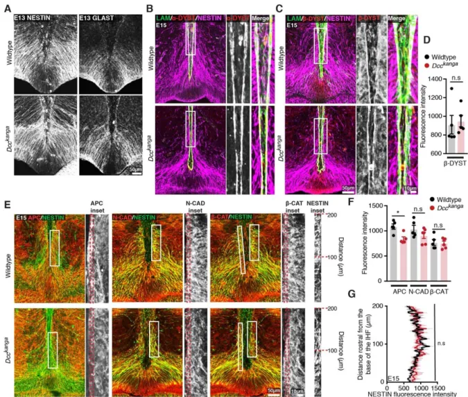
Next, we further investigated whether the aberrant organisation of radial glial
308
processes along the IHF in Dcckanga mice was due to a loss of endfoot adhesion to
309
the IHF pial surface. There was no difference in fluorescence intensity of
Nestin-310
positive MZG processes within 5 µm adjacent to the IHF surface between Dcckanga 311
and wildtype mice, suggesting comparable attachment of radial glial endfeet to the
312
IHF in both strains (Supplementary File 1, Figure 3-figure supplement 1E and G).
313
This was further evidenced by the normal localisation of α- and β-dystroglycan at the
314
pial IHF surface in Dcckanga mice, where these molecules form crucial adhesions
315
between radial glial endfeet and the extracellular matrix (Myshrall et al., 2012;
316
Supplementary File 1; Figure 3-figure supplement 1-D). Moreover, molecules that
317
normally maintain the bipolar morphology of radial glia, such as β-catenin and
N-318
cadherin, were also expressed normally within Dcckanga Nestin-positive radial glia,
319
but adenomatous polyposis coli (APC) was instead significantly reduced in Dcckanga 320
Nestin-positive radial glial endfeet (Supplementary File 1, Figure 3-figure supplement
321
1D and 3-figure supplement 1E; Yokota et al., 2009). APC regulates the growth and
322
extension of basal radial glial processes and cell polarity of radial glia and migrating
323
astrocytes (Etienne-Manneville and Hall, 2003; Yokota et al., 2009). Thus, reduced
324
localisation of APC within Dcckanga radial glial basal endfeet may indicate perturbed
325
regulation of cell process growth and/or cell polarity. Therefore, Dcckanga
Nestin-326
positive radial glia display reduced elongation and reduced occupation of the pial IHF
327
surface compared to wildtype radial progenitors of MZG. Collectively, these results
328
suggest that DCC is not required for radial MZG to adhere to the IHF, but instead
329
regulates the morphology and organisation of radial MZG processes along the pial
330
IHF surface prior to IHF remodelling.
12 332
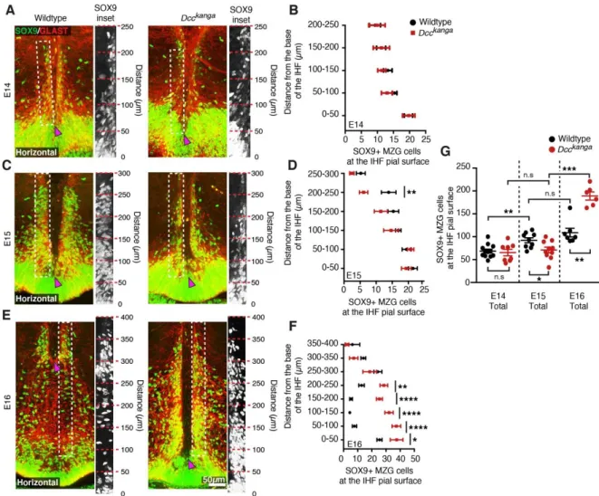
Dcc signalling regulates MZG somal translocation to the IHF prior to IHF
333
remodelling 334
To determine if the aberrant morphology and organisation of radial glial processes
335
observed in Dcckanga mice affects the subsequent distribution of translocated MZG
336
cell bodies at the IHF surface, immunohistochemistry for glial markers Sox9 and
337
Glast was performed in E14-E16 Dcckanga mice. Wildtype MZG undergo substantial
338
somal translocation to the IHF between E14 and E15 (Gobius et al., 2016;
339
Supplementary File 1, Figure 4A, 4C and 4G). In contrast, Dcckanga mice showed
340
reduced somal translocation to the IHF (Supplementary File 1; Figure 4A, 4C and
341
4G), with significantly fewer MZG cells at the IHF pial surface by E15 in Dcckanga 342
compared to wildtype mice (Supplementary File 1; Figure 4B and 4G). When binned
343
along the rostro-caudal axis, we found a significant reduction in the number of cell
344
bodies reaching the rostral IHF pial surface in E15 Dcckanga mice (200-250 µm;
345
Supplementary File 1 and Figure 4C-D). Since MZG progenitors somal translocate
346
toward their basal process attached to the IHF (Gobius et al., 2016), our results
347
suggest that the lack of radial MZG processes occupying the rostral E14 IHF surface
348
in Dcckanga mice results in a decrease of MZG cell bodies present in the
349
corresponding region one day later. There was, however, a significant increase in
350
MZG cell bodies present at the IHF pial surface between E15 and E16 in Dcckanga 351
mice (Supplementary File 1, Figure 4C, 4E and 4G). This suggests that MZG
352
migration towards the IHF is delayed but does eventually occur in Dcckanga mice,
353
albeit after IHF remodelling would normally have been initiated. Dcckanga MZG remain
354
adjacent to the unremodelled IHF at E16 in contrast to wildtype MZG, which are
355
scattered along the midline where IHF remodelling has occurred and continue to
356
expand their domain rostral and dorsal for further IHF remodelling (Gobius et al.,
357
2016). Furthermore, despite DCC having been previously implicated in regulating
358
cell proliferation and cell death (Arakawa, 2004; Llambi et al., 2001; Mehlen et al.,
359
1998), cell birth-dating, differentiation and apoptosis experiments did not reveal any
360
significant differences between the MZG of Dcckanga and wildtype mice
361
(Supplementary File 1 and Figure 4-figure supplement 1). Taken together, these
362
results suggest that the irregular morphology and distribution of radial Dcckanga MZG
363
processes is associated with delayed somal translocation of MZG to the IHF surface,
364
and may prevent the initiation of IHF remodelling.
13
Radial glia in the corticoseptal boundary detach from the pial surface and
366
cluster their processes to form a triangular group of cells known as the glial wedge,
367
while other radial glia in this region translocate their soma to the IHF (similar to MZG
368
cells), where they subsequently form the indusium griseum glia (Shu and Richards,
369
2001; Smith et al., 2006). We investigated whether DCC also regulates the
370
development of these glial populations, which were observed to be abnormal at E15
371
(Figure 3C-D) and secrete axon guidance molecules during CC formation (reviewed
372
in Donahoo and Richards, 2009; Gobius and Richards, 2011; Morcom et al., 2016).
373
In Dcckanga and Dcc knockout mice, the glial wedge was malformed and there was a
374
major reduction in somal translocation of Sox9-positive indusium griseum glia to the
375
IHF surface, which subsequently prevented formation of this glial guidepost cell
376
population (Supplementary File 1 and Figure 4-figure supplement 1G-I). Thus, DCC
377
may play a more general role in regulating the morphological maturation and
378
migration of multiple radial astroglial populations in the developing midline, which are
379
critical for CC formation.
380 381
DCC signalling regulates MZG cell morphology and spatial distribution during 382
IHF remodelling 383
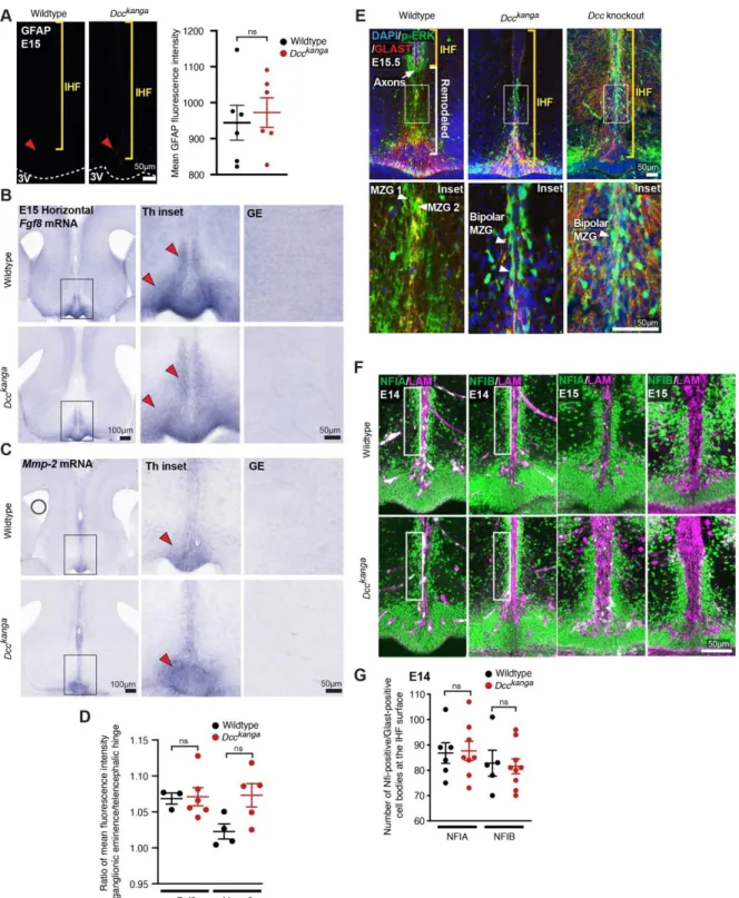
We previously demonstrated that MZG differentiation is controlled by molecular
384
signaling initiated by the morphogen FGF8 via the Mitogen activated protein kinase
385
(MAPK) pathway to NFI transcription factors A and B (Gobius et al., 2016). Members
386
of this signaling pathway (Fgf8, NFIA, NFIB, and p-ERK1/2) were expressed
387
normally in Dcckanga MZG compared to MZG in their wildtype littermates at E15
388
(Figure 5-figure supplement 1B, S5D-F and Supplementary File 1). Further, Dcckanga 389
MZG continue to express Mmp2 mRNA (Figure 5-figure supplement 1C, D and
390
Supplementary File 1), which we previously demonstrated to be expressed during
391
MZG-mediated degradation of the IHF during remodelling (Gobius et al., 2016).
392
Next, we investigated the distribution and maturation of MZG in Ntn1 and Dcc 393
mutant mice at E16 and 17, when wildtype MZG normally differentiate into multipolar
394
astrocytes during IHF remodelling (Gobius et al., 2016). Immunohistochemistry for
395
Nestin, Glast (Figures 3F, 3J) and Gfap (Figure 5A) demonstrated that Dcckanga, Dcc 396
knockout and Ntn1-lacZ MZG remain attached to the caudal IHF pial surface and
397
have not intercalated at stages equivalent to when wildtype MZG have infiltrated and
398
remodelled the IHF (Figures 3F, 3J, 5A and Supplementary File 1). DCC-deficient
14
MZG expressed GFAP at comparable levels to wildtype MZG at E17, and
400
demonstrated no precocious expression of GFAP at E15, similar to wildtype MZG
401
(Figure 5A-B, Figure 5-figure supplement 1A and Supplementary File 1). Therefore,
402
DCC-deficient MZG do not mature precociously prior to migration and IHF
403
remodelling, or fail to differentiate during callosal development. However, Ntn1-lacZ,
404
Dcckanga and Dcc knockout mice demonstrate a significant reduction of
GFAP-405
positive glia at E17 in the region where the CC normally forms in wildtype mice (i.e.,
406
> 450 µm from the third ventricle; Figure 5A-C and Supplementary File 1). Instead,
407
MZG in these mutants remain close to the third ventricle and the majority fail to
408
migrate. To quantify this, we normalised the level of GFAP between sections and
409
calculated a rostro-caudal ratio of this fluorescence. We observed a significant
410
reduction in the rostro-caudal ratio of GFAP fluorescence in Ntn1-lacZ, Dcckanga and
411
Dcc knockout mice compared to controls (Figure 5A-D and Supplementary File 1).
412
Since progressive migration and intercalation of MZG is required for IHF remodelling
413
(Gobius et al., 2016), these results indicate that Ntn1 and Dcc affect IHF remodelling
414
by regulating the morphology and spatial organisation of both radial MZG progenitors
415
and mature MZG, and therefore their ability to intercalate across the IHF, but not
416
their proliferation and adhesion to the pial IHF surface.
417 418
Variable DCC knockdown during midline development causes a spectrum of 419
callosal phenotypes. 420
The current and previous results from our laboratory indicate at least two distinct
421
roles for NTN1 and DCC during CC formation: first, they act on astroglia to facilitate
422
remodelling of the IHF, and second, they regulate the pathfinding of callosal axons to
423
the telencephalic midline (Fothergill et al., 2014). To investigate these roles further,
424
we aimed to disrupt DCC expression specifically within the progenitors of callosal
425
neurons, sparing expression within MZG. We designed two Dcc-targeted
426
CRISPR/CAS9 constructs (Dcc-CRISPR) and acquired a Dcc-targeted shRNA ( Dcc-427
shRNA; Zhang et al., 2018) for targeted in utero electroporation into the E13
428
cingulate cortex in wildtype and Dcckanga mice and successfully labelled callosal
429
axons that reach the contralateral hemisphere by E18 (Figure 6-figure supplement
430
1A). We observed no phenotype in all experimental cases (Figure 6-figure
431
supplement 1A), and instead found these techniques failed to reduce DCC
432
expression sufficiently; the only significant reduction in DCC protein was observed in
15
heterozygous Dcckanga mice electroporated with Dcc-shRNA (average DCC
434
expression reduced to 93.06% compared to the non-electroporated hemisphere;
435
Figure 6-figure supplement 1A-C and Supplementary File 1). In order to knockout
436
DCC more robustly in the cortex, we crossed Dccflox/flox mice (Krimpenfort et al.,
437
2012) with mice carrying Emx1iCre (Kessaris et al., 2006), and the tdTomatoflox_stop 438
reporter allele (Madisen et al., 2010).
439
At birth, we observed a spectrum of callosal phenotypes in Dcc cKO mice,
440
including complete callosal absence (4/12 mice), partial CC absence (5/12 mice),
441
and a normal CC that was comparable to control mice, which do not express
442
Emx1iCre (3/12 mice) based on rostral-caudal CC length across ventral, middle and
443
dorsal horizontal sections (Figure 6A and 6F). Moreover, the HC was significantly
444
reduced in the majority of animals and was absent in one Dcc cKO mouse (Figure
445
6A, 6H and Supplementary File 1). Unexpectedly, we found the IHF was significantly
446
retained across Dcc cKO mice, indicating that IHF remodelling had not been
447
completed (Figure 6A, 6D-E, and Supplementary File 1). The severity of callosal
448
agenesis and HC dysgenesis was significantly correlated with the extent to which the
449
IHF had been remodelled (Figure 6A, 6J, 6K, Figure 6-figure supplement 1D-G and
450
Supplementary File 1). Complete callosal agenesis Dcc cKO mice demonstrated the
451
most severe retention of the IHF, encompassing the majority of the telencephalic
452
midline, while partial callosal agenesis and even full CC Dcc cKO mice demonstrated
453
a retention of the rostral IHF (Figure 6A, 6E and Supplementary File 1). Moreover, in
454
partial callosal agenesis and full CC Dcc cKO mice that demonstrated partial
455
retention of the rostral IHF, callosal axons often crossed the midline more caudal in a
456
region where the IHF had been remodelled compared to control mice (see corpus
457
callosum remnant or CCR in Figure 6A). This suggests that in the absence of their
458
normal substrate, callosal axons are able to adapt and cross the midline in a region
459
where the substrate is available. These results were reflected by a significant
460
increase in the rostro-caudal depth of the partial or full CC in Dcc cKO mice (Figure
461
6A, 6G and Supplementary File 1). In Dcc cKO mice with complete callosal
462
agenesis, callosal axons were unable to cross the midline and accumulated adjacent
463
to the IHF that had not been remodelled (red arrowheads in Figure 6A), similar to
464
Dcckanga and Dcc knockout mice. These results demonstrate that DCC regulates the
465
extent of IHF remodelling throughout the telencephalic midline. The retention of the
466
IHF in these mice was unexpected; we had instead expected that reduced DCC
16
expression in the cortex would cause callosal axon misguidance with normal
468
formation of an interhemispheric substrate. Instead, our results suggest that DCC
469
primarily regulates the formation of the interhemispheric substrate to determine CC
470
size in mice.
471
We next explored whether loss of DCC expression within callosal axons might
472
cause prior callosal axon misguidance that could indirectly impact IHF remodelling.
473
We found that DCC expression was significantly reduced in the cingulate cortex and
474
adjacent intermediate zone in the majority of P0 Dcc cKO mice (mean expression
475
reduced to 80.4% and 62.9% respectively; Figure 6A, 6I and Supplementary File 1),
476
and within E15 Dcc cKO mice (mean DCC expression in the cingulate cortex
477
reduced to 76.6% in Dcc cKO; Figure 6L, 6O and Supplementary File 1), as
478
expected. Surprisingly, we found that TDTOMATO-positive/GAP43-positive axons,
479
which had reduced DCC expression, approached the interhemispheric midline in Dcc 480
cKO mice, similar to their cre-negative littermates (Figure 6M-N, 6P and
481
Supplementary File 1). This suggests that axons with reduced, but not entirely
482
eliminated DCC expression, approach the midline adjacent to the IHF in a timely and
483
spatially appropriate manner, and are unable to cross the midline in regions where
484
the IHF is not remodelled in Dcc cKO mice.
485
Next, we investigated the recombination pattern and development of MZG in
486
Dcc cKO mice. Cre activity, as measured by TDTOMATO expression, was
487
widespread in cells throughout the telencephalic midline, including septal cells and
488
HC axons, resulting in reduced DCC expression in multiple cell types (Figure 6B).
489
Mean DCC expression within the telencephalic hinge was comparable between Dcc 490
cKO mice and their cre littermates (Figure 6L, 6O and Supplementary File 1), but we
491
also observed TDTOMATO-positive/GLAST-positive MZG cell bodies within the
492
telencephalic hinge, and at the IHF surface in Dcc cKO mice (Figure 6Q-R and
493
Supplementary File 1). This suggests the potential for Dcc knockdown in a subset of
494
MZG cells within Dcc cKO mice, which may fail to intercalate across the IHF,
495
possibly causing the IHF remodelling defect observed in P0 Dcc cKO mice.
496
However, unlike Dcckanga mice, we were unable to find a significant population
497
difference in the distribution of GLAST-positive MZG between Dcc cKO mice and
498
their cre-negative littermates, at the level of DCC knockdown observed in this model
499
(Figure 6-figure supplement 1H-I and Supplementary File 1). Thus, the variable
500
callosal and IHF remodelling phenotypes observed in Dcc cKO mice likely arise from
17
varying degrees of DCC knockdown in these models due to the mosaic expression
502
of Emx1iCre within MZG. In order to further explore the role of DCC and the impact of
503
human DCC mutations on the behavior of astroglia, we next investigated the function
504
of human DCC mutations using in vitro assays. 505
506
NTN1-DCC signalling promotes cytoskeletal remodelling of astroglia and 507
neural progenitors 508
Our results suggest that NTN1 and DCC may have important functions in the
509
morphological development of radial glia more broadly. We established in vitro 510
assays to test the function of NTN1-DCC signalling and DCC mutant receptors in
511
regulating the morphology of astroglial-like cells. Such assays can also be used to
512
examine human variants of DCC pathogenic mutations (see next section). To
513
develop these assays, we employed N2A neuroblast cells, which display neural
514
progenitor properties (Augusti-Tocco and Sato., 1969; Shea et al., 1985), as well as
515
U251 glioma cells, which express astroglial markers and display invasive capacity
516
(Zhang et al., 2013) similar to MZG cells. Importantly, endogenous DCC has
517
previously been demonstrated to render several glioma cell lines migratory in
518
response to a gradient of NTN1 as a chemoattractant (Jarjour et al., 2011). Both cell
519
lines were transfected with either full-length DCC fused to a TDTOMATO reporter
520
(pCAG-DCC:TDTOMATO) to express wildtype DCC, or a membrane-targeted
521
TDTOMATO reporter (pCAG-H2B-GFP-2A-Myr-TDTOMATO) as a control and
522
stimulated with NTN1. Moreover, we transfected U251 cells with a DCCkanga
523
construct (pCAG-DCCkanga:TDTOMATO), to test whether the P3 domain was critical
524
for NTN1-DCC signalling effects on cell morphology.
525
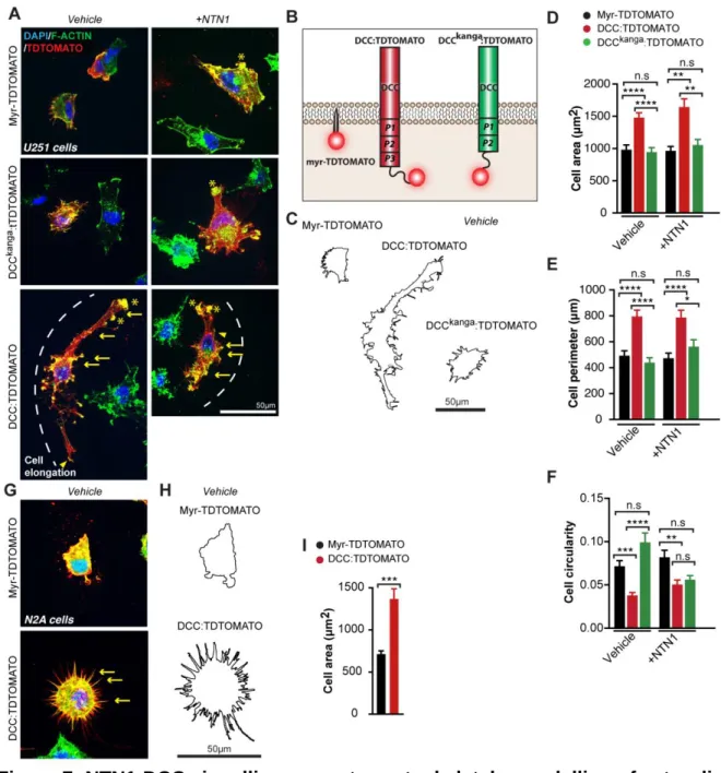
Expression of DCC:TDTOMATO in U251 cells in the absence of ligand
526
(vehicle alone) promoted cell spreading and elongation, reflected by a significant
527
increase in average cell area and cell perimeter, and a significant decrease in cell
528
circularity compared to control (Supplementary File 1 and Figure 7A-F). This effect
529
was not observed following expression of the DCCkanga construct alone
530
(Supplementary File 1 and Figure 7A-F), suggesting that the P3 domain of DCC is
531
critical for inducing changes in glial cell shape. We further confirmed that these
532
morphological changes were due to the presence of the coding region of wildtype
533
DCC, by comparing to cells transfected with plasmids where DCC had been excised
534
and only the TDTOMATO remained (pCAG-TDTOMATO; Supplementary File 1 and
18
Figure 7-figure supplement 1A and Figure 6-figure supplement 17B). A similar effect
536
was observed following DCC overexpression in N2A cells, which also registered a
537
significant increase in average cell area and cell perimeter, and decrease in cell
538
circularity, compared to controls (Supplementary File 1, Figure 7G-I, and Figure
7-539
figure supplement 1G and H), further indicating similar effects on cell morphology in
540
glial and neural progenitor lineages. Interestingly, application of NTN1 did not affect
541
cell shape following DCC expression in either cell line (Supplementary File 1 and
542
Figure 7A-F), suggesting that endogenous NTN1, which is known to be expressed
543
by U251 cells (Chen et al., 2017), may be sufficient for activation of
544
DCC:TDTOMATO receptors, or that NTN1 is not required for this effect. To
545
investigate this, we examined NTN1 expression in these cell lines using western blot
546
analysis. We confirmed that both our cell lines expressed NTN1 endogenously, and
547
that transfection of DCC increased DCC levels but did not affect NTN1 expression
548
(Figure 7-figure supplement 1J). No endogenous DCC was detected by western blot
549
in either cell line (Figure 7-figure supplement 1J). Thus, addition of DCC induced
550
cytoskeletal rearrangements in both N2A and U251 cells, which may involve
551
autocrine NTN1 signalling. Typical features of DCC-expressing cells with or without
552
bath application of NTN1 included actin-rich regions resembling filopodia,
553
lamellipodia, and membrane ruffling in U251 cells, while only filopodia were highly
554
abundant in DCC:TDTOMATO-expressing N2A cells; all of these features were
555
rarely observed in control cells from both cell lines (Figure 7A, G). No difference in
556
cleaved-caspase 3-mediated cell apoptosis was observed following DCC expression
557
in either cell line (Figure 7-figure supplement 1I). This suggests that DCC signalling
558
does not mediate programmed cell death but rather promotes remodelling of the
559
actin cytoskeleton in glioma and neuroblast cells in a similar manner to neurons and
560
oligodendrocytes (Rajasekharan et al., 2009; Shekarabi and Kennedy, 2002).
561 562
Humans with agenesis of the CC carry loss-of-function pathogenic variants in 563
DCC that are unable to modulate cell shape
564
Having established that DCC signalling rearranges the cytoskeleton of astroglial-like
565
cells, and that the P3 domain of DCC is crucial for this function, we next investigated
566
whether DCC mutant receptors from humans with dysgenesis of the CC affected this
567
function. Site directed mutagenesis was performed to introduce missense mutations
568
into the pCAG-DCC:TDTOMATO expression vector in order to model mutated DCC 569
19
receptors found in six families with previously reported cases of complete or partial
570
agenesis of the CC (p.Met743Leu, p.Val754Met, p.Ala893Thr, p.Val793Gly,
571
p.Gly805Glu, p.Met1217Val;p.Ala1250Thr; Marsh et al., 2017; Marsh et al., 2018;
572
Figure 8A and Figure 6-figure supplement 1C). We further included two artificial
573
mutant receptors that were previously shown to perturb NTN1 binding and
574
chemoattraction (p.Val848Arg, p.His857Ala; Finci et al., 2014). First, these mutants
575
were transfected into HEK293T and COS-7 cells that do not endogenously express
576
DCC (Chen et al., 2013; Shekarabi and Kennedy, 2002). Immunoblotting and
577
immunohistochemistry performed without cell permeabilisation revealed that all
578
mutant DCC receptors were appropriately expressed and localised to the cell
579
membrane (Gad et al., 2000; Figure 7-figure supplement 1E). Using a previously
580
established in vitro binding assay (Müller and Soares, 2006; Zelina et al., 2014), we
581
discovered that DCC mutant proteins with altered residues located at the NTN1
582
binding interface (p.V793G and p.G805E) were unable to bind NTN1 (Figure 8B),
583
while all other receptors with altered residues lying outside of the NTN1 binding
584
interface still bound NTN1 (p.M743L, p.V754M, p.A893T and p.M1217;A1250T;
585
Figure 8B). Surprisingly, all eight mutant DCC receptors were unable to modulate
586
cell morphology in the presence of NTN1 (Figure 8C-E; Supplementary File 1).
587
Collectively, our results suggest a model whereby mutations that affect the ability for
588
DCC to regulate cell shape (Figure 8F), are likely to cause callosal agenesis through
589
perturbed MZG migration and IHF remodelling.
590 591
Discussion 592
Genes that encode axon guidance molecules frequently cause callosal dysgenesis
593
when knocked out in mice (Edwards et al., 2014). This has led to the prevalent view
594
that callosal dysgenesis in these mice might be primarily to due defects in callosal
595
axon guidance towards and across the midline. Here, we identified a novel function
596
for the classical axon guidance genes NTN1 and DCC in regulating the morphology
597
of midline astroglia for IHF remodelling prior to CC and HC formation. Importantly,
598
normal astroglial development and IHF remodelling are critical processes that
599
precede and are necessary for subsequent CC axon guidance across the
600
interhemispheric midline (Gobius et al., 2016). We find that defects in IHF
601
remodelling are consistently associated with dysgenesis of the CC and HC in mice
602
and humans with pathogenic variants in Ntn1 or Dcc.
20
Our in vitro assays and analysis of mouse and human cell morphology
604
indicate that the cytoskeletal remodelling function of NTN1-DCC signalling is likely to
605
be crucial for MZG development, IHF remodelling, and subsequent CC formation.
606
The timely differentiation and appropriate distribution of MZG cells at the IHF surface
607
is required for their intercalation and IHF remodelling function (Gobius et al., 2016).
608
Our data suggest a model whereby failed IHF remodelling associated with mutations
609
in Ntn1 and Dcc occurs due to delayed astroglial migration to the IHF as a
610
consequence of perturbed process extension and organisation of MZG precursors.
611
Notably, no medial extension of MZG processes across the basement membrane or
612
perforations in the IHF to allow glia from each hemisphere to interact and intercalate
613
were observed in Ntn1 or Dcc mutant mice at any developmental stage examined.
614
This suggests that NTN1-DCC signalling might also be required for MZG
615
intercalation and removal of the intervening leptomeninges. The DCC homologue
616
UNC-40 is known to facilitate formation of a polarised actin-rich cell protrusion in the
617
Caenorhabditis elegans anchor cell, which breaches the basement membrane rich in
618
UNC-6 (NTN1 homolog), enabling the cell to invade the vulval epithelium (Hagedorn
619
et al., 2013; Ziel et al., 2009). DCC may perform a similar function in MZG by
620
engaging secreted NTN1, which we found to be localised at the IHF basement
621
membrane in agreement with the localisation of radial-glial-derived NTN1 in the
622
spinal cord (Varadarjan et al., 2017), and preferentially polarising actin remodelling
623
and process extension toward the IHF during MZG intercalation. Moreover, callosal
624
axons that also rely on DCC-mediated cytoskeletal remodelling for growth and
625
guidance, may non-cell-autonomously influence the final stages of MZG
626
development via a secreted cue or contact-dependent mechanism. Further
627
dissecting this would ideally involve even greater precision in complete and cell-type
628
specific knockout of DCC and NTN1, since knockdown in a subset of cells or merely
629
lowering the expression level was insufficient to induce a consistent phenotype.
630
Notably, we find that the P3 domain-dependent functions of DCC may be
631
required for astroglial development and IHF remodelling. These functions include
632
receptor dimerisation, interaction with the co-receptor ROBO1, or interaction with
633
effectors FAK, MYO10, and TUBB3 (Fothergill et al., 2014; Li et al., 2004; Qu et al.,
634
2013; Stein and Tessier-Lavigne, 2001; Wei et al., 2011; Xu et al., 2018).
635
Accordingly, mice deficient in Robo1, Fak and Tubb3, as well as their signaling
636
effectors Cdc42, Fyn, Enah and Mena, which normally act downstream of DCC to
21
regulate the cell cytoskeleton, all display dysgenesis of the CC (Andrews et al.,
638
2006; Beggs et al., 2003; Goto et al., 2008; Menzies et al., 2004; Tischfield et al.,
639
2010; Yokota et al., 2010). Similarly, astroglial cells remodel their cytoskeleton to
640
transition from a bipolar to multipolar morphology, and this process is known to
641
involve the intracellular DCC effectors CDC42, RAC1, RHOA, N-WASP and EZRIN
642
(Abe and Misawa, 2003; Antoine-Bertrand et al., 2011; Derouiche and Frotscher,
643
2001; Lavialle et al., 2011; Murk et al., 2013; Racchetti et al., 2012; Shekarabi et al.,
644
2005; Zeug et al., 2018). Whether these molecules serve as downstream effectors of
645
DCC to influence astroglial development and IHF remodelling during CC formation is
646
an interesting question for future research.
647
In addition to NTN1 and DCC, as shown here, mice lacking the axon guidance
648
molecules ENAH, SLIT2, SLIT3, and RTN4R have previously been reported to have
649
incomplete IHF remodelling and disrupted midline glial development associated with
650
callosal dysgenesis (Menzies et al., 2004; Unni et al., 2012; Yoo et al., 2017). Taken
651
together, those studies and ours suggest that other axon guidance genes may play
652
similar roles in astroglial development and IHF remodelling during CC formation.
653
Additional candidate axon guidance molecules that may regulate IHF remodelling
654
include EPHB1, EFNB3, GAP43, HS6ST1, HS2ST1, ROBO1 and VASP, since
655
mouse mutants lacking these molecules display disrupted midline glial development
656
and callosal dysgenesis (Andrews et al., 2006; Conway et al., 2011; Mendes et al.,
657
2006; Menzies et al., 2004; Shen et al., 2004; Unni et al., 2012). Additional
658
molecules of interest are EFNB1, EFNB3, EPHB2, and EPHA4, since these are
659
expressed by MZG (Mendes et al., 2006).
660
In summary we have demonstrated that rather than solely regulating axon
661
guidance during telencephalic commissure formation, Dcc and Ntn1 are critical
662
genes required for IHF remodelling. Moreover, our study provides a novel role for
663
axon guidance receptor DCC in regulating astroglial morphology, organisation and
664
migration. Exemplified by Ntn1 and Dcc, our study provides support for widespread
665
consideration of astroglial development and IHF remodelling as possible underlying
666
mechanisms regulated by these and other classically regarded “axon guidance
667
genes” during CC formation.
668
669
Materials and Methods 670
EXPERIMENTAL MODELS AND SUBJECT DETAILS
22 Animals
672
Dccflox/flox (Krimpenfort et al., 2012), Dcc knockout (Fazeli et al., 1997), Dcckanga 673
(Finger et al., 2002), Emx1iCre (Kessaris et al., 2006), Ntn1-lacZ (Serafini et al., 1996,
674
and tdTomatoflox_stop (Madisen et al., 2010) mice on the C57BL/6J background and
675
CD1 wildtype mice were bred at The University of Queensland. Prior approval for all
676
breeding and experiments were obtained from the University of Queensland Animal
677
Ethics Committee. Male and female mice were placed together overnight and the
678
following morning was designated as E0 if a vaginal plug was detected. Dcc 679
knockout and Dcckanga mice were genotyped by PCR and Ntn1-lacZ mice were
680
tested for the presence of the LacZ gene and deemed homozygous if the β-681
galactosidase enzyme was trapped intracellularly, as previously described (Fazeli et
682
al., 1997; Finger et al., 2002; Fothergill et al., 2014; Krimpenfort et al., 2012; Serafini
683
et al., 1996). Dccflox/flox mice were genotyped by the Australian Equine Genetics
684
Research Centre at the University of Queensland.
685 686
Human subjects 687
Ethics for human experimentation was acquired by local ethics committees at The
688
University of Queensland (Australia), the Royal Children’s hospital (Australia), and
689
UCSF Benioff Children’s Hospital (USA). Genetic studies were performed previously
690
(Marsh et al., 2017). Structural MR images were acquired as previously described
691
(Marsh et al., 2017). In our study, we analysed the brain phenotype of affected
692
individuals in family 2 (carrying DCC p.Val793Gly) and family 9 (carrying DCC 693
p.Met1217Val;p.Ala1250Thr in cis) from our previous study.
694 695
METHOD DETAILS
696
Cell birth-dating and tissue collection 697
For cell birth dating studies, 5-ethynyl-2´-deoxyuridine (EdU; 5 mg per kg body
698
weight, Invitrogen) dissolved in sterile phosphate buffer solution (PBS) was injected
699
into the intraperitoneal cavity of awake pregnant dams. Brains were fixed via
700
transcardial perfusion or immersion fixation with 4% paraformaldehyde (PFA).
701 702
Cell lines 703
HEK293 cells (from ATCC CRL-1573, not authenticated, free of mycoplasma
704
contamination) were used to express alkaline phosphatase-conjugated NTN1
23
(NTN1-AP) in the supernatant of COS-7 cell culture. Although this cell line is
706
commonly misidentified, this did not affect the conclusion of the binding assay done
707
in COS-7 cells. U251 cells were obtained as U-373MG (RRID: CVCL_2219) but
708
subsequently identified as U-251 via PCR-based short tandem repeat profiling. All
709
cell lines were routinely tested for mycoplasma to ensure that cell lines were free of
710
mycoplasma contamination. See the key resources table (supplementary file 2) for
711 more information. 712 713 Cell culture 714
All cell lines were cultured at 37°C within a humidified atmosphere containing 5%
715
CO2 and immersed in Dulbecco's Modified Eagles Medium (DMEM) medium 716
(Invitrogen or HyClone™), supplemented with 10% fetal bovine serum. U251 cells
717
were plated on poly-d-lysine-coated coverslips (via submersion in 0.05 mg/mL
718
solution, Sigma-Aldrich) at 10% confluence 24 hours prior to transfection. The
719
pCAG-TDTOMATO, pCAG-H2B-GFP-2A-MyrTDTOMATO, pCAG-DCC:TDTOMATO
720
and pCAG-DCCkanga:TDTOMATO plasmids (1 µg) were transfected into the plated
721
U251 cells using FuGENE 6 (Promega) in Opti-MEM (Gibco, Life Technologies).
722
Cells were then grown for 20 hours and either fixed with 4% paraformaldehyde/4%
723
sucrose or stimulated with ligand. Since 100ng/mL of recombinant NTN1 is sufficient
724
to induce morphological changes in primary oligodendrocyte precursor cells
725
(Rajasekharan et al., 2009), 200ng of recombinant mouse NTN1 protein (R&D
726
Systems) was diluted in sterile PBS and added to cultures within 2 mL media. When
727
ligand was added, cells were grown for a further 12 hours before fixation with 4%
728
PFA/4% sucrose. N2A cells were cultured and transfected as outlined for the U251
729
cells except that after NTN1 stimulation, cells were cultured for only 8 hours before
730
fixation. The pCAG-DCC:TDTOMATO wildtype and missense mutant receptor
731
constructs (1.764 µg) were also transfected into HEK293T cells cultured on
acid-732
washed coverslips using FuGENE HD (Promega) in Optimem (Gibco, Life
733
Technologies). After 24 hours, cells were fixed with 4% PFA/4% sucrose.
734 735
NTN1-binding assay 736
Supernatant containing alkaline phosphatase-conjugated NTN1 (NTN1-AP)
737
was generated from expression in HEK293T cells as previously described (Zelina et