HAL Id: inserm-00090008
https://www.hal.inserm.fr/inserm-00090008
Submitted on 25 Aug 2006
HAL is a multi-disciplinary open access
archive for the deposit and dissemination of
sci-entific research documents, whether they are
pub-lished or not. The documents may come from
teaching and research institutions in France or
abroad, or from public or private research centers.
L’archive ouverte pluridisciplinaire HAL, est
destinée au dépôt et à la diffusion de documents
scientifiques de niveau recherche, publiés ou non,
émanant des établissements d’enseignement et de
recherche français ou étrangers, des laboratoires
publics ou privés.
Cornel Popovici, Yael Berda, Fabien Conchonaud, Aurélie Harbis, Daniel
Birnbaum, Régine Roubin
To cite this version:
Cornel Popovici, Yael Berda, Fabien Conchonaud, Aurélie Harbis, Daniel Birnbaum, et al.. Direct
and heterologous approaches to identify the LET-756/FGF interactome.. BMC Genomics, BioMed
Central, 2006, 7, pp.105. �10.1186/1471-2164-7-105�. �inserm-00090008�
BioMedCentral
BMC Genomics
Open Access
Research article
Direct and heterologous approaches to identify the LET-756/FGF
interactome
Cornel Popovici, Yael Berda, Fabien Conchonaud, Aurélie Harbis,
Daniel Birnbaum and Régine Roubin*
Address: Institut de Cancérologie de Marseille, Laboratoire d'Oncologie Moléculaire, Institut Paoli-Calmettes et UMR599 INSERM, 27 Bd. Leï Roure, 13009 Marseille, France
Email: Cornel Popovici - popovici@marseille.inserm.fr; Yael Berda - yaelberda@yahoo.com; Fabien Conchonaud - conchonaud@ciml.univ-mrs.fr; Aurélie Harbis - aurelie_harbis@yahoo.fr; Daniel Birnbaum - birnbaum@marseille.inserm.fr;
Régine Roubin* - roubin@marseille.inserm.fr * Corresponding author
Abstract
Background: Fibroblast growth factors (FGFs) are multifunctional proteins that play important
roles in cell communication, proliferation and differentiation. However, many aspects of their activities are not well defined. LET-756, one of the two C. elegans FGFs, is expressed throughout development and is essential for worm development. It is both expressed in the nucleus and secreted.
Results: To identify nuclear factors associated with LET-756, we used three approaches. First, we
screened a two-hybrid cDNA library derived from mixed stages worms and from a normalized library, using LET-756 as bait. This direct approach allowed the identification of several binding partners that play various roles in the nucleus/nucleolus, such as PAL-1, a transcription regulator, or RPS-16, a component of the small ribosomal subunit. The interactions were validated by co-immunoprecipitation and determination of their site of occurrence in mammalian cells. Second, because patterns of protein interactions may be conserved throughout species, we searched for orthologs of known mammalian interactors and measured binary interaction with these predicted candidates. We found KIN-3 and KIN-10, the orthologs of CK2α and CK2β, as new partners of LET-756. Third, following the assumption that recognition motifs mediating protein interaction may be conserved between species, we screened a two-hybrid cDNA human library using LET-756 as bait. Among the few FGF partners detected was 14-3-3β. In support of this interaction we showed that the two 14-3-3β orthologous proteins, FTT-1 and FTT-2/PAR-5, interacted with LET-756.
Conclusion: We have conducted the first extensive search for LET-756 interactors using a
multi-directional approach and established the first interaction map of LET-756/FGF with other FGF binding proteins from other species. The interactors identified play various roles in developmental process or basic biochemical events such as ribosome biogenesis.
Background
FGFs constitute a superfamily of pleiotropic growth
fac-tors involved in multiple cellular processes such as mitogenesis, angiogenesis and mesoderm induction [1]. Published: 03 May 2006
BMC Genomics 2006, 7:105 doi:10.1186/1471-2164-7-105
Received: 13 January 2006 Accepted: 03 May 2006 This article is available from: http://www.biomedcentral.com/1471-2164/7/105
© 2006 Popovici et al; licensee BioMed Central Ltd.
This is an Open Access article distributed under the terms of the Creative Commons Attribution License (http://creativecommons.org/licenses/by/2.0), which permits unrestricted use, distribution, and reproduction in any medium, provided the original work is properly cited.
There are 22 FGFs in humans. Except FGF11-14, they exert their biological activities by acting as extracellular growth factors binding to receptors (FGFR1-4) of the tyrosine kinase receptor superfamily [2]. In addition, FGF1-3 and FGF11-14 are localized in the nucleus and function intra-cellularly [3]. Intracellular FGFs bind to several proteins that play a role in FGF trafficking: FIBP, which allows FGF1 to shuttle between the cytosol and the nucleus [4], synaptotagmin-1, which allows FGF1 exocytosis [5], Cystein Rich FGF receptor (CFR), which forms complexes with various FGFs and allows their secretion [6,7], and LRP-1 and 2 (lipoprotein receptor-related proteins), which in conjunction with DAB-1 (Disabled) regulate EGL-17/FGF export in C. elegans [8]. FGF interactors may also regulate FGF nuclear activity; this is the case of Casein Kinase II regulatory subunits [9] and splicing factor SF3a66 [10], which both interact with FGF2. Finally, pro-teins of the extracellular matrix such as fibstatin and fibrinogen interact with FGF2 [11,12]. LET-756 is one of the two FGFs of C. elegans [13, 14 for reviews]. It is essential for worm development [15]. Like some mammalian FGFs, it acts both intra and extracellu-larly. The molecular motif allowing secretion [16] and some of LET-756 extracellular functions have been described [17,18] but the intracellular functions remain poorly defined, although nuclear localization is probably of importance [19]. To further characterize the functions of LET-756, we used yeast two-hybrid screens to identify proteins that interact with this FGF. We identified several interacting proteins involved in various developmental processes or in basic biochemical events such as ribosome biogenesis, and validated some of the interactions by co-immunoprecipitation and/or colocalization.
Results
Identification of nematode LET-756 binding proteins by yeast two-hybrid library screens
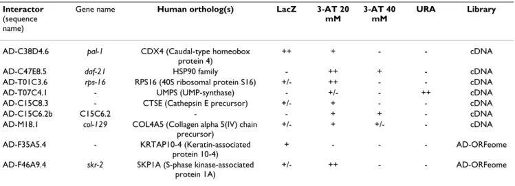
To identify worm proteins that interact with LET-756, we used the two-hybrid system in the MAV103 yeast with LET-756 fused to the Gal-4 DNA binding domain (pDB) as bait and two C. elegans libraries. The latter were either normalized [20] to contain one representative of each expressed gene of the whole genome (ORFeome) or derived from a mixed stage worm population. Library clones were coupled to the Gal-4 activating domain (pAD). The bait did not show any intrinsic transcriptional activation of the three yeast reporter genes. In a screen of approximately 4 × 10⁶ transformants, 41 clones were pos-itive for the two reporters tested, or for only one reporter but with great intensity. The gap repair technique con-firmed 9 clones (Table 1). Sequencing of these clones and blastn or tblastx interrogation of databases revealed uni-dentified sequences (UIS) and sequences coding for pro-teins with known functions: UMP synthase, an enzyme
involved in de novo nucleic acid synthesis, cathepsin (aspartic peptidase A1, pepsinogen family member), RPS-16 (small ribosomal subunit), transcription factor PAL-1 involved in the anterior-posterior development of the male [21], DAF-21, a chaperone of the HSP-90 family, involved in chemosensory transduction and insulin sig-nalization [22,23], COL-129, an isoform of collagen, and SKR-2 (homolog of skp1 of S. cerevisiae), a component of the skp1p/cullin/F-box SCF complex with ubiquitin ligase activity [24,25]. The latter also shows similarities with P19, which is associated with cyclinA/CDK2 complex in humans.
To confirm the results on some on the interactors we judged potentially relevant, pAD interactor clones were picked individually from the ORFeome library and tested directly in yeast two-hybrid system against pDB LET-756. PAL-1 interacted strongly with LET-756 (two positive tests). For the other interactors, only one test was clearly positive (Table 2A).
Identification of LET-756-binding proteins among orthologs of known mammalian FGF interactors
The conservation of signaling pathways between worms and mammals, as well as the conserved FGF structures in different species [26,27], suggested that orthologs of human FGF-binding proteins could interact with LET-756. We used blast interrogation to determine the most conserved orthologs supposed to retain the ancestral func-tion of the known human FGF-interactors. These orthologs were recovered from the ORFeome library and tested in binary interactions (Table 2B). We found kin-10 (CK2β) and rpl-6 positive for two reporter genes, and
kin-3 (CK2α), hsp-6 (mortalin), F14E5.2 (cystein rich FGF
receptor, CFR) and C18A3.3 (NoBP) positive for one reporter gene.
Identification of human LET-756-binding proteins
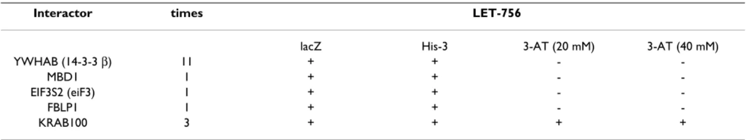
We based our third approach on the assumption that pro-tein interaction motifs may be conserved between species. We made a heterologous screen using a cDNA human library as prey and LET-756 as bait. The yeast two-hybrid system was used with LET-756 fused to LexA DNA binding domain as bait (pDB) for the screening of a human pla-centa cDNA library containing the Gal-4 activating domain (pAD). Table 3 indicates the number of times the interactors were isolated and the strength of the interac-tion. Identified partners were different from those unveiled by the C. elegans screens but showed similar bio-logic activities (Table 4). MBD1 and ZN420 are transcrip-tion factors, 14-3β (YWHAB, tyrosine 3-monooxygenase/tryptophan 5-monooxygenase activa-tion protein, β polypeptide) has chaperone activity, and FBLI1 (Filamin-binding LIM protein 1) is a protein of the Zyxin family.
BMC Genomics 2006, 7:105 http://www.biomedcentral.com/1471-2164/7/105
Finally, the orthologs of human 14-3-3β , ftt-1/par-5 and
ftt-2, and the orthologs of FBL1, zyx-1a and zyx-1b, were
obtained from the nematode ORFeome library and tested for direct interaction in yeast two-hybrid system. Table 2C indicates that FTT-1 and FTT-2, but not ZYX-1a or ZYX-1b, reacted with the LET-756 bait.
Confirmation of interactions by co- immunoprecipitation experiments
A number of interactors we identified have been described as false positives in various studies. It is the case for HSP family members and ribosomal proteins, and to some extent for collagen-related proteins, Zn finger proteins and proteasome subunits [28]. To validate the interaction of LET-756 with the candidate partners, co-immunopre-cipitation experiments were done in Cos-1 cells. Cos-1
Table 2: Direct interactions between LET-756 (DB) and clones derived from the ORFeome library. A) clones detected in the screening of the C. elegans libraries; B) orthologs of clones detected in the screening of the human placenta library; C) orthologs of known mammalian FGF interactors.
Interactors (AD) LET-756 (DB)
LacZ 3-AT URA
A RPS-16 +/- ++ -PAL-1 ++ + -DAF-21 - +* -COL-129 +/- + -SKR-2 +/- +/- -C15C6.2 +/- + -B KIN-10 (CSNK2B, CK2β) +/- + -KIN-3 (CSNK2A, CK2α) - + -HSP-6 (HSPA9B, mortalin) - + -F14E5.2 (GLG1, CFR) - +/-
-C18A3.3 (EBNA1BP2, NoBP) - +/-
-RPL-6 (RPL6) +/- +
-C FTT-1/PAR-5 (YWHAB, 14-3-3β) +/- +
-FTT-2 (YWHAB, 14-3-3β) +/- +
-ZYX-1a, ZYX-1b (ZN420) - -
-*: 3-AT concentration allowing yeast growth was 20mM except for DAF-21 where it was 40 mM
Table 1: LET-756-interacting proteins identified by screening Y2H C. elegans libraries. Bait used was DB-LET-756. Sequences of AD-interactor clones are in (58). Clones were assigned scores for LacZ expression, growth on plates lacking histidine but containing 20 or 40 mM 3-amino triazol (3-AT) and in addition, growth on plates lacking uracil (URA) for the C. elegans libraries
Interactor
(sequence name)
Gene name Human ortholog(s) LacZ 3-AT 20 mM
3-AT 40 mM
URA Library
AD-C38D4.6 pal-1 CDX4 (Caudal-type homeobox protein 4)
++ + - - cDNA
AD-C47E8.5 daf-21 HSP90 family - ++ + - cDNA
AD-T01C3.6 rps-16 RPS16 (40S ribosomal protein S16) +/- ++ - - cDNA
AD-T07C4.1 - UMPS (UMP-synthase) - +/- - ++ cDNA
AD-C15C8.3 - CTSE (Cathepsin E precursor) +/- + - - cDNA
AD-C15C6.2b C15C6.2 - - + + - cDNA
AD-M18.1 col-129 COL4A5 (Collagen alpha 5(IV) chain precursor)
+/- + +/- - cDNA
AD-F35A5.4 - KRTAP10-4 (Keratin-associated protein 10-4)
+ - - - AD-ORFeome
AD-F46A9.4 skr-2 SKP1A (S-phase kinase-associated protein 1A)
cells were transiently cotransfected with HA-tagged part-ner constructs and LET-756::GFP, and the lysates were immunoprecipitated with anti-GFP. Fig. 1 shows the result of a western blot probed first with anti-HA to reveal the co-immunoprecipitated proteins, and second with anti-GFP to normalize the transfection with LET-756. All tested partners immunoprecipitated with LET-756, although with different strength, unrelated to their level of expression (not shown). The 14-3-3β and KIN-10 pro-teins were reproducibly the less efficient. To make sure that overexpression of the two tagged proteins was not responsible for the immunoprecipitation, we used TACC1 as an unrelated HA-tagged control. In similar condition, TACC1 was unable to immunoprecipitate LET-756.
Confirmation of interactions by subcellular colocalization experiments
To further confirm FGF/partner interaction in mamma-lian cells, HA-tagged partners were co-expressed with LET-756::GFP in Cos-1 cells and their respective subcellular localization was examined. As already described [16,19], LET-756::GFP localized in specific regions of the nucleus where splicing factors are concentrated. Immunofluores-cence microscopy using anti-HA antibodies revealed PAL-1 in foci in the nucleus that colocalized with LET-756 (Fig. 2). We have previously established [19] that treatment of LET-756 expressing cells with actinomycin D (a drug inhibiting Pol I and Pol II activities) displaces LET-756 to the perinucleolar and nucleolar compartments. Addition of actinomycin D did not move the PAL-1 protein to the
nucleolus as it did for LET-756 but kept both partners in close association in nucleoplasmic foci. Other partners, such as RPL-6, were delocalized by transfection of LET-756. RPL-6 was localized in large foci mainly in the nucle-oplasm when transfected alone (column I, Fig. 2) but was most often dispersed through the nucleoplasm and asso-ciated with LET-756 when cotransfected with the FGF (Fig. 2, column II to IV). FTT-1/PAR-5 and FTT-2 localized pref-erentially in the cytoplasm when transfected alone, but observed also in the nucleus when transfected together with 756. In addition, vesicles containing both LET-756 and FTT-1 or FTT-2 were visible. KIN-3 colocalized with LET-756 in nuclei and exhibited a strong expression in cytoplasm whether LET-756 was present or not whereas KIN-10 present in nucleoplasm of untransfected LET-756 cells moved with LET-756 in the speckles when co-trans-fected. Upon actinomycin D treatment both LET-756 and KIN-10 formed enlarged speckles and moved to the nucle-olus (Fig. 2). This stricking delocalization observed upon actinomycin D treated KIN-10 co-transfected cells did not occurred with KIN-3. In other instances, the partner mod-ified LET-756 localization: RPS-16 concentrated LET-756 in large foci when both proteins were present in the nucleus (Fig. 2). The protein encoded by C15C6.2 did not show any gross colocalization (Fig. 2). Finally, COL-129 was localized only at the Golgi apparatus, whether LET-756 was present or not, and CFR was localized only in the cytoplasm.
Table 3B: LET-756-interacting proteins identified by screening Y2H human placenta libraries. B: Description of the proteins and identification of the C. elegans ortologs.
Protein Description C.elegans orthologs
YWHAB (14-3-3β) protein with chaperone activity implicated in subcellular compartmentalization of binding partners
PAR-5 or FTT1, FTT-2
MBD1 negative regulator of transcription, the methyl CpG-Binding Domain none EIF3S2 initiator of translation (subunit 9) EIF3-B
FBLI1 protein belonging to the Zyxin family, having 3 LIM domains, involved in protein-protein interaction (filamin-binding LIM protein-1)
ZYX-1
ZN420 zinc finger protein with a Krab domain none
*: 3-AT concentration allowing yeast growth was 20mM except for DAF-21 where it was 40 mM
Table 3A: LET-756-interacting proteins identified by screening Y2H human placenta libraries. A: Number of times the clone was detected during the Y2H library screening and scores for the different reporter genes.
Interactor times LET-756
lacZ His-3 3-AT (20 mM) 3-AT (40 mM)
YWHAB (14-3-3 β) 11 + + -
-MBD1 1 + + -
-EIF3S2 (eiF3) 1 + + -
-FBLP1 1 + + -
-KRAB100 3 + + + +
BMC Genomics 2006, 7:105 http://www.biomedcentral.com/1471-2164/7/105
Discussion
Several growth factors are found in the nucleus in addi-tion to their other localizaaddi-tions. This is the case for LET-756 but not for EGL-17, the other C. elegans FGF [29]. The
role of LET-756 in the nucleus is not known. To help char-acterize this role we searched to identify intracellular binding partners of LET-756. By using different two-hybrid screens in yeast, we identified several proteins
Table 5: C. elegans orthologous genes of known human FGF interactors.
FGF Interactor C. elegans or human orthologs
FGF1 HSPA9B (Stress-70 protein, mortalin) [39] hsp-6 (C37H5.8)
FGF1 NP_060979.2 (PRO1855, LOC55379, ribosome-binding protein p34, RBP34*) [39]
F56A8.3
FGF1 LDL (Low density lipoproteins) [40]
-FGF1 S100A13 (Calcium binding protein S100A13) [41]
-FGF1 SYT1 (Synaptotagmin-1) [5] snt-1 (F31E8.2)
FGF1 FIBP (FGF-1 intracellular binding protein) [4] -FGF1 and FGF2 FGFBP1 (Fibroblast growth factor binding protein 1) [42] -FGF1 and FGF2 CSNK2A (Casein Kinase II, catalytic subunit α) [43] kin-3 (B0205.7)
FGF2 CSNK2B (Casein Kinase II, regulatory subunit β) [9] kin-10 (T01G9.6b)
FGF2 RPL6 (Large ribosomal subunit L6) [36] rpl-6 (R151.3)
FGF2 RPS19 (40S ribosomal protein S19) [37] rps-19 (T05F1.3)
FGF2 CEP57 (Translokin, Centrosomal protein of 57 kDa, Proliferation-inducing protein 8, PIG8) [44]
-FGF2 SMN1 (Survival motor neuron protein) [45, 46] smn-1 (C41G7.1)
FGF2 FGA/FGB/FGG (Fibrinogen, alpha/beta/gamma polypeptides) [12, 47] -FGF2 FN1 (Fibstatin, fragment of fibronectin) [11] C56C10.4 FGF2 API5 (Apoptosis inhibitor 5, Fibroblast growth factor 2-interacting factor,
FIF) [48]
-FGF2 SF3A2 (Splicing factor 3A subunit 2) [10] F11A10.2
FGF2 PTX3 (Long pentraxin 3) [49]
-FGF2 PF4 (CXC chemokine platelet factor 4) [50]
-FGF1, FGF2, FGF3, FGF4 GLG1 (Golgi apparatus protein 1 precursor, Cysteine-rich fibroblast growth factor receptor, CFR-1) [6,7]
F14E5.2
FGF3 EBNA1BP2 (rRNA processing protein EBP2, Nucleolar Binding Protein, NoBP) [51]
C18A3.3
FGF11-14 MAPK8IP2 (Islet-brain-2, JNK MAP kinase scaffold protein 2) [52] jip-1 (F56D12.4)
EGL-17 LRP-1 (Low-density lipoprotein Receptor Related, F29D11.1) [8] lrp-1
EGL-17 LRP-2 (Low-density lipoprotein Receptor Related, T21E3.3) [8] lrp-2
EGL-17 DAB-1 (Drosophila disabled homolog, M110.5) [8] dab-1/dab-2
- No approved gene name
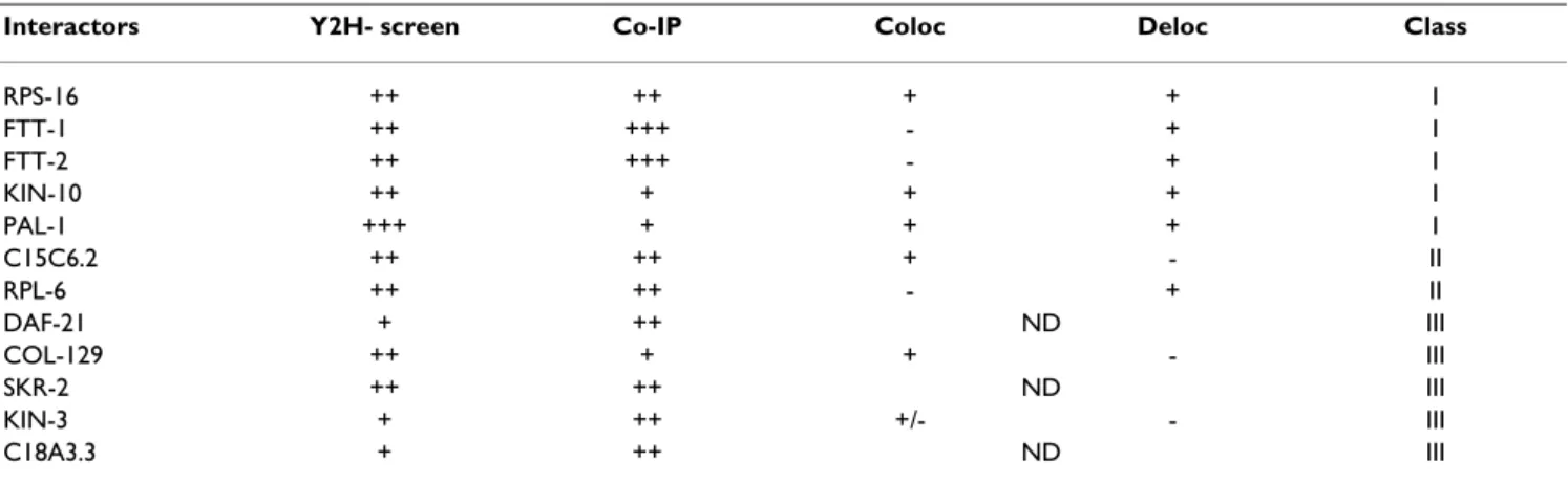
Table 4: Summary of interaction strengths
Interactors Y2H- screen Co-IP Coloc Deloc Class
RPS-16 ++ ++ + + I FTT-1 ++ +++ - + I FTT-2 ++ +++ - + I KIN-10 ++ + + + I PAL-1 +++ + + + I C15C6.2 ++ ++ + - II RPL-6 ++ ++ - + II DAF-21 + ++ ND III COL-129 ++ + + - III SKR-2 ++ ++ ND III KIN-3 + ++ +/- - III C18A3.3 + ++ ND III
Scores obtained in the different assays used: (i) two-hybrid assays, (ii) co-immunoprecipitation (co-IP), (iii) colocalization by immunofluorescence in single transfected cells (Coloc) and (iv) delocalization (Deloc) of one or the two partners induced by the cotranfection. Interactions were classified as I (the strongest) to III (the weakest).
involved in various aspects of protein synthesis or degra-dation. Further analysis by co-immunoprecipitation and colocalization confirmed the interactions identified. We demonstrated that not only LET-756 could interact with mammalian partners as well as their orthologs (e.g 14-3-3β vs FTT-1/PAR-5 and FTT-2) but also that orthologs of mammalian FGF partners could interact with LET-756 (CK2β vs KIN-10). Analyses of these partners could be of interest in the study of mammalian FGFs.
The majority of the proteins we identified are nuclear, which was expected since the two-hybrid system needs the fusion to be targeted to the nucleus. However, some inter-actions have been identified as false positive in other screens [28]. By performing co-immunoprecipitations and studying subcellular localization, we validated the interaction of LET-756 with RPS-16, FTT-1/PAR-5, FTT-2, KIN-10 and RPL-6 with high score, and with PAL-1, DAF-21, SKR-2, KIN-3 and C18A3.3 with lower strength (see Table 6). The function of some interacting partners is rel-evant to FGF biology. The 14-3-3/FTT-1/FTT-2 proteins, which belong to the highly conserved family of chaperone molecules transit to the nucleus and participate in nucleo-cytoplasmic transport, regulating intracellular transduc-tion [30]. We did not find a 14-3-3 conventransduc-tional phos-phorylated binding site on LET-756. However, other domains have been involved in 14-3-3 binding, such as nuclear localization signals (31 for review). No role for FTT-1 or FTT-2 in modulating secretion has been assigned
in C. elegans. It will be interesting to analyze whether interaction of these proteins with the EFVSVA motif of secretion described in LET-756 [16] causes its secretion. The interaction of LET-756 with PAL-1 is of interest because PAL-1 is also highly conserved during evolution. It is the ortholog of caudal (Drosophila), CDX1, 2 and 4 (mammals) and Xcad3 (Xenopus) paraHOX proteins. Caudal proteins are involved in the transcriptional regula-tion of multiple genes that are involved in posterior pat-terning [32]. The interaction of LET-756 with PAL-1 could activate the expression of various genes involved in nem-atode anterior-posterior development as it is the case for the interaction of Xenopus e-FGF with Xcad3 and the resulting activation of HOX genes [33,34]. In addition,
pal-1 mutant exhibits aberrant cell position in posterior
muscle cells [35], a site of LET-756 expression [13,19] as well as in posterior hypodermis, a site of LET-756 action [17]. Both muscle and epidermis evolve from the C line-age. In the absence of PAL-1, the C blastomeres fail to develop. Protein phosphorylation by the coordinated activities of protein kinases and phosphatases is central to many signal transduction pathways. The combined action of LET-756/FGF, EGL-15 receptor, CLR-1 phosphatase (for a review see [14]) and KIN-3 and KIN-10, the respec-tive catalytic and regulatory subunits of CK2, might regu-late various processes involved in proliferation -as it is described for FGF1 and 2 [43,9] – or in other functions. KIN-3 and KIN-10 have been recently implicated in pri-mary cilia biology [55]. Finally, some ribosomal proteins
Co-immunoprecipitation of LET-756::GFP and HA-tagged protein partners Figure 1
Co-immunoprecipitation of LET-756::GFP and HA-tagged protein partners Cos-1 cells were transfected with let-756::gfp alone (0) and with various HA-tagged constructs presumed or not (TACC1) to interact with LET-756. Twenty four
hours later cell lysates were immunoprecipitated with anti-GFP antibodies. Western blots were first revealed with anti-HA antibodies and then with anti-GFP antibodies. An additional band, indicated with an asterisk, was consistently observed with HA- tagged RPL-6. A week and non- reproducible band can also be seen in the FTT-2 co-immunoprecipitation lane. None of these bands arose from possible complexes since 1) electrophoresis was performed in denaturating conditions and 2) antibody detection relied on the tag epitope and not on the endogenous protein.
BMC Genomics 2006, 7:105 http://www.biomedcentral.com/1471-2164/7/105
interact with mammalian FGF [36-38] to regulate their signaling and trafficking to the nucleus; reciprocally, FGFs may regulate ribosome biogenesis and protein synthesis during the G1 phase of the cell cycle. In contrast to these relevant interactors, others appear irrelevant, such as MBD1 since no methylation occurs in C. elegans.
The interactions revealed by the two-hybrid screens are rather weak. This could be due to 1) the high stringency associated with the system using the MAV 103/203 yeast strains; it is worth noting that in large-scale screenings of interactions no partner for LET-756 was found [20]; 2) a bad exposure of the binding site in the fusion proteins; 3) the existence of ternary interactions as seen in the ligand – tyrosine kinase receptor – heparan sulphate complex and 4) the need for post-translational modifications of the proteins that occur in mammalian cells and not in yeast, explaining why better interactions between glyco-sylated LET-756 [16] and various partners were obtained in immunoprecipitation and immunofluorescence exper-iments than in yeast two-hybrid screens.
Finally, it will be interesting to know whether the expres-sion pattern of LET-756, which is mainly muscular and neuronal in the worm, overlap with that of the various partners. Search in the literature (56, 57) was not conclu-sive since the majority of the partners were found in eggs (FTT-1/PAR-5, FTT-2, DAF-21, PAL-1), intestine (HSP-6, SKR-2) or cuticle (COL-129).
Conclusion
We have conducted the first extensive search for LET-756 interactors and established the first interaction map of LET-756/FGF with FGF binding proteins (Fig. 3 and Table 6). This could help understand FGF functions. Proteins of interest were involved in developmental processes or in basic biochemical events such as ribosome biogenesis and protein synthesis. In addition, to get insight in the evolu-tion of the FGF interactome network, which we have illus-trated in Fig. 3, we tested 6 of 20 orthologs of human FGF interactors (Table 6), and found KIN-10 and RPL-6 as new potential interactors. Looking for physical interactions in
Intracellular localization of LET-756::GFP and HA-tagged protein partners Figure 2
Intracellular localization of LET-756::GFP and HA-tagged protein partners Cos-1 cells were transfected with let-756::gfp with or without various HA-tagged encoded constructs. Twenty four hours later cells were either fixed in 4%
parafor-maldehyde and permeabilized in triton or in cold methanol. Cells were incubated for an hour with rat anti-HA antibodies. Sec-ondary goat anti-rat Texas red-coupled antibodies were then added for another 30 min. Coverslips were visualized with a confocal Leica microscope. Cells were transfected with the HA-tagged protein alone (column I) or cotransfected with
let-756::gfp and revealed by direct fluorescence (column II), by anti-HA immunofluorescence (column III) and merge (column IV).
In cells cotransfected with LET-756 and KIN-10 and treated with actinomycin D, both GFP staining and anti-HA immunostain-ing are displaced around and into the nucleolus. The yellow star indicates a neighbor simmunostain-ingle KIN-10 transfected cells. Its locali-zation is not modified by the drugs. Panels A and B each show results for five interactors analyzed.
a physiological system will determine which of these interactions are essential.
In conclusion, 1) combining the yeast two-hybrid screen with bioinformatics and computational biology, we have delineated potential interactors of LET-756, and possibly of the entire FGF family; 2) comparative genomics analy-sis yielded valuable insights into conserved and divergent aspects of function, regulation, and evolution since not all pathways are conserved as demonstrated by the ortholog analysis; 3) the information given herein, although not complete, might be useful for people working in the field.
Methods
Yeast two-hybrid assays
A full-length let-756 transcript was fused in-frame with the coding sequence of the DNA binding domain (DB) of Gal4, and was used in a Y2H screen system as described in [20] for the C elegans libraries. Two worm libraries fused to the activation domain (AD) of Gal4, a cDNA and the AD-ORFeome libraries [53] were screened. The MAV103 yeast strain based Y2H assay contains three reporter genes
(HIS3, lacZ and URA3) [54]. A cDNA human placenta library fused to the activation domain of LexA was also screened. The L40 yeast strain based Y2H assay contains only two reporters (HIS3 and lacZ). In this case, the full length let-756 transcript was cloned into the LexA DNA binding domain bait expression vector pBTM116B Kana. Yeast assays were done using conventional lithium ace-tate-based method. Clones were assigned scores for LacZ expression, growth on plates lacking histidine but con-taining 20 or 40 mM 3-amino triazol and in addition, growth on plates lacking uracil for the C. elegans libraries. To ascertain interactions, the gap repair technique was performed as in [54].
Plasmid construction
To generate prey-tagged expression vectors used in co-immunoprecipitation assay or immunofluorescence, the coding regions of various genes were amplified by PCR using as template the corresponding EST clones obtained from RZPD (Berlin, Germany) and then inserted in the expression vectors using Gateway technology (Invitrogen, Carlsbad, CA).
Interactome of the FGF family Figure 3
Interactome of the FGF family The identified interactors of various FGFs were grouped in six categories, depending of the
BMC Genomics 2006, 7:105 http://www.biomedcentral.com/1471-2164/7/105
LET-756::GFP was obtained as previously described [16].
Cell culture and in vivo interaction assay
Cos-1 cells grown in DMEM supplemented with 10% fetal calf serum were plated in 60-mm dishes at a concentration of 2 × 10⁶ cells/dish and immediately transfected with 1µg DNA in Fugene, according to the manufacturer instruc-tions. Twenty four hours after transfection, cells were lysed in 1 ml triton buffer (10 mM Tris, pH7.4, 100 mM NaCl, 2.5 mM MgCl2, 1% triton, 1 mM EDTA, 10 mM DTT). Detergent insoluble materials were removed by 30 min centrifugation at 13000 rpm at 4°C. Whole cell lysates were first incubated with protein G-sepharose beads and then with the relevant antibody for at least 2 hr. Protein G-sepharose beads were then added for another additional 2 hr and washed 3 times with lysis buffer. Bound proteins were eluted by boiling in SDS sample buffer and resolved on a 10% SDS-PAGE gel and analyzed by Western blots. For immunofluorescence analysis, cells grown on glass coverslips were fixed and permeabilized in 3.7% PAF and 0.1%Triton or in methanol for 6 min at -20°C. Similar results were obtained using these different modes of fixation. Cells were incubated with primary anti-body for 1 hr and then incubated with Texas Red-conju-gated secondary antibody for another hr. Plasmid LET-756::GFP was visualized by autofluorescence. Coverslips were examined using a Leica TCS NT confocal microscope. The following antibodies were used: rat monoclonal anti-HA (12CA5) antibody was from Roche (Indianopolis, IN, USA), rabbit polyclonal anti-GFP from Abcam (Cam-bridge, UK), Texas Red anti-rat antibody from Molecular Probes (Eugene, OR, USA), peroxydase anti-mouse from Santa Cruz (Santa Cruz, CA, USA)
Abbreviations
DMEM, Dulbecco's mofied Eagle's medium; FBS, fetal bovine serum; DTT dithiotreitol; Y2H, yeast two hybrid; GFP, green fluorescent protein
Authors' contributions
CP, YB and AH performed the two-hybrid screenings and the analyses of the data, CP and FC the gap-repair confir-mation of clones, FC and RR the co-immunoprecipitation and immunofluorescence experiments, CP the art work. CP played the major role in the bioinformatics analysis. DB initiated the C. elegans project and helped draft the manuscript. RR conceived and coordinated the study and wrote the manuscript. All authors read and approved the final manuscript.
Acknowledgements
We thank F. Birg and D. Maraninchi for encouragements, P. Pontarotti for helpful discussions, P. Lecine for advices during this study and J. Reboul for the gift of the ORFeome library and advices. The work has been supported
by Inserm, Institut Paoli-Calmettes and grants from the Ligue Nationale Contre le Cancer (Label).
References
1. Goldfarb M: Functions of fibroblast growth factors in
verte-brate development. Cytokine Growth Factor Rev 1996, 7:311-325.
2. Dickson C, Spencer-Dene B, Dillon C, Fantl V: Tyrosine kinase
sig-nalling in breast cancer: fibroblast growth factors and their receptors. Breast Cancer Res 2000, 2:191-196.
3. Goldfarb M: Signaling by fibroblast growth factors: the inside
story. Sci STKE 2001, 106:PE37.
4. Kolpakova E, Wiedlocha A, Stenmark H, Klingenberg O, Falnes PO, Olsnes S: Cloning of an intracellular protein that binds
selec-tively to mitogenic acidic fibroblast growth factor. Biochem J
1998, 336:213-222.
5. LaVallee TM, Tarantini F, Gamble S, Mouta Carreira C, Jackson A, Maciag T: Synaptotagmin-1 is required for fibroblast growth
factor-1 release. J Biol Chem 1998, 273:22217-22223.
6. Burrus LW, Zuber ME, Lueddecke BA, Olwin BB: Identification of
a cysteine-rich receptor for fibroblast growth factors. Mol Cell Biol 1992, 12:5600-5609.
7. Kohl R, Antoine M, Olwin BB, Dickson C, Kiefer P: Cysteine-rich
fibroblast growth factor receptor alters secretion and intra-cellular routing of fibroblast growth factor 3. J Biol Chem 2000, 275:15741-15748.
8. Kamikura DM, Cooper JA: Lipoprotein receptors and a disabled
family cytoplasmic adaptor protein regulate EGL-17/FGF export in C. elegans. Genes Dev 2003, 17:2798-2811.
9. Bonnet H, Filhol O, Truchet I, Brethenou P, Cochet C, Amalric F, Bouche G: Fibroblast growth factor-2 binds to the regulatory
beta subunit of CK2 and directly stimulates CK2 activity toward nucleolin. J Biol Chem 1996, 271:24781-24787.
10. Gringel S, van Bergeijk J, Haastert K, Grothe C, Claus P: Nuclear
fibroblast growth factor-2 interacts specifically with splicing factor SF3a66. Biol Chem 2004, 385:1203-1208.
11. Bossard C, Van den Berghe L, Laurell H, Castano C, Cerutti M, Prats AC, Prats H: Antiangiogenic properties of fibstatin, an
extra-cellular FGF-2-binding polypeptide. Cancer Res 2004, 64:7507-7512.
12. Peng H, Sahni A, Fay P, Bellum S, Prudovsky I, Maciag T, Francis CW:
Identification of a binding site on human FGF-2 for fibrino-gen. Blood 2004, 103:2114-2120.
13. Birnbaum D, Popovici C, Roubin R: A pair as a minimum: The
two fibroblast growth factors of the nematode
Caenorhabdi-tis elegans. Dev Dyn 2005, 232:247-255.
14. Huang P, Stern MJ: FGF signaling in flies and worms: more and
more relevant to vertebrate biology. Cytokine Growth Factor Rev
2005, 16:151-158.
15. Roubin R, Naert K, Popovici C, Vatcher G, Coulier F, Thierry-Mieg J, Pontarotti P, Birnbaum D, Baillie D, Thierry-Mieg D: let-756, a C.
elegans fgf essential for worm development. Oncogene 1999,
18:6741-6747.
16. Popovici C, Conchonaud F, Birnbaum D, Roubin R: Functional
phy-logeny relates LET-756 to FGF9. J Biol Chem 2004, 279:40146-40152.
17. Huang P, Stern MJ: FGF signaling functions in the hypodermis
to regulate fluid balance in C. elegans. Development 2004, 131:2595-2604.
18. Bulow HE, Boulin T, Hobert O: Differential functions of the C.
elegans FGF receptor in axon outgrowth and maintenance of
axon position. Neuron 2004, 42:367-374.
19. Popovici C, Fallet M, Marguet D, Birnbaum D, Roubin R:
Intracellu-lar trafficking of LET-756, a fibroblast growth factor of C.
ele-gans, is controlled by a balance of export and nuclear signals.
Exp Cell Res 2006, 312:1484-1495.
20. Reboul J, Vaglio P, Rual JF, Lamesch P, Martinez M, Armstrong CM, Li S, Jacotot L, Bertin N, Janky R, Moore T, Hudson JR Jr, Hartley JL, Bra-sch MA, Vandenhaute J, Boulton S, Endress GA, Jenna S, Chevet E, Papasotiropoulos V, Tolias PP, Ptacek J, Snyder M, Huang R, Chance MR, Lee H, Doucette-Stamm L, Hill DE, Vidal M: C. elegans
ORFe-ome version 1.1: experimental verification of the genORFe-ome annotation and resource for proteome-scale protein expres-sion. Nat Genet 2003, 34:35-41.
21. Baugh LR, Hill AA, Claggett JM, Hill-Harfe K, Wen JC, Slonim DK, Brown EL, Hunter CP: The homeodomain protein PAL-1
spec-Publish with BioMed Central and every scientist can read your work free of charge
"BioMed Central will be the most significant development for disseminating the results of biomedical researc h in our lifetime."
Sir Paul Nurse, Cancer Research UK
Your research papers will be:
available free of charge to the entire biomedical community peer reviewed and published immediately upon acceptance cited in PubMed and archived on PubMed Central yours — you keep the copyright
Submit your manuscript here:
http://www.biomedcentral.com/info/publishing_adv.asp
BioMedcentral
ifies a lineage-specific regulatory network in the C. elegans embryo. Development 2005, 132:1843-1854.
22. Birnby DA, Link EM, Vowels JJ, Tian H, Colacurcio PL, Thomas JHA:
Transmembrane guanylyl cyclase (DAF-11) and Hsp90 (DAF-21) regulate a common set of chemosensory behav-iors in Caenorhabditis elegans. Genetics 2000, 155:85-104.
23. Inoue T, Takamura K, Yamae H, Ise N, Kawakami M, Tabuse Y, Miwa J, Yamaguchi Y: Caenorhabditis elegans DAF-21 (HSP90) is
characteristically and predominantly expressed in germline cells: spatial and temporal analysis. Dev Growth Differ 2003, 45:369-376.
24. Nayak S, Santiago FE, Jin H, Lin D, Schedl T, Kipreos ET: The
Caenorhabditis elegans Skp1-related gene family: diverse
functions in cell proliferation, morphogenesis, and meiosis. Curr Biol 2002, 12:277-287.
25. Yamanaka A, Yada M, Imaki H, Koga M, Ohshima Y, Nakayama K:
Multiple Skp1-related proteins in Caenorhabditis elegans : diverse patterns of interaction with Cullins and F-box pro-teins. Curr Biol 2002, 12:267-275.
26. Popovici C, Roubin R, Coulier F, Birnbaum D: An evolutionary
his-tory of the FGF superfamily. Bioessays 2005, 27:849-857.
27. Itoh N, Ornitz DM: Evolution of the Fgf and Fgfr gene families.
Trends Genet 2004, 20:563-569.
28. Van Criekinge W, Beyaert R: Yeast Two-Hybrid: State of the
Art. Biol Proced Online 1999, 2:1-38.
29. Burdine RD, Chen EB, Kwok SF, Stern MJ: egl-17 encodes an
inver-tebrate fibroblast growth factor family member required specifically for sex myoblast migration in Caenorhabditis
ele-gans. Proc Natl Acad Sci USA 1997, 94:2433-2437.
30. Aitken A: 14-3-3 and its possible role in co-ordinating multiple
signalling pathways. Trends Cell Biol 1996, 6:341-347.
31. Bridges D, Moorhead GB: 14-3-3 proteins: a number of
func-tions for a numbered protein. Sci STKE 2005, 296:re10.
32. Pellettieri J, Seydoux G: Anterior-posterior polarity in C.
ele-gans and Drosophila – PARallels and differences. Science 2002, 298:1946-1950.
33. Pownall ME, Tucker AS, Slack JM, Isaacs HV: eFGF, Xcad3 and Hox
genes form a molecular pathway that establishes the anter-oposterior axis in Xenopus. Development 1996, 122:3881-3892.
34. Isaacs HV, Pownall ME, Slack JM: Regulation of Hox gene
expres-sion and posterior development by the Xenopus caudal homologue Xcad3. EMBO J 1998, 17:3413-3427.
35. Edgar LG, Carr S, Wang H, Wood WB: Zygotic expression of the
caudal homolog pal-1 is required for posterior patterning in
Caenorhabditis elegans embryogenesis. Dev Biol 2001,
229:71-88.
36. Shen B, Arese M, Gualandris A, Rifkin DB: Intracellular association
of FGF-2 with the ribosomal protein L6/TAXREB107. Bio-chem Biophys Res Commun 1998, 252:524-528.
37. Soulet F, Al Saati T, Roga S, Amalric F, Bouche G: Fibroblast growth
factor-2 interacts with free ribosomal protein S19. Biochem Biophys Res Commun 2001, 289:591-596.
38. Skjerpen CS, Wesche J, Olsnes S: Identification of
ribosome-binding protein p34 as an intracellular protein that binds acidic fibroblast growth factor. J Biol Chem 2002, 277:23864-23871.
39. Mizukoshi E, Suzuki M, Loupatov A, Uruno T, Hayashi H, Misono T, Kaul SC, Wadhwa R, Imamura T: Fibroblast growth factor-1
interacts with glucose-regulated protein GRP75/mortalin. Biochem J 1999, 343:461-466.
40. Ananyeva N, Tjurmin A, Saenko E, Haudenschild C: Low density
lipoproteins interact with acidic fibroblast growth factor and modify its function. Arterioscler Thromb Vasc Biol 2003, 23:601-607.
41. Carreira CM, LaVallee TM, Tarantini F, Jackson A, Lathrop JT, Hamp-ton B, Burgess WH, Maciag T: S100A13 Is Involved in the
Regu-lation of Fibroblast Growth Factor-1 and p40 Synaptotagmin-1 Release in Vitro. J Biol Chem 1998, 273:22224-22231.
42. Tassi E, Al-Attar A, Aigner A, Swift MR, McDonnell K, Karavanov A, Wellstein A: Enhancement of fibroblast growth factor (FGF)
activity by an FGF-binding protein. J Biol Chem 2001, 276:40247-40253.
43. Wiedlocha A, Falnes PO, Madshus IH, Sandvig K, Olsnes S: Dual
mode of signal transduction by externally added acidic fibroblast growth factor. Cell 1994, 76:1039-1051.
44. Bossard C, Laurell H, Van den Berghe L, Meunier S, Zanibellato C, Prats H: Translokin is an intracellular mediator of FGF-2
traf-ficking. Nat Cell Biol 2003, 5:433-439.
45. Claus P, Doring F, Gringel S, Muller-Ostermeyer F, Fuhlrott J, Kraft T, Grothe C: Differential intranuclear localization of fibroblast
growth factor-2 isoforms and specific interaction with the survival of motoneuron protein. J Biol Chem 2003, 278:479-485.
46. Claus P, Bruns AF, Grothe C: Fibroblast growth factor-2(23)
binds directly to the survival of motoneuron protein and is associated with small nuclear RNAs. BiochemJ 2004, 384:559-565.
47. Sahni A, Khorana AA, Baggs RB, Peng H, Francis CW: FGF-2
bind-ing to fibrin(ogen) is required for augmented angiogenesis. Blood 2006, 107:126-131.
48. Van den Berghe L, Laurell H, Huez I, Zanibellato C, Prats H, Bugler B:
FIF [fibroblast growth factor-2 (FGF-2)-interacting-factor], a nuclear putatively antiapoptotic factor, interacts specifi-cally with FGF-2. Mol Endocrinol 2000, 14:1709-1724.
49. Camozzi M, Zacchigna S, Rusnati M, Coltrini D, Ramirez-Correa G, Bottazzi B, Mantovani A, Giacca M, Presta M: Pentraxin 3 inhibits
fibroblast growth factor 2-dependent activation of smooth muscle cells in vitro and neointima formation in vivo. Arterio-scler Thromb Vasc Biol 2005, 25:1837-1842.
50. Chadderton NS, Stringer SE: Interaction of platelet factor 4 with
fibroblast growth factor 2 is stabilised by heparan sulphate. Int J Biochem Cell Biol 2003, 35:1052-1055.
51. Reimers K, Antoine M, Zapatka M, Blecken V, Dickson C, Kiefer P:
NoBP, a nuclear fibroblast growth factor 3 binding protein, is cell cycle regulated and promotes cell growth. Mol Cell Biol
2001, 21:4996-5007.
52. Schoorlemmer J, Goldfarb M: Fibroblast growth factor
homolo-gous factors and the islet brain-2 scaffold protein regulate activation of a stress-activated protein kinase. J Biol Chem
2002, 277:49111-49119.
53. Vaglio P, Lamesch P, Reboul J, Rual JF, Martinez M, Hill D, Vidal M:
The Caenorhabditis elegans ORFeome Database. Nucleic Acids Res 2003, 1:31237-31240.
54. Walhout AJ, Vidal M: High-throughput yeast two-hybrid assays
for large-scale protein interaction mapping. Methods 2001, 24:297-306.
55. Hu J, Bae YK, Knobel KM, Barr MM: Casein Kinase II and
Cal-cineurin Modulate TRPP Function and Ciliary Localization. Mol Biol Cell 2006, 17:2200-2211.
56. [http://nematode.lab.nig.ac.jp/db2/]. 57. [http://elegans.bcgsc.ca/perl/eprofile/index]. 58. [http://www.wormbase.org].