Publisher’s version / Version de l'éditeur:
Journal of molecular biology, 398, 1, pp. 111-121, 2010-03-10
READ THESE TERMS AND CONDITIONS CAREFULLY BEFORE USING THIS WEBSITE. https://nrc-publications.canada.ca/eng/copyright
Vous avez des questions? Nous pouvons vous aider. Pour communiquer directement avec un auteur, consultez la
première page de la revue dans laquelle son article a été publié afin de trouver ses coordonnées. Si vous n’arrivez pas à les repérer, communiquez avec nous à PublicationsArchive-ArchivesPublications@nrc-cnrc.gc.ca.
Questions? Contact the NRC Publications Archive team at
PublicationsArchive-ArchivesPublications@nrc-cnrc.gc.ca. If you wish to email the authors directly, please see the first page of the publication for their contact information.
Archives des publications du CNRC
This publication could be one of several versions: author’s original, accepted manuscript or the publisher’s version. / La version de cette publication peut être l’une des suivantes : la version prépublication de l’auteur, la version acceptée du manuscrit ou la version de l’éditeur.
For the publisher’s version, please access the DOI link below./ Pour consulter la version de l’éditeur, utilisez le lien DOI ci-dessous.
https://doi.org/10.1016/j.jmb.2010.03.006
Access and use of this website and the material on it are subject to the Terms and Conditions set forth at
Structural basis of the regulation of the CbpA Co-chaperone by its
specific modulator CbpM
Sarraf, Naghmeh S.; Baardsnes, Jason; Cheng, Jing; O'Connor-McCourt,
Maureen; Cygler, Miroslaw; Ekiel, Irena
https://publications-cnrc.canada.ca/fra/droits
L’accès à ce site Web et l’utilisation de son contenu sont assujettis aux conditions présentées dans le site LISEZ CES CONDITIONS ATTENTIVEMENT AVANT D’UTILISER CE SITE WEB.
NRC Publications Record / Notice d'Archives des publications de CNRC:
https://nrc-publications.canada.ca/eng/view/object/?id=b1c8f8ac-de34-473d-8848-b0ee52d0583b https://publications-cnrc.canada.ca/fra/voir/objet/?id=b1c8f8ac-de34-473d-8848-b0ee52d0583bStructural Basis of the Regulation of the CbpA
Co-chaperone by its Specific Modulator CbpM
Naghmeh S. Sarraf
1,2⁎
, Jason Baardsnes
1, Jing Cheng
1,2,
Maureen O'Connor-McCourt
1, Miroslaw Cygler
1,3and Irena Ekiel
1,2⁎
1Health Sector, Biotechnology Research Institute, National Research Council of Canada, 6100 Royalmount Avenue, Montreal, Quebec,
Canada H4P 2R2
2Department of Chemistry and Biochemistry, Concordia University, 7141 Sherbrooke W., Montreal, Quebec, Canada H4B 1R6
3Department of Biochemistry, McGill University, Montreal, Quebec, Canada H3G 1Y6 Received 3 February 2010; received in revised form 2 March 2010;
accepted 3 March 2010
Available online 10 March 2010
CbpA, one of the Escherichia coli DnaJ homologues, acts as a co-chaperone in the DnaK chaperone system. Despite its extensive similarity in domain structure and function to DnaJ, CbpA has a unique and specific regulatory mechanism mediated through the small protein CbpM. Both CbpA and CbpM are highly conserved in bacteria. Earlier studies showed that CbpM interacts with the N-terminal J-domain of CbpA inhibiting its co-chaperone activity but the structural basis of this interaction is not known. Here, we have combined NMR spectroscopy, site-directed mutagenesis and surface plasmon resonance to characterize the CbpA/CbpM interaction at the molecular level. We have determined the solution structure of the CbpA J-domain and mapped the residues that are perturbed upon CbpM binding. The NMR data defined a broad region on helices α2 and α3 as involved in the interactions. Site-directed mutagenesis has been used to further delineate the CbpA J-domain/CbpM interface. We show that the binding sites of CbpM and DnaK on CbpA J-domain overlap, which suggests a competition between DnaK and CbpM for binding to CbpA as a mechanism for CbpA regulation. This study also provides the explanation for the specificity of CbpM for CbpA versus DnaJ, by identifying the key residues for differential binding.
Crown Copyright @ 2010 Published by Elsevier Ltd. All rights reserved.
Edited by P. Wright
Keywords:CbpA-J domain; CbpM; NMR structure; site-directed mutagenesis;
SPR
Introduction
Chaperones include a broad collection of proteins that aid nascent polypeptide folding, transition across cellular and organelle membranes, disassembly of
macromolecular complexes or aggregates, targeting for proteolysis, and quality control, as well as the regulation of conformational changes that affect biological functions, and signalling.1,2 The Hsp70
chaperone family represents one of the most potent cellular defences against harsh environmental changes, enabling survival under stress conditions such as heat shock,3hypothermia4and environmental
pollution.5These chaperones are present in all bacteria
and in all cellular compartments of eukaryotes.6Their
activity is controlled by a cycle of ATP binding, hydrolysis, and nucleotide exchange.7 This cycle is
regulated by Hsp40 co-chaperones, which stimulate ATP hydrolysis8and stimulate the activity of Hsp70s
by stabilizing their interaction with substrates.9 Escherichia coli contains three Hsp70 proteins,
DnaK, Hsc62/HscC and Hsc66/HscA.9,10 DnaK is
important for bacterial viability6 and is the most
extensively characterized Hsp70 family member. There are presently six identified Hsp40 co-chaper-ones in E. coli, three of which, DnaJ, CbpA and DjlA, bind to DnaK11 (Fig. 1). These proteins can bind to
*Corresponding authors.Health Sector, Biotechnology Research Institute, National Research Council of Canada, 6100 Royalmount Avenue, Montreal, Quebec, Canada H4P 2R2. E-mail addresses:naghmeh.sarraf@bri.nrc.ca;
irena.ekiel@nrc.ca.
Present address: J. Cheng, Department of Animal Science, MacDonald Campus, McGill University, 21111 Lakeshore Road, Ste-Anne-De-Bellevue, Quebec, Canada H9X 3V9.
Abbreviations used: JdomCbpA, J-domain of CbpA;
MBP, maltose-binding protein; SEC, size-exclusion chromatography; SPR, surface plasmon resonance; MBP, maltose-binding protein; SEC, size-exclusion
chromatography; SPR, surface plasmon resonance; HSQC, heteronuclear single quantum coherence; NOE, nuclear Overhauser effect; NOESY, NOE spectroscopy.
Available online at www.sciencedirect.com
substrate polypeptides and transfer them to Hsp70. Among them, DnaJ is the best characterized as a regulator of various DnaK activities.12,13 The
co-chaperones belonging to DnaJ/Hsp40 class (JDP family) contain an ∼ 70-residue long J-domain signature motif. The J-domain promotes ATP hy-drolysis by Hsp70, and nucleotide exchange factors return the Hsp70 protein to the ATP-bound state, which releases the substrate, and resets the cycle.1,10
The E. coli 34 kDa CbpA was isolated originally by virtue of its retention on a curved DNA-affinity column (named for curved DNA-binding protein).14
It was subsequently shown that CbpA is a major protein associated with E. coli nucleoids during the stationary growth phase.15Over-expression of CbpA
can complement for known phenotypes associated with the loss of DnaJ function, including bacterial temperature sensitivity, as well as failure to replicate mini-F plasmid and bacteriophage λ.16 In all these
cases, however, multiple copies of cbpA are required for the suppression of dnaJ mutant phenotypes, indicating that dnaJ and cbpA are under distinctly different regulation in the cell.16,17 The amino acid
sequence of CbpA is 39% identical with that of DnaJ, but it lacks the 69 amino acid cysteine-rich zinc finger domain of DnaJ. Nevertheless, CbpA, like DnaJ, can function as a DnaK co-chaperone in vitro and in vivo18
constituting a functional homolog of DnaJ.19 The
J-domain is the most conserved part of these proteins, with residues 1–78 of CbpA showing 55% identity with the E. coli DnaJ J-domain.19
In vitro, the CbpA co-chaperone binds efficiently to
protein substrates,14activates the DnaK chaperone for
the remodelling of protein complexes and promotes protein disaggregation.18,20 Like DnaJ, CbpA forms
a homodimer in solution, with dimerization in both proteins mediated through the C-terminal domain.18,19A unique feature of CbpA as compared
to DnaJ is its intimate interaction with a specific inhibitory partner protein, CbpM.11 CbpM is coded
by the yccD gene that lies downstream of cbpA within the same operon and shares homology with proteins encoded by genes located downstream of dnaJ-like
genes in a diverse range of bacteria. The conserved grouping of these genes and the co-expression of these two proteins suggested that they act together.18It has
been shown that CbpM binds specifically to CbpA and inhibits both the co-chaperone activity and the DNA-binding activity of CbpA.21,22It was suggested
that during certain growth or stress conditions CbpA might be released from CbpM and recruited by DnaK to function as a co-chaperone.18
The CbpM interacting region is within the J-domain of CbpA (JdomCbpA) and they bind with
a 1:1 ratio,19 although the stoichiometry of the
complex was not studied in detail. The binding site and precise nature of this interaction at the structural level are not known.19 Also, despite the
high level of sequence conservation between CbpA and DnaJ and their functional overlap, the molecular reasons for the specificity of CbpM for CbpA versus DnaJ are not known.11
Here, we address the question of the structural basis for the specific regulation of CbpA by CbpM. Using an integrated approach that includes NMR spectroscopy, mutagenesis and biophysical charac-terization we have determined the solution structure of JdomCbpAand characterized the most important
residues for the interaction between CbpA and CbpM. We have identified the sequence differences between the J-domains of DnaJ and CbpA that determine the specificity of the regulation by CbpM.
Results
NMR structure of the J-domain from the co-chaperone CbpA
The solution structure of the J-domain of CbpA was determined by NMR spectroscopy (Table 1; Fig. 1. The DnaK chaperone/co-chaperone/modulator
system in E. coli. For DnaJ, CbpA and DjlA, J-domains are shown as black circles. The other domains are not shown in detail, except for the transmenbrane (TM) domain of DjlA. CbpM, the specific modulator for CbpA, is shown as a rounded rectangle.
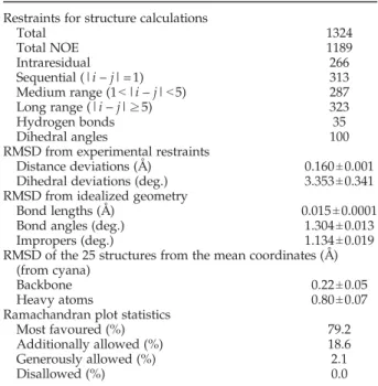
Table 1.Structural statistics for JdomCbpA
Restraints for structure calculations
Total 1324
Total NOE 1189
Intraresidual 266
Sequential (|i – j|=1) 313
Medium range (1b|i – j|b5) 287
Long range (|i – j|≥5) 323
Hydrogen bonds 35
Dihedral angles 100
RMSD from experimental restraints
Distance deviations (Å) 0.160±0.001 Dihedral deviations (deg.) 3.353±0.341 RMSD from idealized geometry
Bond lengths (Å) 0.015±0.0001
Bond angles (deg.) 1.304±0.013
Impropers (deg.) 1.134±0.019
RMSD of the 25 structures from the mean coordinates (Å) (from cyana)
Backbone 0.22±0.05
Heavy atoms 0.80±0.07
Ramachandran plot statistics
Most favoured (%) 79.2
Additionally allowed (%) 18.6
Generously allowed (%) 2.1
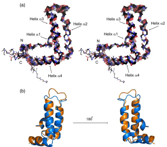
Fig. 2). The NMR structure reveals 4 helices: helix α1 (Y6 – I9), α2 (L18 – K31), α3 (A42 – L56) and α4 (Q60 – M68). The two central helices, α2 and α3, form an antiparallel hairpin, with helices α1 and α4 extend-ing to one side, creatextend-ing an orthogonal bundle (Fig. 2a). The protein shows structural similarity to other J-domains, in particular to the J-domain of DnaJ from E. coli (PDB 1XBL, Fig. 2b),23 with which it
shares 55% sequence identity (the RMSD for the backbone atoms of residues 5 – 68 of both molecules is 1.32 Å). Most of the functionally important residues involved in chaperone binding are located on helices α2 and α3 in both J-domains of CbpA and DnaJ.19,24Superposition of only these two helices in
the two J-domains results in an RMSD of 0.81 Å for the backbone atoms, which indicates a high degree of similarity between these regions in CbpA and DnaJ. A highly basic region, typical for J-domains and composed of side chains from helix α2 (K19 – R31), is even more basic than in DnaJ due to having R30 instead of M30 in DnaJ. In the neighboring loop between helices α2 and α3, however, R36 of DnaJ is
replaced by V36 in CbpA. This loop, which has different lengths in CbpA and DnaJ, contains the highly conserved residues HPDRN (33–37), which are critical for functional activity of DnaJ as a co-chaperone.24 The remaining residues are less well
conserved among J-domains.
Characterization of the interaction between JdomCbpAand CbpM
Interactions between JdomCbpA and CbpM were
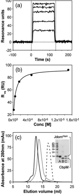
characterized by surface plasmon resonance (SPR), size-exclusion chromatography (SEC) and nuclear magnetic resonance (NMR) spectroscopy. SPR de-termined that the dissociation constant of the complex (KD) is 450 nM (Fig. 3a and b). The complex
was easily detected by SEC and eluted with an apparent molecular mass of ∼55 kDa (Fig. 3c). Densitometry of the individual protein bands from SDS-PAGE for the complex (Fig. 3c) showed a JdomCbpA to CbpM ratio of 1:1, which agrees with
the previously suggested 1:1 ratio in the complex.19
Fig. 2. (a) NMR structure of JdomCbpA. A stereo view of the superposition of the final 20 energy-minimized structures.
The elements of secondary structure are annotated. (b) An overlay of the JdomCbpA(blue) and JdomDnaJ(1XBL) (orange)
structures. The RMSD for the backbone atoms of residues 5 – 68 of both molecules is 1.32 Å. The loop between helices α2 and α3 is disordered in both structures. The lowest energy structure is shown here.
When injected alone, JdomCbpA, a 9.6 kDa domain,
eluted as a monomeric protein with an apparent molecular mass of ∼12 kDa. The elution volume for CbpM (an 11.5 kDa protein) corresponding to ∼36 kDa, indicated oligomerisation. This oligomeric state, which was observed in earlier studies,18 was
preserved under a range of different experimental conditions, including different concentrations of protein (0.04 mM – 1.4 mM) and in the presence of
glycerol (up to 10%, v/v). Although the apparent size of CbpM derived from SEC is close to a trimer, it could represent an elongated dimer. Both sequence analysis (PSI-BLAST) and structure prediction by threading (PSIPRED software)25indicate that CbpM
is similar to the MerR family of transcriptional regulators, which are dimeric.
Mapping the binding region for CbpM in CbpA J-domain by NMR
NMR data for a complex between 15N-enriched
wild type JdomCbpAand unlabelled CbpM indicated
extensive broadening of signals, due to the relatively large size of the complex, and a slow to intermediate exchange regime (data not shown). Analysis of the peak broadening and the chemically shifted peaks revealed that most of the affected residues are in helices α2 and α3 of the J-domain. One mutant of JdomCbpA, RK(30-31)AA, with weakened binding to
CbpM, had superior NMR spectra with sharper signals compared to those of the complex with wild type JdomCbpA. The 1H,15N heteronuclear single
quantum coherence (HSQC) spectra of the 15
N-enriched JdomCbpA-RK mutant were very similar in
peak positions to those for the wild type J-domain with significant chemical shift changes observed only in the direct vicinity of the mutation site (Supplementary Data Fig. S1). Additionally, nuclear Overhauser effect (NOE) patterns detected in 3D
1H,15N NOESY-HSQC spectra for the mutant and
wild-type J-domains were very similar, indicating that the mutation introduced no significant struc-tural perturbation. Therefore, we used the RK(30-31) AA mutant to map and characterize the interaction interface in the JdomCbpA
–CbpM complex.
Titration of15N-enriched RK(30-31)AA JdomCbpA
with an increasing quantity of unlabelled CbpM resulted in signal broadening and shifting of signals at close to equimolar ratios of proteins, indicative of slow to intermediate exchange. To define the binding site on the surface of JdomCbpA, the relative
broadening of signals was analysed as the change in peak intensity for each JdomCbpAresidue in the free
and in the 1:1 complex state with CbpM. The results from this experiment (Fig. 4) indicate that residues 23 – 50 are most affected. This region corresponds to the C-terminal part of helix α2, the N-terminal part of helix α3 and the connecting loop. Although the JdomCbpAregion most affected by CbpM binding is
quite extensive, the results clearly do not represent simple immobilization of the most rigid part of the domain comprising helices α2 and α3, since both the N-terminal half of the second, and C-terminal part of the third helix are much less affected.
Defining the key residues in the J-domain for binding CbpM
On the basis of NMR studies, we proceeded to confirm the CbpM binding site on JdomCbpA. An
initial set of multiple mutants in the JdomCbpA was
designed within the region concluded from NMR. Fig. 3. Characterization of CbpM binding to JdomCbpA
by SPR and SEC. (a) Representative sensorgram for binding of JdomCbpAto immobilized CbpM. The
steady-state assay was used to analyse the results and double referencing was used to subtract the buffer effects. (b) The binding isotherm for binding of JdomCbpAto immobilized
CbpM C) SEC profiles of JdomCbpA(broken line), CbpM
(dotted line) and the complex (continuous line) on a Superdex 75 column. Tricine-SDS-PAGE for: lane A, JdomCbpA; lane B, CbpM; and lane C, the complex is
shown in the inset. Because both proteins are nearly the same size, the His-tag was not cleaved from JdomCbpAto
This set consisted of alanine (or valine) mutations on helices α2 and α3 (YRR(25–27)VAA, RK(30–31)AA, RF(45–46)AA) as well as substitutions by corres-ponding DnaJ residues in the loop between the helices (VSK(36–38)RNQ and VSKEPD(36–41) RNQGDKE). Complex formation, characterized by SPR and SEC indicates considerably impaired binding to CbpM for all of these mutants (Fig. 5b;
Supplementary Data Fig. S2).
An additional set of point mutations was designed to refine the binding site and to identify the key interacting residues within the region affected by CbpM binding. This was accomplished by generat-ing mutants Y25A, R26A, A29I, H33A, D35A, E43A, R45A, F46A and F46Y. Additionally, to explore the functional consequences of the sequence differences between JdomCbpA and JdomDnaJ, replacement of
residues by their equivalents in JdomDnaJ was
carried out (R26K, R30M, V36R and R45K). The folded state of all purified mutants was verified by NMR (Supplementary Data Fig. S3). SPR and SEC were used to characterize the interaction between each JdomCbpA mutant and CbpM, (Fig. 5;
Supple-mentary Data Fig. S2).
The majority of mutations had substantial influ-ence on binding affinities with KDvalues changing
by up to two orders of magnitude (Table 2). Five of them, Y25A, R26K A29I, H33A and F46A, showed strongly impaired binding to CbpM, with an affinity at least 50-fold lower than that of the wild type JdomCbpA (ΔΔG
binding N 2 kcal mol- 1). These complexes could not be detected by SEC ( Supple-mentary Data Fig. S2). The second group included mutants R26A and V36R with binding affinities between 10- and 30-fold lower than that of wild type JdomCbpA (ΔΔG
binding 1 – 2 kcal mol- 1). The
third group, mutants R45A, R45K, R30M and F46Y, showed an affinity for binding to CbpM similar to that of the wild type JdomCbpA (ΔΔG
binding 0.2 –
1 kcal mol- 1).
These results complement the NMR data in defining the key residues of JdomCbpA involved
in the interaction with CbpM (Fig. 6) and identify the specific differences between JdomDnaJ and
JdomCbpA sequences, which contribute to large
differences in their binding to CbpM.
Discussion
It is well established that CbpM regulates CbpA through physical interaction with JdomCbpA.18,19
The focus of this study was to provide the molecular basis for this interaction. As the first step, we solved the solution structure of JdomCbpA by NMR
spec-troscopy. As expected from the 55% sequence identity between the J-domains of CbpA and DnaJ, the JdomCbpA structure is highly similar to that of
DnaJ. Importantly, the positioning of conserved amino acids and the functionally conserved residues involved in co-chaperone activity, located on helices α2 and α3, are similar in both structures (Fig. 2b).
We combined NMR and site-directed mutagenesis to map the JdomCbpA
–CbpM interface. NMR anal-ysis of the perturbation of JdomCbpAresidues upon
titration with CbpM suggests that the binding site is within the 27 residue segment T23–A50. This segment includes a large part of α2, the loop between helices α2 and α3 and the N-terminal portion of α3. We have focused on the central region, Y25–F46, and investigated single and mul-tiple amino acid mutations within this region in order to identify the residues that are important for the interactions with CbpM. Some of the residues that were mutated are shown in Fig. 6. They are coloured on the basis of their influence on the interaction upon mutation.
Mutation of five residues, Y25, R26, A29, H33 and F46, lead to highly impaired binding to CbpM, evaluated qualitatively by SEC and quantitatively by SPR, with ΔΔGbinding N 2 kcal mol- 1. These residues constitute the hotspots in the binding interface of JdomCbpA. Mutating some of the
neighbouring residues, V36 and R30, also results in a weaker binding to CbpM (a less pronounced effect for the latter). All these residues are highly conserved in JdomCbpA of different species. They
Fig. 4. (a) Superposition of two-dimensional 1H-15N
HSQC spectra of 15N-RK(30-31)AA-J domCbpA alone
(black contours) and in the presence of CbpM (grey contours). (b) Ratio of peak intensities at 1:1 CbpM to RK (30-31)AA-J domCbpA plotted against J-domain residue
number. The broken line is the arbitrary cutoff for selection of the most affected residues upon binding to CbpM.
are all structurally proximal and form a contiguous surface on JdomCbpA (Fig. 6, shown in red and
orange).
The binding region includes the loop between α2 and α3. Replacing the first residue of the loop, V36, with arginine (DnaJ equivalent) lowers the binding affinity more than 10-fold and indicates that this valine residue contributes substantially to the binding. Replacing the first three amino acids in this loop with their equivalents in DnaJ (VSK(36–38) RNQ) results in an even more impaired binding (Table 2). Somewhat surprisingly, replacing this whole loop with its equivalent in DnaJ has less effect. These effects could reflect conformational differences in the loop between helices α2 and α3.
Alanine replacement of two J-domain residues in the close vicinity of the binding site, E43 and D35, results in a slightly higher binding affinity for CbpM. These residues are likely to be located on the edge of the CbpM binding site. The increased affinity might be due to either an electrostatic effect or to a better spatial accommodation of a smaller side chain. Replacing one of the hot spots, F46, for tyrosine has only a small effect on the binding, even though the alanine mutation in the same position
has a major effect on binding. This indicates that the presence of an aromatic ring or a large hydrophobic side chain is required to maintain the interaction with CbpM. Consistent with our definition of the binding site, mutating R45 to lysine or alanine has little effect on the interaction because this residue is located on the opposite side of the JdomCbpArelative
to the hotspots.
In summary, our mutagenesis results defined the CbpM binding site on the surface of JdomCbpA
(Fig. 6) and identified five hot spots (Y25, R26, A29, H33 and F46) at this interface. These results explain why earlier studies,19 which focused mostly on the
N-terminal and C-terminal regions of JdomCbpA
(Supplementary Data Fig. S4) did not identify the binding interface.
CbpA and CbpM are both highly conserved in a wide range of bacteria,18 which suggests that the
specific regulatory mechanism of CbpM is also conserved.19It is known that CbpA and CbpM are
co-expressed and accumulate to similar levels in the stationary phase of cell growth.22It was shown also
that CbpM regulates the DNA-binding activity of CbpA,18 and it was suggested that the inhibitory
effect of CbpM on CbpA might prevent DnaK from Fig. 5. Characterization of the interaction between CbpM and JdomCbpAmutants by SPR. (a) Three representative
sensorgrams for binding of J domCbpAmutants (F46Y, R26A and Y25A) to immobilized CbpM. (b) The binding isotherms
interacting with the specific set of substrates in the cell.22However, the molecular mechanism
underly-ing this inhibition is not clear.
To investigate the role of JdomCbpA in the
regulatory mechanism of CbpA inhibition by CbpM, it would be useful to know the binding site of DnaK on CbpA. Although this binding site was not studied experimentally, the high levels of sequence and structure conservation among J-domains implies that the mode of DnaK binding is shared by different J-domains. Indeed, many hybrid proteins with swapped J-domains retained co-chaperone functions.10 Therefore, the well
charac-terized DnaK/DnaJ complex can serve as a model for analysing DnaK/CbpA complexes.24,26
A previous study using scanning mutagenesis and
in vivo assays identified eight residues in JdomDnaJ
that were essential for its binding to DnaK24
(Supplementary Data Fig. S4). With the exception of a part of the loop between the second and the third helix (RN(36,37) in DnaJ corresponding to VS(36,37) in CbpA), all other residues important for binding to DnaK, including Y25, K26, HPD(33–35) and F46 are either identical or similar in CbpA and DnaJ. We expect that all these residues should be functionally important for DnaK binding in CbpA as well. Indeed, at least three of them, H33, V36 and S37, were shown to contribute to the co-chaperone activity of CbpA.19
We mutated all these residues in JdomCbpA (with
exception for P34). Nearly all these mutants showed impaired binding to CbpM (Table 2), indicating an overlap of the binding sites on JdomCbpA for both CbpM and DnaK. Binding of DnaJ to DnaK is weak (KD 500 nM for ATP-loaded DnaK)27,28 and the
formed complex is transient. In fact, the CbpA/ DnaK complex might be even weaker because such a
complex has not been detected biochemically in
vitro.18CbpM binds with sub-micromolar affinity to
JdomCbpA(K
D450 nM) and potentially with higher
affinity to full-length CbpA, which dimerizes via a C-terminal domain. Therefore, considering the over-lapping regions for binding of CbpM and DnaK on JdomCbpA, we propose that CbpM competes with the
chaperone for CbpA. This competitive blocking of chaperone/co-chaperone binding by CbpM is a plausible mechanism of regulation.
An alternative regulatory mechanism was sug-gested for the Thermus thermophilus chaperone system.34 This system possesses a small regulatory
protein, DafA, which stabilizes a resting tripartate complex between DnaK, DnaJ and DafA.29,30On the
basis of some sequence similarity between CbpM and DafA, such an alternative model can be considered for regulation of CbpA by CbpM. However, we could not detect the binary or ternary complexes involving
E. coli CbpM and DnaK with or without CbpA or
JdomCbpAby SEC (data not shown), consistent with
earlier observations.18 Thus, despite the apparent
similarity between CbpM and DafA, they appear to use different mechanisms for control of chaperone/ co-chaperone function.
Our competition model of CbpA regulation is focused on direct interactions between JdomCbpA
and CbpM, because it has been shown that the J-domain of CbpA is sufficient for CbpM interaction.19
However, CbpM interaction with CbpA is known to inhibit the DNA binding of CbpA.21,22DNA does not
bind the domain but the region C-terminal to the J-domain of CbpA,19suggesting additional role of this
region in regulation by CbpM.
Despite the fact that the J-domain is highly conserved between CbpA and DnaJ in E. coli, CbpM Table 2.Binding of CbpM to wild type (WT) or mutants of JdomCbpA
JdomCbpAvariant Position on the structure SECa K
D(μM)b ΔΔGbinding(kcal mol- 1)c
WT - ++ 0.45±0.1 0
E43A Helix α3 ++ 0.33±0.05 –0.15
D35A Loop between α2 and α3 ++ 0.15±0.01 –0.62
R45K Helix α3 ++ 0.63±0.02 0.23
R45A Helix α3 ++ 0.85±0.04 0.40
R30M Helix α2 ++ 1.56±0.10 0.76
F46Y Helix α3 ++ 2.14±0.06 0.95
V36R Loop between α2 and α3 + 5.6±0.5 1.51
VSKEPD(36-41)RNQGDKE Loop between α2 and α3 + 9.92±1.43 1.85
R26A Helix α2 + 11.5±0.75 1.94
VSK(36-38)RNQ Loop between α2 and α3 + 20.7±9.1 2.26
RK(30-31)AA Helix α2 + N 20 N 2.26 YRR(25-27)VAA Helix α2 – N 20 N 2.26 RF(45-46)AA Helix α3 – N 20 N 2.26 Y25A Helix α2 – N 20 N 2.26 R26K Helix α2 – N 20 N 2.26 A29I Helix α2 – N 20 N 2.26
H33A Loop between α2 and α3 – N 20 N 2.26
F46A Helix α3 – N 20 N 2.26
a ++, strong binding; +, weak binding; –, very weak or no binding.
b Standard deviations for the WT corresponds to three independent runs from three different protein samples, and for the mutants
correspond to three independent runs from the same samples. Each WT and mutant protein run was performed over a freshly immobilized CbpM surface.
c ΔΔ
Gbindingwas calculated from ΔΔGbinding=ΔGwildtype- ΔGmutantwhere ΔGwildtypeand ΔGmutantcorrespond to the free energies of
interacts specifically with CbpA, and not DnaJ.18,19To
address the issue of specificity of CbpM, we mutated all the residues that are different in the two co-chaperones in the region affected by CbpM binding to their equivalents in DnaJ. These residues included R26, R30, V36, VSK(36–38), VSKEPD(36–41) and R45. The SPR results (Table 2) identified R26 and, to a lesser extent, V36 as the key residues for CbpM-binding specificity. For position 26, the amino acid type seems to be particularly important. CbpM displays a N50-fold difference in affinity for two similar side chains in this position, arginine (CbpA) versus lysine (DnaJ) and a 25-fold lower affinity of binding for an alanine mutation. These results indicate that the interface on the CbpM side is very discriminative, and does not accommodate the side chain of lysine despite its charge similarity to arginine. The two key residues R26 and V36 identified above are generally conserved in CbpA proteins from a wide range of bacteria, while DnaJ proteins have conserved K and R residues in these positions, respectively. Combined, these sub-stitutions could create a N4 kcal/mol difference between binding of CbpM to CbpA versus DnaJ. Therefore, subtle sequence difference between the two co-chaperones lead to substantial differences in
affinity, providing a basis for the observed regulatory specificity of CbpM limited to CbpA.
The present study provides new insights into the interplay between the interactions of the chaperone DnaK, co-chaperone CbpA and its regulator CbpM. Our results identify the binding site for CbpM on JdomCbpA for the first time, and provide the
quantitative characterization of this interaction and the effects of CbpA interface mutations. On the basis of our biophysical and structural studies we propose a competition model in which a shared interface exists for DnaK and CbpM on JdomCbpA. Despite the high
level of similarity of this region in CbpA and DnaJ, CbpM interacts specifically with CbpA. Our data explain how subtle sequence differences between the two proteins can lead to their differential regulation.
Materials and Methods
Plasmids and bacterial strains
Constructs expressing JdomCbpA were prepared by
generating a PCR fragment coding for CbpA (residues 2–79) from E. coli K12 genomic DNA, which were ligated Fig. 6. The essential residues on JdomCbpA for binding to CbpM.
Hot spots, identified by mutations Y25A, R26A, A29I, H33A and F46A, are shown in red. Positions with moderately weaker binding upon mutation, V36R and R30M, are shown in orange. Residues for which mutations to alanine en-hanced binding, D35 and E43, are shown in blue, and the loop be-tween helices α2 and α3 is shown in yellow.
into pFO1, a derivative of the pET15b vector (Novagen), to obtain an N-terminal His8-tagged thrombin-cleavable
construct. Site-directed mutagenesis was done with the QuikChange (Stratagene) kit as recommended by the manufacturer. A CbpM PCR fragment (residues 2–101) amplified from E. coli K12 genomic DNA was ligated into pJW271, a derivative of the pMAL-c2X vector (New England BioLabs Inc.) to obtain a tobacco etch virus protease-cleavable N-terminal MBP fusion protein. After verification by DNA sequencing, the constructs were transformed into E. coli Rosetta pLysS (Novagen) for protein expression.
Protein expression and purification
CbpM, the JdomCbpAand corresponding mutant
pro-teins were expressed in Luria-Bertani (LB) medium at 37 °C and induced with 0.5 mM isopropyl-β-D -thiogalac-topyranoside at 22 °C for 18 h.15N- and13C,15N-labelled
JdomCbpA was expressed in minimal (M9) medium
enriched with [15N]ammonium chloride and [13C]glucose
and supplemented with thiamine and trace metals. Expression was done in induced cultures overnight at 22 °C, cells were harvested and suspended in buffer A (50 mM Tris–HCl pH 8, 250 mM NaCl, 1 mM DTT) containing 10 mM imidazole.
Recombinant proteins were purified by standard affinity chromatography using Ni2+-NTA affinity resin
(Qiagen). The proteins were eluted with buffer A contain-ing 350 mM imidazole, and tags were cleaved uscontain-ing either thrombin or tobacco etch virus protease depending on the construct while being dialysed against buffer A at 4 °C for 18 h. After cleavage, the tag was removed from the protein samples using amylose (New England BioLabs Inc.) and/ or Ni2+-NTA affinity resin and proteins were subjected to
size-exclusion chromatography.
Size-exclusion chromatography (SEC)
Individual proteins and the complexes between CbpM and wild type or mutant JdomCbpAwere separated on a
Superdex 75 column (GE Healthcare) equilibrated in buffer A at a flow rate of 0.4 ml/min at 22 °C. For each complex, individual proteins were first injected onto the column, the fractions were concentrated and a mixture of CbpM with each CbpA mutant protein was injected following incubation of the complex for 10 min at 22 °C. JdomCbpA was always in excess on a molar basis
compared to CbpM in the mixture. NMR spectroscopy
Samples for NMR measurements were prepared in buffer B (50 mM sodium phosphate buffer pH 6.8, 250 mM NaCl, 1 mM DTT, 0.02% (w/v) NaN3) at protein
concentrations of 1.2 – 1.5 mM. All NMR data were collected at 300 K on Bruker Avance500 spectrometer equipped with a triple-resonance cryoprobe and with z-gradient pulse field z-gradient accessories.
Sequence-specific backbone and aliphatic side chain chemical shift assignments were obtained using combined HNCA, CBCA(CO)NH, HNCACA, H(CCCO)NH, HBHA (CO)NH and (H)CC(CO)NH experiments. The1H
chem-ical shift assignments for aromatic side chains were based primarily on 2D DQF COSY, TOCSY and NOESY experiments. The 1H chemical shifts were referenced
directly to internal DSS at 0 ppm and the1C and the15N
chemical shifts were referenced indirectly to DSS.31NMR
spectra were processed using XWINNMR (Bruker Bios-pin) and analysed with programs NMR-Pipe,32 CARA
and XEASY.33
Structure calculations
The 3D 1H-15N NOESY-HSQC, 3D 1H-13C
NOESY-HSQC (in 2H
2O) and 2D homonuclear NOESY
experi-ments were used to collect NOE-restraints for structure calculation. A mixing time of 100 ms was used in all experiments. Dihedral restraints were derived from13Cα, 13Cβ,13C′,1Hαand15N chemical shifts using the program
TALOS.34 Structure calculations were performed using
CYANA 2.1.34 NOE upper limit distances were used
together with dihedral angles in the standard CYANA protocol of seven interactive cycles of calculations. Automatic NOE assignments from CYANA were manu-ally verified/corrected. The 20 final structures with lowest energy or violations were retained for refinement. Hydrogen bond constraints derived from1H,15N HSQC
exchange experiment were added at the last stage of calculations. A summary of the results is given in Table 1. The structure was refined using Xplor-NIH NMR.35,36
NMR analysis of JdomCbpA-CbpM complexes
The interaction between 15N-labeled JdomCbpA and
CbpM was studied by recording the HSQC spectra of
15N-labeled JdomCbpA, titrated with unlabelled CbpM.
The line broadenings were estimated by measuring the ratios of the intensities for each peak, in the absence of CbpM and in the presence of a 1:1 complex.
Surface plasmon resonance (SPR) biosensor steady-state assays
All SPR assays were done with a ProteOn XPR36 instrument (Bio-Rad Laboratories Ltd., Mississauga, Ontario) with running buffer comprised of buffer C (50 mM sodium phosphate buffer pH 7.4, 100 mM NaCl, 1 mM DTT) containing 0.005% (v/v) Tween 20. A fresh surface of immobilized CbpM was prepared before each replicate series of injections using standard amine coupling procedures. To activate the GLC sensor chips surface (Bio-Rad Laboratories Ltd.), equal volumes of 0.1 M sNHS and 0.5 M EDC (ProteOn amine coupling kit, Bio-Rad Laboratories Ltd.) were mixed and injected for 300 s in the ligand direction at 30 μL/min. This was followed by an injection of 60 μM CbpM in running buffer at 25 μL/min until 200 ∼ 350 RUs had been captured. The remaining activated groups on the surface were quenched using a 300 s injection of 1 M ethanolamine at 30 μL/min. Three blank injections of running buffer alone preceded each replicate series of analyte injections in order to stabilize the baseline. Wild type JdomCbpA and mutants
were injected at a flow rate of 50 μL/min for 120 s at 25 °C in the analyte direction, which runs perpendicular to the CbpM immobilization.
Threefold dilutions of each analyte with a concentration range of 15.0 μM – 0.19 μM, and one buffer blank for double referencing were injected simultaneously over the CbpM surface. Each analyte injection was allowed to return to baseline without regeneration, and was followed with a running buffer blank injection.
Each set of analyte sensorgrams was double-referenced using the inter-spot reference and buffer blank analyte
injection, and the steady-state KD values were
deter-mined using the ProteOn Manager v2.1 software. The plateau RU values were used to generate binding isotherms, and KDfrom each was determined using the
equilibrium fit (one site ligand). The KD values from
three independent runs, each over a freshly-prepared CbpM surface, were used to generate the standard deviation values.
Protein Data Bank accession number
The coordinates for JdomCbpA have been deposited in
RSCB-PDB under the PDB accession number 2KQX.
Acknowledgements
We thank Dr Allan Matte for useful discussions, Dr Ovidiu Minailiuc for help with structure calcula-tions and refinement, and Laura McDonald for careful reading of the manuscript. The financial support for this work was provided by CIHR. The NRC publication number is NRC 50685.
Supplementary Data
Supplementary data associated with this article can be found, in the online version, atdoi:10.1016/ j.jmb.2010.03.006
References
1. Young, J. C., Agashe, V. R., Siegers, K. & Hartl, F. U. (2004). Pathways of chaperone-mediated protein folding in the cytosol. Nat. Rev. Mol. Cell. Biol. 5, 781–791.
2. Bukau, B., Weissman, J. & Horwich, A. (2006). Molecular chaperones and protein quality control.
Cell, 125, 443451.
3. Swain, J. F., Dinler, G., Sivendran, R., Montgomery, D. L., Stotz, M. & Gierasch, L. M. (2007). Hsp70 chaperone ligands control domain association via an allosteric mechanism mediated by the interdomain linker. Mol. Cell, 26, 2739.
4. Rada, A., Tonino, P., Anselmi, G. & Strauss, M. (2005). Is hypothermia a stress condition in HepG2 cells? Expression and localization of Hsp70 in human hepatoma cell line. Tissue Cell, 37, 59–65.
5. Mukhopadhyay, I., Nazir, A., Saxena, D. K. & Chowdhuri, D. K. (2003). Heat shock response: hsp70 in environmental monitoring. J. Biochem. Mol.
Toxicol. 17, 249–254.
6. Bukau, B. & Horwich, A. L. (1998). The Hsp70 and Hsp60 chaperone machines. Cell, 92, 351–366. 7. Minami, Y., Hohfeld, J., Ohtsuka, K. & Hartl, F. U.
(1996). Regulation of the heat-shock protein 70 reaction cycle by the mammalian DnaJ homolog, Hsp40. J. Biol. Chem. 271, 19617–19624.
8. Takayama, S., Xie, Z. & Reed, J. C. (1999). An evolutionarily conserved family of Hsp70/Hsc70 molecular chaperone regulators. J. Biol. Chem. 274, 781–786.
9. Qiu, X. B., Shao, Y. M., Miao, S. & Wang, L. (2006). The diversity of the DnaJ/Hsp40 family, the crucial partners for Hsp70 chaperones. Cell. Mol. Life Sci. 63, 2560–2570.
10. Hennessy, F., Nicoll, W. S., Zimmermann, R., Cheetham, M. E. & Blatch, G. L. (2005). Not all J-domains are created equal: implications for the specificity of Hsp40-Hsp70 interactions. Protein Sci. 14, 1697–1709.
11. Genevaux, P., Georgopoulos, C. & Kelley, W. L. (2007). The Hsp70 chaperone machines of Escherichia
coli: a paradigm for the repartition of chaperone
functions. Mol. Microbiol. 66, 840–857.
12. Karzai, A. W. & McMacken, R. (1996). A bipartite signaling mechanism involved in DnaJ-mediated activation of the Escherichia coli DnaK protein.
J. Biol. Chem. 271, 11236–11246.
13. Kelley, W. L. (1998). The J-domain family and the recruitment of chaperone power. Trends Biochem. Sci. 23, 222–227.
14. Ueguchi, C., Kakeda, M., Yamada, H. & Mizuno, T. (1994). An analogue of the DnaJ molecular chaperone in Escherichia coli. Proc. Natl Acad. Sci. USA, 91, 1054–1058.
15. Ali Azam, T., Iwata, A., Nishimura, A., Ueda, S. & Ishihama, A. (1999). Growth phase-dependent varia-tion in protein composivaria-tion of the Escherichia coli nucleoid. J. Bacteriol. 181, 6361–6370.
16. Ueguchi, C., Shiozawa, T., Kakeda, M., Yamada, H. & Mizuno, T. (1995). A study of the double mutation of dnaJ and cbpA, whose gene products function as molecular chaperones in Escherichia coli. J. Bacteriol. 177, 3894–3896.
17. Wegrzyn, A., Taylor, K. & Wegrzyn, G. (1996). The cbpA chaperone gene function compensates for dnaJ in lambda plasmid replication during amino acid starvation of Escherichia coli. J. Bacteriol. 178, 5847–5849.
18. Chae, C., Sharma, S., Hoskins, J. R. & Wickner, S. (2004). CbpA, a DnaJ homolog, is a DnaK co-chaperone, and its activity is modulated by CbpM.
J. Biol. Chem. 279, 33147–33153.
19. Bird, J. G., Sharma, S., Roshwalb, S. C., Hoskins, J. R. & Wickner, S. (2006). Functional analysis of CbpA, a DnaJ homolog and nucleoid-associated DNA-binding protein. J. Biol. Chem. 281, 34349–34356.
20. Gur, E., Biran, D., Shechter, N., Genevaux, P., Georgopoulos, C. & Ron, E. Z. (2004). The Escherichia
coliDjlA and CbpA proteins can substitute for DnaJ in
DnaK-mediated protein disaggregation. J. Bacteriol. 186, 7236–7242.
21. Chenoweth, M. R., Trun, N. & Wickner, S. (2007). In vivo modulation of a DnaJ homolog, CbpA, by CbpM.
J. Bacteriol. 189, 3635–3638.
22. Chenoweth, M. R. & Wickner, S. (2008). Complex regulation of the DnaJ homolog CbpA by the global regulators sigmaS and Lrp, by the specific inhibitor CbpM, and by the proteolytic degradation of CbpM.
J. Bacteriol. 190, 5153–5161.
23. Pellecchia, M., Szyperski, T., Wall, D., Georgopoulos, C. & Wuthrich, K. (1996). NMR structure of the J-domain and the Gly/Phe-rich region of the
Escherichia coli DnaJ chaperone. J. Mol. Biol. 260, 236–250.
24. Genevaux, P., Schwager, F., Georgopoulos, C. & Kelley, W. L. (2002). Scanning mutagenesis identifies amino acid residues essential for the in vivo activity of the Escherichia coli DnaJ (Hsp40) J-domain. Genetics, 162, 1045–1053.
25. Bryson, K., McGuffin, L. J., Marsden, R. L., Ward, J. J., Sodhi, J. S. & Jones, D. T. (2005). Protein structure prediction servers at University College London.
Nucleic Acids Res. 33, W36–W38.
26. Greene, M. K., Maskos, K. & Landry, S. J. (1998). Role of the J-domain in the cooperation of Hsp40 with Hsp70. Proc. Natl Acad. Sci. USA, 95, 6108–6113. 27. Mayer, M. P., Laufen, T., Paal, K., McCarty, J. S. &
Bukau, B. (1999). Investigation of the interaction between DnaK and DnaJ by surface plasmon reso-nance spectroscopy. J. Mol. Biol. 289, 1131–1144. 28. Suh, W. C., Lu, C. Z. & Gross, C. A. (1999). Structural
features required for the interaction of the Hsp70 molecular chaperone DnaK with its cochaperone DnaJ. J. Biol. Chem. 274, 30534–30539.
29. Dumitru, G. L., Groemping, Y., Klostermeier, D., Restle, T., Deuerling, E. & Reinstein, J. (2004). DafA cycles between the DnaK chaperone system and translational machinery. J. Mol. Biol. 339, 1179–1189. 30. Motohashi, K., Yohda, M., Endo, I. & Yoshida, M.
(1996). A novel factor required for the assembly of the DnaK and DnaJ chaperones of Thermus thermophilus.
J. Biol. Chem. 271, 17343–17348.
31. Wishart, D. S., Bigam, C. G., Yao, J., Abildgaard, F., Dyson, H. J., Oldfield, E. et al. (1995). 1H, 13C and 15N chemical shift referencing in biomolecular NMR.
J. Biomol. NMR, 6, 135–140.
32. Delaglio, F., Grzesiek, S., Vuister, G. W., Zhu, G., Pfeifer, J. & Bax, A. (1995). NMRPipe: a multidimen-sional spectral processing system based on UNIX pipes. J. Biomol. NMR, 6, 277–293.
33. Christian Bartels, T.-H. X., Billeter, M., Güntert, P. & Wüthrich, K. (1995). The program XEASY for com-puter-supported NMR spectral analysis of biological macromolecules. J. Biomol. NMR, 6, 1–10.
34. Cornilescu, G., Delaglio, F. & Bax, A. (1999). Protein backbone angle restraints from searching a database for chemical shift and sequence homology. J. Biomol.
NMR, 13, 289–302.
35. Schwieters, C. D., Kuszewski, J. J., Tjandra, N. & Clore, G. M. (2003). The Xplor-NIH NMR molecular structure determination package. J. Magn. Reson. 160, 65–73.
36. Schwieters, C. D., Kuszewski, J. J. & Clore, G. M. (2006). Using Xplor-NIH for NMR molecular structure determination. Prog. NMR Spectrosc. 48, 47–62.