Publisher’s version / Version de l'éditeur:
Vous avez des questions? Nous pouvons vous aider. Pour communiquer directement avec un auteur, consultez la
première page de la revue dans laquelle son article a été publié afin de trouver ses coordonnées. Si vous n’arrivez
Questions? Contact the NRC Publications Archive team at
PublicationsArchive-ArchivesPublications@nrc-cnrc.gc.ca. If you wish to email the authors directly, please see the first page of the publication for their contact information.
https://publications-cnrc.canada.ca/fra/droits
L’accès à ce site Web et l’utilisation de son contenu sont assujettis aux conditions présentées dans le site LISEZ CES CONDITIONS ATTENTIVEMENT AVANT D’UTILISER CE SITE WEB.
PLoS ONE, 12, 6, 2017-06-26
READ THESE TERMS AND CONDITIONS CAREFULLY BEFORE USING THIS WEBSITE.
https://nrc-publications.canada.ca/eng/copyright
NRC Publications Archive Record / Notice des Archives des publications du CNRC : https://nrc-publications.canada.ca/eng/view/object/?id=056fbbb8-1f53-4a8f-86b6-5f5a42a79bb1 https://publications-cnrc.canada.ca/fra/voir/objet/?id=056fbbb8-1f53-4a8f-86b6-5f5a42a79bb1
Archives des publications du CNRC
This publication could be one of several versions: author’s original, accepted manuscript or the publisher’s version. / La version de cette publication peut être l’une des suivantes : la version prépublication de l’auteur, la version acceptée du manuscrit ou la version de l’éditeur.
For the publisher’s version, please access the DOI link below./ Pour consulter la version de l’éditeur, utilisez le lien DOI ci-dessous.
https://doi.org/10.1371/journal.pone.0179644
Access and use of this website and the material on it are subject to the Terms and Conditions set forth at
Application of laboratory and portable attenuated total reflectance
infrared spectroscopic approaches for rapid quantification of alpaca
serum immunoglobulin G
Elsohaby, Ibrahim; Burns, Jennifer B.; Riley, Christopher B.; Shaw, R.
Anthony; Mcclure, J. Trenton
Application of laboratory and portable
attenuated total reflectance infrared
spectroscopic approaches for rapid
quantification of alpaca serum
immunoglobulin G
Ibrahim Elsohaby1,2*, Jennifer B. Burns1, Christopher B. Riley1,3, R. Anthony Shaw4, J.
Trenton McClure1
1Department of Health Management, Atlantic Veterinary College, University of Prince Edward Island, Charlottetown, Prince Edward Island, Canada, 2 Department of Animal Medicine, Division of Infectious Diseases, Faculty of Veterinary Medicine, Zagazig University, Zagazig City, Sharkia Province, Egypt, 3Institute of Veterinary, Animal and Biomedical Sciences, Massey University, Palmerston North, New Zealand, 4 National Research Council of Canada, Medical Devices Portfolio, Winnipeg, Manitoba, Canada *ielsohaby@upei.ca
Abstract
The objective of this study was to develop and compare the performance of laboratory grade and portable attenuated total reflectance infrared (ATR-IR) spectroscopic approaches in combination with partial least squares regression (PLSR) for the rapid quantification of alpaca serum IgG concentration, and the identification of low IgG (<1000 mg/dL), which is consistent with the diagnosis of failure of transfer of passive immunity (FTPI) in neonates. Serum samples (n = 175) collected from privately owned, healthy alpacas were tested by the reference method of radial immunodiffusion (RID) assay, and laboratory grade and por-table ATR-IR spectrometers. Various pre-processing strategies were applied to the ATR-IR spectra that were linked to corresponding RID-IgG concentrations, and then randomly split into two sets: calibration (training) and test sets. PLSR was applied to the calibration set and calibration models were developed, and the test set was used to assess the accuracy of the analytical method. For the test set, the Pearson correlation coefficients between the IgG measured by RID and predicted by both laboratory grade and portable ATR-IR spectrome-ters was 0.91. The average differences between reference serum IgG concentrations and the two IR-based methods were 120.5 mg/dL and 71 mg/dL for the laboratory and portable ATR-IR-based assays, respectively. Adopting an IgG concentration<1000 mg/dL as the cut-point for FTPI cases, the sensitivity, specificity, and accuracy for identifying serum sam-ples below this cut point by laboratory ATR-IR assay were 86, 100 and 98%, respectively (within the entire data set). Corresponding values for the portable ATR-IR assay were 95, 99 and 99%, respectively. These results suggest that the two different ATR-IR assays per-formed similarly for rapid qualitative evaluation of alpaca serum IgG and for diagnosis of
a1111111111 a1111111111 a1111111111 a1111111111 a1111111111 OPEN ACCESS
Citation: Elsohaby I, Burns JB, Riley CB, Shaw RA, McClure JT (2017) Application of laboratory and portable attenuated total reflectance infrared spectroscopic approaches for rapid quantification of alpaca serum immunoglobulin G. PLoS ONE 12 (6): e0179644.https://doi.org/10.1371/journal. pone.0179644
Editor: Reza Khodarahmi, Kermanshah University of Medical Sciences, ISLAMIC REPUBLIC OF IRAN Received: December 20, 2016
Accepted: June 1, 2017 Published: June 26, 2017
Copyright:© 2017 Elsohaby et al. This is an open access article distributed under the terms of the
Creative Commons Attribution License, which permits unrestricted use, distribution, and reproduction in any medium, provided the original author and source are credited.
Data Availability Statement: All relevant data are within the paper.
Funding: This research was funded by the Atlantic Canada Opportunities Agency.
Competing interests: The authors have declared that no competing interests exist.
IgG<1000 mg/dL, the portable ATR-IR spectrometer performed slightly better, and provides more flexibility for potential application in the field.
Introduction
Immunoglobulins are glycoproteins produced by B-lymphocytes, and are a crucial component of the host’s adaptive immune system [1]. Camelids have an epitheliochorial microcotyledon-ary placenta that does not allow the transplacental transfer of immunoglobulins from the dam to the fetus [2]. Therefore, newborn camelids are born essentially hypogammaglobulinemic and rely on the transfer of immunoglobulins through colostrum intake and enteric absorption for passive immunity [2,3]. Immunoglobulin G (IgG) is the predominant class of colostral immunoglobulins involved in the transfer of passive immunity to newborn crias [4]. Inade-quate transfer of IgG (<1000 mg/dL) to neonatal crias is know as failure of transfer of passive immunity (FTPI) and is associated with increased incidences of infections including septice-mia, diarrhea, pneumonia, arthritis, omphalitis and meningitis [5–7]. The reported prevalence of FTPI in neonatal camelids in the United States is 21% [8]. Therefore, early and accurate diagnosis of FTPI in camelids is an integral part of most camelid husbandry programs that can reduce morbidity and mortality rates for crias [9].
Several diagnostic tests are available for measuring serum IgG concentration and assessing FTPI in llamas and alpacas including: radial immunodiffusion (RID) assay, immunoturbidi-metric assay, gamma-glutamyl transferase, zinc sulfate turbidity, glutaraldehyde coagulation, sodium sulfate precipitation, and refractometry [8,10,11]. Although RID is the historical direct and quantitative reference method for measuring IgG concentrations, it has significant drawbacks including high cost, time required to obtain results (18–24 h), requirements for a high level of technical skills to perform, utilization of labile reagents, and poor adaptation to field settings [12,13]. The other methods described above have been used for measuring cam-elid IgG concentrations and the diagnosis of FTPI with varying degrees of accuracy [8,14]. However, data supporting the use of these assays are sparse. Thus, there is still a requirement for rapid, accurate, and automated methods to quantify camelid serum IgG concentration and diagnose FTPI.
Infrared (IR) spectroscopy is recognized as an analytical tool suitable for qualitative and quantitative determination of individual components within complex biological samples including serum, plasma, milk and urine [15–17]. IR spectroscopy is simple, rapid, accurate and requires minimal or no sample preparation [18,19]. Modern computing technology has catalyzed the adoption of IR spectroscopy as a routine practical analytical/diagnostic tool by integration with chemometric tools such as partial least squares regression (PLSR) and princi-pal component analysis (PCA) [20]. The most common IR spectroscopic sampling techniques are transmission and attenuated total reflectance infrared (ATR-IR) methods [21,22]. Techni-cal difficulties commonly encountered with transmission techniques include practiTechni-cal issues associated with filling and cleaning short-path length cells (for liquid samples), uncertainties in optical path length (for dried films), and the time required for sample submission and prep-aration [21,22]. In comparison, ATR-IR spectroscopy by its nature does not have issues asso-ciated with optical path length or sample thickness. Previous reports have illustrated the use of transmission-IR spectroscopy in combination with PLSR to quantify IgG concentrations in bovine [23,24], equine [25] and camelid species [26]. However, it is less amenable for field use on the farm, veterinary clinics or small laboratories. Miniaturization of the IR spectroscopy
components has allowed the development of small compact laboratory and portable IR spec-trometers that can be used in field settings, while offering simplicity, selectivity, and perfor-mance similar to that of the benchtop instruments [27]. The objective of the present study was to investigate the feasibility and compare the performance of laboratory grade and portable ATR-IR spectrometers combined with multivariate analysis (PLSR) for the rapid quantifica-tion of alpaca serum IgG concentraquantifica-tions and the identificaquantifica-tion of low IgG (<1000 mg/dL), which is consistent with a diagnosis of FTPI in neonates.
Materials and methods
Serum samples
Serum samples from 175 privately owned alpacas were used. These were collected between 2009 and 2011 from six farms in Ontario and New Brunswick, Canada (n = 82) and three farms in South Australia, Australia (n = 93). Age and sex data were only available for 129/175 alpacas. Of these 129 alpacas, 81 were female and 48 were male. Of the 129 animals with data on their ages, four alpacas were <2 months of age, 5 were 2–3 months of age, 15 were 3–6 months of age, 12 were between 6 months and 1 year of age, and 93 were >1 year of age. Blood samples were collected from alpacas via jugular venipuncture into a sterile, plastic vacutainer tube without anticoagulant, then centrifuged at 1500 g for 10 min at room temperature. These serum samples were collected for a previous study [26] and were stored frozen at −80˚C at the University of Prince Edward Island (UPEI). The research protocol was reviewed and approved by the UPEI Animal Care Committee and the Animal Ethics Committee of the University of Adelaide.
Reference RID assay
Serum samples were left to thaw for 60 min at room temperature (20–24˚C) and then vortexed at maximum of 2700 rpm for 10 s. The IgG concentrations of alpaca sera were measured by the reference method using a commercial camelid IgG RID kit (Triple J Farms; Bellingham, WA, USA) [26]. Serum samples and RID standards were tested undiluted in replicates of five. The average of the assay standards was used to build a RID calibration curve that was then used to determine the IgG concentration of each sample. Serum samples with an IgG concen-tration greater than the manufacturer’s upper standard value (>3000 mg/dL) were diluted (1:1 or 1:2) with 0.85% saline and retested.
ATR-IR spectroscopy
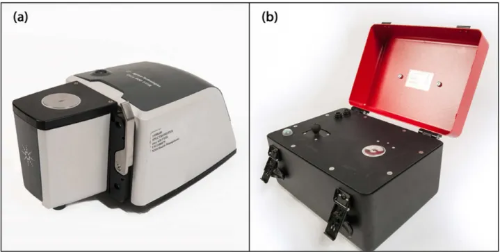
Infrared spectra were acquired using both laboratory Cary 630 (Fig 1a) and portable 4500 series (Fig 1b) attenuated total reflectance mid-infrared spectrometers (Agilent Technologies, Dansbury, Connecticut, USA). The laboratory Cary 630 spectrometer is interfaced with a cus-tomized triple-reflection diamond ATR sampling accessory with a 1-mm diameter sampling surface, and a 200 μm active area providing a 2 μm depth of penetration for IR energy at 1700 cm-1. However, the portable 4500 series spectrometer is interfaced with a triple-reflection dia-mond crystal ATR accessory with a 2-mm diameter sampling surface and a 200 μm active area providing a 6 μm effective penetration depth for IR energy at 1700 cm-1.
Thawed serum samples were diluted (1:1) with deionized sterile water and vortexed at a maximum of 2700 rpm for 10 s. From each diluted sample a 5 μL aliquot was placed onto the diamond crystal of the spectrometer and dried by a stream of air from a domestic hair dryer. Samples were fully dried within 3–5 min, forming a thin film on the crystal. The IR spectra were then collected using Agilent MicroLab PC software (Agilent Technologies, Dansbury,
Connecticut, USA). At a spectral resolution of 8 cm-1, 512 scans were co-added to optimize the signal to noise ratio over the wavenumber range of 4000–650 cm-1. Two independent spectra were collected from each serum sample with each spectrometer. The spectrometer crystal was cleaned with 100% ethanol and a laboratory wipe between each sample application and a new background spectrum (I0) was performed before applying the next serum sample. A total of
350 (175 x 2) ATR-IR spectra were collected from each spectrometer, saved in spectrum (SPC) format, and then converted into printable (PRN) formatted data using GRAMS software (GRAMS/AI version 7.02, Thermo Fisher Scientific). The PRN format spectral data were imported into MATLAB1(version R2016a, MathWorks, Natick, MA) for data analyses.
Spectral pre-processing
Spectra from each spectrometer were subjected to several trial pre-processing methods prior to provisional PLSR analyses, including Savitsky-Golay smoothing (2ndorder polynomial function with 9 points), first-order and second-order derivatization [28] and two different nor-malization methods (Standard normal variate (SNV) and vector nornor-malization) [29,30], then followed by spectral region selection. Only the combined spectral regions 3700–2600 cm-1and 1800–1300 cm-1were selected for further analysis [26]. Subsequently, spectrum outlier detec-tion was performed using Dixons Q-test [31,32] at each wavenumber. Spectra were designated as outliers and excluded from further analysis if more than 50% of absorbance values were out-side the 95% confidence level. The average of the duplicate spectra for each serum sample (after removal of outliers if applicable) was used for subsequent analysis.
Calibration models development
A multivariate regression method (PLSR) was used to create calibration models for each ATR-IR spectrometer separately. The 175 serum samples were sorted based on their IgG Fig 1. Attenuated total reflectance infrared spectrometers.(a) laboratory Cary 630 and (b) portable 4500 series (Agilent Technologies, Dansbury, Connecticut, USA).
concentration (lowest to highest) obtained from the reference RID based method and linked to their corresponding pre-processed spectra, and then split into test and calibration sets. The spectrum of every third serum sample was selected as a member of the test set (n = 58); the remaining samples (n = 117) were assigned to the calibration set. This ensured that the test set encompassed the full range of IgG values for evaluating the predictive performance of the cali-bration models. The calicali-bration set (n = 117) was further randomly split into training (n = 59) and validation (n = 58) data sets for model development.
A previously described PLSR approach was applied to the training set to develop 30 trial calibration models with the number of PLS factors ranging from 1 to 30 [26]. Each trial model was used to quantify the IgG concentrations of the validation set of serum samples. This proce-dure was repeated 10,000 times, utilizing randomly assigned splits of the calibration data set into new training and validation sets. The root mean squared error for the Monte Carlo cross-validation value (RMMCCV) [33,34] was calculated for each of the 30 trial calibration models, and the optimal number of PLS factors was chosen based on the lowest RMMCCV value. Once the number of PLS factors had been determined, the training and validation sets were recombined to build the final calibration model with the optimal number of PLS factors.
Evaluation of trial calibration models
The predictive performance of each provisional calibration model was evaluated using the test data set. The level of agreement between IgG concentrations measured by the reference RID method and those predicted by the calibration models was assessed for both the calibration and test data sets by scatter plot, and determination of Pearson correlation coefficients [35]. The differences and interchangeability between the IgG measured by RID and ATR-IR meth-ods for the test set were further evaluated by the Bland-Altman plot [36,37].
The potential utility of each ATR-IR calibration model was further evaluated by calculation of the ratio of predictive deviation (RPD) and the range error ratio (RER) [38]. For RPD values
<2, the calibration model was considered to be poorly predictive, those between 2.0 and 2.5 as
adequate for qualitative evaluation or for screening purposes, values >2.5 (or RER >10) were regarded as acceptable for quantification, and values >3 (or RER >20) were considered that the calibration model was suitable for accurate quantitative analysis [38].
The practical applicability of the ATR-IR calibration models for the diagnosis of FTPI (serum IgG <1000 mg/dL) in alpaca serum was evaluated. The diagnostic sensitivity, specific-ity and accuracy were calculated using 2 x 2 tables (diagt command in Stata vs 14.0 statistical software, StataCorp, College Station, TX) for both the test set and entire data set. Sensitivity (Se) was defined as the proportion of samples with IgG <1000 mg/dL, as determined by RID, that was correctly classified as positive by the ATR-IR assay. Conversely, specificity (Sp) was defined as the proportion of samples with IgG >1000 mg/dL that was correctly classified as negative by the ATR-IR assay. Accuracy was defined as the percentage of all samples that were correctly classified by the ATR-IR assay.
Results
Serum IgG determined by RID method
The RID-determined IgG concentrations of the 175 alpaca serum samples ranged from 394 to 6327 mg/dL, with an average of 2654 mg/dL and standard deviation of 1305 mg/dL. The mean, SD and range of the RID-determined IgG concentration for the calibration and test data sets are presented inTable 1.
ATR-IR spectra
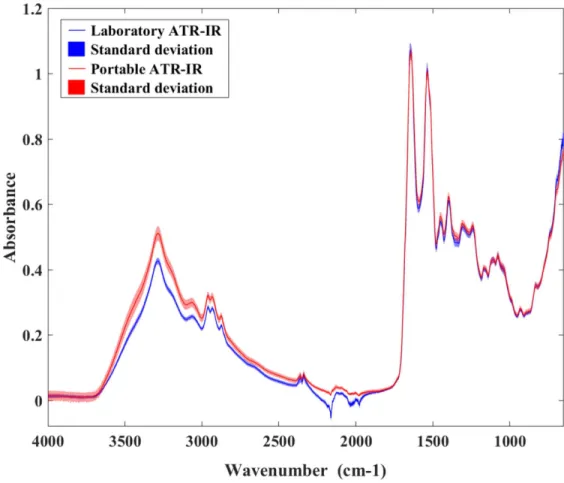
The IR spectrum of alpaca serum collected from both the laboratory (Cary 630) and portable (4500 series) ATR-IR spectrometers over the wavenumber range of 4000–650 cm−1showed distinct bands characteristic of functional group vibrations (Fig 2). A broad strong absorption band centered at 3300 cm−1was associated with N–H stretching vibration (Amide A) of the protein amide linkages, and strong absorptions centered around 1650 and 1550 cm−1were associated with protein C = O stretching (Amide I) and N–H bending (Amide II) vibrations, respectively [15,18]. Both laboratory and portable ATR-IR spectrometers provided similar spectral patterns but the portable ATR-IR generated slightly higher signal intensities compared to the laboratory grade spectrometer, due to a slightly longer effective optical pathlength for that particular ATR element/geometry (Fig 2). The 2200–1900 cm−1range is compromised in Table 1. Descriptive statistics for the 175 alpaca serum samples in the calibration and test data sets (RID-determined serum IgG values).
Datasets RID-IgG concentration (mg/dL)
n Mean SD Range
Calibration set 117 2665 1320 5934
Test set 58 2632 1285 5172
https://doi.org/10.1371/journal.pone.0179644.t001
Fig 2. The average reflectance spectra and their corresponding standard deviation values of alpaca serum samples obtained from the laboratory and portable ATR-IR spectrometers.
both systems due to very strong absorptions by the diamond coating on the ATR elements. However, the effect was most clearly evident for the Cary 630 spectra.
Calibration models
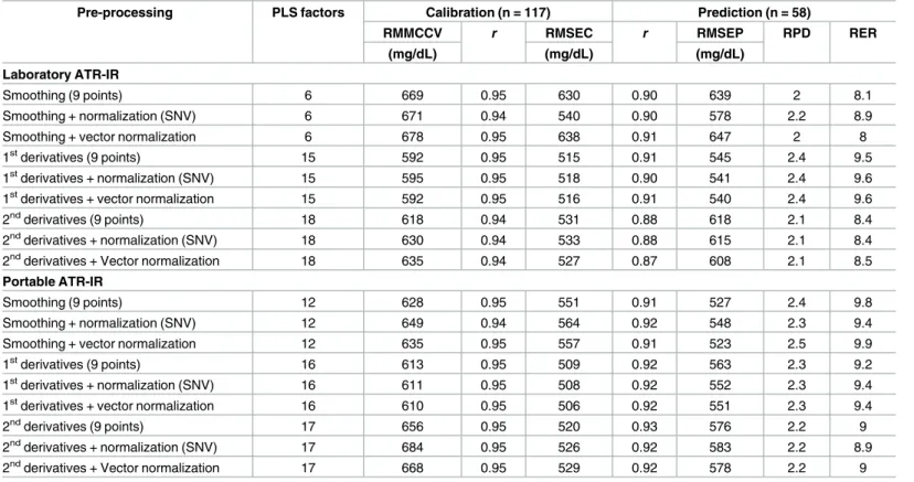
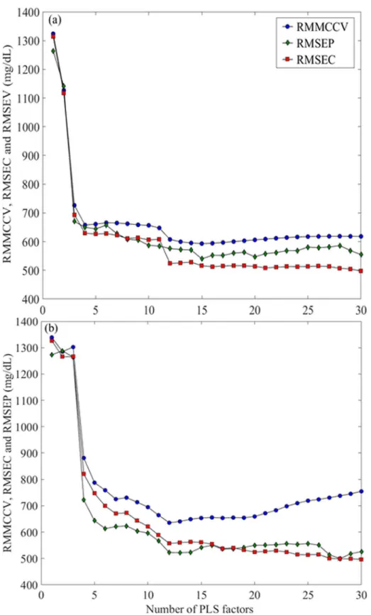
The parameters and performance characteristics of the PLS calibration models that were built using a different combination of spectral pre-processing techniques are presented inTable 2. The optimal PLS calibration models for the laboratory grade and portable ATR-IR spectrome-ters were obtained using data from first order derivatives spectra and vector normalization, and smoothed spectra and vector normalization, respectively. The optimum number of PLS factors for these models were 15 for the laboratory grade ATR-IR (Fig 3a) and 12 for the porta-ble ATR-IR spectra (Fig 3b), based on the lowest IgG RMMCCV (592 and 635 mg/dL, respec-tively).Fig 3displays the root mean squared error of calibration (RMSEC) and root mean squared error of prediction (RMSEP) plotted against the number of PLS factors.
Calibration models performance
Scatter plots (Fig 4) show the level of agreement between serum IgG concentrations measured by the reference RID assay and those predicted by both the laboratory grade (Fig 4a) and portable (Fig 4b) ATR-IR based assays. For both laboratory grade and portable ATR-IR spec-trometers, the Pearson correlation coefficient (r) for the calibration data set was 0.95; Table 2. Comparison of analytical methods based upon various spectral pre-processing protocols.
Pre-processing PLS factors Calibration (n = 117) Prediction (n = 58)
RMMCCV r RMSEC r RMSEP RPD RER
(mg/dL) (mg/dL) (mg/dL) Laboratory ATR-IR
Smoothing (9 points) 6 669 0.95 630 0.90 639 2 8.1
Smoothing + normalization (SNV) 6 671 0.94 540 0.90 578 2.2 8.9
Smoothing + vector normalization 6 678 0.95 638 0.91 647 2 8
1stderivatives (9 points) 15 592 0.95 515 0.91 545 2.4 9.5
1stderivatives + normalization (SNV) 15 595 0.95 518 0.90 541 2.4 9.6 1stderivatives + vector normalization 15 592 0.95 516 0.91 540 2.4 9.6
2ndderivatives (9 points) 18 618 0.94 531 0.88 618 2.1 8.4
2ndderivatives + normalization (SNV) 18 630 0.94 533 0.88 615 2.1 8.4 2ndderivatives + Vector normalization 18 635 0.94 527 0.87 608 2.1 8.5 Portable ATR-IR
Smoothing (9 points) 12 628 0.95 551 0.91 527 2.4 9.8
Smoothing + normalization (SNV) 12 649 0.94 564 0.92 548 2.3 9.4
Smoothing + vector normalization 12 635 0.95 557 0.91 523 2.5 9.9
1stderivatives (9 points) 16 613 0.95 509 0.92 563 2.3 9.2
1stderivatives + normalization (SNV) 16 611 0.95 508 0.92 552 2.3 9.4 1stderivatives + vector normalization 16 610 0.95 506 0.92 551 2.3 9.4
2ndderivatives (9 points) 17 656 0.95 520 0.93 576 2.2 9
2ndderivatives + normalization (SNV) 17 684 0.95 526 0.92 583 2.2 8.9 2ndderivatives + Vector normalization 17 668 0.95 529 0.92 578 2.2 9 PLS = Partial least squares; RMMCCV = Root mean squared error of the Monte Carlo cross validation value; r = Pearson correlation coefficient; RMSEC = Root mean squared error of calibration; RMSEP Root mean squared error of prediction; RPD (ratio of predictive deviation) = SD divided by RMSEP; RER (range error ratio) = Range divided by RMSEP; SNV Standard normal variate.
Fig 3. Plots of RMMCCV, RMSEC and RMSEP against the number of partial least squares (PLS) factors.The optimum number of PLS factors for (a) laboratory grade and (b) portable attenuated total reflectance infrared (ATR-IR) assays were determined to be 15 and 12, respectively, based on the lowest RMMCCV.
Fig 4. Scatter plots comparing Immunoglobulin G (IgG) measured by the reference radial
immunodiffusion (RID) assay to those provided by (a) laboratory grade and (b) portable ATR-IR based assays for the calibration and test sets.
corresponding value for the test data set was 0.91. The Bland-Altman plots (Fig 5) revealed that the average of the differences between RID-determined IgG concentration and the labora-tory ATR-IR assay was 121 mg/dL (Fig 5a), and 71 mg/dL for the portable ATR-IR assay (Fig 5b), indicating no signficant bias between these methods. The 95% confidence interval for lab-oratory grade and portable ATR-IR spectrometers ranged from –950 to 1191 mg/dL and –960 Fig 5. Bland-Altman plots for the test (n = 58) data set.(a) for laboratory ATR-IR assay, the dashed lines represent the 95% confidence limits of agreement (– 950 to 1191 mg/dL), while the solid line represents the mean difference between laboratory ATR-IR and RID assays (120.5 mg/dL). (b) for portable ATR-IR assay, the dash lines represent the 95% confidence limits of agreement (– 960 to 1101 mg/dL), while the solid line represents the mean difference between portable ATR-IR and RID assays (71 mg/dL).
to 1101 mg/dL, respectively. The RPD and RER values for laboratory grade and portable ATR-IR based assays were 2.4 and 9.6, and 2.5 and 9.9, respectively, indicating that both ATR-IR assays were acceptable for qualitative evaluation or for screening of alpaca serum IgG concentrations.
The diagnostic test characteristics (Se, Sp and accuracy) of the laboratory and portable ATR-IR assays for diagnosis of serum IgG <1000 mg/dL (FPTI cut-off) were determined sepa-rately for the test sets and for the entire data sets (Table 3). Within the whole data set, 21 out of 175 serum samples had RID-IgG concentration <1000 mg/dL, resulting in a true low IgG prevalence of 12%. The number of serum samples that had IgG concentrations <1000 mg/dL according to the laboratory grade (n = 18) and portable (n = 21) ATR-IR based assays, resulted in apparent prevalences of 10% and 12%, respectively. For the laboratory grade ATR-IR based assay, there were no false positives and three false negatives identified. For the portable ATR-IR based assay, one false positive and one false negative were identified (Table 3).
Discussion
This study showed that IR spectroscopy, using either a laboratory grade or portable ATR-IR spectrometer, is an adequate method for rapid qualitative evaluation and for screening of alpaca serum IgG concentrations. The performance of the IR-based analytical methods depended marginally on the choice of IR spectrometer, and more critically on spectral pre-processing strategies. In the present study, the IR-based assays showed performance compara-ble to those previously reported for transmission-IR spectroscopy [26]. In contrast to transmis-sion spectroscopy, the ATR-IR based approaches have the advantage of reduced sample preparation time (<5 min), no requirement for a transmission substrate, and reduced impreci-sion associated with variability in sample volume and film thickness, issues that can contribute to imprecision in transmission-IR based assays [24,39].
The optimal analytical methods were confirmed as adequate for qualitative evaluation or for screening of alpaca IgG concentration by both their predictive accuracy (i.e., RMSEP) and by standard tests to characterise PLSR-based calibrations (RPD and RER values) [38]. Spectral derivation and smoothing contributed to optimization of the analytical methods using the lab-oratory grade and portable spectrometers, respectively (Table 2). Similar results were previ-ously reported in related studies that used either transmission or ATR-IR spectrometers [24, 26]. Whether smoothed or derivatized spectra were used, vector normalization proved more beneficial for prediction of IgG concentration in alpaca serum than standard normal variate (SNV). Interestingly, the opposite behaviour was observed previously for analogous transmis-sion IR-based IgG assays [26]. The optimum PLS model for the portable ATR-IR based assay required fewer PLS-factors than did that of the laboratory ATR-IR based assay (Fig 3). This Table 3. Diagnostic test characteristics for assessment of IgG<1000 mg/dL, the cut-off for failure of transfer of passive immunity (FTPI).
Dataset n Test characteristics
TP FP TN FN Se Sp Accuracy Laboratory ATR-IR Test set 58 5 0 51 2 71% 100% 97% All data 175 18 0 154 3 86% 100% 98% Portable ATR-IR Test set 58 6 1 50 1 86% 98% 97% All data 175 20 1 153 1 95% 99% 99%
n = number of samples; TP = true positives; FP = false positives; TN = true negatives; FN = false negatives; Se = sensitivity; Sp = specificity.
could be attributed to better coupling of the optical element within the optical train for the dedicated ATR (portable 4500 series) instrument as compared to the ATR module within the laboratory spectrometer [40]. Further, the larger number of PLS-factors suggests that the labo-ratory ATR-IR assay may be less robust than the portable ATR-IR counterpart, by virtue of its reliance upon additional factors that are relatively noisy [41].
The portable ATR-IR assay showed higher utility (higher RPD and RER values), and more flexibility for field application [40,41] than the laboratory grade ATR-IR assays reported in the present study, and that previously reported for bovine serum IgG [24]. Correlation between the RID-based IgG analyses and ATR-IR based assays was better than that previously reported for transmission-IR based IgG assays for alpaca [26] and equine [25] sera, but worse than that achieved for transmission and ATR-IR based bovine IgG assays [24]. Limitations in the predic-tive ability of both laboratory grade and portable ATR-IR assays in this study may be attributed to the source of the samples. Serum samples were collected from two different geographical regions and the IgG content was highly variable, ranging from 394 to 6327 mg/dL. Further, only 12% (21 out 175) of serum samples having IgG concentration less than 1000 mg/dL. As a result, the developed assays were weighted toward higher IgG concentrations.
The correlation with RID-IgG concentrations was lower than that reported for comparison of RID-IgG to immunoturbidimetric methods [10], but higher than that reported for compari-sons of serum total protein refractometry versus RID-IgG [8]. The Bland-Altman plots (Fig 5) revealed a non-significant bias for the portable ATR-IR-based assay as compared to the lab sys-tem counterpart. However, the bias for both ATR-IR based assays was higher than that previ-ously reported (20.2 mg/dL) for the transmission-IR based assay [26].
Both ATR-IR assays showed excellent Sp, but the Se of the portable ATR-IR assay was higher than that of the laboratory grade ATR-IR assay (Table 3). When compared with reported Se and Sp of other methods available to assess FTPI in neonatal camelids, these results are equivalent to or better than most [8,10,26,42]. Both ATR-IR spectroscopic assays cor-rectly classified the majority of FTPI cases as such, and the misclassified samples had RID IgG concentrations very close to the diagnostic cut-off of 1000 mg/dL. This suggests that the likeli-hood of misdiagnosis is small; the false negative samples correspond to only partial FTPI, with minimal risk of morbidity and mortality [2,7].
Indeed, the transferability into field conditions (veterinary clinics and large farms) remains open for discussion and a further in depth investigation is needed to better evaluate the porta-ble ATR-IR assay for rapid field evaluation of FTPI. However, this study is a proof of concept, with respect to the emerging technologies for automated measurement of alpaca serum IgG concentration and diagnosis of FTPI.
Conclusions
This study supports the applicability of both laboratory grade and portable ATR-IR spectros-copy as the basis for rapid qualitative evaluation of alpaca serum IgG concentration and diagnosis of FTPI. While the laboratory grade and portable ATR-IR based assays perform sim-ilarly, the portable ATR-IR assay has the potential advantage of enabling determination of IgG concentration in the field.
Acknowledgments
The authors thank Cynthia Mitchell for her technical assistance. The authors also thank Pro-fessor Michael Reichel (University of Adelaide, Australia) and Dr. Nicole Jewett (New Bruns-wick, Canada) for providing the samples used in this study. Also, we would like to thank the
anonymous reviewers for their helpful comments in the preparation of this article. This study was partially supported by the Atlantic Canada Opportunities Agency.
Author Contributions
Conceptualization:Ibrahim Elsohaby, Christopher B. Riley, R. Anthony Shaw, J. Trenton McClure.
Data curation:Ibrahim Elsohaby, Jennifer B. Burns.
Formal analysis:Ibrahim Elsohaby.
Funding acquisition:Christopher B. Riley, J. Trenton McClure.
Investigation:Ibrahim Elsohaby.
Methodology:Ibrahim Elsohaby.
Project administration:J. Trenton McClure.
Resources:Christopher B. Riley, R. Anthony Shaw, J. Trenton McClure.
Software:Ibrahim Elsohaby, R. Anthony Shaw.
Supervision:J. Trenton McClure.
Validation:Ibrahim Elsohaby, J. Trenton McClure.
Visualization:Ibrahim Elsohaby, J. Trenton McClure.
Writing – original draft:Ibrahim Elsohaby.
Writing – review & editing:Ibrahim Elsohaby, Jennifer B. Burns, Christopher B. Riley, R. Anthony Shaw, J. Trenton McClure.
References
1. Rehman S, Bytnar D, Berkenbosch JW, Tobias JD. Hypogammaglobulinemia in pediatric ICU patients. J Intensive Care Med. 2003; 18: 261–264.https://doi.org/10.1177/0885066603255905PMID:
15035760
2. Wernery U. Camelid Immunoglobulins and Their Importance for the New-Born–A Review. J of Vet Med. Series B. 2001; 48: 561–568.https://doi.org/10.1111/j.1439-0450.2001.00478.x
3. Chappuis G. Neonatal immunity and immunisation in early age: lessons from veterinary medicine. Vac-cine. 1998; 16: 1468–1472.https://doi.org/10.1016/S0264-410X(98)00110-8PMID:9711790 4. Walker P, Tibary A. Neonatal care of camelids: a review and case reports. J Camel Pract Res. 1999; 6:
255–263.
5. Adams R, Garry F. Llama neonatology. Vet Clin N Am-Food A. 1994; 10: 209–227.
6. Dolente BA, Lindborg S, Palmer JE, Wilkins PA. Culture-Positive Sepsis in Neonatal Camelids: 21 Cases. J Vet Intern Med. 2007; 21: 519–525. PMID:17552461
7. Whitehead CE. Management of neonatal llamas and alpacas. Vet Clin N Am-Food A. 2009; 25: 353– 366.
8. Weaver DM, Tyler JW, Marion RS, Wallace LM, Nagy JK, Holle JM. Evaluation of assays for determina-tion of passive transfer status in neonatal llamas and alpacas. J Am Vet Med Assoc. 2000; 216: 559– 563. PMID:10687013
9. Fowler ME. Medicine and surgery of camelids. 3rd ed. Ames: Blackwell Publishing John Wiley & Sons; 2010.
10. Pinn TL, Gagliardo LF, Purdy SR, Appleton JA, Stokol T. Comparison of three immunoglobulin G assays for the diagnosis of failure of passive transfer of immunity in neonatal alpacas. J Vet Diagn Invest. 2013; 25: 91–98.https://doi.org/10.1177/1040638712472182PMID:23345272
11. Johnston NA, Parish SM, Tyler JW, Tillman CB. Evaluation of serum gamma-glutamyltransferase activ-ity as a predictor of passive transfer status in crias. J Am Vet Med Assoc. 1997; 211: 1165–1166. PMID:
12. Garmendia AE, Palmer GH, DeMartini JC, McGuire TC. Failure of passive immunoglobulin transfer: a major determinant of mortality in newborn alpacas (Lama pacos). Am J Vet Res. 1987; 48: 1472–1476. PMID:3674556
13. Hutchison JM, Salman MD, Garry FB, Johnson LW, Collins JK, Keefe TJ. Comparison of two commer-cially available single radial immunodiffusion kits for quantitation of llama immunoglobulin G. J Vet Diagn Invest. 1995; 7: 515–519.https://doi.org/10.1177/104063879500700416PMID:8580175 14. Weaver DM, Tyler JW, Scott MA, Wallace LM, Marion RS, Holle JM. Passive transfer of colostral
immu-noglobulin G in neonatal llamas and alpacas. Am J Vet Res. 2000; 61: 738–741. PMID:10895892 15. Shaw RA, Mantsch HH. Infrared spectroscopy in clinical and diagnostic analysis. In: In: Meyerse R.A.,
editor. Encyclopedia of Analytical Chemistry: Applications Theory and Instrumentation. Chichester, UK: Wiley Online Library; 2000. pp. 83–102.
16. Shaw RA M H. Infrared spectroscopy in clinical and diagnostic analysis. In: Robert Meyers, editor. Encyclopedia of Analytical Chemistry: Applications Theory and Instrumentation. UK: John Wiley and Sons Ltd; 2008. pp. 83–102.
17. De Marchi M, Toffanin V, Cassandro M, Penasa M. Invited review: Mid-infrared spectroscopy as pheno-typing tool for milk traits1. J Dairy Sci. 2014; 97: 1171–1186.http://dx.doi.org/10.3168/jds.2013-6799. PMID:24440251
18. Shaw RA, Kotowich S, Leroux M, Mantsch HH. Multianalyte serum analysis using mid-infrared spec-troscopy. Ann Clin Biochem. 1998; 35 (Pt 5): 624–632.
19. Reid LM, O’donnell CP, Downey G. Recent technological advances for the determination of food authenticity. Trends Food Sci Technol. 2006; 17: 344–353.
20. Rodriguez-Saona L, Allendorf M. Use of FTIR for rapid authentication and detection of adulteration of food. Annu Rev Food Sci Technol. 2011; 2: 467–483. https://doi.org/10.1146/annurev-food-022510-133750PMID:22129392
21. Sun D. Infrared spectroscopy for food quality analysis and control. Academic Press, New York, USA; 2009.
22. Smith BC. Fundamentals of Fourier transform infrared spectroscopy. 2nd ed. CRC Press, Boca Raton, FL; 2011.
23. Elsohaby I, Riley CB, Hou S, McClure JT, Shaw RA, Keefe GP. Measurement of serum immunoglobulin G in dairy cattle using Fourier-transform infrared spectroscopy: A reagent free approach. Vet J. 2014; 202: 510–515.https://doi.org/10.1016/j.tvjl.2014.09.014PMID:25438729
24. Elsohaby I, Hou S, McClure JT, Riley CB, Shaw RA, Keefe GP. A rapid field test for the measurement of bovine serum immunoglobulin G using attenuated total reflectance infrared spectroscopy. BMC Vet Res. 2015; 11:218.https://doi.org/10.1186/s12917-015-0539-xPMID:26289208
25. Riley CB, McClure JT, Low-Ying S, Shaw RA. Use of Fourier-transform infrared spectroscopy for the diagnosis of failure of transfer of passive immunity and measurement of immunoglobulin concentrations in horses. J Vet Intern Med. 2007; 21: 828–834. PMID:17708406
26. Burns J, Hou S, Riley C, Shaw R, Jewett N, McClure J. Use of Fourier-Transform Infrared Spectroscopy to Quantify Immunoglobulin G Concentrations in Alpaca Serum. J Vet Intern Med. 2014; 28: 639–645.
https://doi.org/10.1111/jvim.12258PMID:24417433
27. Sorak D, Herberholz L, Iwascek S, Altinpinar S, Pfeifer F, Siesler HW. New developments and applica-tions of handheld Raman, mid-infrared, and near-infrared spectrometers. Appl Spectrosc Rev. 2012; 47: 83–115.
28. Savitzky A, Golay MJ. Smoothing and differentiation of data by simplified least squares procedures. Anal Chem. 1964; 36: 1627–1639.
29. Barnes R, Dhanoa M, Lister SJ. Standard normal variate transformation and de-trending of near-infra-red diffuse reflectance spectra. Appl Spectrosc. 1989; 43: 772–777.
30. Barnes R, Dhanoa M, Lister S. Letter: Correction to the description of Standard Normal Variate (SNV) and De-Trend (DT) ransformations in Practical Spectroscopy with Applications in Food and Beverage Analysis–2nd Ed. J Near Infrared Spec. 2004; 1:185–186.
31. Dean R, Dixon W. Simplified statistics for small numbers of observations. Anal Chem. 1951; 23: 636– 638.
32. Rorabacher DB. Statistical treatment for rejection of deviant values: critical values of Dixon’s" Q" param-eter and related subrange ratios at the 95% confidence level. Anal Chem. 1991; 63: 139–146.
33. Xu Q, Liang Y. Monte Carlo cross validation. Chemometrics Intellig Lab Syst. 2001; 56: 1–11.http://dx. doi.org/10.1016/S0169-7439(00)00122-2.
34. Picard Richard R. and Cook R. Dennis. Cross-validation of regression models. J Amer Statist Assoc. 1984; 79: 575–583.
35. Lawrence I, Lin K. A concordance correlation coefficient to evaluate reproducibility. Biometrics. 1989: 255–268. PMID:2720055
36. Altman DG, Bland JM. Measurement in medicine: the analysis of method comparison studies. Statisti-cian. 1983; 32: 307–317.
37. Bland JM, Altman DG. Comparing two methods of clinical measurement: a personal history. Int J Epide-miol. 1995; 24: S7–S14. PMID:7558556
38. Williams PC, Sobering D. How do we do it: a brief summary of the methods we use in developing near infrared calibrations. In Davies AM, Williams P, editors. Near Infrared Spectroscopy: The Future Waves. 1996. pp. 185–188.
39. Hou S, Shaw R, Riley C. Chemometric analysis of attenuated total reflectance infrared spectral data for quantitation of immunoglobulin G in equine plasma and serum. Chemometrics Intellig Lab Syst. 2016; 156: 108–114.
40. Ayvaz H, Rodriguez-Saona LE. Application of handheld and portable spectrometers for screening acryl-amide content in commercial potato chips. Food Chem. 2015; 174: 154–162.https://doi.org/10.1016/j. foodchem.2014.11.001PMID:25529665
41. Wang T, Tan SY, Mutilangi W, Aykas DP, Rodriguez-Saona LE. Authentication of Whey Protein Pow-ders by Portable Mid-Infrared Spectrometers Combined with Pattern Recognition Analysis. J Food Sci. 2015; 80: C2111–C2116.https://doi.org/10.1111/1750-3841.13006PMID:26352755
42. Hawkins DM. The problem of overfitting. J Chem Inf Comput Sci. 2004; 44: 1–12.https://doi.org/10. 1021/ci0342472PMID:14741005