HAL Id: hal-02478087
https://hal.inria.fr/hal-02478087
Submitted on 17 Feb 2020
HAL is a multi-disciplinary open access
archive for the deposit and dissemination of
sci-entific research documents, whether they are
pub-lished or not. The documents may come from
teaching and research institutions in France or
abroad, or from public or private research centers.
L’archive ouverte pluridisciplinaire HAL, est
destinée au dépôt et à la diffusion de documents
scientifiques de niveau recherche, publiés ou non,
émanant des établissements d’enseignement et de
recherche français ou étrangers, des laboratoires
publics ou privés.
Lesion-Robust White-Matter Bundle Identification
through Diffusion Driven Label Fusion
Guillermo Gallardo, Gaston Zanitti, Alfred Anwander, Mat Higger, Sylvain
Bouix, Samuel Deslauriers-Gauthier, Demian Wassermann
To cite this version:
Guillermo Gallardo, Gaston Zanitti, Alfred Anwander, Mat Higger, Sylvain Bouix, et al..
LesionRobust WhiteMatter Bundle Identification through Diffusion Driven Label Fusion. ISMRM 2020
-28th Annual Meeting & Exhibition, Aug 2020, Sydney / Virtual, Australia. �hal-02478087�
Lesion-Robust White-Matter Bundle Identification through Diffusion Driven
Label Fusion
Introduction.
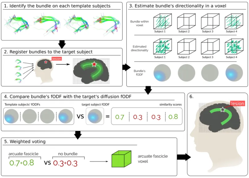
Pathologies such as infiltrative tumors disrupt the structure of white matter in the brain, resulting in cognitive deficits. Inferring which pathways are affected by the lesion is key for both pre and post-treatment planning. In the presence of lesions, tractography algorithms[1] fail to wholly track white matter pathways, regardless if these are unaffected by the lesion[2]. We solve this by harnessing aggregated healthy subject information. Given a set of labeled major bundles in a group of healthy subjects, we non-linearly register them to our patient’s brain and combine them using a novel label fusion algorithm. A major advantage of label fusion techniques is their high accuracy even when inferring from few subjects[3]. Extant label fusion techniques rely on tissue contrast, not taking into account the fibrous structure of white matter. Leveraging that brain structures constrain the diffusion of water particles differently, we propose a novel diffusion driven technique to improve the localization of brain pathways (Fig. 1). We show its feasibility and advantages in both synthetic and human data.
Methods.
Let Ls, s ∈ S be a set of 3D volumes in a common space representing the labeling of template subjects S. Let {li} be the set of labels representing fiber bundles and grey matter structures in the brain. We propose a weighted voting technique to label a target voxel x:
L∗(x) =l∈labels X s∈S p(L(x) = l|Ls(x))p(D(x)|Dsl(x)). p(L(x) = l|Ls(x)) = ( 1, if Ls(x) = l 0, otherwise. p(D(x)|Dsl(x)) = hF (x), Fsl(x)i, if Ls(x) = l, and l 6= 0 hF (x), U i, if Ls(x) = 0 0, otherwise
The term p(L(x) = l|Ls(x)) represents the ”vote” of each template[4], being 1 if the label is present in the template’s voxel and 0 if not. The term p(D(x)|Dsl(x)) weighs the vote based on how much the voted bundle resembles the target’s diffusion data. It express the probability of seeing the target’s diffusion, D(x), given that the template’s tract l is present in the voxel, Dsl(x).
We characterize D(x) by fitting a Constrained Spherical Deconvolution (CSD) model[5] to the target’s diffusion data and estimating a fiber orientation density function (fODF). The fODF Fx(θ, φ) represents the fraction of fibers within the voxel x aligned along the spherical coordinate (θ, φ). Then, we characterize the within-voxel directionality of the template’s bundle by looking at the entry and exit points (Fig. 1.3) of its streamlines. We estimate a fODF from these directions by means of CSD as with the diffusion data. If the template has no bundle in the voxel, a uniform fODF, U , is used. Finally, we define p(D(x)|Dsl(x)) as the inner product between the diffusion-based fODF, F (x), and the bundle fODF, Fsl(x), with both fODFs normalized such that hF (x), F (x)i = 1.
Experiments and Results.
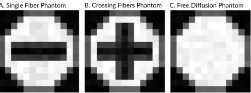
Our technique performs a weighted voting, were votes for bundles aligned with the target’s diffusion should get higher votes. To assess that our technique weights the votes correctly we created 3 synthetic diffusion datasets using Phantomas[6]. The first phantom possess only one bundle, the second possess two crossing bundles and the third no bundles (Fig. 2A-C). We generated 30 DWIs per phantom using a SNR=20, a resolution of 1mm per voxel, and 10 voxels per dimension. We used the DWIs to computed the weight a vote for a bundle and its planer rotations would obtain (Fig. 2D). Figure 2D shows that
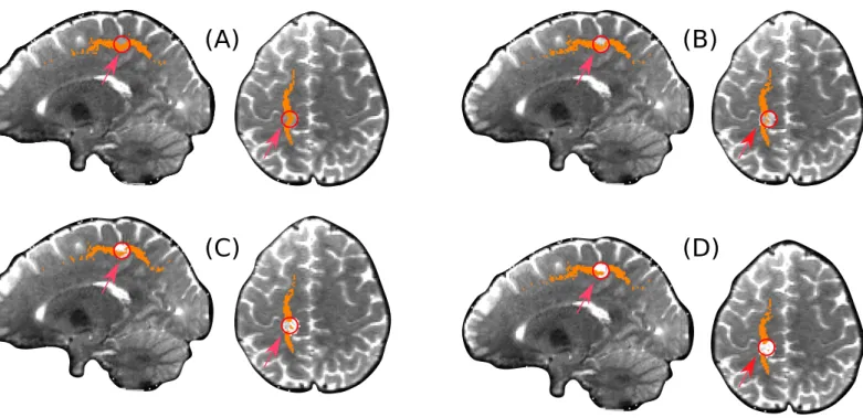
To compare how the labeling behaves in the presence of lesions, we simulated a disruptive lesion in the Superior Longi-tudinal Fascicle (SLF) bundle (Fig. 3). We did so by selecting a spherical region of 4mm where the SLF passes and mixing the diffusion signal with isotropic diffusion. Figure 3 shows that the more isotropic the signal, the fewer voxels are labeled within the lesion, allowing to identify the lesion within the tract.
Conclusion
We presented a label fusion technique to localize fiber bundles when whole-bundle tracking is hampered. Through 3 exper-iments we showed that our technique: weights votes correctly; is more conservative than the traditional majority voting, a desirable feature in clinical applications; and contrary to normal voting, it does not label voxels within a lesion, allowing to identify both the affected tract and the lesion interrupting it.
Figure 1: Pipeline of our technique, we show the simplified case of localizing only one bundle, affected by a disruptive lesion. (1) The bundle of interest is localized in healthy subjects and (2) registered to the target subject. (3) For each template bundle its directionality is represented by means of an fODF, its (4) similarity with the templates diffusion fODF is used as a weight to (5) vote for the presence of the bundle. Repeating this in every voxel allows to localize the bundle in the template’s brain and the lesion.
Figure 2: Experiment on synthetic data. A. Phantom with only one fiber bundle. B. Phantom with crossing fibers. C. Phantom with no fiber (isotropic diffusion). D. Weights (blue line) a vote for a specific fiber bundle and its planar rotations would get compared against the vote for ”non bundle”. The weights are higher when the structure being voted is consistent with the underlying diffusion.
Table 1: Sensitivity and specificity of our proposed method (Weighted) and Majority Voting (Majority) when inferring single bundles from 9 subjects. The inferred bundles are: Superior Longitudinal Fasciculus (SLF) I, II and III, Inferior Longitudinal Fasciculus (ILF), Cortico Spinal Tract (CST) (TP), and Inferior Occipito Frontal Fascicle (IOFF).
Figure 3: Lesions were simulated in a specific region (red circle) by mixing isotropic diffusion signal within the region. The figures shows the voxels marked as SLF at different values of signal mixing. (A) 25% of isotropic signal, (B) 50% of isotropic signal, (C) 75% of isotropic signal and (D) 100% of isotropic signal. Results show that fewer voxels as the lesion grows in intensity.