HAL Id: hal-03104655
https://hal.sorbonne-universite.fr/hal-03104655
Submitted on 9 Jan 2021
HAL is a multi-disciplinary open access
archive for the deposit and dissemination of
sci-entific research documents, whether they are
pub-lished or not. The documents may come from
teaching and research institutions in France or
abroad, or from public or private research centers.
L’archive ouverte pluridisciplinaire HAL, est
destinée au dépôt et à la diffusion de documents
scientifiques de niveau recherche, publiés ou non,
émanant des établissements d’enseignement et de
recherche français ou étrangers, des laboratoires
publics ou privés.
Sequence variations of ACVRL1 play a critical role in
hepatic vascular malformations in hereditary
hemorrhagic telangiectasia
Sophie Giraud, Claire Bardel, Sophie Dupuis-Girod, Marie-France Carette,
Brigitte Gilbert-Dussardier, Sophie Riviere, Jean-Christophe Saurin, Mélanie
Eyries, Sylvie Patri, Evelyne Decullier, et al.
To cite this version:
Sophie Giraud, Claire Bardel, Sophie Dupuis-Girod, Marie-France Carette, Brigitte
Gilbert-Dussardier, et al.. Sequence variations of ACVRL1 play a critical role in hepatic vascular
malforma-tions in hereditary hemorrhagic telangiectasia. Orphanet Journal of Rare Diseases, BioMed Central,
2020, 15 (1), pp.254. �10.1186/s13023-020-01533-2�. �hal-03104655�
R E S E A R C H
Open Access
Sequence variations of
ACVRL1 play a
critical role in hepatic vascular
malformations in hereditary hemorrhagic
telangiectasia
Sophie Giraud
1, Claire Bardel
2,3,4, Sophie Dupuis-Girod
1,5, Marie-France Carette
6, Brigitte Gilbert-Dussardier
7,8,
Sophie Riviere
9, Jean-Christophe Saurin
3,10, Mélanie Eyries
11, Sylvie Patri
12, Evelyne Decullier
13,
Alain Calender
1,3,14and Gaëtan Lesca
1,3*Abstract
Background: Hereditary Hemorrhagic Telangiectasia (HHT) is an autosomal dominant disorder characterized by multiple telangiectases and caused by germline disease-causing variants in theENG (HHT1), ACVRL1 (HHT2) and, to a lesser extentMADH4 and GDF2, which encode proteins involved in the TGF-β/BMP9 signaling pathway. Common visceral complications of HHT are caused by pulmonary, cerebral, or hepatic arteriovenous malformations (HAVMs). There is large intrafamilial variability in the severity of visceral involvement, suggesting a role for modifier genes. The objective of the present study was to investigate the potential role ofENG, ACVRL1, and of other candidate genes belonging to the same biological pathway in the development of HAVMs.
Methods: We selected 354 patients from the French HHT patient database who had one disease causing variant in eitherENG or ACVRL1 and who underwent hepatic exploration. We first compared the distribution of the different types of variants with the occurrence of HAVMs. Then, we genotyped 51 Tag-SNPs from the Hap Map database located in 8 genes that encode proteins belonging to the TGF-β/BMP9 pathway (ACVRL1, ENG, GDF2, MADH4, SMAD1, SMAD5, TGFB1, TGFBR1), as well as in two additional candidate genes (PTPN14 and ADAM17). We addressed the question of a possible genetic association with the occurrence of HAVMs.
Results: The proportion of patients with germlineACVRL1 variants and the proportion of women were significantly higher in HHT patients with HAVMs. In the HHT2 group, HAVMs were more frequent in patients with truncating variants. Six SNPs (3 inACVRL1, 1 in ENG, 1 in SMAD5, and 1 in ADAM17) were significantly associated with HAVMs. After correction for multiple testing, only one remained significantly associated (rs2277383).
Conclusions: In this large association study, we confirmed the strong relationship betweenACVRL1 and the development of HAVMs. Common polymorphisms ofACVRL1 may also play a role in the development of HAVMs, as a modifying factor, independently of the disease-causing variants.
Keywords: HHT, Rendu-Osler,ACVRL1, Modifier gene, Hepatic arteriovenous malformation
© The Author(s). 2020 Open Access This article is licensed under a Creative Commons Attribution 4.0 International License, which permits use, sharing, adaptation, distribution and reproduction in any medium or format, as long as you give appropriate credit to the original author(s) and the source, provide a link to the Creative Commons licence, and indicate if changes were made. The images or other third party material in this article are included in the article's Creative Commons licence, unless indicated otherwise in a credit line to the material. If material is not included in the article's Creative Commons licence and your intended use is not permitted by statutory regulation or exceeds the permitted use, you will need to obtain permission directly from the copyright holder. To view a copy of this licence, visithttp://creativecommons.org/licenses/by/4.0/. The Creative Commons Public Domain Dedication waiver (http://creativecommons.org/publicdomain/zero/1.0/) applies to the data made available in this article, unless otherwise stated in a credit line to the data.
* Correspondence:gaetan.lesca@chu-lyon.fr
1Hospices Civils de Lyon, Service de Génétique, Groupement Hospitalier Est,
69677 Bron, France
3Université Claude Bernard Lyon 1, Université de Lyon, Villeurbanne, France
Background
Hereditary Hemorrhagic Telangiectasia (HHT, Rendu-Osler-Weber syndrome, ORPHA 774) is an autosomal dominant systemic disease whose primary pathogenic expression is arteriovenous malformation. Common manifestations are recurrent epistaxis beginning in child-hood and cutaneous telangiectases, which are clusters of abnormally dilated capillaries. Visceral complications in-clude intestinal bleeding and pulmonary (PAVM), hep-atic (HAVM) and central nervous system arteriovenous malformations [1].
HHT is related to deleterious variants in 2 major genes: ENG (OMIM 131195) that encodes endoglin and which is located on chromosome 9q and causes HHT1 (OMIM 187300), and ACVRL1 (OMIM 601284) that en-codes activin receptor-like kinase type 1 (ALK1), which is located on chromosome 12q and causes HHT2 (OMIM 600376) [2, 3]. Disease-causing variants in the MADH4 gene (OMIM 60093), which encodes SMAD4 and is located on chromosome 18q, have been found in patients with juvenile polyposis and HHT symptoms [4]. Furthermore, missense variants in GDF2 gene (OMIM 605120), which encodes bone morphogenetic protein-9 (BMP9) and is located on chromosome 10q, have been found in 3 individuals with a vascular phenotype over-lapping with HHT, characterized by epistaxis and tel-angiectasia without solid-organ involvement [5]. About 5–10% of the patients fulfilling the HHT clinical features do not have a germline pathogenic variant in one of these genes [6].
Mutations in two other genes, RASA1 (OMIM 139150) and EPHB4 (OMIM 600011), were reported in patients with overlapping, but clinically distinct disorders, named capillary malformation-arteriovenous malformation 1 (OMIM 608354) and 2 (OMIM 618196), respectively [7,8].
Endoglin and ALK1 are predominantly expressed in endothelial cells and are receptors of the transforming growth factor-β (TGF-β) family that mediates responses to BMP9, and to a lesser extent, to BMP10 and TGF-β1/ β3 [9,10]. Both endoglin and ALK1 bind BMP9 and, in association with BMPR2, trigger signaling pathways mainly via SMAD1 and SMAD5, which form hetero-meric complexes with SMAD4 and translocate into the nucleus where they regulate the transcriptional activity of target genes.
HAVMs consist of arteriovenous shunting responsible for high-output cardiac failure, ascites, and biliary ische-mia with sepsis. Liver transplantation remains the main treatment option in advanced stages although new treat-ments such as anti-vascular endothelial growth factor therapies are emerging [11, 12]. The higher prevalence of HAVMs in patients with germline pathogenic ACVR L1 variants, compared to ENG, has been suggested by several studies [13–16] and confirmed by a prospective
study including 102 HHT patients [17]. In the latter study, based on data from hepatic Doppler sonography, 71% patients had HHT2, and 63% of these had HAVMs.
The wide intrafamilial clinical variability in HHT, in-cluding liver involvement, suggests a role for modifier genes, in addition to the ENG or ACVRL1 disease-causing variants. The objective of the present study was to investigate the potential role of genetic variations in ACVRL1, ENG, and a selection of other genes encoding proteins of the TGFβ/BMP9 pathway in the develop-ment of HAVMs.
Methods
Patients
This is an non-interventional study including DNA from 354 patients with a diagnosis of HHT confirmed by mo-lecular genetics (107 with ENG disease-causing variants and 247 with ACVRL1 disease-causing variants) and who benefited from systematic hepatic evaluation by ultrasound Doppler (n = 247), contrast abnominal CT (n = 51), or both exams (n = 56). The patients were col-lected through the three laboratories involved in HHT genetic diagnosis in France: Lyon (n = 248), Paris (n = 91), and Poitiers (n = 15). The number of patients with MADH4 disease-causing variants was too low (< 30) for statistical analyses. Patients had given written consent for the study of genetic factors involved in HHT accord-ing to the French bioethics law. Genetic and clinical data have been collected in the French HHT patient database (CIROCO) from 2005 to 2013.
Patients were classified into three groups according to clinical data or to liver imaging (if asymptomatic) that has previous shown to reflect disease severity [17]: i) group 1 had no liver abnormality: diameter of the mon hepatic artery < 6.5 mm (which was the most
com-monly indicated measure in hepatic exploration),
maximum velocity in the hepatic artery < 120 cm/s, ii) group 2 had isolated enlargement of the diameter (be-tween 6.5 and 10.5) or of the velocity of the hepatic ar-tery (≥ 120 cm/s), iii) group 3 had either a common hepatic artery diameter higher than 10.5 cm, heart failure due to hepatic shunt, or had undergone liver transplant-ation for severe liver involvement.
Analysis of disease-causing ENG and ACVRL1 variants
DNA was extracted from patient blood according to standard procedures. ENG and ACVRL1 were analyzed by sequencing on a 3130XL sequencer (Thermo Fisher Scientific, Illkirch-Graffenstaden, France). Large rear-rangements were identified by Quantitative Multiplex PCR Short Fragments (QMPSF) or Multiplex Ligation PCR Amplification (MLPA) using the P093 Salsa MLPA HHT/PPH1 probe set (MRC-Holland, Amsterdam, The
Netherlands) on a 3130XL sequencer. The detailed de-scription of analysis has been previously reported [18].
SNP selection and genotyping
As potential candidate modifier genes for HHT, we selected ACVRL1, ENG and MADH4, the ligands (BMP9/GDF2 and TGFB1) and the receptor of the TGF-β signaling pathway (TGFBR1), and two main transcription factors implicated in the TGF-β signaling pathway (SMAD1 and SMAD5). Forty-eight Tag-SNPs from the HapMap project were chosen using Haploview V4.1 software (https://www.broadinstitute.org/ haploview/haploview) [19]. All SNPs with a maximum r2 value of 0.8 (pairwise tagging approach) and a minor allele frequency (MAF) of at least 5% in CEU HapMap individuals ( https://ftp.ncbi.nlm.nih.gov/hapmap/genotypes/2010-05_pha-seIII/hapmap_format/polymorphic/) were selected: ACVRL1 (rs7956340, rs11169953, rs11169954, rs1150051, rs2277383, rs706819, rs2293092), ENG (rs10987759, rs4836585, rs16930129, rs10121110, rs2005129, rs11792480, rs17557600, rs3739817, rs10819309, rs10987746, rs1330684), GDF2 (rs3758496, rs12252199, rs9971293, rs9421799), MADH4 (rs10502913, rs948588, rs12458752), SMAD1 (rs6537355, rs959641, rs2118438, rs714195, rs1016792, rs13120142, rs12505085), SMAD5 (rs5008734, rs746994, rs746993, rs17749249), TGFB1 (rs2241715, rs4803455, rs8110090, rs11466338, rs11466345), and TGFBR1 (rs10988705, rs10819638, rs6478974, rs10739778, rs10512263, rs928180, rs10733710).
In addition, we also genotyped two SNPs of the PTPN14 gene (rs2936018 and the Tag-SNP rs3002297 chosen with Haploview) and one SNP in the ADAM17 gene that have been shown to be associated with PAVMs in HHT1 patients [20,21].
For genotyping, DNA was amplified by PCR (PCR conditions available upon request). Analysis of 36 SNPs was performed by High Resolution Melting (HRM) on a Light Scanner Instrument according to the manufac-turer’s protocol (Idaho Technology, Salt Lake City, UT, USA) when heterozygous genotypes could be easily dis-tinguished from the homozygous one. For the 15 other SNPS, since HRM was not conclusive, analysis was per-formed by direct sequencing on a 3130XL sequencer.
Statistical analysis
Identification of modifier genes
In order to look for modifier gene for hepatic involve-ment in HHT, patients of group 1 (no liver disease) were compared to patients of group 2 + 3 (mild or severe hep-atic involvement). The association between liver disease severity and the different SNPs was tested using a mixed-effects logistic regression model including sex, gene responsible for HHT (ENG or ACVRL1) and age at diagnosis of hepatic abnormality for patients of groups 2 and 3, or age at the last normal hepatic exploration for
patients of group 1 (referred to as“age at hepatic screen-ing” in the following sections) as covariates.
To improve the convergence of the model, age at hep-atic exploration was centered. An individual random ef-fect was added to take family relationships into account. The analysis was performed using the R package pedi-greemm [22].
In a first step, all 51 SNPs were tested in turn. Their genotypes were modeled using an additive effect (geno-types were coded 0 for homozygous for the major allele, 1 for heterozygous and 2 for homozygous for the minor allele). As many tests were performed, the risk of false positive results was controlled using the False Discovery Rate (FDR) [23]. For all SNPs with p≤ 0.05 (prior to the correction for multiple testing), a second model was fit-ted to estimate separately the effect of being homozy-gous for the minor allele or heterozyhomozy-gous compared to the reference (homozygous for the major allele).
Analysis of clinical variables
The effect of clinical variables (sex, age at hepatic screening, type of pathogenic variant, gene responsible for HHT) on the phenotype (groups 1, 2, and 3) was tested with two sided Cochran-Armitage trend tests (R package DescTools) for qualitative data or one-way ana-lysis of variance for quantitative data. The proportion of missense variant in HHT1 and HHT2 was compared with Pearson Chi-Squared test. Some patients being re-lated, the independence assumption may not exactly be met so the effect of these variables was confirmed in multivariate mixed-effect logistic regression models. In addition, when the type of variant is studied, there might be a bias due a founder mutation [24] that was found in 55 HHT2 patients. Analyses were thus performed both with and without these 55 patients.
Results
Clinical data
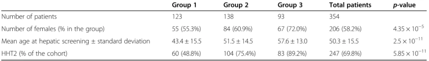
A total of 354 patients were analyzed: 206 females and 148 males. The proportion of females and the age of pa-tients at hepatic exploration increased significantly in function of the severity of HAVM (Table 1). This was also found in mixed logistic models (p < 10− 5 for age at hepatic screening, and p < 10− 3for sex).
Distribution of the ACVRL1 and ENG disease-causing variants
On a total of 107 HHT1 patients, 44 (41.1%) had hepatic involvement, including 10 individuals of group 3. There were 247 HHT2 patients: 187 (75.7%) had hepatic dis-ease including 83 of group 3. The proportion of HHT2 patients increased significantly according to the severity of HAVM (Table 1 and p < 10− 5 in the mixed logistic model).
The distribution of the types of disease-causing vari-ants for ACVRL1 (HHT2) and ENG (HHT1) was differ-ent: the proportion of missense was higher in the ACVR L1 group (Table2, all patients: p < 0.0002, founder muta-tion bias removed: p < 1,7 × 10− 8) in accordance with data from the international mutation database (http:// arup.utah.edu/database/HHT).
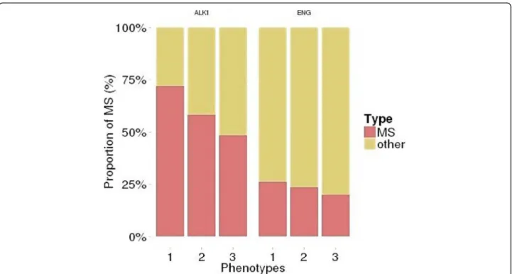
In the HHT2 group, the proportion of truncating vari-ants increased with the severity of liver involvement. The increase was not statistically significant when all HHT2 patients were considered (p = 0.052) but when the bias due to the founder mutation is removed, it is significant (p = 0.01254) phenotype (Fig.1).
Association with modifier genes
A significant association (p = 0.0001) was found between an ACVRL1 SNP (rs2277383) and HAVM. This remained sig-nificant after correction for multiple testing with the FDR (p = 0.0052). There was also an association between HAVM and 3 other ACVRL1 SNPs (rs11169953, rs1150051, rs2293092), one ENG SNP (rs3739817), one SMAD5 SNP (rs5008734), and one ADAM17 SNP (rs10495565), but none of them achieved significance after correction for multiple testing: an additional table file shows this in more details [Additional file1].
When we analyzed the data within each HHT genetic subgroup, rs2277383 was not significantly associated with HAVM in the HHT1 patients. On the other hand, one ACVRL1 SNP (rs1150051) and one ADAM17 SNP (rs10495565) were associated with HAVM in HHT1 pa-tients, but the association did not reach significance after correction for multiple testing by FDR. In contrast, in HHT2 patients, there was a significant association be-tween rs2277383 and HAVM (p = 10− 4before correction for multiple testing and p = 0.026 after correction) and an association with 2 other ACVRL1 SPNs (rs11169953, rs2293092), and the same SMAD5 SNP (rs5008734),
which did not reach significance after correction for multiple testing.
Discussion
In the present study we investigated the role of different genetic factors in the occurrence of HAVM in HHT pa-tients. We confirmed the more severe hepatic involve-ment in females than in males, already observed in previous studies, with a growing proportion of women with the severity of hepatic disease (Table1) [15,16,25]. One striking characteristic of HHT is the wide interfa-milial and intrafainterfa-milial variability in the clinical expres-sion of visceral AVMs. Interfamilial variability suggests that some disease-causing variants may have different consequences compared to others while intrafamilial variability is rather in favor of a role for additional fac-tors, such as modifier genes.
We first studied the relationship between the germline pathogenic variant and the risk of HAVM. In a previous study of genotype-phenotype correlation in HHT we ob-served a higher frequency of hepatic involvement in HHT2 patients compared to HHT1 patients; although the difference did not reach statistical significance [15], it was supported by other studies [16,26]. In the previ-ous study only 57% (197/343) of the patients had sys-tematic hepatic exploration and the criteria for hepatic involvement and severity were not well defined at that time [15], whereas, in this study, hepatic exploration fol-lowing the criteria defined in 2008 was performed in all patients [17]. Clinical records were revisited and patients were classified into 3 severity groups. Based on this im-proved classification of patients we could confirm the higher frequency of HAVMs in HHT2 patients. Among HHT2 patients, the proportion of truncating variants in-creased with the severity of liver involvement. Signifi-cance was reached when we excluded the patients with the c.1112dup/p.Thr372fs variant, which results from a regional founder effect and may introduce confounding
Table 1 Clinical characteristics in the three phenotype groups of hepatic involvement
Group 1 Group 2 Group 3 Total patients p-value Number of patients 123 138 93 354
Number of females (% in the group) 55 (55.3%) 84 (60.9%) 67 (72.0%) 206 (58.2%) 4.35 × 10⁻⁵ Mean age at hepatic screening ± standard deviation 43.4 ± 15.5 51.5 ± 14.5 57.6 ± 13.0 50.3 ± 15.5 2.5 × 10⁻¹¹ HHT2 (% of the cohort) 60 (48.8%) 104 (75.4%) 83 (89.2%) 247 (69.8%) 5.85 × 10⁻¹¹
Table 2 Type of variant according to the disease-causing gene
Type of variant
Missense In-frame indels Splice-site Non sense Frameshift Large deletion or duplication Total ENG* (HHT1) 25 (24.0%) 4 (3.8%) 16 (15.3%) 20 (19.2%) 33 (30.8%) 6 (5.6%) 104 ACVRL1 (HHT2) 112 (45.3%) 4 (1.6%) 19 (7.7%) 25 (10.1%) 85 (34.4%) 2 (0.8%) 247
* 3ENG variants which affected the initiation codon were excluded because their effect was not demonstrated
factors. No association with the type of variant was ob-served for the HHT1 group.
The large intrafamilial variability of HAVM in HHT suggests a role for modifier genes or for non-genetic fac-tors. The higher prevalence of hepatic involvement in fe-males, which is well known and observed herein, suggests a role for hormonal factors or other sex-determined genetic factors. However, it is of note that previous studies, based on immunochemistry analyses of oestrogen and progesterone receptors expression in pa-tients with HHT has brought controversial results and do not support a role for hormonal receptors, at least in the mucosa from nasal telangiectases [27,28].
Modifying genetic factors may be located in trans, i.e. on the allele of ENG or ACVRL1, which does not carry the disease-causing pathogenic variant or in a different gene. We tested the hypothesis of a modifier role for ACVRL1, ENG, and six other genes encoding proteins of the TGFβ/BMP9 pathway. Six SNPs (3 in ACVRL1, 1 in ENG, 1 in SMAD5, and 1 in ADAM17) were significantly associated with HAVMs but after correction for multiple testing, only one remained significantly associated (rs2277383). This SNP is located in the last intron of ACVRL1 and is not predicted to affect splicing. The fre-quency of HAVMs was lower in HHT2 patients with the less frequent allele (G). Since this SNP is located in in-tron 9, 155 bases before exon 10 (c.1378-155 T/G) this association could be due to linkage disequilibrium with the c.1112dup founder pathogenic variant that was present in 53 patients (21%). In order to test this
hypothesis, we performed another analysis excluding these patients. The association between this SNP and HAVM was still highly significant. These results suggest that the (G) allele could be a protective factor towards hepatic involvement, at least in HHT2 patients. This SNP could have an effect on the regulation of expression of the mutated allele of ACVRL1. Alternatively, it could be in linkage-disequilibrium with another SNP with a functional effect. A recent study suggested an association between the ACVRL1 SNP (c.314-35A > G) and the oc-currence of pulmonary and hepatic involvement in HHT1 patients [29]. This is rather in favor of the hy-pothesis of common mechanisms for the development of arteriovenous malformation in different organs. In that study, however, hepatic involvement was determined ac-cording to clinical screening only and no systematic echography evaluation was performed as was the case herein. This methodological difference may have influ-enced the results since liver involvement is age-dependent in HHT and evolved asymptomatically for many years.
In the present study we also included two SNPs of PTPN14 and 1 SNP of ADAM17, which were previously suggested to play a role in HHT [20,21]. PTPN14 is lo-cated in a region orthologous to mouse Tgfbm2 that is a modifier locus for the phenotype of Tgfb−/− mice [30]. Association of two SNPs of this gene with the occur-rence of PAVMs was reported in a cohort of 721 Dutch patients and replicated in a cohort of 222 French pa-tients [20]. In the present study we did not detect any
association between these two SNPs and hepatic involve-ment. ADAM17 is located in a region orthologous to Tgfbm3b, another modifier locus for the Tgfb−/− mice.
Three SNPs of this gene were shown to be associated with the presence of PAVMs in HHT1 patients in the Dutch and French cohorts [21]. In the present study, we found a trend towards an association between HAVM and the common (G) allele of the ADAM17 SNP (rs10495565) in HHT1 but not in HHT2 patients. Func-tional data provided supporting evidence for PTPN14 in-volvement in the endoglin/ALK1 biological pathways in the lung [31]. The latter study also found that genetic variations in the wild-type ENG allele, inherited from the unaffected parent, may modify the risk for pulmon-ary AVMs in HHT1 patients. The pulmonpulmon-ary AVM-at-risk allele ENG-rs10987746-C associates with a slight but significant reduction in ENG transcript levels in hu-man lymphoblastoid cell lines, providing supporting evi-dence that genetic variation within this gene may influence its own expression. In the present study, we also found a trend towards an association of one ENG SNP (rs3739817) with HAVM in the whole HHT group, but not in the HHT1 or HHT2 subgroups. A recent study in 752 HHT patients failed to find significant asso-ciation for candidate genes with arteriovenous malfor-mations of different organs, including HAVMs [32]. However, it is of note that the characterization of liver involvement was different in that study, based on hepatic or cardiac symptoms and signs only. The data presented herein suggest that the possible effects of modifier genes could be different according to the genetic type of HHT (HHT1 versus HHT2), resulting from a yet unknown mechanism. The identification of genetic factors that in-fluence the clinical outcome of a disease with wide intra-familial and interintra-familial clinical variability, such as HHT, may lead to a better understanding of patho-physiological mechanisms and to pave the way for the development of targeted treatments.
Limitations of the study
The fact that the significant association between the ANP rs2277383 and HAMV was not observed in the HHT1 subgroup may probably be due to the lower num-ber of patients. The present results need to be confirmed by studies performed in larger cohorts of patients or supported by biological experiments on cellular or ani-mal models.
Conclusion
Our data confirm that ACVRL1 variants are highly asso-ciated with the risk of HAVMs, and that this risk may be higher in HHT2 patients. In addition to the disease-causing variant, common polymorphisms of ACVRL1 may also play a role in the modulation of this risk, at
least in the HHT2 group. These results emphasize the major role of ACVRL1 in the development of HAVMs.
Supplementary information
Supplementary information accompanies this paper athttps://doi.org/10. 1186/s13023-020-01533-2.
Additional file 1. Acknowledgements
We thank C Auboiroux and B Chambe for technical support, P Robinson for manuscript proofreading and the patients enrolled in this study.
Authors’ contributions
SG and GL conceptualized and designated the study; SG, MFC, BGD, SR, JCS, ED, ME, SP and SDG acquired the data; SG, GL and CB analyzed the data and interpret the findings; CB performed statistical analysis; all authors
contributed to the draft of the manuscript and approved the final version of the manuscript; SG obtained Grant Support.
Funding
This work was supported by the Projet Hospitalier de Recherche Clinique Interrégional 2009 D50675 (Hospices Civils de Lyon).
Availability of data and materials
The datasets used and/or analyzed during the current study are available from the corresponding author on reasonable request.
Ethics approval and consent to participate
Patients had given written consent for the study according to the French bioethics law.
Consent for publication
This manuscript does not contain any individual person’s data in any form. All data are analyzed as aggregates.
Competing interests
The authors declare they have no conflict of interest. Author details
1Hospices Civils de Lyon, Service de Génétique, Groupement Hospitalier Est,
69677 Bron, France.2Service de Biostatistique-Bioinformatique, plateforme de séquençage à haut débit, Hospices Civils de Lyon, Lyon, France.3Université
Claude Bernard Lyon 1, Université de Lyon, Villeurbanne, France.4CNRS UMR 5558, Laboratoire de Biométrie et Biologie Evolutive, Equipe
Biotatistique-Santé, F-69100 Villeurbanne, France.5Centre de Référence National pour la maladie de Rendu-Osler, Groupement Hospitalier Est, Bron, France.6Service de Radiologie, Hôpital Tenon, Paris, France.7Service Génétique, CHU de Poitiers, Poitiers, France.8EA3808, Université de Poitiers,
Poitiers, France.9CHU de Montpellier, Service de Médecine Interne, Hôpital St Eloi, Montpellier, France.10Hospices Civils de Lyon, Service de
Gastro-Entérologie, Hôpital E. Herriot, Lyon, France.11Assistance
Publique-Hôpitaux de Paris, Département de Génétique, GH Pitié-Salpêtrière, Paris, France.12CHU la Milétrie, Laboratoire de Génétique, Poitiers, France.
13Unité de recherche clinique du pole IMER of the Hospices Civils de Lyon,
Lyon, France.14Equipe EA7426, Immunopathologie des voies respiratoires, Université Lyon 1, Lyon, France.
Received: 8 April 2020 Accepted: 7 September 2020
References
1. Plauchu H, de Chadarevian JP, Bideau A, Robert JM. Age-related clinical profile of hereditary hemorrhagic telangiectasia in an epidemiologically recruited population. Am J Med Genet. 1989;32:291–7.
2. McAllister KA, Grogg KM, Johnson DW, Gallione CJ, Baldwin MA, Jackson CE, Helmbold EA, et al. Endoglin, a TGF-β binding protein of endothelial cells, is the gene for hereditary hemorrhagic telangiectasia type 1. Nat Genet. 1994; 8:345–51.
3. Johnson DW, Berg JN, Baldwin MA, Gallione CJ, Marondel I, Yoon SJ, et al. Mutations in the activin receptor-like kinase 1 gene in hereditary haemorrhagic telangiectasia type 2. Nat Genet. 1996;13:189–95. 4. Gallione CJ, Repetto GM, Legius E, Rustgi AK, Schelley SL, Tejpar S, et al. A
combined syndrome of juvenile polyposis and hereditary hemorrhagic telangiectasia associated with mutations in MADH4 (SMAD4). Lancet. 2004; 363:852–9.
5. Wooderchak-Donahue WL, McDonald J, O’Fallon B, Upton PD, Li W, Roman BL, et al. BMP9 pathogenic variants cause a vasculazt-anomay syndrome with phenotypic overlap with hereditary hemorrhagic telangiectasia. Am J Hum Genet. 2013;93:530–7.
6. McDonald J, Wooderchak-Donahue W, Van Sant WC, Whitehead K, Stevenson DA, Bayrak-Toydemir P. Hereditary hemorrhagic telangiectasia: genetics and molecular diagnostics in a new era. Front Genet. 2015;6:1. 7. Eerola I, Boon LM, Mulliken JB, Burrows PE, Dompmartin A, Watanabe S,
et al. Capillary malformation-arteriovenous malformation, a new clinical and genetic disorder caused by RASA1 mutations. Am J Hum Genet. 2003;73: 1240–9.
8. Amyere M, Revencu N, Helaers R, Pairet E, Baselga E, Cordisco M, et al. Germline loss-of-function mutations in EPHB4 cause a second form of capillary malformation-arteriovenous malformation (CM-AVM2) deregulating RAS-MAPK signaling. Circulation. 2017;136:1037–48.
9. Blanco FJ, Santibanez JF, Guerrero-Esteo M, Langa C, Vary CP, Bernabeu C. Interaction and functional interplay between endoglin and ALK-1, two components of the endothelial transforming growth factor-beta receptor complex. J Cell Physiol. 2005;204:574–84.
10. David L, Mallet C, Mazerbourg S, Feige JJ, Bailly S. Identification of BMP9 and BMP10 as functional activators of the orphan activin receptor-like kinase 1 (ALK1) in endothelial cells. Blood. 2007;109:1953–61. 11. Dupuis-Girod S, Ginon I, Saurin JC, Marion D, Guillot E, Decullier E, et al.
Bevacizumab in patients with hereditary hemorrhagic telangiectasia and severe hepatic vascular malformations and high cardiac output. JAMA. 2012; 307:948–55.
12. Khalid SK, Garcia-Tsao G. Hepatic vascular malformations in hereditary hemorrhagic telangiectasia. Semin Liver Dis. 2008;28:247–58.
13. Berg J, Porteous M, Reinhardt D, Gallione C, Holloway S, Umasunthar T, et al. 2003. Hereditary hemorrhagic telangiectasia: a questionnaire based study to delineate the different phenotypes caused by endoglin and ALK1 mutations. J Med Genet 2003;40:585–590.
14. Kuehl HK, Caselitz M, Hasenkamp S, Wagner S, Harith El HA, Manns MP, et al. Hepatic manifestations is associated with ALK1 in hereditary hemorrhagic telangiectasia: identification of five novel ALK1 and one novel ENG mutation. Hum Mutat. 2005;25:320.
15. Lesca G, Olivieri C, Burnichon N, Pagella F, Carette MF, Gilbert-Dussardier B, et al. Genotype-phenotype correlations in hereditary hemorrhagic telangiectasia: data from the French Italian HHT network. Genet Mol. 2007;9: 14–22.
16. Letteboer TG, Mager JJ, Snijder RJ, Koeleman BP, Lindhout D, Ploos van Amstel JK, Westermann CJ. Genotype-phenotype-relationship in hereditary hemorrhagic telangiectasia. J Med Genet. 2006;43:371–7.
17. Gincul R, Lesca G, Gelas-Doe B, Rollin N, Barthelet M, Dupuis-Girod S, et al. Evaluation of previously nonscreened hereditary hemorrhagic telangiectasia patients shows frequent liver involvement and early cardiac consequences. Hepatology. 2008;48(5):1570–6.
18. Lesca G, Burnichon N, Raux G, Tosi M, Pinson S, Marion MJ, et al. Distribution of ENG and ACVRL1 (ALK1) mutations in French HHT patients. Hum Mutat. 2006;27:598.
19. Barret JC, Fry B, Maller J, Daly MJ. Haploview: analysis and visualization of LD and haplotype maps. Bioinformatics. 2005;21:163–5.
20. Benzinou M, Clermont FF, Letteboer TG, Kim JH, Espejel S, Harradine KA, et al. Mouse and human strategies identify PTPN14 as a modifier of angiogenesis and hereditary hemorrhagic telangiectasia. Nat Commun. 2012;3:616. 21. Kawasaki K, Freimuth J, Meyer DS, Lee MM, Tochimoto-Okamoto A, Benzinou M, et al. Genetic variants of Adam17 differentially regulate TGFbeta signaling to modify vascular pathology in mice and humans. Proc Natl Acad Sci U S A. 2014;111:7723–8.
22. Vazquez AI, Bates DM, Rosa GJM, Gianola D, Weigel KA. Technical note: an R package for fitting generalized linear mixed models in animal breeding1. J Anim Sci. 2010;88:497–504.
23. Benjamini Y, Hochberg Y. Controlling the false discovery rate: a practical and powerful approach to multiple testing. J R Statistic Soc B. 1995;57:289–300.
24. Lesca G, Genin E, Blachier C, Olivieri C, Coulet F, Brunet G, et al. Hereditary hemorrhagic telangiectasia: evidence for regional founder effects of ACVRL1 mutations in French and Italian patients. Eur J Hum Genet. 2008;16:742–9. 25. Mora-Lujan JM, Iriarte A, Alba E, Sanchez-Corral MA, Cerda P, Cruellas F,
et al. Gender differences in hereditary hemorrhagic telangiectasia severity. Orphanet J Rare Dis. 2020;16:63.
26. Bayrak-Toydemir P, McDonald J, Markewitz B, Lewin S, Miller F, Chou LS, et al. Genotype-phenotype correlation in hereditary hemorrhagic telangiectasia: mutations and manifestations. Am J Med Genet A. 2006;140: 463–70.
27. Eivazi B, Werner JA, Roessler M, Negm H, Teymoortash A. Lack of significant estrogen and progesterone receptor expression in nasal telangiectasias in hereditary hemorrhagic telangiectasia: an immunohistochemical analysis. Acta Otolaryngol. 2012;132:86–9.
28. Pau H, Carney AS, Walker R, Murty GE. Is oestrogen therapy justified in the treatment of hereditary haemorrhagic telangiectasia: a biochemical evaluation. Clin Otolaryngol Allied Sci. 2000;25:570–6.
29. Pawlikowska L, Nelson J, Guo DE, McCulloch CE, Lawton MT, Young WL, et al. The ACVRL1 polymorphism is associated with organ vascular malformations in hereditary hemorrhagic telangiectasia patients with ENG mutations, but not in patients with ACVRL1 mutations. Am J Med Genet. 2015;167A:1262–7.
30. Tang Y, McKinnon ML, Leong LM, Rusholme SA, Wang S, Akhurst RJ. Genetic modifiers interact with maternal determinants in vascular development of Tgfb1(−/−) mice. Hum Mol Genet. 2003;12:1579–89. 31. Letteboer TGW, Benzinou M, Merrick CB, Quiley DA, Zhau K, Kim IJ, et al.
Genetic variation in the functional ENG allele inherited from the non-affected parent associates with presence of pulmonary arteriovenous malformation in hereditary hemorrhagic telangiectasia 1 (HHT1) and may influence expression of PTPN14. Front Genet. 2015;6:67.
32. Pawlikowska L, Nelson J, Guo DE, CE MC, Lawton MT, Kim H, et al. Association of common candidate variants with vascular malformations and intracranial hemorrhage in hereditary hemorrhagic telangiectasia. Mol Genet Genomic Med. 2018;6:350–6.
Publisher’s Note
Springer Nature remains neutral with regard to jurisdictional claims in published maps and institutional affiliations.