Université de Monfréal
Implication de la voie de dégradation ubiquitine-dépendante dans la pathologie des maladies de surcharge lysosomale
par Panojot Bffsha
7 f
cj. Département de BiochimieFaculté de Médecine
Mémoire présenté à la Faculté des études supérieures en vue de l’obtention du grade de
Maître ès sciences en Biochimie
Mai, 2005
© Panojot Bifsha, 2005
Université de Monfréal Faculté des études supérieures
2
Direction des bibliothèques
AVIS
L’auteur a autorisé l’Université de Montréal à reproduire et diffuser, en totalité ou en partie, par quelque moyen que ce soit et sur quelque support que ce soit, et exclusivement à des fins non lucratives d’enseignement et de recherche, des copies de ce mémoire ou de cette thèse.
L’auteur et les coauteurs le cas échéant conservent la propriété du droit d’auteur et des droits moraux qui protègent ce document. Ni la thèse ou le mémoire, ni des extraits substantiels de ce document, ne doivent être imprimés ou autrement reproduits sans l’autorisation de l’auteur.
Afin de se conformer à la Loi canadienne sur la protection des
renseignements personnels, quelques formulaires secondaires, coordonnées ou signatures intégrées au texte ont pu être enlevés de ce document. Bien que cela ait pu affecter la pagination, il n’y a aucun contenu manquant. NOTICE
The author of this thesis or dissertation has granted a nonexclusive license allowing Université de Montréal to reproduce and publish the document, in part or in whole, and in any format, solely for noncommercial educational and research purposes.
The author and co-authors if applicable retain copyright ownership and moral rights in this document. Neither the whole thesis or dissertation, nor substantial extracts from it, may be printed or otherwise reproduced without the authot’s permission.
In compliance with the Canadian Privacy Act some supporting forms, contact information or signatures may have been removed from the document. While this may affect the document page count, it does flot represent any loss of content from the document.
Université de Monfréal faculté des études supérieures
Ce mémoire intitulé:
Implication de la voie de dégradation ubiquitine-dépendante dans la pathologie des maladies de surcharge lysosomale
présenté par: Panojot Bffsha
a été évaluéparunjury composé des personnes suivantes:
Président-rapporteur : Alain Moreau Directeur de recherche : Alexei Pchejetski Membre dujury : Marie-Josée Hébert
ACKNOWLEDGEMENTS
This work was supported in part by the operating grant from Canadian
Institutes of Health Research MT-38107, Genome Canada/Genome Quebec
grantand by the equipment grant from Canadian Foundation for Innovation
to Dr Alexey V. Pshezhetsky. It is also worth mentioning the contribution of Dr Rob Sladek and the Genome Quebec Microarray Facility for the gene microarray studies. first and foremost, many thariks to Dr Pshezhetsky for offering me a position in bis prestigious laboratory, for bis relentless efforts towards my supervision and bis overail support in conducting research. I greatly appreciate the predoctoral summer studentship offered by the Canadian Society for Mucopolysaccharide and Related Disease, which allowed me to initiate these studies. Tins work would flot have been possible without the contribution of every laboratory team member and the collaboration of select members of the scientific community, who provided helpful suggestions and who were wffling to share their resuits. Consequently, I would like to thank Mme Karine Landry for helping me to integrate into the lab and for her enormous contribution in the realization of tins project in biochemistry techniques (immunocytochemistry, westerns, Real-time PCR). Dr Volkan Seyrantepe supervised me in matters of molecular biology such as gene cloning techniques (UCH-L1 subcloning and siRNA studies). Dr Mila Ashmarina helped in the interpretation of microarray results and the elaboration of the main hypothesis. Mme Stéphanie Trndel helped in the mass spectrometry analysis of mouse brains and Mme Christianne Quiniou in the westerns of apoptotic proteins. Dr Roy Gravel, of the University of Calgary, provided Sandhoff mouse brains. I apologize if the name of certain individuals could not be included herein, but I wil neyer cease to recognize thefr benevolence. Lastly, I would like to thank my parents for their encouragement in pursuing graduate studies.
Table of contents
LIST 0F FIGURES .v
LIST 0F TABLES vii
ABBREVIATIONS viii
ABSTRACT 1
RÉSUMÉ 2
LITERATURE REVIEW 3
LYSOSOMAL STORAGE DISORDERS 3
-Endosomal-lysosomal system 4
-Concept of lysosomal storage 7
-Pathologicalfeatures 9
-Cettular pathology 11
-Animal modets ofÏysosomat storage disorders 14
—Diagnosisanti strategiesof treatment 17
PROGRAMMED CELL DEATH (APOPTOSIS) 18
-Apoptotic protease activation 18
-Regutators of apoptosis 19
-Two pathways of caspase activation 20
—77w consequences of caspase activation 23
-Caspase-independent pathways 23
UBIQUflIN-PROTEASOMAL SYSTEM tUFS) 24
-Deubiquitinating enzymesanti UCH-L1 25
-Animal model for UCH-L1 deflciency (GAD mouse, 27
-Rote of ubiquitinationinapoptosis 29
-Involvement of UPSin neurodegeneration 32
INTRODUCTION 37
MATERIALS AND METHODS 41
-Cett culture 41
-mRNA quantification by real-time PR 41
-Confocal immunofluorescence microscopy 42
-Enzymatic assays 44
-Western blotting 45
-Proteasomal activity assays 46
{TCH-U overexpression studiesantiapoptosis
detection 47
-Quantification of UcH-U protein inbrain
-UcH-L1 small interferingRNAgene
silencing in human skin fibroblasts .50
-Detection ofpro- and anti-apoptotic
proteins in UcH-L1 siRNA-treated cetis 53
RESULTS AND DISCUSSION 54
UCH-L1 AND LYSOSOMALSTORAGE 54
-Reduced UcH-L1 mRNA levels in fibroblasts
from LSD patients and Sandhoffmouse brains 54
-Reduced ubiquitin hydrolase activity and
UCH-Ll protein levels infibroblasts 0f LSD patients 58
-Reduced UCH-L1 protein levels in Sandhoffmouse brains 59
-E-64-induced lysosomat storage
resuits in reduced expression of UcH-Ll 62
UBIQUffIN-PROTEASOME PATHWAY IN LYSOSOMAL STORAGE 64
-Decreased proteasomal activity in
human flbroblasts with lysosomal storage 64
-Impaired proteasomal activity in Sandhoffmouse brains 67
-Lysosomal storage inducesfonnation ofubiquitin
pro tein aggregates and reduced level offree ubiquitin 67
APOPTOSIS AND LYSOSOMAL STORAGE 69
-Fibroblasts with Ïysosomal storage display higher apoptosis rates 71
-Overexpression of UCH-L1 in fibroblasts with
lysosomat storage rescues tlwmfrom apoptosis 73
-Suppression of UCH-L1 in normal
fibroblasts induces caspase-mediated apoptosis 74
-Apoptotic proteins bax, bd-2 and p53
are increased in UCH-L1 suppressedfibroblasts 79
CONCLUSIONS AND PERSPECTIVES 82
LIST 0F FIGURES
FIGURE 1: The Ïysosomal network
6
FIGURE 2: Molecularelements of the apoptotic cascade
21
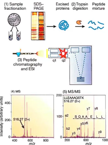
FIGURE3: Generic mass spectrometry (MS)-based proteomics experiment 49
FIGURE4: Mechanisrn ofRNA interference (RNAi) 52
FIGURE5:Lysosomat storage bodies in the celis of patients affected with siatidosis
and sialic acid storage disease 55
FIGURE 6: Suppression of UCH-L1 in culturedflbroblasts obtainedfrom LSD
patients 56
fIGURE7: Immunohistochemical detection of UcH-L1 in control ceÏÏs and cells
obtainedfrom LSD patients 57
FIGW?.E8: Ubiquitin hydrolase activity in control cells and cells ofLSD patients 58 FIGU1?E9: Comparison of UcH-L1 cDNA and protein levels in total brains of
Sandhoffmouse model 59
FIGURE 10: Western blots for UCH-L1 in Sandhoffmouse brains 60
FIGURE 11: Identification and quantification ofUCH-L1 protein in brain lysates of
Sandhoffmouse model by tandem mass spectrometnj 61
FIGURE12: Ubiquitinated lysosomat storage bodies in cultured skin flbrobïasts
treated with E-64 63
FIGURE 13: Decreaseofproteosomal activity in cuÏtured skinflbroblastsfrom
LSDpatients 65
FIGURE 14:Decrease ofproteasomal activity in brain tissues ofSandhoffmice 66 FIGURE 15: Detection ofubiquinated proteins in celts with lysosomal storage 68
FIGURE 16: Ubiquitin western blot offlbroblasts ofLSD patients 69
FIGURE 17:Development oflysosomat storage, UcH-L1 deflciency and apoptosis in
cuÏtured skin flbroblasts treated with E-64 70
FIGURE18: E-64-treatedflbrobÏats show decreased UCH-L1 protein and increased
apoptosis 72
FIGURE 19:Devetopment of apoptosis in cultured skin flbroblastsftom LSD
patients 73
FIGURE20: Suppression ofE-64-induced apoptosis in cuttured skin flbroblasts 75 FIGURE21: Inhibition of UCH-L1 expression in cultured human skin flbroblasts by
siRNA 77
FIGURE22: Induction of apoptosis by siRNA-induced inhibition ofUCH-L1
expression 76
FIGURE23: Expression ofpro- and anti-apoptotic proteins in cultured skin flbroblasts
treated with UCH-L2 siRNA 81
FIGURE24: Proposed scheme of UCH-L1-mediated apoptosisfollowing lysosomal
LIST 0F TABLES
TABLE I: Non-exhaustive list ofLSD with defective enzyme(s) and corresponding
metabolite accumulation 8
TABLE II: CÏinïcopathologi calL$D phenotypes 10
TABLE III: Non-exhaustive list of mouse modeÏs of human LSD 15
TABLE IV: Neurodegenerative disorderswithubiquitin-immunoreactivityin ceÏt
inclusions 34
TABLEV: Major groups ofproteins whose expression was changedin both SIASD
anti sialidosisfibroblasts 40
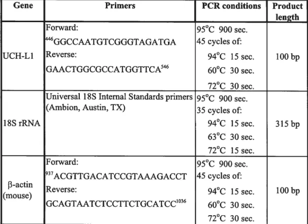
TABLE W: PCR primersanticonditions used to measure concentration of UcH-L1
mRNA in cutturedfibroblasts anti mouse brain 42
TABLE VII: siRNA primers used to downregulate UCH-L1 in cultured
ABBREVIATIONS
AD Alzheimer’s disease AMC 7-amido-4-methylcoumarin CAS Caspase DAPI 4,6-diamidino-2-phenylindole; DMSO Dimethylsulfoxide DTT Dithiothreitol E-64 (epoxysuccinyl-leucylamido-(4-guanidino)butane EDTA Ethylenediaminetetraacetic acidFMK fluoromethyketone
GAPDH Glyceraldehyde-3-phosphate dehydrogenase
GI Gaucher disease type I
Gil Gaucher disease type II
GM1 GM1-gangliosidosis
GS Galactosialidosis
HEX Hexosaminidase
HPLC High performance liquid chromatography
LC-MS/MS Liquid chromatography-tandem mass spectromefry LSD Lysosomal storage disorder/ disease
MA Morqulo syndrome type A
MB Morquio syndrome type B
MEM Minimum essential medium
PAGE Polyacrylamide gel elecfrophoresis PBS Phosphate-buffered saline
PD Parkinson’s disease
SDS Sodium dodecyl sulfate
SIASD Sialic acid storage dïsease
SL Sialidosis
TBS Tris buffered saline
UCH-L1 UbiquitinC-terminal hydrolase Li (PGP 9.5)
ABSTRACT
Increased programmed ccli death is an important pathological feature underlying the tissue and organ maffuncifon seen in lysosomal storage disorders (LSD). Using expression microarrays, differentially expressed genes that were common to cultured fibroblasts from patients affected with several different LSDs were identified. These studies, conffrmed by biochemical experiments, demonsfrated that lysosomal storage is associated with down regulation of the most abundant cellular ubiquitin C-terminal hydrolase, UCH-L1 (PGP 9.5), in cultured fibroblasts of patients representing 8 different LSDs as well as in the brain tissues of a mouse model of Sandhoff disease with a disrupted hexosaminidase B gene. Induction of lysosomal storage by treatment of cultured fibroblasts with the lysosomal cysteine protease inhibitor E-64 reduced UCH-L1 transcripts, protein levels and enzyme activity. Ail celis and tissues exhibiting lysosomal storage contained ubiquitinated protein aggregates, and showed reduced levels of free ubiquitin, decreased proteasomal activity, and increased numbers of apoptotic celis. The increased rate of apoptosis seen in E64-freated fibroblasts was reversed by fransient transfection with a UCH-L1 expression vector. In confrast, siRNA mediated down-regulation of UCH-L1 resulted in high apoptosis rates with concomitant increase in the expression of pro-apoptotic proteins such as Bax and p53. Taken together, these data suggest that UCH Li deficiency and partial impairment of the ubiquitin-dependent protein degradation pathway may represent a common mechanism for the pathogenesis of lysosomal storage disorders.
Keywords: Lysosome, proteasome, UCH-Li, ubiquitin, apoptosis, inclusions, neurodegeneration
RÉSUMÉ
La mort programmée accrue de cellules est un dispositif pathologique important sous-tendant les défauts de fonctionnement du tissu et de l’organe dans les désordres de surcharge lysosomale (SL). Nous montrons que lapoptose dans les cellules avec de la SL se réalise par l’entremise d’un affaiblissement partiel de la voie de dégradation ubiquitine-dépendante. En utilisant des micropuces d’expression, nous avons évalué les changements secondaires des profils d’ARN messager dans les fibroblastes cultivés de patients affectés avec plusieurs différentes maladies de SL et avons identifié les gènes différenfiellement exprimés qui étaient communs à ces conditions. Ces études, confirmées par des expériences biochimiques, ont démontré que la SL est associée avec la sous-régulation de la plus abondante hydrolase d’ubiquitine C-terminale, soit UCH-L1 (PGP 9.5), dans tous les patients représentant huit désordres de SL différents. Les niveaux de transcrits UCH Li, de protéine et d’activité ont été également réduits dans des cellules normales en lesquelles la SL a été induite par l’inhibiteur lysosomal de cystéine protéases, E-64. Toutes les cellules montrant de la SL contenaient des agrégats de protéines ubiquitinées, en plus des niveaux réduits d’ubiquitine libre, inhibition partielle de l’activité du proteasome et un nombre accru de cellules apoptotiques. Le taux d’apoptose accru observé chez les fibroblastes traités avec E-64 a pu être reversé lors d’une transfection temporaire avec le vecteur d’expression pour UCH-L1. D’autre part, la suppression de UCH-L1 par interférence d’ARN dans des fibroblastes normaux a précipité ces derniers vers l’apoptose avec une élévation concomitante de l’expression des protéines pro-apoptotiques Bax et p53. Nous speculons que l’insuffisance en UCH-Li ainsi que la dérégulation partielle de la voie de dégradation ubiquitine-dépendante représentent un mécanisme commun pour la pathogénèse des maladies de surcharge lysosomale.
Mots-clés Lysosome, protéasome, UCH-L1, ubiquitine, apoptose, inclusions, neurodégénération
LYSOSOMAL STORAGE DISORDERS
Cellular composition is determined by the maintenance of a balance between degradation and synthesis of intracellular components. This homeostasis can lie perturbed when either process is impaired especially during adaptive changes, leading to ceil and tissue deregulation. Component turnover is not only important for the sake of salvaging damaged or mmecessary by-products, but is also essential in the redirection of ongoing processes toward new needs. For example, protein degradation rates are increased during muscle hypertrophy and liver regeneration.
In lysosomal storage disorders (LSD), defects in lysosomal enzymes, in lysosomal transmembrane fransporters, in hydrolase cofactors or in the biogenesis of the organelle itseff, causes the sequesfration of the corresponding unprocessed subsfrates inside the lysosomes and renders virtually impossible the further reutilization of the monomeric components. This affects the architecture and function of the celis, tissues and organs (extralysosomal effects) even though in very few cases the accumulated substrate may lie directly cytotoxic. The signs associated with LSDs are often neurological, but cari also lie multisystemic, with skeletal, CNS, cardio vascular and ocular defects.
The clinical course of these diseases is chronic and progressive, very often lethal before or in early adulthood. Over 40 types of lysosomal storage diseases have been identffied, with a collective incidence of 1 in 7000-8000 (Winchester et ai, 2000) live bfrths, and they have been grouped according to similarities in the biochemistry of accumulated materials, the clinical presentation and the enzymatic deficiencies. In terms of the storage material, LSDs can 5e divided into three large groups, the sphingolipidoses, mucopoly
saccharidoses, glycoproteinoses and several other indïvidual enfities (Table I,
p.8). The inheritance is autosomal recessive for most LSDs, except for MPS II and Fabry disease. Most of the LSDs can be diagnosed by the assay of enzymes in circulating leucocytes, biopsy material or cultured skin flbroblasts. Almost ail of the genes responsible for the variety of LSDs have been cloned and tMs information is being used for genotype-phenotype correlation, genetic counseling and selection of patients for novel forms of therapy.
Endosomat-tysosomat system
Lysosomes are organelles specialized for the infracellular digestion of macromolecules and are filed with hydrolytic enzymes operating optimally at an acïdic pH of about 5.0. Lysosomes contain about 40 types of hydrolytic enzymes or acid hydrolases, including proteases, nucleases, glycosidases, lipases, phospholipases, phosphatases, and suffatases. The internai pH of the lysosome is consequently maintained low by an integral lysosomal membrane ATP-driven hydronium ion (Hj pump. The limilÏng membrane of the lysosome or late endosome contains also a set of higffly glycosylated integral membrane proteins, designated as lysosomal-associated membrane proteins (LAMPs) (such as LAMP-1, LAMP-2, and CD63/LAMP-3), lysosomal membrane glycoproteins (LGPs) and lysosomal integral membrane proteins (LIMPs) (Eskelinen et ai, 2003). Additional lysosomal membrane proteins mediate transport of ions, amino acids, and other solutes across the lysosomal membrane and contribute to the maintenance of an acidic luminal pH in the range of 4.6 —5.0. Similar to ail other secretory proteins, lysosomai enzymes or acid hydrolases are synthesized in the endoplasmic reticulum and fransported to the Golgi apparatus where ffiey undergo a variety of post transiational modifications, of which is the aftachment of terminal mannose
6-phosphate groups to specific oligo-saccharide side chains that can 5e recognized by M6P receptors found on the iimer surface of the Golgi membrane, and then carried from the trans Goigi network to late endosomes by means of clathrin-coated transport vesicies.
Lysosomes are morphologically heterogeneous, often resembling other organelies of the endocytic and secretory pathways including melanosomes, lytic granules, major histocompatibiity complex (MHC) ciass II
compartments, platelet dense granules, basophil granules, and neutrophil azurophil granules. Therefore, lysosomes are currently distinguished from other organelles on the basis of an operational definition, which describes them as membrane-Sound acidic organelles that contain mature acid dependent hydrolases and LAMPs, but lack mannose 6-phosphate receptors (MPRs) (Komfeld and Meilman, 1989).
In ail eukaryotic celis, there are two major pathways of membrane traffic reaching lysosomes, the endocytic pathway (heterophagy) and autophagy (Fig. 1, p.6). The former delivers cargo from outside of the cell and
plasma membrane, while the latter transports materials in the cytoplasm and into lysosomes. Endosomes also relay newiy synthesized lysosomai enzymes from the secretory pathway to lysosomes (biosynthetic pathway).
It is well known that receptors endocytosed from the plasma membrane are either recycled to celi surface or transported to lysosomes for degradation. This sorting event, which takes place in the endosomes, is critical, since it acts as a filter of cellular signal transduction (Yoshimori,
2002). Ligand-mediated endocytosis is characteristically an early response in
the signaling pathways triggered by a diverse group of cell surface receptors, including hetero-frimeric guanine nucleotide-bïnding protein (G protein) coupled receptors (GPCRs), receptor tyrosine kinases (RTKs) and cytokine receptors.
fIGURE1 The lysosomal network.
From the celi surface, the endocytic pathwayextendsto lysosomes. The biosynthetic pathway
bridges the secretOry paffiway and the endocytic pathway. Autophagy delivers materials from the cytoplasm to lysosomes. Lysosomes are usually meeting places where severai streams of infracellular traffic converge. Endocytosed molecules are irdtially delivered in vesicles to small, irregularly shaped infracellular organelles cafled early endosomes. Some of these ingested molecules are selectively reftieved and recycled to the plasma membrane, while offiers pass on into ffie mildly acidic (pH —6) late endosomes to be digested by lysosomal hydrolases. These are progressively delivered to the endosome from the Goigi apparatus. Mature lysosomes form from the Iate endosomes, accompanied by a further decrease in internai pH. Lysosomes are thought to be produced by a graduai maturation process, during which endosomal membrane proteins are selectiveiy retrieved from ffie developing lysosome by transport vesicles that deliver these proteins back to endosomes or the frans Golgi network. A second pathway to degradation in lysosomes is used in ail ceil types for the disposai of obsolete parts of the ceil itseff, a process called autophagy. The process seems to begin with ffie enclosure of an organefle by membranes of unknown origin, creating an autophagosome, which then fuses with a lysosome (or a late endosome). Professionai phagocytes (macrophages and neutrophils in vertebrates) enguif objects to form a phagosome, which is then converted to a iysosome in the manner of the autophagosome. Lysosomai secretion (aiso called defecation) of the undigested contents is a minor paffiway that enables ail ceils under stress to elirninate indigestibie debris. Some ceil types, however, contain specialized lysosomes such as melanocytes that have acquired the necessary machinery for fusion wiffi the plasma membrane.
Concept of tysosomat storage
In general, the distribution of the stored material, and hence which organs are affected in LSDs, is determined by two interrelated factors: (1) the tissue where most of the material to 5e degraded is found and (2) the location where most of the degradation normally occurs (Winchester et ai, 2000). for example, the brain is rich in glycosphingolipids due to their presence in neuronal membranes, and hence defective hydrolysis of these molecules, as occurs in GM1 and GM2 gangliosidoses such as Krabbe, Niemann- Pick and Gaucher diseases, resuits primarily in storage within neurons and neurologic symptoms. In contrast, glycosaminoglycans (mucopolysaccharides) are primarily structural molecules produced by most ceils and are found mainly on the surface of ceils and in the matrix. Thus, defects in the degradation of mucopolysaccharides affect vfrtually every organ because they are widely disfributed in the body. Celis of the mononuclear phagocyte system are involved in the degradation of a variety of subsfrates through the lysosomal pathway and organs rich in phagocytic ceils, such as the spleen and liver, are frequently enlarged in several forms of LSDs.
Normally, LDSs show allelic variation (different mutations of the same gene) and significant clinical heterogeneity. The severity of the disease, which influences the time of onset and the pace of the regression, appears to 5e correlated with the residual enzyme activity, which itself depends on the nature of the molecular defect (even though clear genotype-phenotype correlations have often not been established, suggesting the existence of “modifying” genes). Such a model, elaborated by Conzelmann and Sandhoff (1983-1984) has found experimental support in several sphingolipidoses.
Disease Enzyme Deficiency Major Accumulating Metabolïtes Glvcogenosis Type 2—Pompe disease a-1,4-Clucosidase (lysosomal glucosidase) Glycogen Sphiiiglipidoses GM1 gangliosidosis GM1 ganglioside Il-galactosidase GM1 ganglioside, gal actose-containing oligosaccharides CM2 gangliosidosis Tay-Sachs disease Hexosamhuidase-a subunit GM2 ganglioside Sandhoff disease Hexosaminidase-13 subunit CM2 ganglioside, globoside GM2 gangliosidosis, variant AB Ganglioside activator protein GM2 ganglioside Suif atidoses Metachromafic leukodysfrophy Arylsulfatase A Sulfatide Multiple sulfatase deficiency Arylsulfatases A, B, C; steroid sulfatase; iduronate suliatase; heparan N-Sulfatide, steroid sulfate, heparan sulfate, sulfatase dermatan sulfate Krabbe disease Galactosylceramidase Galactocerebroside Fabiy disease u-Galactosidase A Ceramide inhexoside Gaucher disease Glucocerebrosidase Glucocerebroside Niemann-Pick disease: types A and B Sphingomyelinase Sphingomyelin Mucopo1ysaccharidoes (MPS) MPS I H (Hurler) u-L-Iduronidase Dermatan sulfate, heparan sulfate MPS II (Hunter) L-Iduronosulfate sulfatase Mucoiip.idoses (ML) 1-ceil disease (ML II) and pseudo-Hurler polydysfrophy Deficiency of phosphorylating enzymes essential for ffie formation of Mucopolysaccharide, glycolipid mannose-6-phosphate recognition marker; acid hydrolases lacking the recognifion marker cannot be targeted to the lysosomes but are secreted extracellularly Other Diseases of Complex Carbohychates Fucosidosis a-Fucosidase Fucose-containing sphingolipids and glycoprotein fragments Mannosidosis a-Mannosidase Mannose-containing oligosaccharides Aspartylglycosaminuria Aspartylglycosamine amide hydrolase Aspartyl-2-deoxy-2-acetamido-glycosylamine Other Lysosomal Storage Dieases Wolman disease Acid lipase Cholesterol esters, friglycerides AcM phosphate deficiency Lysosomal acid phosphatase Phosphate esters
D
D
D
TABLE I Non-exhaustive list of LSD with defective enzyme(s) and corresponding metabolite accumulationTwenty percent of normal enzyme activity is usually adequate to carry out cellular function. Consequently, heterozygotes (carriers of LSDs) whose enzyme activity is about 50 percent of normal are clinically unaffected
(Grabowski and Hopkin, 2003). Symptoms develop when residual enzyme
acffvity falis below a threshold of 15 to 20 percent. Therefore, mutations that
leave no residual enzyme activity cause severe, early-onset illness. Milder
mutations cause insidious, late- or aduit-onset illness. But, what is the link between (specific) subsfrate storage in the lysosomes and tissue lesions? Although no well-established mechanism has yet been put forward to answer this question, it is likely that LSDs are accompanied by dysregulated celi growth or death.
Patho logical features
Manifestations of neurological deterioration associated with many LSDs begin in infancy or childhood. Very often it begins with a delay and then arrest of psychomotor development, neurological regression, buindness, and seizures. Inexorable progression leads to a vegetative state. LSDs have diverse and distinct chuical manifestations. Some of them share certain cili-ilcal and pathological features grouped into four basic clinical-pathological phenotypes: leukodysfrophy, mucopolysaccharidosis, storage histiocytosis and neuronal lipidosis, the latter being the most prevalent phenotype (Table II, p.lO).
The pathology, in the neuronal lipidosis phenotype, primarily involves the gray matter. Storage causes neuronal ballooning and torpedo-like swellings of proximal axons and dendrites. This process leads to loss of neurons and their axons, but myelin is not primarily affected (Neufeld and Muenzer, 2001). Storage in retinal ganglion ceils causes blindness. Other celis
and organs that do flot process large amounts of gangliosides are either normal or show mfld storage without celi damage. The prototype of the
TABLE II OEnicopaffiological LSD phenotypes
PHENOTYPE PATHOLOGY CLINICAL FINDINGS LSDs NEURONAL UPIDOSIS Storage in the neuronal Neurological regression, Gangliosidoses,
body and processes seizures, blindness mucopolysacchafldoses, neuronal ceroid Iipofuscinoses
LEUKODYSTROPHY Storage in Neurological regression, Gangliosidoses (metachromatic
oligodendrocytes and spasticity, peripheral Ieukodystrophy, Krabbes disease) Schwann ceils neuropathy
MUCOPOLYSACCHARIDOSIS Storage in exfraneural Visceromegaly, soft tissue Mucopolysacchafldoses, tissues swelling, skeletal dyspiasia, glycoproteinoses, GM1
heart disease gangliosidosis
STORAGE HISTIOCYTOSIS Storage in histiocytes Hepatosplenomegaly, Gangliosidoses (Gaucher disease, hematopoiefic abnormalities Niemann-Pick disease)
neuronal lipidosis phenotype is Tay-Sachs disease, a form of GM2 gangliosisosis prevalent among Ashkenazi Jews, first described more than 100 years ago (Neufeld and Muenzer, 2001). Some LSDs impair enzymes that are important for turnover of myelin lipids and damage myelin-producing celis. This results in loss of myelin (leukodystrophy) manifested by neurological deterioration and spasticity. The mucopolysaccharidoses and glycoproteinoses affect neurons as well but have also severe skeletal and visceral manifestations, which constitute the mucopolysaccharidosis phenotype. In general, the celis that are most severely affected by LSDs are neurons, because they process large amounts of gangliosides and once lost they cannot 5e replaced, and histiocytes, because their main function is lysosomal degradation, and they decompensate if thefr lysosomal enzymes are deficient. Several LSDs show lipid storage in histiocytes throughout the lymphoid and hematopoietic tissues. Such storage histiocytosis causes hepatosplenomegaly, bone manow depression, bone damage and other
manifestations. In some LSDs, notably Gaucher disease, storage histiocytosis is the main abnormality.
Cet!utar pathotogy
The major classes of storage diseases can 5e distinguished by the ulfrastructural appearance of their storage material. Glycosphingolipidoses present membranous swirls, mucopolysaccharidoses with multilamellar stacks (“zebra bodies”), fucosidosis and mannosidosis with watery or wispy material (“open” inclusions), and so forth (Suzuki, 1976). On elecfron microscopic examination, the stored products are membrane-Sound because they are contained within lysosomes. The explanation for the characteristic appearances of storage bodies was that the accumulation of undegraded ceil material past a certain crifical point was believed to lead to physicochemical interactions with other components within the lysosome (e.g., cholesterol) and to formation of the characteristic morphological appearance of the residual or storage body for a given disease (Suzuki, 1976). Early studies have shown that residual bodies in storage diseases like Tay-Sachs and other neurolipïdoses contained acid phosphatase activity, suggesting a lysosomal status (Walkley, 1998). Arguments that the storage bodies sometimes lacked histochemical evidence of lysosomal enzymes, or even lysosomal-like delimiting membranes, and thus might not 5e lysosomal, were eventually interpreted as simpiy late disease-associated alterations in the lysosome.
Cytotoxicity following mechanical disruption of ceils has often been invoked as a plausible explanation for the cell dysfunction in lysosomal diseases. Given the observations that there are ceil-selective consequences of the storage disease process, it was reasonable to assume that significant extra lysosomal events are set in motion by deficiencies of single lysosomal enzymes. At least four possible mechanisms that reach beyond the mere
mechanical disruption hypothesis can 5e cited (Walkley, 1998): (i) Material stored within the lysosome may escape and in turn have deieterious effects elsewhere in the celi, either by direct toxicity or by impinging on other metabolic pathways to alter celi function. (ii) The massive accumulation of unprocessed compounds within the lysosome (e.g., gangliosides) may deprive the celi of certain precursor molecules, leading to a compensatory upregulafion or dysfunction of other metabolic pathways or organeiles. (iii)
Abnormalffles of the lysosomal system may lead to a reduced entry of materials into lysosomes and to subsequent increases of this compound elsewhere in the ceil, with defrimental consequences. For example, plasma membrane receptor endocytosis is partially disrupted and the related signaling pathway remains activated. (iv)Defective lysosomai enzyme activity may adverseiy effect the normal functioning of other related organelies, most notably endosomes and the whole lysosomal network dynamics (Fig. 1, p.6).
Generaliy exacerbated apoptosis has been observed in celis or tissues from patients or animais affected with neurolipidoses; that is, in Tay-Sachs, Sandhoff (Huang et al, 1997; Wada et ai, 2000), and Krabbe diseases (Taniike et ai, 1999; Jatana et al, 2002), prosaposin deficiency (Tohyama et al, 1999), neuronal ceroid iipofuscinoses (Lane et al, 1996) and metachromatic leuko dysfrophy (Coenen et ai, 2001). Although the molecular mechaiiism of this increased (neuronal) celi death is not yet estabiished, it has been postulated that lysosphingoiipids, which accumulate in the affected cells, may mediate cytotoxicity, possibly through inhibition of protein kinase C (Hannun et ai, 1987). Interestingly, ceiluiar sphingosine ieveis are eievated in Niemann—Pick disease type C mice, in which increased neuronal ceil death has been also observed (Wu et ai, 1999). Although the exact contribution of sphingoiipids to apoptosis needs further characterization (mostly in terms of enzymes
involved and cellular targets), evidence has accumulated that some of these lipidic molecules act as bioregulators of ceil growth and death. There is littie information regarding possible alterations in the apoptotic program in the visceral forms of lysosomal disorders. Nevertheless, increased apoptosis has been described in chondrocytes from animais affected with mucopoly saccharidosis type W (Simonaro et ai, 2001). Cystinotic ceils aiso exhibit an increased sensffivity to apoptosis (Park et ai, 2002) that might 5e related to the (thioi-related) redox status of these celis.
Neurons in storage diseases not only exhibit somatic swelling secondary to accumulation of material, but they reveai a number of other somatic, dendritic and axonal changes. In Tay-Sachs disease for example, cortical neurons were described as being swoilen and having enlargements within the basilar dendrites, but spinal motoneurons only exhibited sweiling without dendrific changes (Walkley et, 1998). This concept of neuron type specific changes was extended to the other storage diseases. Two distinct types of celiular alterations emerged as characteristic of many storage diseases, namely meganeurites and axonai spheroids. Meganeurites were parasomatic enlargements within the axon hillock and appeared to occur secondary to storage as part of a volume expansion by the neuron. They were found only on certain types of neurons while other types appeared to undergo simple somatic enlargement secondary to storage. There are realiy two classes of meganeurites (Walkley et al, 1987; Walkley et al, 1988), named spiny and aspiny. Spiny and aspiny meganeurites can occur on specific populations of neurons, whereas in other diseases, like Baffen disease, only aspiny meganeurites were found. On the other hand, axonal spheroids are enlargements of the distai segment of the axon that contain storage material inconsistent with the specific defective lysosomal hydrolase, as opposed to meganeurite storage material composition. The ultrasfructural appearance of
spheroid material is the same across many types of storage disorders. The close similarity of accumulated material in studies of a distai crush or low temperature lesions in axons to that of spheroids in storage disease (Parton et ai, 1992) suggests that the latter may be secondary to a block in retrograde transport. Spheroids are capabie of causing significant interference with the efficacy or timing of action potential propagation (Walkley et ai, 1991). In animal models of GM1 and GM2 gangliosidosis, Niemann- Pick disease, CL mannosidosis, and mucopolysaccharidosis, there is a striking correlation between the location and incidence of axonal spheroids and the type and severity of clinical neurological disease (March et ai, 1997).
Anima! modets of tysosomat storage disorders
The animal models of the LSDs should be defined as those in wInch ffie genes that are orthologous to the genes that cause human lysosomal disorders are functionally defective. This is to make the distinction with modeis generated by exogeneous administration of metabolites in high doses to artificialiy induce a lysosomai storage condition by perturbing the balance of subsfrate influx over ïts efflux. Although spontaneously occurring genetic lysosomal storage diseases are as rare in other mammalian species as in man, the advent of gene targeting technology has revoiutionized the state of animal models of genetic diseases and neariy ail lysosomal storage diseases have been duplicated in the mouse (Table III, p.l5). These animal models cari overcome many of the limitations inherent in studies of human patients such as rarity of ffie disease, exfremeiy complex genetic background and iogistical and ethical constraints in the design and execution of experiments with human subjects.
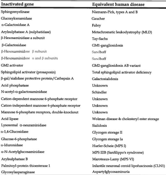
TABLE III Non-exhaustive list of mouse models of human LSD
Inacfivated gene Equivalent human disease
Sphingomyelinase Niemann-Pick, types A and B
Glucosylceramidase Gaucher
u-Galactosidase A fabry
Arylsuiphatase A (suiphatidase) Metachromatic Ieukodystrophy (MLD)
P-Hexosaminidase a suburnt Tay-Sachs
-Ga1actosidase GM1-gangliosidosis
[—lrTexesam.i.nidase f.lsuhunit Sandhoff
[i-Hexosaminidase u and 3 suhunits Sandhoff
GM2 activator GM2-gangliosidosis AB variant
Sphingolipid activator (prosaposin) Total sphingolipid activator deficïency
3-gal/sialidase protective protein/Cathepsin A Galactosïalidosis
Acid phosphatase Unknown
N-acetyl-Œ-galactosaminidase Schindier
Cation-dependent mannose 6-phosphate receptor Unknown
Cation-mdependent mannose 6-phosphate receptor Unknown
Mannose 6-phosphate receptors, double-knockout Unknown
Acid lipase Wolman disease & cholesteryl ester storage
Lysosomal Œ-neuraminidase Sialidosis
cx4,4-Glucosidase Glycogen storage II
Glucose-6-phosphatase Glycogen storage la
u-Iduronidase Hurler-ScheiefMPS I)
a-N-Acetylglucosaminidase MPS IIIB (Sanfilippos syndrome)
Arylsulphatase B Maroteaux-Lamy (MPS VI)
Palmitoyl-protein thioesterase 1 Infantile neuronal ceroid lipofuscinosis (CLN1)
Glycosylasparaginase Aspartylglycosaminuria
Adapted fromJ. Inher. Metab. Dis.21 540-547 andJ Gene Med 2004;6: 481—506
Genetic lysosomal diseases have been known to occur spontaneously
in many mammalian species, but most of them occur among larger animais that caimot 5e easiiy amenabie to genetic manipulation such as mice or rats.
Only two spontaneous mouse models of weil-delineated genetic lysosomal diseases are known; 13-giucuronidase deficiency (mucopolysaccharidosis VII)
and the twitcher mutant (galactosylceramidase deficiency, Krabbe disease).
respective human defects in ail aspects (Ellinwood et ai, 2004) and so happens with some of ffie induced mutants, such as acid sphingomyelinase deficiency, t3-hexosamÏnidase f3-subunit (Sandhoff disease), galactosialidosis and total sphingolipid activator deficiency. Conversely, a subgroup of those induced mouse models shows minor deviations from human disease, including the acid -galactosidase deficiency and f3-hexosaminidase a-subunit deficiency (Tay-Sachs disease) among others.
Whfle mouse models have become necessary in the clarification of the pathogenetic mechanism of disease and the exploration of therapeutic approaches, species differences in the brain development and metabolic pathways must 5e always remembered if the ultimate goal of the study is application to human patients. When mouse models exhibit phenotypic differences to those of human diseases, several factors are considered as the underlying mecharilsm of ffiose differences. 0f primary importance is the lag in developmental processes that occur at different lime intervals between humans and mice, notabiy the absolute and relative time scales with respect to the lifespan, and the variability in metabolic pathways (Suzuki et ai, 1998). for example, neuronal proliferation and arborization in mice are initiated 7-8 days after birth, as opposed to prenatal completion in humans. At the same time period, human newborn brains are already well into the active phase of myelination in the telencephalon. An excellent example that illustrates the effect of metabolic differences on the displayed phenotype is the Tay-Sachs disease, which shows dramaticaily different clinical manifestations between human and the genetically equivalent mouse disease. In humans, the gangliosides containing a single sialic acid residue, known as GM1 and GM2, are degraded almost exclusively by hydrolysis of the respective terminal sugars, galactose and N-acetylgalactosamine. These reactions are catalyzed by lysosomal J3-galactosidase and (3-hexosamiriidase-A, in the presence of
their substrate specific cofactors saposin-B and GM2-activator protein, respectively (Huang et ai, 1997). In contrast, in the mouse, GM1- and GM2-gangliosides are readily degraded also by sialidase through sialic acid removal to the respective derivatives, GAl and GA2, thus providing an alternative route for their catabolism.
Diagnosis and strategies oftreatment
Theoretically, two approaches can lie envisaged in order to restrict the amount of stored material inside the lysosome. First, we can provide the missing enzyme either by direct administration, gene therapy or bone marrow transplantation. Second, we can alleviate the amount of undigested substrate by pharmacologically decreasing its rate of synthesis or by restricting its ftow to the lysosome. In case of a lysosomal transporter defects, equaliy valid would lie the possibiity to divert the transport of products or by enhancing their export from the lysosome. for example, in cystinosis, there is an intra-lysosomal accumulation of cysteine. Intralysosomal cysteine levels can be decreased by oral administration of high doses of cysteamine, a basic compound that reacts with the accumulated cysteine to form a mixed disulfide resembling the structure of lysine, which is further transported outside of the lysosome by the corresponding lysine amino acid transporter (Winchester et ai, 2003).
The most widely used procedure for the diagnosis of LSDs is enzyme assay and for most LSDs, this cari lie performed on leukocytes with fast turnaround (Neufeld et ai, 2001). Cultured fibroliiasts are required in a few LSDs. Cultured amniocytes or chorionic villus cells may be used for prenatai diagnosis. Biochemicai determination of storage products is cumbersome, but has some applications. for instance, demonstration of glucosaminoglycans (GAG) in urine is a useful screening test for GAG storage. Storage of
abnormal products can be detected in the ceils of the patient by light and electron microscopy. In addition to neurons, gangliosides and ceroid lipofuscin are stored in somatic celis and may be detected by nerve, muscle, skin, conjunctival, and other biopsies. Tissue diagnosis (detection of specific storage materials by electron microscopy) is sifil the standard for some NCLs because biochemical assays of the affected proteins are not yet available. The gene mutations of LSDs can be detected by DNA analysis. Mutation analysis is used mainly for carrier detection and prognosis.
PROGRAMMED CELL DEATH (APOPTOSIS)
Celi death is a part of normal physiology for most metazoan species. During development, redundant or unwanted cells are removed through programmed cell death, making important contributions to morphogenesis, organogenesis and other processes. However, excessive apoptosis, for example, has been implicated in neurodegenerative diseases such as Alzheimer’s disease, Parkinson’s disease, amyotrophic lateral sclerosis and several hereditary diseases that are caused by extended polyglutamine repeats (Yuan et al, 2000). Among the features of cells undergoing apoptosis, as opposed to necrosis, are chromatin condensation, nuclear fragmentation, plasma membrane blebbing, ceil shrinkage and ultimately shedding of membrane-delimited ceil fragments known as apoptotic bodies (Arends and Wyllie, 1991).
Apoptotic protease activation
The biochemical events that occur during apoptosis are the result of hydrolysis of cellular proteins by a family of cysteine proteases called caspases, which cleave specific target proteins at aspartic acid residues. 0f the 11 human caspases, 7 have defrnite (caspases 3, 6, 8, and 9) or probable
(caspases 2, 7, and 10) roles in apoptosis, whereas the remainder is thought to lie invoived in cytokine processing and the regulation of inflammation
(Honig et ai, 2000). Active caspases are teframeric enzymes consisting of two large 17- to 37-kDa and two smail 10- to 12-kDa subunits. Caspases are normally held in an inactive form by an integral hihibitory domain that becomes the target of proteolysis upon their activation. Each caspase is synthesized as a single-chain zymogen that contains an N-terminal prodomain foliowed by one large and one smail subunit. Maturation of the procaspases invoives cleavage at criticai aspartate residues, that is, at the same type of bonds that caspases themselves cleave. This feature of caspase activation provides the opportunity for autoactivation as well as the possibility of caspase cascades. The advantage to the cell of this sfrategy is that no protein sythesis is required to activate the apoptotic pathway given that ail the components are already present.
Regutators of apoptosis
The Bd-2 family of proteins comprises both anti-apoptotic and pro apoptotic members and has a pivotai role in controlling programmed celi death by reguiating mitochondriai integrity and mitochondria-mediated caspase activation (Adams et ai, 2001). There are at least 17 famiiy members, which can be divided into three groups based on the presence or absence of four conserved Bd-2 homology (BH) domains. The N-terminai BH4 domain is present in ail anti-apoptotic family members, inciuding Bd-2, Bd-w, Bcl-xL, Mci-1, A-1, Boo/Diva, and Nrf3. Pro-apoptotic family members, ail of which lack the BH4 domain, fali into one of two categories. Either they are smaii family members containing only the BH3 domain (e.g., Bid, Bad, Bik, Bim, Blk, Bmf, Hrk, Bnip3, Nix, Noxa, and PUMA) or are iarger and resemble Bd-2 more closeiy but merely lack the BH4 domain (e.g., Bax, Bak, and Bok). The
pro-apoptofic “BH3 orily” proteins present in higher eukaryotes (e.g., Bid, Bim, Bmf) fransiocate from cytoplasmic sites to the outer surfaces of mitochondria in response to apoptotic stimuli. The ratio between anti apoptotic and pro-apoptotic Bd-2 members is a crucial factor that helps to determine the susceptibiity of a ceil to a death signal (Hengartner, 2000).
In addition to Bd-2 family members, a famÏly of polypeptides called IAP (irihibitor of apoptosis) proteins also regulates apoptotic processes (Deveraux et ai, 1999; Salvesen et ai, 2002). Human members of this family include clAPi, cIAP2, XIAP, NAIP and livin. Common structural features include one or more baculovirus inhibitor repeat (BIR) motifs and RING domains. The zinc finger-like BIR domains bind the surfaces of caspases, allowing sequences between the BIRs to block the caspase active site. In this manner, XTAP, clAPi, and cIAP2 are able to block caspase 8- and 9- initiated events in vitro and to inhibit the activities of purified caspases 3, 7, and 9. The RING domain acts as an ubiquitin ligase and presumably facifitates proteasome-mediated degradation of whatever these IAP proteins bind
(Kaufmann et al. 2001).
Two Pathways of Caspase Activation
Apoptosis-signaling pathways can 5e categorized into three phases: initiation and caspase activation, mitochondrial commitment and effector events (Fig. 2, p.2l). According to current understanding, there are at least two major pathways of apoptotic caspase activation (Hengartner, 2000). One, calied ffie “death receptor” or “exfrrnsic” pathway, ufflizes caspase 8 and/or 10 as the initiator caspase in a protease cascade. The other, called the “mitochondrial” or “infrinsic” pathway, invoives caspase 9. Though the initiation and early spectrum of caspase activation differ between these two
pathways, both these signal transduction pathways converge at the activation of caspases 3 and 6, whichare the two widely studied effector caspases.
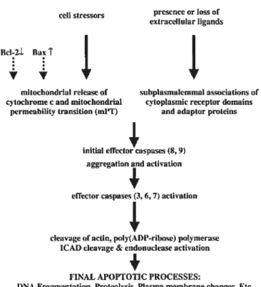
APOPTOTIC SIGNAL
ccli stressors prescnce or loss of extracellutar ligands
Bcl-2L Baxt
mitochonddal relcaseof subplasmalemmal associations ut cytochromee ami mitochondrial cytoplasmic receptor domains
pcrmeahility transition (nil’T) and adapb.r proteins
initial ettcctnr caspasrs (5, 9) aggregation and activation
ettector caspases 3, 6, 7) activatîon
cleavageofactin,polytAt)P—rihnse) polynierase
1CAD cleavage & endonuclease activation
+
FINAL APOPTOTIC PROCESSES:
DNA Fragmentation, Proteoivsis, Plasma membrane changes, Etc.
FIGURE2 Molecular elements of the apoptofic cascade (Honig et ai, 2000)
The “extrinsic” or “death receptor” pathway begins with ligation of specific celi surface receptors. In the case of fas, binding of its cognate ligand (Fas ligand) resuits in an alteration of the so-called “death domain” in the cytoplasmic tau of the receptor to permit binding of the death domain containing adaptor molecule FADD. As a consequence of this binding, the “death effector domain” of FADD acquires the ability to bind homologous domains in procaspase 8, thereby drawing this zymogen to the site. Molecular interactions within this assembly, which is called a DISC, resuit in cleavage of procaspase 8 and release of mature caspase 8 to the cytosol, where
it cleaves both procaspase 3 and the small “BH3 only” Bd-2 family member Bid (Hacker, 2000). Depending on the ratio of these cleavages, apoptosis might be friggered directly or might depend on activation of the mitochondriai pathway (intrinsic pathway).
Induction of apoptosis by a variety of toxic insuits, including radiation and chemotherapeutic agents, is accompanied by changes in mitochondriai function. These stimuli cause several mitochondrial changes, inciuding decreases of mitochondrial membrane potential and the release of polypeptides that normaily reside in the mitochondrial inter-membrane space. Among the polypeptides released is cytochrome c. Once in the cytopiasm, this protein binds to a scaffoiding protein called apoptotic protease-activating factor-1 (Apaf-1). In the presence of cytochrome c and dATP, Apaf-1 undergoes a conformationai change that facilitates binding of a protein interaction domain calied a caspase recruitment domain (CARD) on Apaf-1 to a similar motif present in the prodomain of procaspase 9. The resuit is the formation of a Mr -700,000 complex calied an apoptosome that has enhanced abiity to cleave pro-caspases 3 and 7 to active enzymes (Dimmeler et ai, 1999). These caspases then contribute to the characteristic apoptotic phenotype. Another mechanism that friggers apoptosis in response to celi stress operates through the transcription factor p53, which is a major player in the events of ceil cycle controi mainly in response to DNA damage. DNA repair mechanisms are activated, but so is transcription of the gene for another bd-2 family protein cailed Bax. Like Bad, Bax ailows cytochrome c to escape from mitochondria, so if the DNA is not repaired in time, BAX concentrations increase, cytochrome cescapes, and apoptosis ensues.
The Consequences of Caspase Activation
In the last stages of apoptosis, once caspase 3 has been activated, biochemical changes that characterize the apoptotic phenotype begin to occur. Ffrst, caspase 3 directly participates in endonuclease activation. The nuclease caspase-activated deoxyribonuclease (CAD/DFF4O) is a consti tutively expressed nuclear protein that is ordinarily complexed with its inhibitor ICAD. Caspase 3-mediated cleavage of ICAD resuits in the liberation of CAD, which begins to digest the chromatin (Hacker, 2000).
Because chromatin in the linker regions is more accessible than chromatin wound around histones, the net resuit is a characteristic intemucleosomal pallem of DNA degradation. This intemucleosomal degradation is accompanied by chromatin condensation, another hallmark of apoptosis. Caspase 3 also cleaves procaspase 6, liberating the active form of this enzyme. Together, caspases 3 and 6 cleave structural proteins of the nucleus (lamins and NuMA), facilitating nuclear fragmentation. In addition, these two proteases (primarily caspase 3) cleave more than 200 other cellular subsfrates, thereby inhibiting DNA repair and celi cycle progression, inactivating signal transduction pathways that are critical for survival, and activating a series of enzymes thatare thought to participate in cellular disassembly.
Caspase-Independent Pathways
Even though caspases appear to 5e critical for cleavage of key subsirates during apoptosis, other effector molecules might also contribute to the apoptotic phenotype. For example, apoptosis-inducing factor (AIT) is an oxidoreductase that is released from mitochondria into the cytosol during apoptosis (Daugas et al., 2000). It subsequently localizes to nuclei, where it is capable of generating large (>50 kb) DNA fragments and inducing chromatin condensation by unknown mechanisms. It is also important to recognize that
some caspase-independent ceil deaths appear to be necrotic, that is, to involve ATP depletion and loss of membrane integrity. When caspases are inhibited, the primary lesions induced by various treatments (e.g., microtubule disruption or DNA damage) and the cytochrome e release that follows sifil occurs. These changes might 5e sufficiently disruptive to cellular metabolism so that celis wii ultimately die even if they cannot activate caspases.
Attempts have been made to order caspase-independent ceil death according to the celiular organelles involved such as the mitochondria, lysosomes and the endoplasmic-reticulum (Ferri et ai, 2001). Interestingly, active participation of lysosomal proteases such as cathepsins B and D has been observed in cell death induced by several stimuli, including oxidative stress, TNF-Œ, bile salt-induced apoptosis and chemotherapeutic drugs (Broker et ai, 2005). Indeed, cathepsin B has been shown to act as an effector protease, dowristream of caspases in certain ceil types (Foghsgaard et ai, 2001), and is capable of executing ceil death independent of the apoptotic machinery in WEHI-S fibrosarcoma and non-small celi lung cancer (NSCLC) ceils (Broker et ai, 2005). Other reports have shown that lysosomal proteases can promote celi death more indirectly by friggering mitochondrial dysfunction and subsequent release of mitochondrial proteins probably through Bld fransiocation after lysosomal disruption by lysosomofropic agents (Cirman et ai, 2004). Finally, lysosomal proteases have been reported to directly cleave and activate caspases, thereby confirming that lysosomal permeabilization often is an early event in the apoptotic cascade (Ferri et ai, 2001).
UBIQUITIN-PROTEASOMAL SYSTEM (UPS)
Protein degradation by the ubiquitin/proteasome system plays a primordial role in a broad array of basic cellular processes such as the ceil cycle, signal transduction and development. Considering these numerous processes, it is flot surprising that the system has been implicated in the pathogenesis of many diseases such as Parkinson’s disease, Alzheimer’s disease, ischaemia reperfusion injury, aging in the central nervous system and diabetes. Proteins modffied by multiubiquitin chains are the preferred substrates for degradation by the 26S proteasome, a multicatalytic protease complex. The proteasome ïs a massive complex consisting of more than 20 protein subunits and having a sedimentation coefficient of 26 (26S) (Ciechanover et al., 2000). It has a cap and barrel structure, with the cap (referred to as the 19$ component of the proteasome) being the part that orients the ubiquitinated proteins in preparation for degradation by the 20$ barrel component, wMch contains the proteases. This multicatalytic protease is characterized by three major activifies, that is the tryptic, chymotryptic and peptidylglutamyl hydrolytic activifies.
Ubiquitination of proteins is a complex ATP-dependent process in which ubiquitin is sequentially activated by ubiquitin-activating enzymes (El), transferred to ubiquitin-conjugating enzymes (E2) and then ligated to protein substrates by ubiquitin ligases (E3). Ubiquitin is joined to the subsfrate through the formation of an isopeptide bond between the C terminus of ubiquitin (G1y76) and the side-chain of a lysine residue of the substrate. An addffional conjugation factor, named F4, was recently described and is involved in polyubiquitin chain elongation of at least a subset of subsfrates in conjunction with El, E2 and E3.
Although targeting proteins for destruction by the proteasome is the best-characterized task of ubiquitin, other functions are being discovered at a
rapid rate. Examples for such proteolysis-independent functions are
ubiquitin-dependent endocytosis, trarscriptional confrol and DNA repafr
(Varshavsky, 1997). Many of these are regulated by alternative types of ubiquitin modification, such as monoubiquitylation (in which only one ubiquitin moiety is added), or by a non-canonical multiubiquitin chain. The proteasome also plays an essentiai role in maintaining celi homeostasis by degrading many rate limiting enzymes and crifical regulatory proteins.
Deubiquitinating enzymes and UCH-L1
Deubiquitinating enzymes (DUB) are proteases that specifically hydrolyse ester, thiol ester and amide bonds to the carboxyl group of glycine residue 76 of ubiquitin (Wing, 2003). These enzymes, in anaiogy with the role of phosphatases in reversibie phosphorylation, perform the reversible ubiquitination of proteins and can have important regulatory functions. They are involved in processing of ubiquitin gene products, in negativeiy regulating the functions of ubiquitination, in regenerating free ubiquitin after protein degradation by the proteasome or the lysosome and in salvaging ubiquffin from possible infraceilular adduct formation (Wilkinson, 1997). Ail eukaryotes contain DUBs encoded by at ieast two gene families, the ubiquitin carboxyl-terminai hydrolase (UCH) and the ubiquitin-specific processing protease (UBP) famiiies, which comprise in total more than 60 known members. The products of the UCH family were named for their activity in hydroiysing smail amides and esters at the C-terminus of ubiquitin and have also been shown to remove peptides and smail proteins, whereas the UBPs are responsible for ubiquitin proprotein and ubiquitin fusion protein processing.
Three human UCH isozymes have been cioned and they exhibit marked tissue specificity. For instance, UCH-L3 is an isoform specificaily
expressed in hematopoetic ceils, wheres UCH-L1 is higffly expressed in neurons (Doran et ai, 1983) and to ceils of the diffuse neuroendocrine system and their tumors. Day and Thompson (1987) cloned UCH-L1 cDNA and the deduced protein, which they called PGP9.5, contains 212 amino acids. Day et al. (1990) determined that the UCH-L1 gene contains 9 exons and spans 10 kb. The 5-prime region contains elements common to many genes and other elements that are shared with the 5-prime regions of the genes encoding neurofilament neuron-specific enolase (ENO2) and THY1 antigen. By Northern blot analysis, Leroy et ai. (1998) detected a 1.3-kb transcript expressed only in brain and further confirmed the gene structure. Examination of specific brain regions revealed expression in ail areas tested, particularly in the substantia nigra. Ubiquitin C-terminal hydrolase Li represents i to 2% of total soluble brain proteins (Wilkinson et ai., 1989).
Its occurrence in Lewy bodies and its function in the proteasome pathway made UCH-L1 a compelling candidate gene in Parkinson disease. In a German family with Parkinson disease, Leroy et al. (1998) identified a missense mutation in the UCH-L1 gene, i1e93 to met (193M), which caused a partial ioss of the catalytic activity of this thioi protease. They suggested that this could lead to aberrations in the proteolytic pathway and aggregation of proteins. In other studies, Lincoin et al. (1999) sequenced the entire coding region of the UCH-Li gene in 11 families with a paftern of Parkinson’s disease consistent with autosomai dominant inheritance. Although they found polymorphisms in noncoding regions, the only amino acid change was S18Y. The S18Y allele was found in approximateiy 20% of chromosomes in a Caucasian population, suggesting that it is unlikely to 5e pathogenic. Lincoin et al. (1999) concluded that the 193M variant must be a rare cause of Parkinson’s disease or a harmless substitution whose occurrence in the family reflected chance. However, Liu et al. (2002) found that UCH-Li, especiaily
variants linked to higher susceptibility to Parkinson disease, caused the accumulation of aipha-synuclein in cultured ceils, an effect that could not 5e explained by its recognized hydrolase activity. Therefore, UCH-L1 exhibited an additional dimerization-dependent ubiquityl ligase activity. Interestingly, the polymorphic variant of UCH-L1 associated with decreased risk for Parkinson disease, serl8 to tyr (S18Y), had reduced ligase activity compared with the wildtype enzyme, but it had comparable hydrolase activity. The authors concluded that the ligase and hydrolase activifies of UCH-L1 may play roles in proteasomal protein degradation, a process critical for neuronal health. Osaka et al. (2003) recently demonstrated that monoubiquitin is stabiized by its direct association to UCH-L1. This team investigated the effect of a UCH-L1 loss of function on ubiquitin levels, as in the gad (gracile axonal dystrophy) mouse, where neuronal monoubiquitin levels were reduced compared to wild-type.
Animal mode! for UCH-L1 deficiency (GAD mouse)
The gracile axonal dysfrophy (gad) mouse is an autosomal recessive mutant that shows sensory ataxia at an early age, followed by motor ataxia later (Yamazaki et ai, 1988). Pathologically, the mutant is characterized by ‘dying-back’ type axonal degeneration and formation of spheroid bodies in nerve terminals. Pathologic observations in the human have associated brain aging and neurodegenerative diseases with progressive accumulation of ubiquitinated protein conjugates. In gad mice, accumulation of amyloid f3 protein and ubiquitin-posifive deposits occur retrogradely along the sensory and motor nervous systems.
Suh et ai. (1995) showed that the gad mutation is located on mouse chromosome 5. Later, Saigoh et al. (1999) found that the gad mutation is caused by an in-frame deletion including exons 7 and 8 of the UCH-L1 gene,
encoding the ubiquitin carboxy-termirial hydrolase selectively expressed in the nervous system and testis. The gad allele encodes a truncated UCH-L1
protein lacking a segment of 42 amino acids containing a catalytic residue. Since this protein is thought to stimulate protein degradation by generating free monomeric ubiquitin, the gad mutation appears to affect protein turnover. The findings showed ffiat altered function of the ubiquitin system directly causes neurodegeneration and suggestes that the gad mouse provides a useful model for investigating human neurodegenerative disorders.
Kurffiara et al. (2000) showed that mice homozygous for a targeted deletion of the related UCH-L3 gene are indistinguishable from wildtype. To assess whether the two hydrolases have redundant function, Kurihara et al. (2001) generated mice homozygous for both UCH-L1 (gad) and UCH-L3Q3-7). The double homozygotes weighed 30% less than single homozygotes and displayed an earlier onset of lethality, possibly due to dysphagia. Axonal degeneraffon of the nucleus tractus solitarius and area postrema of the medulla was noted in these mice. 11e double homozygotes also displayed a more severe axonal degeneration of the gracile tract of the medulla and spinal cord than had been observed in UCH-L1 (gad) single homozygotes. In addition, degeneration of dorsal root ganglia celi bodies was detected in both the double homozygotes and UCH-L3Q\3-7) single homozygotes. Given that both UCH-L1 (gad) and UCH-L3Q\3-7) single homozygotes displayed distinct degenerative defects that were exacerbated in the double homozygotes, the authors concluded that UCH-L1 and UCH-L3 may have both separate and overlapping functions in the maintenance of neurons of the gracile tract, nucleus tractus solitarius, and area postrema.
Rote of ubiquitination in apoptosis
The relation between ubiquitin and apoptosis was first suggested when increases in ubiquitin expression were observed during programmed ceil death in the intersegmental muscle of insects (Schwartz et ai, 1990) and mature lymphocytes. This hypothesis was further reinforced by evidence 1irking proteasome inhibition to the activation of celi death in human leukemic HL6O celis (Drexier, 1997). Today, it is widely accepted that ubiquitin is critically involved in the regulation of molecules at every phase of apoptosis. Important regulators of apoptosis, including the Bd-2 family of proteins, the lAPs and regulators of the iriliibitor of nuclear factor-KB kinase (IKK) have been identilied as new sulistrates of the ubiquination system. In addition, the tumour suppressor p53 and other celi-cycle proteins that are akeady known to be subsfrates of the proteasome have now been assigned specific functions in the regulation of apoptosis. Moreover, lAPs, which are pivotally involved in the negative regulation of pathways that induce death, have themselves been shown to take part in the ubiquitylation of apoptotic substrates.
Despite the amount of work correlating the variation in ubiquitin levels (and ubiquitination paffem), proteasomal subunit composition and proteasomal activity to apoptosis, proteasomal inhibition does not exhibit a clear directional correlation. That is to say that the downsfream effects to proteasomal inhibition, in terms of apoptosis, may be either positive or negative in a ceil-type dependent maimer without negating the possibffity that it changes the celi susceptibiity to apoptotic stimuli. These seemingly contTadictory results might be due to the unspecific nature or concentrations of the inhibitors that were used, or to differences between the celi types (Orlowski et ai, 1999). An illustration of the above assertion is that most stages of lymphocytes and most leukemic ceil mes become sensitized to
apoptosis following proteasome inhibition, whilst primary thymocytes do not in response to dexamethasone, y-irradiation, phorbol ester or etoposide freatment (Lee et ai, 2003). As a general ruie, in most celi lis, proteasome inhibitors frigger apoptosis.
As mentioned eariier, the JAP famiiy inhibits apoptosis primarily by inactivating and degrading pro-apoptotic proteins and presents the only known subset of apoptosis-signaiing molecuies that contain E3 iigase acffvity (Deveraux et ai, 1999) by the presence of RING domains. The mechanism of IAP-mediated inhibition of apoptosis predominantly lies in its interaction with caspases such as casp 3, casp 7, casp 9, which resuits in their ubiquitination and subsequent degradation by the UPS.
Ubiquitination of the Bd-2 proteins is known to target for degradation both pro- or anti-apoptotic family members. Recent reports indicate that the ubiquitin/proteasome system decisiveiy influences the deiicate balance between these two fractions of the Bci-2 famiiy. Degradation of pro-apoptofic members such as Bid, Bax and Bak has been reported to promote celi survival and converseiy, apoptotic progression has been found to requfre poiy ubiquitin-mediated Bd-2 degradation. Proteasomes have aiso been noted to modulate apoptosis by affecting the iffe time of the ‘only’ members BH3-interacting-domain death agonist (Bid) and Bd-2 interacting kiiler (Bik).
The p53 tumor suppressor protein is one of the best-known pro apoptotic proteins acting at the transcriptionai levei and its induction occurs foiiowing infrmnsic stressors, such as DNA damage, and ieads to a hait in celi cycle progression and/or apoptosis activation. Foliowing activation by various stresses, p53 downreguiates anti-apoptotic proteins, such as Bd-2 and the transcription factor (3-catenin (Sadot et ai, 2001), and upregulates several death-promoting moiecuies such as the adaptor protein Apaf-1, Bax, Noxa, Puma, etc (Lee and Peter, 2003). The role of each of these proteins in
p53-mediated apoptosis seems to be ceil-type dependent. In addition, p53 might also confribute to the induction of apoptosis through franscriptionally independent activities (Ryan et al. 2001). The mechanisms that underlie these functions remain poorly understood, but might point to a direct role for p53 at mitochondria and the relocalization of death receptors to the ceil surface. For example, p53 was shown to directly interact with Bax protein leading to its activation and to ffie subsequent permeabifization of the mitochondrial membrane (Chipuk et ai, 2004). The ieveis of p53 protein are usually quite low, as its rapid degradation prevents the accumulation of p53 in normal proliferating ceils. The stabiity of p53 is highly regulated, primarily by its abiity to bind the cellular proto-oncogene Mdm2, which functions as an ubiquitin ligase for p53, mediating its ubiquitination and proteasome degradation. The expression of Mdm2, in turn, is regulated by p53, producing a feedback loop that tightly regulates p53 function.
Several stress-induced signaling pathways leading to ffie inhibition of MDM2-mediated degradation of p53 have been identified. Kinases activated by genotoxic damage, including ATM and ATR, Chkl, Chk2 as well as other sfress-related kinases, have been shown to phosphorylate and thus stabffize p53 (Abraham, 2001). Furthermore, the response to p53 phosphorylation on a particular residue may depend on the specific stress signal initiating the phosphorylation event, underscoring the complexity of p53 phosphorylation and stabiization. Regulation of p53 nuclear localization also piays an important role in confrolling p53 function and its abiity to fransactivate responsive genes. The abffity of p53 to 5e exported from the nucleus is certainly enhanced by Mdm2 ubiquitin ligase activity (Ryan et ai, 2001).
Unlike polyubïquitination, the functional consequences of monoubi quitination are diverse and include receptor endocytosis, virus budding, transcription, DNA repair and caspase recruitment during apoptosis.
Concerning apoptosis, four proteins are targeted by monoubiquitination: ifistone H2A, caspase-3, caspase-7 and DEDD. The role of monoubi quitination of the above proteins in apoptosis remains speculative since this field is stiil under development, but a cunent hypothesis is that a lattice formation of ubiquitinated proteins forms and may act as a signaling platform (Lee and Peter, 2003).
Involvernent ofZIPS in neurodegeneration
Genetic data from various species clearly hidicate the existence of a link between proteasomal dysfunction and neuronal disorders. Moreover, an increasing body of evidence supports a crucial role for the apoptotic system in the manifestation of many of the neurodegenerative diseases, although our knowledge about the mechauisms that are involved in these disorders is sifil incomplete.
Neuronal death takes place within hours to days after a stroke, but may occur over years in Alzheimer disease (AD). Mter a traumatic injury or stroke, neurons die rapidly because of excitotoxicity (typically occurring within 48 h) and apoptosis (typically occurring within 4-14 days) (Honig and Rosenberg, 2000). There can also 5e extensive apoptosis of astrocytes and oligodendrocytes during tifis period. In confrast, neuronal death in diseases such as AD or Parkinson disease (PD) occurs over a period of years as a resuit of necrosis induced by oxidative stress and the accumulation of protein aggregates. The steady accumulation of protein aggregates in late-onset neurodegenerative diseases presents an ongoing insuit to neurons that causes steady, progressive injury and steady, progressive cell death. Conversely, preventing the accumulation of toxic protein aggregates appears to 5e the most effective sfrategy for inhibiting neurodegeneration in these progressive, late-onset neurodegenerative diseases (Orr et al, 2000). The toxic aggregates