HAL Id: tel-01674220
https://tel.archives-ouvertes.fr/tel-01674220
Submitted on 2 Jan 2018HAL is a multi-disciplinary open access archive for the deposit and dissemination of sci-entific research documents, whether they are pub-lished or not. The documents may come from teaching and research institutions in France or abroad, or from public or private research centers.
L’archive ouverte pluridisciplinaire HAL, est destinée au dépôt et à la diffusion de documents scientifiques de niveau recherche, publiés ou non, émanant des établissements d’enseignement et de recherche français ou étrangers, des laboratoires publics ou privés.
Phenotypic plasticity in the symbiotic cnidarian
Anemonia viridis : stress response at multiple levels of
structural complexity
Patricia Nobre Montenegro Ventura
To cite this version:
Patricia Nobre Montenegro Ventura. Phenotypic plasticity in the symbiotic cnidarian Anemonia viridis : stress response at multiple levels of structural complexity. Agricultural sciences. COMUE Université Côte d’Azur (2015 - 2019), 2016. English. �NNT : 2016AZUR4136�. �tel-01674220�
Université Côte d‟Azur – UFR Sciences
École Doctorale des Sciences Fondamentales et Appliquées THÈSE
Pour obtenir le titre DOCTEUR EN SCIENCES
DE L‟UNIVERSITÉ DE NICE – SOPHIA ANTIPOLIS Spécialité: Sciences de l‟Environnement
Présentée par Patrícia VENTURA
PLASTICITÉ PHÉNOTYPIQUE
CHEZ LE CNIDAIRE SYMBIOTIQUE ANEMONIA VIRIDIS: ANALYSE DE LA RÉPONSE AU STRESS
A DIFFÉRENTS NIVEAUX DE COMPLÉXITE STRUCTURALE
Phenotypic plasticity in the symbiotic cnidarian Anemonia viridis: stress response at multiple levels of structural complexity
Soutenue le 12 Décembre 2016 devant le jury composé de :
M. Mario GIORDANO Docteur Rapporteur
M. Jean-Christophe PLUMIER Professeur Rapporteur
M. Denis ALLEMAND Professeur Examinateur
M. Matthieu ROULEAU Docteur Examinateur
Mme. Stéphanie BARNAY-VERDIER Docteur Co-directrice thèse
2 ACKNOWLEDGMENTS
Mes premières remercîment sont pour vous, Paola Furla and Stéphanie Barnay-Verdier, pour m‟avoir accepté comme étudiant en thèse.
Paola, io sono arrivata in laboratorio di « sorpresa » e tu mi aiutato fin dal inizio. In questi tre anni ho potuto ammirare la tua conoscenza scientifica e non hai mai smesso di sorprendermi. Ho imparato tante cose con te, hai sempre saputo rispondere hai miei dubbi e mi hai sempre encoraggiato e motivato durante il PhD. Sarai sempre un esempio per me in futuro.
Stéphanie, tu m‟as guidé dans les mondes des cellules ; jusqu‟au début de la thèse un « univers parallèle » pour moi. Merci pour ta patience pendant ces trois années, j‟ai tellement appris avec toi. C‟était une long journée parfois, mais ton soutien m‟as toujours permis de maintenir la confiance, même quand tout que nous restait c‟était Cédric, le champignon.
Merci à Pr. Dominique Higuet, directeur de l‟UMR 7138, pour m‟avoir accueillir dans le laboratoire.
Merci aux membres du comité de thèse pour tous les bons conseils, discussions que m‟ont beaucoup aidé à progresse pendant ces 3 ans. Merci Eric Röttinger, Matthieu Rouleau, Sylvie Tambutté et Aldine Amiel.
Thank you to all the members of my jury that accepted to read my thesis and be part of my jury: Dr Mario Giordano, Pr Jean-Christophe Plumier, Pr Denis Allemand, Dr Matthieu Rouleau.
Je voudrais aussi remercier les membres de l‟equipe SYMAR.
PLM, ça me rend triste que tu n‟aies pu voir la fin de ma thèse (tu m‟aurais certainement aidés avec « les jumelles »). C‟est aussi dommage que tu ne puisses pas témoigner mon progrès sur mon français (combien de blagues j‟ai écouté pour l‟accent portugais). En plus, pendant le voyage en voiture jusqu‟à Lyon pour le congrès, tu m‟as torturé pendant 5 heures à écouter « Les fables de la fontaine ». Impossible de t‟oublier.
Thamilla, « mistinguette ». De formatrice implacable au début à une amie à la fin. Comme avec moi beaucoup des choses se résument souvent à la nourriture, merci de m‟avoir choisir à chaque fois les meilleures dattes. « Merci » de m‟avoir abandonné au sport après 2 séances (depuis 1 mois de torture pour me convaincre à y aller). Il va falloir faire une santé avec la liqueur au chocolat, je pense que 1µl ça suffit pour rigolé jusqu‟à faire mal aux abdo.
3 Cédric si je devrais résume ces trois ans en deux mots, je dirai « Gaufres, HAWAI ». Merci pour m‟introduire au fabuleux gout des vraies gaufres de Liégé avec le sucre perlé qui caramélise au bon moment. Avec toi j‟ai appris plein de choses important sur la culture belge : quand on invite un belge pour un apéro le soir, il a déjà diné ! C‟est quand notre prochaine soirée au "Fût et à Mesure" ?! Ou tu préfères plutôt aller au North Shore ? Barbara ta arrivée tard au laboratoire mais grâce à tes qualité comme concaner/concierge j‟ai l‟impression que t‟étais là dès le début, que rien t‟échappe. Merci pour partager avec moi tes connaissances en génétique, j‟oublierai jamais ton expression quand t‟as appris à transformer un fichier fastq (qui au début j‟ai compris face cul) en fasta. Les batailles d‟épées ont était top, principalement quand la chef était pas là. T‟es troooooop geek ! Ludmilla, I remember being very happy when I heard that a polish girl was coming in the laboratory; finally, someone with who to speak English. It was great to have met you, even if in part-time each year.
Brigitte, toujours très gentille et prête pour nous aider, même si quand tu parles au téléphone pour avoir quelque chose tu deviens une lionne. Très bon achat le Thermomix, ça nous manque.
Didier, acclimatation ou adaptation? Merci pour ton aide dans plusieurs discussions pendant ma thèse et aussi pour ton aide pour la statistique.
Richard, merci pour tes efforts pour nous apprendre le monde de la bioinformatique. Les blagues machistes c‟était bien qu‟au début je ne comprenais rien de tout.
Chef minou, plus qu‟une chef. Désolée que le Portugal ait battu la France. C‟était top partagé le bureau avec toi, nous avons tellement rigolé en trois ans. Merci pour avoir enrichi mon vocabulaire avec des mots personnalisés comme "groupir" que personne comprend. Je ferai jamais plus une manip sans pensé à "ce partir mon kiki".
Additionally, I would like to thank to many other people that one way or another made part of my PhD.
Fabrizio (grande), grazie per tutte le aventure in montagna, per avere la pazienza di essere accompagnato da una ragazza di mare, che con le scarpe di tutti giorni provava ad arrampicarsi in montagna, grazie per avermi insegnato come trovare funghi. Grazie a te ho trovato il mio primo (ed unico) porcino. Ti ricordi come era favoloso?
Fabrizio (piccolo), dopo averti conosciuto a Lecce ed averte ritrovato a Nizza, c‟è una cosa che ancora non è chiara, ma sei più Napolitano o Romano?! (quasi quasi direi un
4 Australiano natto nel posto sbagliato). Grazie per avere datto un nuovo senzo alla Pasqua, di portarci sempre cose buone da mangiare in quest‟anni.
Giulia, dopo avere provato a farmi fare i più strani balli africani, c‟e l‟hai fatta. Hai solo sbagliato di continente, dovresti avere sempre cominciato dal Sud America. Grazie per essere stata sempre la forza motrici del gruppo, e per farmi vedere che esistono persone molto più sbadate di me .
Pierre et Marianne, mon couple d‟amis français préfère. Nous avons passé des très bons moments ensemble, des aventures à la montagne, sur la plage, de super barbecues et aventure culinaires ensemble. Pierre merci pour tes efforts de pêché du poisson, je suis content que maintenant t‟as beaucoup plus de succès. Vive l‟Atlantique ! Marianne, merci pour ton aide quand je suis arrivé, tu m‟as tellement aidé avec le français. Merci pour toutes les moelleux au chocolat que t‟as préparé À bientôt en Bretagne.
Sylvaine, Vizinha ! How good it is to have a real vizinha! So much energy, always smiling, don‟t you ever lose that. So little time together and so many funny stories to laugh: Bongo, Bat catcher, crazy people in Nice. “Seu Luiz, seu Luiz, la la la la”.
Claudia, abbiamo fatto questa strada allo stesso momento, e condiviso le paranoie della tese, principalmente in questo ultimo anno. Grazie per avere sempre provato ad aiutarmi con il mio italiano bizarro , anche se come direbbe Giulia, il mio umore portoghese, tradotto in italiano non sia sempre facile. Grazie anche di averme lasciato sostituire Giulia con i cioccolatini.
Emna, toujours très sympa, avec un mot d‟encouragement, merci pour ta simplicité. J‟espère un jour visité la Tunisie est te connaître un peu de plus.
Daniela, I am still waiting for a Mauritius dinner . It was great to have met you, thanks for always correcting my English and for all the laugh and support.
Simona, grazie per avermi fatto conoscere tanti posti belli in Sardegna, per tutto il cibo buono che ci hai sempre portato di Sardegna, per le tante chiachiere e risate in laboratorio. Paolo, o dovrei simpaticamente dire “Il cattivo”, grazie per il challenge che è sempre stato discutere la conservazione del tonno e del baccalà con te e per le cene di pesce sostenibile. Pauline, sans toi, tout le monde au laboratoire aurait toujours pensé que je ne suis pas une fille sourient, merci pour les avoir assuré que je rigole aussi.
Alexis, quand je t‟ai connu il y a trois ans l‟OM encore jouait au foot. Ton humour particulier et tes blagues on était toujours un défi, mais c‟est tellement bon !
Fabrice, merci pour ton aide, principalement quand je désespère avec la pH metrie . Merci pour toutes les blagues et les discussions sur le Portugal et la France.
5 Pierre Vandenbussche, j‟ai souffert avec toi et les araigne mais t‟es toujours tellement très gentil, que je peux facilement oublier (ça serait plus facile si tu fais disparaitre l‟araigne peluche).
Merci à tous les stagiaires qui m‟ont beaucoup aidé pendant ces trois ans, Nicolas, Gaële, Leila, Laura, Valentine. Vous avez contribué avec votre intérêt, passion et dédicace. Merci à Maeva et Magali de la plateforme de microscopie de l‟IBV et aussi à Sophie de la plateforme de microscopie électronique.
Obrigada aos meus amigos, que me encorajaram sempre e me receberam a cada regresso a casa come se nunca tivesse partido. Sempre juntos não importa a estrada que nos separe. Obrigada aos meus pais, por tudo. Por todos os valores que me transmitiram, por me terem sempre encorajado a seguir os meus sonhos, e por nunca me deixarem faltar nada. Obrigada mamã, por nunca me teres deixado contentar com um não, e por teres sempre feito a distância parecer mais pequena pelo correio. Obrigada papá, por teres lutado e por fazeres sempre um esforço de apanhar um avião para me vir ver (tens medinho!).
La strada è molto più bella se percorsa a due. Grazie per credermi sempre, di darmi tutta la forza, di starmi sempre vicino. Grazie per tutto che abbiamo condiviso, per farmi sentire sempre di più parti di un mondo a due, “lisboeta” e “palermitano”.
6
TABLE OF CONTENTS
LIST OF FIGURES ... 8
LIST OF PERSONAL PUBLICATIONS AND COMMUNICATIONS ... 10
LIST OF ABBREVIATIONS ... 11
Preamble ... 12
1. Chapter 1 - General introduction ... 13
1.1 The Phylum Cnidaria ... 13
1.1.1 Biodiversity ... 13
1.1.2 Ecological importance ... 13
1.1.3 General anatomy ... 14
1.1.4 Cellular anatomy ... 14
1.2 Physiological properties of cnidarians ... 15
1.2.1 Diversity of natural fluorescence... 16
1.2.2 Cnidarian longevity and regeneration ... 17
1.2.3 Environmental adaptation ... 20
1.3 Our cnidarian study model: the sea anemone Anemonia viridis ... 25
1.4 Thesis objectives ... 26
2. Chapter 2 – In vivo studies of phenotypic plasticity... 27
2.1 Introduction ... 27
2.1.1 Stress response: general context ... 27
2.1.2 Ocean acidification ... 28
2.2 Impact of ocean acidification on marine organisms ... 29
2.2.1 Impact of ocean acidification on calcification ... 30
2.2.2 Ocean acidification and carbon-concentrating mechanisms ... 32
2.3 Role of phenotypic plasticity in the response to OA... 37
Abstract ... 39
Introduction ... 39
Materials and Methods ... 40
Results ... 43
Discussion ... 45
References ... 48
Supplementary materials ... 49
2.4 Impact of multiple stressors in cnidarians ... 51
Material and Methods ... 52
Results ... 52
7
2.5 Conclusions ... 55
3. Chapter 3 – Development of a new in vitro cellular tool for the study of cnidarian phenotypic plasticity ... 57
3.1 Cnidarian cellular phenotypic plasticity ... 57
3.2 Vertebrate cell culture ... 59
3.2.1 Origin and contribution of vertebrate cell cultures ... 59
3.2.2 Diversity of vertebrate cell culture methodologies... 60
3.3 Invertebrate cell cultures ... 62
3.3.1 Terrestrial cell cultures ... 62
3.3.2 Marine cell cultures ... 62
3.4 Cnidarian cell culture development ... 63
3.4.1 State of the art ... 63
3.4.2 Anemonia viridis cell culture establishment ... 65
3.5 Characterization and validation of cnidarian primary cell culture ... 65
Introduction ... 67
Materials and Methods ... 69
Results ... 72
Discussion ... 77
References ... 80
Supplementary materials ... 81
3.6 Further research on diversification of cell culture ... 82
3.6.1 In vitro primary cell culture assays from separated monolayers: epiderm vs. gastroderm . 82 3.6.2 Hanging drop culture assays for isolation and cultivation of cnidarian pluripotent cells . 86 3.7 Conclusions ... 89
4. Chapter 4 – General conclusions and perspectives ... 91
5. References ... 96
8
LIST OF FIGURES
Figure 1.1 – Phylogenetic relationship between the different classes of the phylum Cnidaria. ... 13
Figure 1.2 Simplified diagram of a polyp and medusa body plan, showing diploblastic tissue organization. ... 14
Figure 1.3 - Cross section of Hydra body plan showing the different cell types present both in the epiderm and in the gastroderm. ... 15
Figure 1.4 - The jellyfish Aequorea Victoria (classe Hydrozoa) expressing the green fluorescence protein (GFP). ... 16
Figure 1.5 - Schematic representation of the mechanisms contributing to the aging process in mammals (López-Otín et al. 2013). ... 18
Figure 1.6 - Schematic representation of the diploblastic organization of a polyp from a symbiotic cnidarian. ... 20
Figure 1.7 - Organic carbon supply and use in a symbiotic cnidarian. ... 21
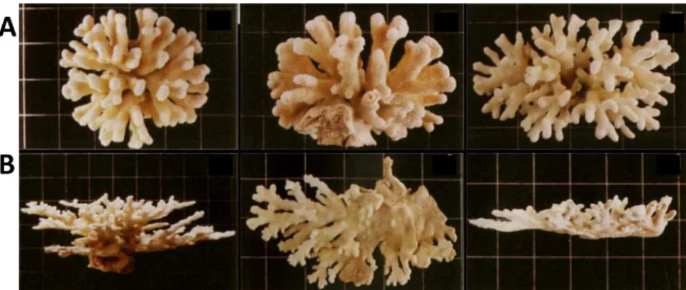
Figure 1.8 - Example of Stylophora pistilata adapted ecomorphs. ... 23
Figure 1.9 - Bleaching phenomenon in cnidarian-dinoflagellate symbiosis. ... 24
Figure 1.10 - The snakelocks anemone, Anemonia viridis.. ... 25
Figure 2.1 - Chemical reactions during absorption of atmospheric carbon dioxide in the seawater. 28 Figure 2.2 - Chemical equilibrium of dissolved inorganic carbon of seawater in a closed system. .... 29
Figure 2.3 - Scanning electron microscopy photographs of coccolithophores under OA. ... 31
Figure 2.4 - Model of dissolved inorganic carbon absorption in non-calcifying symbiotic cnidarians ... 33
Figure 2.5 - Model of CCM in diatoms. ... 34
Figure 2.6 - CO2 vents naturally bubbling CO2 from seafloor acidify water. ... 36
Figure 2.7 - The effect of in situ long-term exposure to „control‟ (C, black) and „high‟ (H, white) pCO2 conditions on the mean Symbiodinium density (a) mean Symbiodinium chlorophyll a content (b), and mean carbonic anhydrase (CA) activity (c) in A.viridis at the Vulcano CO2 vent. ... 44
Figure 2.8 - The effect of laboratory short-term exposure to control (C) and high (H) pCO2 on the mean Symbiodinium density (a) mean Symbiodinium chlorophyll a content (b), on the net photosynthesis rates (c) and on the CA activity (d) in A.viridis. ... 45
Figure 2.9 - The effect of laboratory short-term exposure to control, high pCO2, elevated temperature and combined treatment on (a) the mean Symbiodinium density, (b) on the net photosynthetic rate. ... 53
Figure 2.10 - The effect of laboratory short-term exposure to control, high pCO2, elevated temperature and combined treatment on the carbonic anhydrase activity in A.viridis. ... 53
Figure 2.11 - Variation of gene expression of carbonic anhydrase genes after in situ long-term exposure to high pCO2 in Vulcano CO2 vents. ... 56
Figure 3.1 - Schematic representation of the actin cytoskeleton reorganization during symbiosis establishment. ... 57
Figure 3.2 - Gastrodermal cells of A. viridis with and without Symbiodinium. ... 58
9
Figure 3.4 - Hanging drop culture allowing maintenance of a 3D structure. ... 61
Figure 3.5 - Cell proliferation during oral regeneration of the sea anemone Nematostella vectensis. ... 64
Figure 3.6 - Observation of culture cells of regenerated tentacles of A. viridis. ... 73
Figure 3.7 - Cell contribution in primary cell cultures along the 31 days of culture. ... 73
Figure 3.8 - Determination of (a) cell viability and (b) cell growth rate of suspension and total cells of A. viridis primary cell cultures along the 31 days of culture. ... 74
Figure 3.9 - Cell proliferation of A. viridis primary cells during the first two weeks of culture. ... 75
Figure 3.10 - Determination of epithelial tissue origin of A. viridis primary cell cultures. ... 76
Figure 3.11 - Assessment of cell viability in total cells of A. viridis primary cell cultures in response to thermal stress. ... 77
Figure 3.12 - Comparison of A. viridis primary cell culture contribution following the establishment of an epidermal, gastrodermal and total cells cultures. ... 83
Figure 3.13 - Determination of cell viability during a 31 d cell culture issued from epidermal or gastrodermal monolayers. ... 84
Figure 3.14 - Observation of drop-cultured cells after 7 days in drop cultures (17 d from the beginning of the primary cell culture). ... 87
Figure 3.15 - Determination of cell viability (a) and cell growth rate (b) of drop-cultures. ... 87
Figure 3.16 - Determination of epithelial tissue origin of A. viridis drop-cultured cells. ... 88
Figure 4.1 - Summary of the perspectives for the powerful new tool developed during this PhD, cnidarian primary cell culture of A. viridis. ... 95
Figure S1 – Relative differential expression of AvCa2mE and AvCa2mG genes in epiderm and in gastroderm. ... 77
Figure A.1 - Alignment of A. viridis α-CA sequences. ... 119
Figure A.2 - Phylogenetic tree analysis for Cnidarian α-CA proteins. ... 120
LIST OF TABLES
Table 1. Meta-analysis of the effects of OA in marine calcifiers and non-calcifiers. ... 30Table 2. Seawater physico-chemical parameters measured at Vulcano (in situ) at both control and high pCO2 sites and during the 21 days of laboratory experiment. ... 41
Table 3. Summary of aquarium set-up treatments. ... 52
Table 4. Amount at 3 d of dissociated cells issued from whole tentacle, epiderm and gastroderm. . 83
Table 5. Comparison of primary cell cultures issued from whole tentacle, epiderm and gastroderm. ... 85
Table S1. Comparison of the responses of anemones in control condition with tentacles removed at each time point versus anemones from which we remove tentacles only on a specific sampling point ... 48
10
LIST OF PERSONAL PUBLICATIONS AND COMMUNICATIONS
AUTHOR‟S PUBLICATIONS:
Ventura, P., Jarrold, M., Merle, P-L., Barnay-Verdier, S., Zamoum, T., Rodolfo-Metalpa, R., Calosi, P., Furla, P. (2016). Resilience to ocean acidification: decreased carbonic anhydrase activity in sea anemones under high pCO2 conditions. Marine Ecology Progress
Series.
Ventura, P., Toullec, G., Chapron, L., Meunier, V., Furla, P., Barnay-Verdier, S. (submitted). New perspectives from gastrodermal cnidarian primary cell cultures.
ORAL COMMUNICATIONS
2016: Ventura, P., Toullec, G., Chapron, L., Furla, P., Barnay-Verdier, S. Stress response of gastrodermal primary cell culture from the temperate symbiotic cnidarian, Anemonia viridis. 13th International Coral Reef Symposium (ICRS 2016), Hawaii (USA).
2013: Ventura, P., Merle, P-L., Zamoum, T., Rodolfo-Metalpa, R., Guibert, I., Furla, P. Activity of carbonic anhydrase in response to pCO2 changes in the cnidarian-dinoflagellate symbiosis. CEPA, Lyon (France).
POSTER COMMUNICATIONS
2015Ventura, P., Toullec, G., Chapron, L., Furla, P., Barnay-Verdier, S. Establishment and characterization of primary cell culture from the temperate symbiotic cnidarian Anemonia viridis. “The origin of metazoans”, Giens (France).
Ventura, P., Toullec, G., Chapron, L., Furla, P., Barnay-Verdier, S. Cnidarian primay cell culture. International Workshop “Animal evolution: new perspectives from early emerging metazoans”, Tutzing (Germany).
Ventura, P., Jarrold, M., Merle, P-L., et al. Short-term acclimation and long-term adaptation to ocean acidification of symbiotic cnidarian Anemonia viridis. 8th Congress of the
11
LIST OF ABBREVIATIONS
ABH: Adaptive Bleaching Hypothesis BSA: Bovine Serum Albumin
CA: Carbonic Anhydrase
CCM: Carbon Concentrating Mechanism DIC: Dissolved Inorganic Carbon DTT: Dithiothreitol
EDTA: Ethylenediaminetetraacetic acid EdU: 5-Ethynyl-2'-Deoxyuridine FP: Fluorescent Protein
GFP: Green Fluorescent Protein GIM: Grace‟s Insect Medium
GMIM: Grace‟s Modified Insect Medium H3P: Phophorylated Histone H3
MAA: Mycosporine-Like Amino Acid OA: Ocean Acidification
PBS, PBT: Phosphate Buffered Saline, Phosphate Buffered Saline and Tween
PCR, RT-PCR, qRT-PCR: Polymerase Chain Reaction, Reverse transcription polymerase chain reaction, Quantitative Reverse transcription polymerase chain reaction
pHi: intracellular pH
SASW: Sterile Artificial Seawater
SCaFSW: Sterile Calcium-Free Artificial Seawater ROS: Reactive Oxygen Species
12 “For such a large number of problems there will be some animal of choice or a few such animals on which it can be most conveniently studied.” August Krogh Principle, 1929
Preamble
Biologists have always looked for an easy model organism to study biological processes and able to address fundamental questions. In general, a good animal model would have the following characteristics: (i) structural simplicity, but at the same time containing basic cellular processes that more complex organisms have; (ii) accessible for research and simple to manipulate; (iii) easy and economical to grow in the laboratory; (iv) easily amenable to genetic manipulation. Today we recognize the invaluable knowledge model organisms have brought to science. Classical model organisms, such as the fruitfly Drosophila melanogaster, the nematode worm Caenorhabditis elegans, the yeast Saccharomyces cerevisiae and the mouse Mus musculus have largely contributed to the core of biological knowledge (Bolker 2012). However, they are unable to answer all the biological questions we pose and answers are limited by their characteristics (Cook et al. 2016). The study of alternative models allowed overcoming the scientific and technical obstacles. Among them, xenopus (Xenopus laevis) and sea urchins eggs (e.g. Lytechinus pictus, Arbacia punctulata, Paracentrotus lividus) allowed breakthroughs in the identification of cell cycle regulation proteins mainly due to the facility in laboratory use, with production of high quantities of large eggs, robust zygotes and synchronous cell cycle (Siefert et al. 2015, Cormier et al. 2016, Sluder 2016). Sea slugs (Aplysia species) with their simple nervous system composed of a small number of large cells, allowed the discovery of the mechanisms behind learning, and short, long-term memory (Carew & Kandel 1973). Zebrafish (Danio rerio) has become undoubtedly the most popular fish model in many research fields, especially for developmental biology, toxicology and genetic research (Ribas & Piferrer 2014). More recently, with the development of new cellular technologies and new generation sequencing approaches (allowing the access to their complete genome), emerging model organisms arose to the rank of true alternative models (Cook et al. 2016). However, the ideal candidate species will have a significant impact because they present the traits for which the questions addressed mattered. It would be a mistake to think we could answer all the biological questions from a limited number of species.
In the present work, we chose to work on diploblastic organisms, from the phylum Cnidaria, displaying attractive properties (from ecological to cellular characteristics), which allow using them as model species for biomedical and environmental research.
13
1. Chapter 1 - General introduction
1.1 The Phylum Cnidaria
The Phylum Cnidaria includes sea anemones, corals, hydroids and jellyfish, in approximately 9 000 species (Technau & Steele 2011), inhabiting aquatic environments but predominantly marine habitats.
1.1.1 Biodiversity
Cnidarians have complex life cycles being present in the form of a polyp (attached to the substrate) or a medusa (mainly planktonic). When reproducing sexually, the medusa produces the gametes, which after fertilization, form a planktonic larva, planula, which then settles and metamorphoses into a polyp. The polymorphism is the criteria that allow distinguishing between the 5 classes of Cnidaria: Hydrozoa, Cubozoa, Scyphozoa, Staurozoa (all included in the subphylum Medusozoa) and Anthozoa (Fig. 1.1; Technau & Steele, 2011). Hydrozoans (hydroids) have generally both a tiny polyp and medusa stage; Cubozoans (box jellyfish) and Scyphozoans (true jellyfish) are predominantly in the medusa form; Staurozoans (stalked jellyfish) have an attached medusa and not alternate between polyps and medusa stage; Anthozoans (sea anemones and corals) are exclusively in the form of solitary or colonial polyps.
Figure 1.1 – Phylogenetic relationship between the different classes of the phylum Cnidaria (modified from Technau & Steele 2011).
1.1.2 Ecological importance
Cnidarians evolved approximately 500 million years ago (Carthwright et al. 2007) and are the simplest living metazoans. Cnidarians have a global distribution, in polar, temperate and tropical latitudes. Their range of distribution goes from shallow coastal waters to the deep sea, although they are more abundant at shallow warmer waters of tropical regions.
14 Tropical coral reefs are a hotspot of biodiversity with ecological and economic relevance (revised in Dubinsky 1990). Coral reefs are a source of shelter, food and a nursery ground for fish species and hundreds of other species. Also, they are an important source of food for humans and are an important economical source of tourism in these regions. Yet, the ecological importance of cnidarians does not reside only on the coral reefs. Cnidarians are carnivorous, and play a role in the food web, as predator and prey. They prey mainly on plankton but also on small crustaceans, fish larvae and are preyed upon by molluscs, fishes, crustaceans and sea turtles (Mitchell et al. 1988). Moreover, they are an important source of bioactive compounds (see segment 1.2.1).
1.1.3 General anatomy
Cnidarians are simplistic organisms with external radial symmetry and an adult dimorphism (polyp and medusa). The gastrovascular cavity has only one central opening, working both as a mouth and an anus. A crown of tentacles surrounds the mouth (Fig.1.2).
Figure 1.2 Simplified diagram of a polyp and medusa body plan, showing diploblastic tissue organization (modified from Sabourault et al. 2009).
1.1.4 Cellular anatomy
Being diploblastic animals, cnidarians are composed by two epithelial monolayers, the epiderm facing the seawater and the gastroderm facing the gastrovascular cavity, both separated by the acellular mesoglea.
Mainly located in the epiderm, cnidarians possess specialized stinging cells, cnidocytes, which are used for predation and defence. Moreover, in the epiderm we can find other cell types: (1) epithelio-muscular cells (longitudinal fibers) used for movement and contraction,
15 (2) interstitial cells (for tissue renewal, see below, 1.2.1), (3) mucous gland cells, which produce mucus for feeding and protection and, (4) neuronal and sensory cells (Hündgen 1984). In the gastroderm, cell types are (1) gland cells which produce digestive enzymes, (2) neuronal cells, and (3) epithelio-muscular cells (circular fibres) used for movement and phagocytosis (Technau & Steele 2011) (Fig. 1.3).
Figure 1.3 - Cross section of Hydra body plan showing the different cell types present both in the epiderm and in the gastroderm (modified from Technau & Steele 2011).
Epithelial cells of cnidarians show a high diversity of functions. Epithelial cells from the epiderm have predominantly a protective function while those from the gastroderm are responsible for food uptake and digestion. In some cnidarians, others gastrodermal cells also have a symbiotic function, hosting unicellular algae (see below segment 1.2.3).
1.2 Physiological properties of cnidarians
Cnidarians possess striking physiological properties, which make them attractive model species for biomedical and environmental researches. Indeed, cnidarians (1) express high diversity of fluorescence proteins, which allowed the development of new biotechnological tools; (2) have great longevity and high regeneration capacity, allowing studies on aging and on the processes of regeneration; and (3) some species are adapted to life in symbiosis with photosynthetic unicellular algae, making them good bioindicators of the health of ecosystems.
16 1.2.1 Diversity of natural fluorescence
In 1962, Shimomura and co-authors, while working on the bioluminescence of the jellyfish Aequorea victoria (class Hydrozoa), isolated and identified two proteins responsible for the bioluminescence, a calcium binding protein (aequorin) and a green fluorescent protein (GFP; (Shimomura et al. 1962) (Fig. 1.4).
Figure 1.4 - The jellyfish Aequorea Victoria (classe Hydrozoa) expressing the green fluorescence protein (GFP). Photo by Phil Blackburn.
It was much later that (Prasher et al. 1992) cloned the GFP gene in order to better understand the mechanisms leading to light generation. This GFP found in cnidarians is unusual and different from other natural pigments, since it does not require added substrate or co-factors other than oxygen to produce its green fluorescence (Chalfie et al. 1994, Heim et al. 1994). This constituted an advantage for the studies in living cells since until then there was no non-invasive technique available, able to maintain the integrity of tissues or cells. It was Chalfie et al. (1994) who first used GFP as a marker for gene expression in living cells of the nematode worm, Caenorhabditis elegans. The authors used GFP gene as a reporter gene attached to a gene of interest and monitored the GFP fluorescence as a proxy of gene expression in living cells or tissues. A. victoria GFP has since then become an invaluable versatile marker in biological research. It has been widely used in protein dynamics, fluorescence microscopy, cell biology and biotechnology, molecular biology, cancer research and many more (see (Zimmer 2002, 2009) for a review). Additionally, other fluorescent proteins (FP) homologous to GFP have been found in cnidarians, mostly in species from class Anthozoa (Lukyanov et al. 2005). Five colour classes are now defined thanks to their respective emission: cyan, green, 2 red classes and yellow (Remington 2011). The discovery of other FP has enlarged the applicability of
GFP-17 like proteins to live cell imaging, allowing the development of multicolour labelling experiments. Each FP has a particular characteristic that confers an advantage in respect to others. For instance, the red FP, which present reduced background fluorescence, is used for the localization of cells in the whole organisms (Wiedenmann et al. 2011).
Fluorescent proteins are only an example of how cnidarians can be an important source of marine natural products for the biological research. Indeed, over 3000 natural products have been identified from cnidarians, with many applications (Rocha et al. 2011). Some examples are secondary metabolites, such as terpenoids, which have an anti-inflammatory potential and diterpenoids, which have been tested as an antitumor drug (see Rocha et al. 2011 for a review). Therefore, cnidarians show a biomedical and biotechnological potential to be exploited by worldwide researchers.
1.2.2 Cnidarian longevity and regeneration
Cnidarians are amongst the longest living animals, and in some cases immortality has been hypothesized. Nevertheless, the range of life span shows a high plasticity within cnidarians. Some polyps of hydrozoans live only a few days (e.g. Campanularia flexuosa; (Strehler & Crowell 1961)), whereas the deep-sea corals Gerardia sp. and Leiopathes sp. are longest lived, 2742 and 4265 years, respectively (Roark et al. 2009). In addition, the hydrozoan species Turritopsis dorhnii (previously known as T. nutricula), is considered immortal, by reverse development of sexually mature medusa into clonal polyps (Piraino et al. 1996).
Most living organisms get old, in a process known as aging. Aging represents one of the biological processes from which we still lack a full answer, mainly due to the diversity of aging across the tree of life. Attempts have been made to define the mechanisms leading to aging, and (López-Otín et al. 2013) categorized nine main molecular and cellular events occurring in cells aging (cf Fig.1.5).
18 Figure 1.5 - Schematic representation of the mechanisms contributing to the aging process in mammals (López-Otín et al. 2013).
These mechanisms are involved at different levels of cellular complexity (from nucleus to the cell tissue). In the nucleus, aging process includes epigenetic alterations (DNA methylation, chromatin remodelling and transcriptional alterations), genomic instability (genomic damages) and telomere attrition (shortening of nucleoprotein structures located at the extremities of the chromosomes). At the organelle level, aging is due to the mitochondria dysfunction (reducing energy source and overproducing reactive oxygen species). At the cellular level, aging is provoked by cellular senescence (the arrest of the cell cycle, i.e. leading to cell death), stem cell exhaustion (depletion of undifferentiated cells capable of differentiation into specific cell types ensuring cell renewal) and loss of proteostasis (accumulation of protein degradation and misfolding of proteins). Finally, aging is the result of altered intercellular communication (perturbation of cell signalling, which leads to failures in the response to inflammation) (López-Otín et al. 2013).
Conversely, in cnidarians, the longevity has been suggested to be explained by some specific strategies overcoming the aging processes. Specifically, mechanisms leading longevity in cnidarians could be linked to the presence, in adult organisms, of stem cells with high and continuous cellular renewal potential. This propriety could then be responsible of two cnidarian specificities: asexual reproduction (responsible of clonal and colonial growth) and tissue regeneration (the process by which animals regrow lost body parts or entire organisms from small body fragments).
From all the above-mentioned mechanisms in cnidarians, regeneration is the most studied. Regeneration can happen in two ways, (1) morphallaxis, i.e. regeneration by reorganization
19 and differentiation of pre-existing cells and (2) epimorphosis, which involves cell proliferation, i.e. cell growth and division leading to an increase in the number of cells (Li et al. 2015). For a long time, the high regenerative capacity of cnidarians was attributed to the presence of stem cells, meaning that regeneration was only done by morphallaxis. In Cnidarians, stem cells were first identified in the hydrozoan genus Hydractinia and later on Hydra (Weismann, 1883). Both groups possess stem cells in the interstitial space of epithelial cells, commonly referred as i-cells (Frank et al. 2009). The totipotency associated to i-cells is responsible for the high regenerative capacity of Hydra, which is able to regenerate two entirely new individuals from head and foot, the head will regrow a foot, and the foot will regrow a head (reviewed in (Holstein et al. 2003b)). Recognized stem cell genes (e.g. piwi, vasa, PL 10) have been used to identify stemness in cnidarian tissues (e.g. Seipel et al. 2003, Siebert et al. 2014) . For instance, piwi is present in the all developmental stages of the hydrozoan Podocoryne carnea with higher expression in the egg and medusa (Seipel et al. 2013) and vasa, piwi and PL 10 have been identified in the epiderm and gastroderm of gastrozooids of the hydrozoan species Nanomia bijuga (Siebert et al. 2014). Nevertheless, recent studies have also shown that other cnidarian species (e.g. the anthozoan Nematostella vectensis) are not able to regenerate without differentiated cell proliferation (Passamaneck & Martindale 2012, DuBuc et al. 2014), showing that regeneration by epimorphosis is also present in cnidarians.
Although regeneration has greatly contributed to our understanding on the mechanisms of longevity in cnidarians, others studies on aging processes could give us a new insight into the extreme life span of cnidarians. One of them is the dynamics of the telomere maintenance in cnidarians. One of the aging mechanisms is the telomere shortening, prevented by the presence of a telomerase. The telomerase is only expressed in germ cells, stem cells and tumours and its activity is responsible for the synthesis of new telomere repeats. In cnidarians, telomerase activity has also been identified (Traut et al. 2007, Ojimi et al. 2009, Zielke & Bodnar 2010); however telomere dynamics over time and their relation with events leading to cell cycle arrest and apoptosis has still to be determined. A relevant way to study telomere and telomerase activity related with cnidarian longevity, growth and stress response could be by the development of in vitro cnidarian culture cells and the analysis of the chromosome dynamics during cell divisions and time. Cell cultures would allow the study of telomerase activity within different types of cells, dividing cells and to determine extension of life span in culture.
20 1.2.3 Environmental adaptation
Cnidarians, as mention above, have a worldwide distribution inhabiting temperate, tropical, deep and surface waters. Colonization of different ecosystems implied adaptation to local environmental conditions, and more recently, anthropogenic threats. One of the ways through which cnidarians adapted was the establishment of mutualistic symbiosis with unicellular phototrophs, an association estimated to have been established 225 million years ago (Rosen 2000).
Symbiosis
Some cnidarians live in association with a photosynthetic dinoflagellate from the genus Symbiodinium spp., and form one of the most studied symbioses in the marine realm - cnidarian-dinoflagellate symbiosis. Symbiodinium are unicellular algae that can be found as free-living species or associated with other unicellular organisms (e.g. dinoflagellate, foraminifera) but also cnidarians, molluscs (e.g. giant clams) and sponges (Trench 1993). The symbiosis between Symbiodinium and Anthozoa – the class of cnidarians comprising sea anemones and corals - is one of the most ecologically significant symbioses, due to its role in the formation of tropical coral reefs, and the great biodiversity herein present (Dubinsky 1990). It is a mutualistic endosymbiotic intracellular association, where the symbiont (Symbiodinium) is located intracellularly in the gastrodermal cells of the host cnidarian, enveloped by a symbiosome membrane that separates the symbiont from the host cytoplasm (Fig. 1.6).
Figure 1.6 - Schematic representation of the diploblastic organization of a polyp from a symbiotic cnidarian.
(A) Location of the symbiont Symbiodinium in the gastroderm tissue (modified from Sabourault et al. 2009). (B) Symbiodinium cells in division isolated from the sea anemone Anemonia viridis.
21 The genus Symbiodinium is divided into different phylogenetic groups, referred as clades (9 clades; A-I). Within each clade, there is additional genetic diversity which supports the distinction of strains or sub-types (e.g D1, G2; (Coffroth & Santos 2005), (Pochon et al. 2014). Their genetic diversity could be linked with the capacity to inhabit multiple environments, and the specificity of tolerance to environmental changes (e.g. (Brading et al. 2011, Oakley et al. 2014).
Symbiodinium are acquired by vertical or horizontal transmission. The former implies the maternal transmission of symbionts directly by asexual or sexual reproduction through incorporation of symbionts into the released eggs. The horizontal transmission involves the release of symbiont-free eggs and the acquisition of the symbiont by phagocytosis, is done externally from the environment, at each generation (Smith and Douglas, 1987). The mechanisms of symbiont recognition have not yet been determined but it is suggested to involve the recognition of specific glycoproteins, more precisely the binding of symbiont glycans by host lectins (Vidal-Dupiol et al. 2009).
-
Benefits of this symbiosisThe success of this symbiosis is dependent on the mutual exchanges promoted by both partners. While in the host, Symbiodinium continue to photosynthesize and transfer until 90% of the organic carbon produced during photosynthesis, contributing to host metabolism, reproduction, growth and calcification (Fig.1.7) (Furla et al. 2005, Davy et al. 2012).
Figure 1.7 - Organic carbon supply and use in a symbiotic cnidarian.
Ci: inorganic carbon (carbon dioxide, bicarbonate); Co: organic carbon (sugars, lipids, amino acid) (Casado-Amezúa et al. 2014).
22 In this way, the host, although capable of heterotrophic nutrition, can satisfy its metabolic needs by autotrophy. From all the photosynthate products, glycerol, glucose and lipids are the most translocated (Trench 1971, Sutton & Hoegh-Guldberg 1990, Burriesci et al. 2012). In opposite direction, the host will contribute to symbiont photosynthesis with essential nutrients derived from its metabolism or from the external medium (such as nitrogen, phosphorus, inorganic carbon) (Muscatine 1990). This association constitutes a new biological entity, with original metabolic properties, the holobiont, which is notably responsible of the colonization of nutrient-poor environments.
- Adaptations to symbiosis
Life in symbiosis between an animal host and a free-living algal species can be both beneficial as risky. In order to establish this successful mutualistic relationship, cnidarians evolved and acquired several mechanisms which allowed adaptation to the constrains imposed by the symbiont. For example, the animal host is constricted to inhabit shallow areas in the euphotic zone where light exposure is enough to fulfil photosynthetic requirements of the Symbiodinium; implying nevertheless a major exposure to UV radiations (UVR). Potential photo-damage is prevented by the presence in the holobiont of mycosporine-like amino acids (MAAs, a family of aromatic amino acids), as they absorb UVR and dissipate it as heat (Shick & Dunlap 2002). Although MAAs are more concentrated in the host tissues, it is not clear who is responsible for the biosynthesis of the MAAs, symbionts or host (Shick & Dunlap 2002, Roth 2014). However, recent studies on the coral Acropora digitifera showed that the animal genome possesses the genes necessary for their biosynthesis (Shinzato et al. 2011). Another constraint that cnidarian hosts have to overcome is the circadian variations of intracellular O2 concentrations. Daily, the symbiotic cnidarians fluctuate from hyperoxia state (i.e. high [O2]) at day-time due to symbiont
photosynthesis, to hypoxia (i.e. low [O2]) during night-time, due to the host and symbiont
respiration (Richier et al. 2003). During photosynthesis, the high concentrations of generated O2 lead to the production of reactive oxygen species (ROS; (Dykens et al. 1992).
Consequently, the host evolved high diversity of anti-oxidant defences to overcome the increased production of ROS, such as superoxide dismutases, catalases, peroxidases which neutralize oxidant species, reducing its toxicity (Furla et al. 2011, Pey et al. 2016). Finally, for photosynthesis to take place, CO2 most arrive continuously to the symbionts. Due to the intracellular location of the symbionts in the most inner layer of tissue, the gastroderm,
23 access to inorganic carbon is dependent on the host. The host has then selected mechanisms of inorganic carbon absorption, further described in chapter 2 (Furla et al. 2005).
Therefore, the cnidarians have evolved a great flexibility in functions to adapt themselves to the continuous constrains imposed by the presence of the intracellular symbiont and to ensure the success and the stability of the symbiosis.
Local environmental responses
Cnidarians are not restricted to stable environments with a limited range of conditions. During evolution they were able to exploit different ecological niches, from fresh to marine waters, such as shallow waters in tropical reefs and open ocean where abiotic conditions (e.g. temperature, nutrient availability, pH, light intensity and water movement) differ substantially. For instance, the range of light exposure goes from full sunlight in shallow waters (e.g. cnidarians in tropical coral reefs), to darkness in the deep-sea (cold-water corals). That capacity suggests a high level of phenotypic plasticity, a mechanism by which a same genotype can produce different phenotypes influenced by the environment. It can reflect changes in morphology, physiology, life history and behaviour of a genotype (Pigliucci 2001, 2005).
One proof of this high phenotypic plasticity is the morphological changes observed in many symbiotic corals species due to abiotic factors, such as light and water movement (Todd 2008). For example, the coral Porites sillimaniani, which is an explanate (flat) coral, was observed to form branches when exposed to high light conditions, while it remained flat at low light (Muko et al. 2000). Another example is the coral Stylophora pistallata, which presents a gradual change of the colony morphs with the depth (Fig. 1.8; Furla & Allemand 2009). This morphological plasticity ensures the efficiency of light exposure for the symbiont photosynthesis.
Figure 1.8 - Example of Stylophora pistilata adapted ecomorphs (Modified from Gattuso, PhD Thesis, 1987).
A
24 (A) Shallow ecomorphs. (B) Deep-sea ecomorphs.
Moreover, cnidarians are adapted not only to different habitat constrains but also to fluctuating environments. In the current scenario of environmental changes, due to natural or anthropogenic threats (e.g. pollution, sedimentation, ocean acidification, rise of sea water temperature), this capacity is called into question. Again, cnidarian phenotypic plasticity could be involved through acclimation process, with a short-term reversible response, conferring resilience to a particular stressor. For instance, Mitchelmore et al. (2003) have shown that in the sea anemone Anthopleura elegantissima following an induced stress of cadmium, capacity to acclimate to these toxic conditions was possible due to the presence of higher levels of symbiont metal-binding antioxidant glutathione.
With the increasing rise in seawater temperature, we are seeing a recurrent phenomenon in coral reefs known as bleaching. Bleaching is the loss of the symbionts and/or loss of photosynthetic pigments, leading to the disruption of cnidarian-dinoflagellate symbiosis and to high mortality rates (Fig. 1.9a; Hoegh-Guldberg 1999). Bleaching phenomenon is mainly considered as a pathogenic state induced by a former dysfunctioning of the photosynthetic complex of the symbiont following cellular damages to symbiont and host cells (Fig. 1.9b).
Figure 1.9 - Bleaching phenomenon in cnidarian-dinoflagellate symbiosis.
(a) On the left a healthy fire coral with its symbionts and on the right a bleached fire coral, where the whitish coral results from the loss of symbionts, therefore the pigments responsible for the colour (source: XL Catlin Seaview Survey). (b) Cellular mechanisms leading to the loss of symbiont in cnidarians. Five mechanisms are in situ degradation, exocytosis of the symbiont to the gastrovascular cavity, host cell detachment with the symbiont into the gastrovascular cavity, formation of apoptosis body leaving the host cell and finally host cell necrosis. H-normal host cell, Sy-symbiosome, S-Symbiodinium, M-mesoglea (source: Weis 2008).
However, bleaching is hypothesized as one adaptive strategy to changing environmental conditions: the adaptive bleaching hypothesis (ABH) (Buddemeier et al. 2004, Fautin &
25 Buddemeier 2004). According to the ABH, bleaching represents a strategy of host to switch from a less favourable symbiont partner to a more favourable one, in a way that symbionts shape the host phenotype. In other words, colonies of corals can be inhabited by different sub-types of Symbiodinium (e.g. Chen et al. 2005). Each of these sub-types can have environmental optima; therefore, during a stress event, the most sensitive species to that particular stress can trigger cellular mechanisms leading to the loss of that specific sub-type, favouring growth of another sub-type. For example, Brading et al. (2011) showed that Symbiodinium sub-types are differentially sensible to increase pCO2, clade A1 and B1 are not
affected, and clades A13 and A2 were favoured by this increase. Therefore, dynamics changes in Symbiodinium could be an important strategy to respond to fluctuating environmental conditions.
Nowadays, as environmental stressors become more and more recurrent, and are expected to increase in intensity (Hoegh-Guldberg et al. 2007, Doney et al. 2009), it is fundamental to understand the limit of the extended cnidarian phenotypes allowing them to acclimate and/or adapt to predicted future conditions.
1.3 Our cnidarian study model: the sea anemone
Anemonia
viridis
The snakelocks sea anemone Anemonia viridis (Forskål, 1775) (class Anthozoa) (Fig. 1.10) was adopted as our model species.
Figure 1.10 - The snakelocks anemone Anemonia viridis. Photo by Alexis Pey.
It is a temperate species found abundant in shallow waters (up to 20 m) of the Mediterranean, Eastern Atlantic, English Channel and North Sea. It is a non-calcifying
26 symbiotic cnidarian that leaves in association with the dinoflagellate Symbiodinium temperate A clade (Casado-Amezúa et al. 2014). Reproduction in this species is done asexually (by fission) or sexually, and the symbionts are transmitted vertically.
The use of A. viridis as model species presents several advantages: (1) long and non-retractable tentacles gives access to large amounts of tissue; (2) A. viridis has a high tentacle regenerative capacity, (3) facility to obtain aposymbiotic (deprived of symbionts) individuals in laboratory, (4) easy dissociation into epiderm, gastroderm and symbionts, and (5) the absence of a calcium carbonate skeleton favours tissue dissociation. Besides, in the last years, numbers of studies on A. viridis have increased knowledge on its physiology (Furla et al. 1998, 2000, Laurent et al. 2014), biochemistry (e.g. Richier et al. 2003), genetics (e.g. Ganot et al. 2011), cellular biology (Barnay-Verdier et al. 2013) and stress response (Richier et al. 2006, Moya et al. 2012, Suggett et al. 2012, Jarrold et al. 2013, Borell et al. 2014, Horwitz et al. 2015). Evidence from these studies suggests an extended plasticity of A. viridis, at different levels of biological complexity (from whole organism to the cell), as mechanism enabling its success under environmental changes and then justifying the use of A. viridis as a model for our studies.
1.4 Thesis objectives
In this PhD thesis we wanted to investigate the extent of the phenotypic plasticity of A. viridis to environmental stresses at different temporal scales (i.e. short and long term stress exposures) and at multiple levels of structural complexity (i.e. whole organism and isolated cells).
Specific goals of this thesis were:
(1) in vivo, identification of the mechanisms behind the physiological plasticity that enable non-calcifying photosynthetic anthozoans to survive and thrive under future ocean acidification (OA) conditions and assess the plasticity under synergetic effects (OA and increased temperature) (Chapter 2).
(2) in vitro, the development of animal primary cell culture as model to determine plasticity to environmental stresses at the cellular level (Chapter 3).
These chapters must be seen as two pieces of a puzzle that in the discussion session (Chapter 4) are combined to draw a comprehensive conclusion concerning the phenotypic plasticity of cnidarians.
27
2. Chapter 2 –
In vivo
studies of phenotypic plasticity
2.1 Introduction
2.1.1 Stress response: general context
Every organism, in the course of its life cycle, faces a change in its natural environment (e.g. arrival of a new predator, abiotic changes). The capacity of an organism genotype to change its phenotype (physiology, behaviour, morphology, development) in response to environmental changes is commonly referred to as phenotypic plasticity. The responses of the organism to a changing environment can then happen in one of four ways: (1) migration to a more favourable environment, (2) adaptation via genetic selection (adaptive phenotypic plasticity), (3) acclimatization via phenotypic plasticity (quick response) and (4) extinction (Hoffman & Parsons 1991, Foo & Byrne 2016). Adaptive phenotypic plasticity corresponds to a modification of the genome leading to a long-term phenotypic change. It is advantageous since it potentiates tolerance to multiple environments by increased fitness (Pigliucci 2001, Ghalambor et al. 2007). Acclimatization represents a short-term plastic change (without genomic modifications), where a single genotype produces a range of phenotypes as a result of an environmental perturbation (Foo & Byrne 2016).
Nowadays, symbiotic cnidarians are exposed to numerous threats (e.g. increased sea water temperature, ocean acidification, pollution, sedimentation, pathogens) that have the potential to disrupt the stability of the symbiotic relationship, possibly causing the breakdown of this symbiosis and ultimately the death of both partners (Hoegh-Guldberg et al. 2007). An environmental perturbation inducing a stress state corresponds to a change for which values fall outside the optimum response range of each species. Here, we are interested in assessing phenotypic plasticity (through adaptation or acclimatization) in a symbiotic cnidarian dealing with global change. Currently, the Intergovernmental Panel on Climate Change (IPCC 2013) described the environmental stressors involved in the global change. Among them, ocean acidification (OA) and global warming are the two major factors affecting marine biodiversity. In that context, we investigated OA and/or ocean warming on a symbiotic cnidarian by understanding the role of the phenotypic plasticity. The model species of choice, Anemonia viridis has the great advantage of inhabiting natural acidified sites, Vulcano Island, South of Italy. In that particular site, submarine gas vents release CO2 creating a natural pCO2 gradient suitable for long-term exposure studies on
phenotypic plasticity in response to OA. This allows us to compare phenotypic plasticity of A. viridis, with short-term high pCO2 laboratory controlled exposure.
28 2.1.2 Ocean acidification
Since the Industrial Revolution, human activity had a huge impact on the excessive amounts of CO2 being released to the atmosphere. Since the oceans act as a sink of
atmospheric CO2, accumulation of CO2 is also happening in the oceans. Estimates are that the oceans absorbed up to one-third of atmospheric CO2 released due to human activities
(Hoegh-Guldberg & Bruno 2010). When CO2 reaches the ocean, it reacts with water
forming carbonic acid (H2CO3), which dissociates into bicarbonate (HCO3-) and protons
(H+), decreasing pH of the seawater while increasing the acidity of oceans, in a process
known as OA (Fig. 2.1) (Caldeira & Wickett 2005, Doney et al. 2009). The term OA does not imply that the ocean will become acidic (pH < 7). Instead, pH is expected to decrease from current levels of 8.08 to 7.6-7.7 by 2100 (IPCC, 2013).
Figure 2.1 - Chemical reactions during absorption of atmospheric carbon dioxide in the seawater.
The increase in dissolved carbon dioxide in the seawater will increase the amount of hydrogen ions, in a process known as OA (source: University of Maryland).
In the current seawater-carbonate chemistry scenario, dissolved inorganic carbon (DIC) concentration is uneven, with bicarbonate (HCO3-) present in higher concentration (ca.
2.1 mM), representing ~ 90% of total DIC while CO2 is present in low concentration (ca.
12 µM), approximately 1%; carbonate ion (CO32-) accounts for the other 9% (Doney et al.
2009). However, under OA, i.e. lower pH, the chemical equilibrium of the seawater will change, with the increase of HCO3- and CO2 and the decrease in CO32- concentration (Fig.
29 Changes in the chemical composition of seawater may impact the marine organisms by modifying the biological processes linked to inorganic carbon availability (i.e. calcification and carbon-concentrating mechanisms, cf below).
Figure 2.2 - Chemical equilibrium of dissolved inorganic carbon of seawater in a closed system (adapted from Holmen 1992).
2.2 Impact of ocean acidification on marine organisms
Marine organisms, from algae to fishes, show different sensitivities to OA, and studies predict that there will be winners and losers under future elevated CO2 conditions (Table 1;
Ries et al. 2009, Fabricius et al. 2011). Therefore, it is correct to assume that OA will shape marine communities. Indeed, OA may negatively impact a wide range of species across various phyla, especially calcifying species from plankton to corals. Conversely, some non-calcifying species are able to cope with OA, in natural or experimental conditions, with potential benefits on productivity (Doney et al. 2009, Fabricius et al. 2011). However, most of the studies on the effect of OA in marine organisms have been focused on the ecological impact rather than the mechanisms that trigger the OA response.
In marine biota unicellular eukaryotes and cnidarians are two major representative groups for studies of OA impact on calcifying and non-calcifying organisms.
30 Table 1. Effects of OA in marine calcifiers and non-calcifiers .
Table was built from meta-analysis of Kroeker et al. 2013 and supplemented with data for sea anemones from independent studies (Suggett et al. 2012, Towanda & Thuesen, 2012, Jarrold et al. 2013). n.d. – non-determined by insufficient number of studies; no effect – no significant changes; - decrease; + increase (simplified from Kroeker et al. 2013).
Taxa Parameter Effect
C A L C IF IER S Calcifying algae Survival n.d Calcification No effect Growth n.d Photosynthesis - Abundance - Corals Survival No effect Calcification - Growth No effect Photosynthesis No effect Abundance - Coccolithophores Survival n.d Calcification - Growth No effect Photosynthesis No effect Abundance No effect Molluscs Survival - Calcification - Growth - Photosynthesis - Abundance No effect Echinoderms Survival No effect Calcification No effect Growth - Photosynthesis - Abundance No effect N O N -C A L C IF IER S Fish Survival n.d Growth No effect Abundance n.d Fleshy algae Survival n.d Growth + Photosynthesis No effect Abundance No effect Seagrass Survival n.d Growth n.d Photosynthesis No effect Abundance n.d Sea anemones Survival n.d Growth +
Photosynthesis +/no effect
Abundance +
2.2.1 Impact of ocean acidification on calcification
Bio-calcification is the process by which calcifying organisms form a calcium carbonate (CaCO3) skeleton, by the combination of calcium ion (Ca2+) with CO32. Current changes in
seawater chemistry, with increased CO2, HCO3-, H+ and the decrease of CO32-, may have an
impact in the formation of a skeleton by calcifying species. Briefly, when the CO2 reaches the seawater, it will react with water and form H2CO3, which will then dissociate into H+
31 and HCO3-. The increased concentration of H+ will then bind with CO32-, outcompeting
the association of Ca2+ from the surrounding water and CO
32-, thus reducing the availability
of ion carbonate in the water. An excessive environmental acidification could even act on biological processes of calcification if the organism overcomes its capacity to maintain pH homeostasis (intracellular and/or in calcification site). Therefore, shell-forming marine organisms may see their skeleton reduced.
Effect on calcification of free-living unicellular eukaryotes
Coccolithophores and foraminifera are two of the most studied calcareous unicellular eukaryotes, and are responsible for the majority of pelagic CaCO3. While studies have
shown multiple responses to calcification in coccolithophores (Meyer & Riebesell 2015), with reduced calcification (e.g. 25% to 66% decrease; Fig. 2.3; Riebesell et al. 2000, Sciandra et al. 2003), increased calcification (doubling values of calcification; Iglesias-Rodriguez et al. 2008) or no significant changes in calcification (Langer et al. 2006, Oviedo et al. 2016), impact of increased pCO2 in foraminifera seems to be always deleterious (e.g
(Spero et al. 1997, Bijma et al. 1999, Uthicke et al. 2013). In fact, studies on volcanic CO2
seeps, where concentrations of CO2 range from present day to those expected for 2100,
have shown a decrease in foraminifer‟s densities and diversity, as it happened before in the Paleocene, where there was a decrease in calcification, hypothesizing that this group can be
menaced due to OA (Uthicke et al. 2013, Stillman & Paganini 2015).
Figure 2.3 - Scanning electron microscopy photographs of coccolithophores under OA. (b, e) Emiliania huxleyi and (c, f) Gephyrocapsa oceanica. Coccolith structure and degree of calcification under control pCO2 levels of about 300 ppmv (b, c) and high pCO2 levels, 780-850 ppmv (source:
Riebesell et al. 2000).
Effect on calcification of corals
Corals are one of the highest producers of CaCO3 in the oceans. Therefore, the predicted
decrease in carbonate ion concentration will significantly reduce the capacity of corals to build their calcium carbonate skeleton (e.g. Gattuso et al. 1998, Hoegh-Guldberg et al. 2007). For instance, Kroeker et al. 2013 showed that on average, OA have a deleterious
32 effect of 32% on coral calcification. However, the impacts on coral calcification seem to be species specific, as not all corals seem to be impacted by OA future scenarios (Reynaud et al. 2003, Doney et al. 2009). A few studies have shown that cold-water corals (e.g. Maier et al. 2013, Rodolfo-Metalpa et al. 2015) and diverse coral communities naturally exposed to OA (Shamberger et al. 2014) seem to have a greater capacity to cope with changes in DIC chemistry.
2.2.2 Ocean acidification and carbon-concentrating mechanisms
Microalgae are the most important primary producers in the ocean. They absorb inorganic carbon and fix CO2 into organic compounds through Rubisco (ribulose bisphosphate
carboxylase/oxygenase), an enzyme with affinity not only to CO2 but also to O2. To
overcome this limitation they possess carbon-concentrating mechanisms (CCM) that efficiently exploit HCO3- pool (largest oceanic DIC pool) in the seawater, and convert it in
CO2 in order to increase the concentration of available for the active site of the Rubisco
(Giordano et al. 2005). An additional component of CCM is carbonic anhydrase (CA) an enzyme responsible for the acceleration of the rather slow inter-conversion between carbon species.
Symbiotic cnidarians have inside their host cells dinoflagellates from the genus Symbiodinium. While, free living Symbiodinium have direct access to DIC from the seawater, once inside host cells, several membranes form a barrier to the diffusion of DIC. One of the crucial contributions of the animal host to the symbiont is then the provision of DIC allowing the process of photosynthesis to proceed. There are two possible sources of DIC to the symbiont, (1) animal host‟s metabolism, i.e. respiration and (2) seawater. Animal host respiration rates are generally lower in respect to the net photosynthetic rates (Furla et al. 2005), indicating the presence of an external source of inorganic carbon. In addition, Symbiodinium Rubisco has low affinities to CO2 therefore efficient allocation of CO2 to the
site of fixation is crucial (Rowan et al. 1996). CO2 diffuses passively through host
membranes but the low concentration in the seawater makes it insufficient to optimal photosynthesis, making HCO3- the major DIC source for carbon fixation. However, host
lipid membranes are impermeable to HCO3- and therefore it must be actively transported
through host membranes.
Similarly to microalgae, symbiotic cnidarians possess CCM that efficiently exploits HCO3
-pool in the seawater and increases the concentration of CO2 reaching Rubisco (Allemand et al. 1998). Bicarbonate is actively transported by the presence of an H+-ATPase pump
33 external medium, and consequent protonation of HCO3- to carbonic acid (H2CO3) (Furla et
al. 2000, Bertucci et al. 2010). A membrane-bond carbonic anhydrase (CA) is responsible for the reversible conversion of HCO3- to CO2, which can then freely diffuse into host cells
cytoplasm. Once in the cytoplasm, CO2 is trapped and converted to HCO3- by another CA,
in agreement to intracellular pH (Venn et al. 2009). The transport of HCO3- to the
gastrodermal cells may involve the presence of bicarbonate active transporters (Zoccola et al. 2015). Once HCO3- finally reaches the gastrodermal cells, a CA located close to the
symbiont membrane protonates HCO3- into CO2, and this is made available to
photosynthesis (Fig. 2.4) (Furla et al. 2000a).
Figure 2.4 - Model of dissolved inorganic carbon absorption in non-calcifying symbiotic cnidarians (modified from Furla et al. 2000 and Zoccola et al. 2015).
CA = carbonic anhydrase
Key enzymes involved in the CCM are the carbonic anhydrases. They are present in almost all organisms (Eubacteria, Archaea and Eukaryota) and comprise six distinct classes: -CAs, -CAs, -CAs, -CAs, -CAs and -CAs (Le Goff et al. 2016). They are located in the cytosol, mitochondria, and membrane or can be secreted. In cnidarians, CA have been molecularly identified and divided according with their phylogeny into three distinct groups, secreted or membrane-bound, cytosolic and mitochondrial proteins and carbonic anhydrase related proteins (Bertucci et al. 2013). The -CA are present in all metazoans and in cnidarians their role has been mainly investigated in biocalcification and photosynthesis, both processes related with the inorganic carbon uptake (Bertucci et al. 2013, (Le Roy et al. 2014). For instance, the STPCA (Stylophora pistillata CA), located in the calicoblastic epiderm (site of calcification), is directly linked to calcification, while the two CA transcripts identified in A. viridis (AvCa2m, AvCa2c; Ganot et al. 2011) are upregulated