i Université de Montréal
An fMRI study of emotional episodic memory in schizophrenia: Effects of diagnosis and sex
par Nadia Lakis
Département de Psychiatrie Faculté de Médecine
Mémoire présenté à la Faculté de Médecine en vue de l’obtention du grade de Maitrise
en Sciences Biomédicales option Psychiatrie
April 2012
ii Université de Montréal
Faculté des études supérieures et postdoctorales
Ce mémoire intitulé :
An fMRI study of emotional episodic memory in schizophrenia: Effects of diagnosis and sex
Présentée par : Nadia Lakis
a été évaluée par un jury composé des personnes suivantes :
Dr. Sandra Boye, président-rapporteur Dr. Adrianna Mendrek, directeur de recherche
iii Résumé
La schizophrénie est une psychopathologie largement hétérogène caractérisée entre autres par d’importantes défaillances dans le fonctionnement cognitif et
émotionnel. En effet, par rapport à la population générale, forte proportion de ces individus présentent une mémoire déficitaire pour les événements émotionnels. À ce jour, le peu d’études qui se sont penchées sur la mémoire émotionnelle épisodique dans la schizophrénie, ont uniquement mis l’emphase sur l'effet de la valence des stimuli (c’est-à-dire le caractère agréable ou désagréable du stimulus). Toutefois, aucune n’a investigué spécifiquement l’intensité de la réaction aux stimuli (c’est-à-dire une faible par rapport à une forte réaction) malgré quantité de preuves faisant montre, dans la population générale, de différents processus de mémoire émotionnelle pour des stimuli suscitant une forte réaction par rapport à ceux évoquant une faible réponse. Ce manque est d’autant plus flagrant étant donné le nombre d’études ayant rapporté un traitement et un encodage atypiques des émotions spécifiquement au niveau de l’intensité de la réponse subjective chez des patients atteints de schizophrénie. Autre fait important, il est étonnant de constater l’absence de recherches sur les différences de sexe dans la mémoire émotionnelle étant donné l’ensemble des divergences entre hommes et femmes atteints de schizophrénie au niveau de la prévalence, de l’âge de diagnostic, de la manifestation clinique, de l’évolution de la maladie, de la réponse au traitement et des structures cérébrales. Pour pallier à ces lacunes, ce mémoire a évalué : (1) l’effet de la valence des stimuli et de l'intensité de la réaction émotionnelle au niveau des fonctions cérébrales correspondant à la mémoire émotionnelle chez des patients atteints de schizophrénie comparativement à des participants sains; et (2) les possibles différences de sexe dans les processus cérébraux impliqués dans la mémoire émotionnelle chez des patients atteints de schizophrénie par rapport à des volontaires sains.
iv Ainsi, la première étude a comparé les activations cérébrales de patients
atteints de schizophrénie par rapport à des participants sains au cours d’une tâche de mémoire émotionnelle dont les stimuli variaient à la fois au niveau de la valence et de l'intensité de la réaction subjective. 37 patients atteints de schizophrénie ainsi que 37 participants en bonne santé ont effectué cette tâche de mémoire émotionnelle lors d’une session d’imagerie par résonance magnétique fonctionnelle (IRMf). Pour toutes les conditions étudiées (images négatives, positives, de faible et de forte intensité), le groupe atteint de schizophrénie a performé significativement moins bien que les
volontaires sains. Comparativement aux sujets sains, ils ont montré moins d’activations cérébrales dans les régions limbiques et préfrontales lors de la reconnaissance des images négatives, mais ont présenté un patron d'activations similaire à celui des participants sains lors de la reconnaissance des images chargées positivement (activations observées dans le cervelet, le cortex temporal et préfrontal). Enfin, indépendamment de la valence des stimuli, les deux groupes ont démontré une
augmentation des activations cérébrales pour les images de forte intensité par rapport à celles de plus faible intensité.
La seconde étude a quant à elle exploré les différences de sexe potentielles au niveau des activations cérébrales associées à la mémoire émotionnelle dans la
schizophrénie et dans la population en général. Nous avons comparé 41 patients atteints de schizophrénie (20 femmes) à 41 participants en bonne santé (19 femmes) alors qu’ils effectuaient la même tâche de mémoire émotionnelle mentionnée plus haut. Or, pour cette étude, nous nous sommes concentrés sur les conditions suivantes : la reconnaissance d’images positives, négatives et neutres. Nous n'avons pas observé de différences entre les hommes et les femmes au niveau des performances à la tâche de mémoire pour aucune des conditions. En ce qui a trait aux données de neuroimagerie, comparativement aux femmes en bonne santé, celles atteintes de schizophrénie ont
v
montré une diminution des activations cérébrales dans les régions corticales du système limbique (p. ex. cortex cingulaire moyen) et dans les régions sous-corticales (p. ex. amygdale) lors de la reconnaissance d'images négatives. Pour ce qui est de la condition positive, elles ont présenté, comparativement au groupe de femmes saines, des diminutions d’activations spécifiquement dans le cervelet ainsi que dans le gyrus frontal inférieur et moyen. Les hommes atteints de schizophrénie, eux, ont montré une augmentation d’activations par rapport aux hommes sains dans le gyrus préfrontal médian lors de la reconnaissance des stimuli négatifs ; ainsi que dans les régions pariétales, temporales et limbiques lors de la reconnaissance des stimuli positifs. Dans un autre ordre d’idées, notre analyse corrélationnelle a mis en évidence, chez les femmes, un lien significatif entre l’activité cérébrale et les symptômes au cours de la mémoire des stimuli positifs, alors que chez les hommes atteints schizophrénie, ce lien a été observé au cours de la mémoire des stimuli négatifs.
Bref, l’ensemble de nos résultats suggère, chez les patients atteints de schizophrénie, un fonctionnement cérébral atypique spécifiquement lors de la reconnaissance d’images négatives, mais un fonctionnement intact lors de la
reconnaissance de stimuli positifs. De plus, nous avons mis en évidence la présence de différences de sexe dans les activations cérébrales associées à la mémoire épisodique émotionnelle soulignant ainsi l'importance d’étudier séparément les hommes et les femmes atteints de schizophrénie dans le cadre de recherches sur les plans cognitif et émotionnel.
Mots-clés : Différences de sexe, mémoire émotionnelle, valence, intensité, schizophrénie, IRMf
vi Abstract
Schizophrenia is characterized by prominent disturbances in cognitive and emotional functioning. For instance, individuals with schizophrenia are often impaired in their memory for emotional events compared to healthy subjects. To date, the limited research on emotional episodic memory in schizophrenia has focused on the effect of valence of affective stimuli (e.g., pleasant vs. unpleasant), while overall ignoring the effect of arousal (e.g., low vs. high) despite evidence of distinct emotional memory processes for high versus low arousing stimuli in the general population, as well as reports of abnormal processing of arousing stimuli in schizophrenia. What’s more, there has yet to be examination of sex differences in the behavioral and neural correlates of emotional memory in this complex psychiatric disorder, which is astonishing considering the substantial evidence of sex differences in almost all features of schizophrenia from prevalence, mean age at onset, clinical presentation, course of illness, response to treatment and brain structure. Accordingly, this thesis examined: (1) the effect of both affective valence and arousal intensity on the brain activations associated with emotional memory in patients with schizophrenia and in healthy control participants and (2) potential sex differences in brain function during emotional memory.
The first study aimed to compare cerebral activations in patients with
schizophrenia and healthy controls during memory retrieval of emotional images that varied in both valence and arousal. Using fMRI, 37 patients with schizophrenia (% male = 51; mean age =32.46) were compared to 37 healthy participants (% male = 51; mean age =31.81) while performing an emotional memory task. patients with schizophrenia performed worse than healthy controls in all experimental conditions. They showed less cerebral activations in limbic and prefrontal regions than controls during retrieval of negatively valenced stimuli, but had a similar pattern of brain activations to controls during retrieval of positively valenced stimuli (particularly in the high arousal condition)
vii in the cerebellum, temporal and prefrontal cortex. Both groups demonstrated increased brain activations in the high relative to low arousing conditions.
The second study explored potential sex differences in the brain activations associated with the recognition of emotional images in schizophrenia and healthy controls. 41 patients with schizophrenia (20 women) were compared to 41 healthy participants (19 women) while performing a yes/no recognition paradigm with positive, negative and neutral images in an fMRI scan. We did not observe sex differences in performance. Compared to healthy women, women with schizophrenia showed a decrease in brain activations in cortical (e.g. middle cingulate) and subcortical limbic structures (e.g. amygdala) during recognition of negative images and decreased activations during the positive condition in the cerebellar vermis, middle and inferior frontal gyrus. Men with schizophrenia had increased activations compared to healthy men in the medial prefrontal gyrus during recognition of negative stimuli and a substantial increase in brain activity during the recognition of positive pictures in parietal, temporal and limbic structures. Correlation analysis revealed significant relationships between brain function and symptoms during positive emotional memory in women and during negative emotional memory primarily in men.
Taken as a whole, our results suggest atypical brain function during retrieval of negative pictures, but intact functional circuitry of positive affect during episodic memory retrieval in patients with schizophrenia compared to healthy subjects. Moreover, our findings revealed sex differences in the brain activations associated with emotional recognition memory in patients with schizophrenia; which further highlights the importance of investigating men and women with schizophrenia separately in the context of emotional and cognitive tasks.
viii Table of contents
Title ……... i
Identification of jury …………...ii
Résumé ...iii
Abstract ...vi
Table of contents ...viii
List of tables ... x
List of figures...xi
Abbreviations ...xii
Thank you ...xii
Introduction ... 1
I. Schizophrenia: An overview...1
Evolution of the concept of schizophrenia: Kraepelin-Bleuler-Schneider….1 Epidemiology of schizophrenia ...2
Clinical features of schizophrenia...3
What causes schizophrenia? ... 4
Treatment without a cure ... 6
Outcome – what are the consequences of schizophrenia? ... 8
II. Sex differences in schizophrenia ...11
Epidemiology ………... 11
Age of onset …...12
Premorbid history …….……... 13
Birth and family history …... 13
Clinical expression ………... 14
Course of illness …...………... 16
ix
Cognition and emotion ……….………... 18
Brain structure ……...………..………... 20
Brain function ………...……….………... 21
III. Emotional memory in schizophrenia ... 24
Behavioral findings ………..………...…………... 24
Neuroimaging findings ……….………... 27
IV. Hypotheses ………... 29
Article 1: Neural correlates of emotional recognition memory in schizophrenia: effects of valence and arousal ………..… 32
Article 2: Sex differences in the cerebral activations associated with emotional Recognition memory in schizophrenia ….……… 73
V. General discussion ………... 119
VI. Conclusion ... 128
x List of tables Article 1 Table 1………... 61 Table 2………... 62 Table 3………... 63 Table 4………...64 Table 5………... 65 Article 2 Table 1………...102 Table 2………...103 Table 3………...105 Table 4………...107 Table 5………...108 Table 6………...109 Table 7………...109
xi List of figures Article 1 Figure 1………...57 Figure 2………..……….57 Figure 3………..……….58 Figure 4………..……….59 Figure 5………..……….60 Article 2 Figure 1………..……….98 Figure 2………..……….99 Figure 3………..………100 Figure 4………..………101
xii Abbreviations
BA Brodmann’s area
BOLD Blood Oxygen Level Dependent CIHR Canadian Institute of Health Research
DSM Diagnostic and statistical manual of mental disorders ERP Event-related potentials
EPI Echo planar imaging
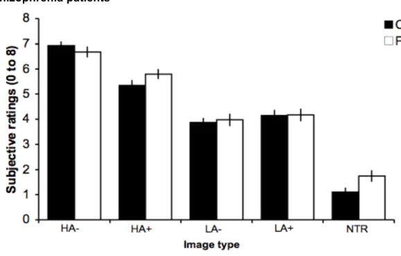
fMRI Functional magnetic resonance imaging HA- High arousal negative
HA+ High arousal positive LA- Low arousal negative LA+ Low arousal positive HC Healthy controls
HM Healthy men
HW Healthy women
IAPS International Affective Picture System
LA Low arousal
MNI Montreal Neurological Institute NEG Negative
NOC National Occupational Classification NTR Neutral
PANSS Positive and negative syndrome scale POS Positive
SCID Clinical interview for DSM-IV SCZ Schizophrenia patients SD Standard deviation SES Socioeconomic status
SPSS Statistical Package for the Social Sciences SZ-M Schizophrenia men
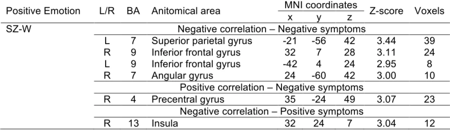
SZ-W Schizophrenia women ROI Region of interest
xiii Thank you
I am one of the fortunate ones to have had so many people in my life who have been there for me and who have supported me throughout this journey.
First and foremost, to all the participants and particularly the patients who took time out of their lives to take part in this study: THANK YOU!
Adrianna, I really cannot thank you enough for being the most amazing supervisor. Your invaluable knowledge, guidance, passion for research, and friendship has given me the confidence and the know-how to complete a thesis that I cannot be more proud of. Your belief in me will forever be appreciated.
This research would not have developed the way it did if it hadn’t also been for the mentorship support of Adham Mancini-Marie, Jose Jiménez and Stéphane Potvin – thank you for your patience and knowledge in answering all my questions regarding fMRI data analysis and interpretation.
I would like to thank my family, who has been there for me through thick and thin (and I know it hasn’t always been easy). Mom and Dad, I will never be able to thank you enough for everything you’ve done for me, for always being proud of me no matter what. To my sisters and brother: Julie, Kelly, Sophie and Junior thank you so much for all your encouragement. A special thank you to my nephews who have never failed to put a smile on my face through moments of stress: Shakeem, Shaundre, Omar, Salamah, and of course to my nieces on the way, Leila Michelle and Keyarrah. Last but definitely not least, I would like to thank my boyfriend Tarek for
supporting me in my research every step of the way. Saying thank you simply does not seem like enough, but it will have to do.
1 Introduction
I. Schizophrenia: An overview
Evolution of the concept of schizophrenia: Kraepelin-Bleuler-Schneider
Kraepelin (1919), Bleuler (1911) and Schneider (1959) were instrumental figures in the conceptualization of schizophrenia, even though their different ideas led to
inconsistencies in defining the illness over the past century (Hoenig, 1983). Kraepelin (1919) believed that schizophrenia comprised a unique disease entity with a single etiology and a distinct pathology. Because the cause was unknown, classification depended largely on the course and outcome of groups of symptoms. More specifically, Kraepelin noted a tendency towards deterioration and an outcome of mental dullness or dementia in patients with an adolescent or early adult onset of catatonia, hebephrenia, and paranoid dementia – a group he termed dementia praecox.
The term schizophrenia, which literally means “a mind that is torn asunder”, was later coined by Euegen Bleuler (1911). In contrast to Kraepelin’s neurodegenerative disorder of adolescents, Bleuler defined a set of basic symptoms considered to be unique to the disorder and present in all patients with the disorder. Moreover, he considered course and outcome of the illness to be variable (e.g. stable outcome, improvement after onset). Rather than the essence of schizophrenia being defined by delusions and hallucinations, Bleuler believed that loosening of associations, blunted affect, ambivalence and autism (now considered negative symptoms) were the fundamental symptoms of the illness.
Subsequently, Jaspers (1946) deemed that a deficit in compassionate interactions in individuals with schizophrenia and an “un-understandability” of the individual experience as significant features of the disorder. Building on this concept, Schneider (1959) defined the first-rank symptoms of schizophrenia (e.g. thoughts experienced as spoken aloud, voices making references in the third person), which
2 were thought by Schneider and others (Mellor, 1970) to be pathognomonic. However, with time this assertion was challenged in light of Schneiderian first-rank symptoms being present in a variety of other nonorganic psychotic psychiatric disorders (Janowsky & Risch, 1979; O'Grady, 1990).
Clearly, these three viewpoints of describing this complex and heterogeneous psychiatric disorder are distinctive. The tools currently used to define schizophrenia including DSM-IV-TR (Diagnostic and Statistical Manual of Mental Disorders) and ICD-10 (International Classification of Diseases) criteria incorporate Kraepelinian chronicity, Bleulerian negative symptoms and Schneiderian positive symptoms. However, the use of different combinations and variable interpretations of these elements with restricted psychopathological validity has led to widespread criticisms (Poland et al., 1994). The epidemiology of schizophrenia
Over the years there have been major epidemiological findings in the occurrence of schizophrenia across populations, time and socio-demographic
characteristics (Tandon et al., 2008). Schizophrenia manifests itself in less than 1% of individuals in a population at some point in their lives (Perala et al., 2007; Stilo & Murray, 2010). The most recent studies suggest that the rate of 1% could be an overestimation and that the prevalence of the disorder is typically higher in developed than in developing countries, and higher in migrant groups than in native-born
populations (McGrath et al., 2008; Saha et al., 2005). Additionally, a higher prevalence of schizophrenia among lower as opposed to higher socio-economic classes within communities has been steadily reported over the past century (Tandon et al., 2008).
The incidence of schizophrenia, defined as the number of new cases for a given population per year, has shown prominent discrepancies among scientific studies. A concise overview of three related systematic reviews on the incidence, prevalence, and mortality associated with schizophrenia has reported that the median incidence of
3 schizophrenia was 15.2/100,000 persons and that the central 80% of estimates varied greatly over a range of 7.7-43/100,000 persons (McGrath et al., 2008). Moreover, significant variations in the incidence of schizophrenia have been documented such that urbanicity, migration and male gender have been linked with an increased risk of
developing the disorder (Saha et al., 2006). A relatively consistent description of
schizophrenia has been documented over the last two centuries and its occurrence has been relatively stable despite changes in specific diagnostic criteria (Bleuler, 1950; Kraepelin, 1971). However, the incidence rates of schizophrenia are suggested to have fluctuated over time with some studies proposing an increase in schizophrenia (Bray et al., 2006) and others implying a decrease in the incidence of schizophrenia with time (Woogh, 2001). It is likely that changes in the tools used to diagnose schizophrenia and the manner in which cases are detected makes these assumptions difficult to interpret (Kendell et al., 1993).
The clinical features of schizophrenia
Schizophrenia is presently defined by a diverse set of signs and symptoms that are broadly classified into positive, negative, cognitive, disorganization, mood and motor symptom dimensions; with differing degrees of psychopathological expression observed across patients and throughout the course of illness (Tandon et al., 2010). Positive symptoms include delusions, hallucinations and other reality distortions while negative symptoms encompass a blunting or loss of a range of affective functions such as a loss of motivation, lack of experiencing pleasure, avolition and blunted affect. The
occurrence of all types of hallucinations is about 50% in those with the disorder and of this percentage, 50% encompass auditory hallucinations (Cutting, 1990). Delusions, on the other hand, are reported to occur at some stage during the course of schizophrenia in more than 90% of those with the diagnosis (Cutting, 2002). With regards to negative
4 symptoms, flattening of affect is observed in approximately 50 % of acute (Andreasen, 1979) or chronic patients with schizophrenia (McCreadie, 1982).
Studies have shown that negative symptoms are generally more stable than positive symptoms and are least likely to improve over the course of illness (Addington et al., 1991; Andreasen & Flaum, 1991). For instance, Arndt and colleagues (1995) observed that while both negative and positive symptoms were already prominent at the time of the patients’ first episode, during the follow-up period, the negative symptoms remained relatively stable while the positive symptoms declined. In addition, it has been suggested that negative symptoms are more closely linked to prognosis, in spite of the suffering that may be associated with psychotic symptoms (Fuller et al., 2002). Many studies have shown that the level of negative symptoms is significantly associated with a poor level of social functioning (Biehl et al., 1986; Breier et al., 1991; Fenton &
McGlashan, 1992; Keefe et al., 1987); albeit others have observed similar relationships between high levels of positive symptoms and social functioning (Breier et al., 1991; Keefe et al., 1987). A surprisingly consistent pattern has emerged between impairments in cognitive ability and symptom profiles, such that cognitive impairment correlates with negative and disorganized symptoms (Andreasen, Flaum, et al., 1990; Bilder et al., 1985; Braff, 1989; Keilp et al., 1988; Merriam et al., 1990) but not with psychotic symptoms in patients with schizophrenia (Bilder, et al., 1985; O'Leary et al., 2000). What causes schizophrenia?
Schizophrenia is a complex, incapacitating and heterogeneous psychiatric disorder, and to date the etiology of the illness remains a mystery. Even so, several theories on the origin of schizophrenia have been put forward.
The risk of developing schizophrenia increases considerably in an individual who has an affected family member, a risk that intensifies as the degree of genetic similarity with the affected family member increases (i.e. a first degree relative) (Kendler
5 & Diehl, 1993; Sullivan et al., 2003). Twin studies of schizophrenia show consistently higher concordance rates in monozygotic (around 50%) than in dizygotic twins (around 17%) (Cardno and Gottesman, 2000). Correspondingly, both individual twin studies and meta-analyses of twin studies estimate the heritability of schizophrenia to be roughly 80% (Riley and Kendler, 2006). Nonetheless, more than two thirds of individuals with schizophrenia do not present with a family history of the illness (Kendler et al., 1993) and no gene appears to be either sufficient or necessary for the development of schizophrenia (Tandon et al., 2008). Thus, the prevalent genetic opinion is that schizophrenia is a polygenic/multifactorial disease (Lichtermann et al., 2000). Other than genes, both pre- and perinatal complications (e.g. maternal influenza, fetal hypoxia) and other environmental elements (e.g. season of birth, cannabis use) have been linked to a substantial increase in the risk of developing the disorder (McGrath, 2011; Mittal et al., 2008; Tandon, et al., 2008; van Os et al., 2008). After decades of research what can be said with confidence is that both genetic and environmental elements are at play (Tandon et al., 2008).
It has been suggested that schizophrenia is "not the result of a discrete event or illness process at all, but rather one end of the developmental spectrum that for genetic and/or other reasons 0.5% of the population will fall into." (Weinberger, 1987).
Developmental changes in the brain that span from the fetal period through young adulthood are now a vital feature of most etiological theories of schizophrenia and this neurodevelopmental hypothesis of schizophrenia is rarely challenged in the literature (Gourion et al., 2004; Walker et al., 2010; Weinberger, 1996). Simplified, the broad neurodevelopmental view of schizophrenia dictates that both genetic and prenatal factors confer vulnerability of schizophrenia by inducing pathologic processes beginning before the brain approaches its adult state in adolescence (Fatemi & Folsom, 2009; Rapoport et al., 2005). Developing in utero as early as late first or early second
6 trimester (Fatemi et al., 2005), these neurodevelopmental anomalies are proposed to lead to the activation of pathologic neural circuits during critical developmental time points in adolescence (sometimes due to elevated stress), ultimately guiding the
manifestation of positive and/or negative symptoms (Fatemi & Folsom, 2009; Keshavan & Hogarty, 1999; Walker, et al., 2010). Yet, precisely how and in what way genetic and environmental factors might interact to cause schizophrenia and the neurobiological mechanism that may mediate such interactions remains unknown.
Treatment without a cure
As far as we know, there is no cure for schizophrenia. In spite of this, existing multi-modal regimens that embrace the use of antipsychotic medications, psychosocial interventions, as well as support with housing and financial sustenance are accessible to individuals with schizophrenia (Tandon, et al., 2010).
About half a century ago, antipsychotic drugs were established into clinical practice sparking the revolution in the pharmacotherapy of schizophrenia (Delay et al., 1952). First generation antipsychotic drugs (i.e. typical antipsychotic) have been shown to be effective in relieving the positive symptoms of schizophrenia and in preventing their reappearance in several patients (for a review see Miyamoto et al., 2005), while having little therapeutic effects on negative symptoms, mood symptoms and cognitive deficits (Fleischhacker, 1995; Hawkins et al., 1999; Tandon et al., 2009). Instead, second-generation or atypical antipsychotic drugs such as clozapine are reported to have superior therapeutic benefits for cognitive and negative symptoms (Keefe et al., 1999; Meltzer & McGurk, 1999); a conjecture that is today continuously debated (Faber et al., 2011; Keefe et al., 2007; Remington & Kapur, 2000). Interestingly, several recent studies appear to imply that atypical antipsychotic agents are no more effective than typical drugs and are not associated with better cognitive or social outcomes (Jones et al., 2006; Keefe, et al., 2007; Lieberman et al., 2005; Swartz et al., 2007). While typical
7 antipsychotics cause a range of side effects such as acute extrapyramidal symptoms and tardive dyskinesia (Miyamoto et al., 2002; Tandon, et al., 2010) the atypical agents produce other serious side effects including agranulocytosis, weight gain and diabetes (Lieberman & Safferman, 1992; McIntyre et al., 2001; Melkersson & Dahl, 2004). Regardless, the finding that atypical drugs increase efficacy in treatment refractory patients (Essali et al., 2010; McEvoy et al., 2006) and lower the risk of extrapyramidal symptoms (Pierre, 2005) have given the scientific community hope that a superior antipsychotic treatment for individuals with schizophrenia is achievable (Tandon et al., 2010). No matter the type of drug used, antipsychotic medications are consistently reported to be superior to placebos in reducing overall symptom severity and the risk of relapse in patients with schizophrenia (Leucht et al., 2003). It is noteworthy to keep in mind; however, that despite the reported therapeutic benefits of antipsychotic
medications, the extent to which their use impacts the mortality, social functioning and overall quality of life in patients with schizophrenia are less clear and research on this topic has yielded inconsistent findings (Lehman et al., 2004; Ren et al., 2009; Thirthalli et al., 2010; Weinmann et al., 2009).
As stated by Fenton and Schooler (2000) “patients are more than the sum of their symptoms”. These authors emphasize that the management of this disabling psychiatric disorder extends beyond the use of antipsychotic drugs. The literature has yielded cumulative evidence of the efficacy of various psychosocial interventions such as psychoeducation (Giron et al., 2010; Pitschel-Walz et al., 2001), Cognitive Behavior Therapy (Gould et al., 2001; Zimmermann et al., 2005), Social Skills Training (Kurtz & Mueser, 2008), Assertive Community Treatment (Nelson et al., 2007) and Cognitive Remediation approaches (McGurk et al., 2007) on various symptom domains, albeit not all studies have found these therapies to be useful (Tandon et al., 2010). The potential benefit of using psychosocial therapies as adjuncts to pharmacotherapy in alleviating
8 certain symptoms, improving compliance to medication, social functioning and quality of life in individuals with schizophrenia is particularly important in light of evidence that 30% of individuals with the disorder are resistant to the classical antipsychotic drugs used today (Meltzer, 1997).
Outcome – what are the consequences of schizophrenia?
Many studies have examined the outcome or “consequences” of schizophrenia. Still, the data on longitudinal outcome have consistently run into tribulations concerning definitions of sample and measures used to assess outcome, among many other confounds and biases, making comparison between studies complicated (Allardyce & van Os, 2010).
The consequences of Kraepelin’s schizophrenia were depicted as encompassing a permanent and pervasive impairment in mental functions – an inexorably deteriorating disorder (Kraepelin, 1919). Subsequently; however, he acknowledged that recovery was possible in dementia praecox (Kraepelin, 1920); a premise that has been reinforced by a growing body of research in which varying degrees of recovery have been observed in patients with schizophrenia. A systematic review of prospective studies analyzing first-episode psychosis published between 1966 and 2003 have outlined a superior outcome for 42% of the population, an intermediate outcome for 35%, and a poor outcome for 27% (Menezes et al., 2006). These authors also established a relationship between a developing country of origin with good outcome and the use of typical antipsychotic medication (versus atypical or a
combination of both) and being treatment-naive at study entry with poor outcome. This review corresponds somewhat to other studies that have reported different degrees of partial or full recovery in individuals with the disorder as well as cases in which the illness has resolved completely or ended in a severe defect state (Harrison et al., 2001; Hegarty et al., 1994; Jobe & Harrow, 2005).
9 Outcome, in the framework of schizophrenia, is a multi-faceted concept
encompassing several distinct areas of psychopathology, social functioning, quality of life and societal impact (Tandon et al., 2008). For instance, individuals with
schizophrenia show an increased prevalence of comorbid psychiatric and medical illnesses (e.g. substance abuse, cardiovascular disease) (Bermanzohn et al., 2000; Leucht et al., 2007), have a decreased probability of employment and substantial impairments in quality of life (Eack & Newhill, 2007; Folsom et al., 2005) as well as an increased risk of suicide and death (Fenton, 2000; Palmer et al., 2005; Saha et al., 2007; Seeman, 2007). Given the disabling characteristics of schizophrenia, it is understandable that the illness has profound impacts on affected individuals and their loved ones. A revealing example is the observation that families of affected individuals report both a higher subjective and objective burden in concurrence with less support from social networks and professionals (Magliano et al., 2005).
At present, an acute illness onset, better premorbid functioning, improved
cognitive abilities, absence of substance abuse, female sex and a later age of onset are associated with a superior outcome in patients with schizophrenia (Breier, et al., 1991; Flyckt et al., 2006; Riecher-Rossler & Rossler, 1998; Shepherd et al., 1989).
Summary
Schizophrenia manifests itself in less than 1% of the population and is broadly defined by a diverse set of positive, negative, disorganization, mood and motor symptom dimensions. Although its etiology remains a mystery, both genetic and environmental elements are at play. To date, multi-modal treatments exist for
individuals with schizophrenia that encompasses the use of antipsychotic medications and psychosocial interventions. The benefit in using psychosocial therapies as adjuncts to pharmacological treatment is advantageous since about one third of patients with schizophrenia are resistant to the classical antipsychotics used today. Finally, research
10 has documented diverse outcomes in patients with schizophrenia varying from superior to intermediate to poor.
11 II. Sex differences in schizophrenia
Why is it crucial to investigate sex differences in schizophrenia? Most
importantly, understanding sex differences in various clinical features of the disorder such as its clinical expression, course of illness and response to treatment will not only provide an opportunity to broaden our understanding of the complex inner workings of this psychiatric disorder but may also provide a unique opportunity to delineate more appropriate treatment regimens based on sex. Considering the vast growth of this field in the past century, this thesis focuses on some of the most prominent areas of sex differences in schizophrenia.
Epidemiology
The incidence of schizophrenia is generally higher in men than women. A systematic review of 158 studies reported a median male:female rate ratio of 1.40 (McGrath et al., 2004). In addition, ratio estimates from various different types of studies (e.g. methodologically rigorous studies, those before 1980, those using DSM-III criteria) lie between 1.27 and 1.54 and significantly exceed 1 highlighting an increased
incidence of schizophrenia in men versus women (Abel et al., 2010). In the midst of studies that have found significant sex differences in favor of males, some have found the opposite pattern or no significant sex difference in incidence rates (Al Mousawi & Dunstan, 1998; Hambrecht et al., 1994; Jablenksy, 1992). Of interest, the different criteria used to diagnose schizophrenia over the years has affected the incidence rates among men and women. Lewine and colleagues (1984) found that the stricter the diagnostic criteria used, the more men than women were diagnosed with schizophrenia. For instance, schizophrenia was only diagnosable before the age of 45 according to DSM-III criteria, which lead to a greater exclusion of women and resulting in a higher proportion of men being diagnosed (Castle et al., 1993).
12 In contrast to the incidence data, studies have more or less consistently reported a lack of sex differences in prevalence rates (McGrath, et al., 2008; Perala, et al., 2007; Saha, et al., 2005). This has been found to be the case for various prevalence
measures including combined point, period and lifetime prevalence estimates (Saha et al., 2005). To date it is unclear how to resolve the paradox of a robust sex difference in the incidence but lack of sex difference in prevalence of schizophrenia (Abel et al., 2010).
Age of onset
An earlier age of onset in men compared to women with schizophrenia is perhaps one of the most robust findings of sex differences in schizophrenia (Review by Leung & Chue, 2000; Salem & Kring, 1998). Hafner et al. (Hafner, an der Heiden, et al., 1998; Hafner, Maurer, et al., 1998) reported a peak of onset in men between the ages of 15-25 followed by a stable decline, while women have a broader peak between the ages of 15-30 with a second smaller peak between 45-49 years of age. The consistent finding of an earlier age of onset in men of about 3-5 years has been shown to exist regardless of culture (Hambrecht et al., 1992; Lewine, 1981), definition of onset (Hafner et al., 1989; Hafner et al., 1993) and definition of illness (Hafner et al., 1989; Lewine et al., 1981). Nonetheless, there exist factors that have contributed to the elimination of sex differences in age of onset. Similar to the sex difference in incidence rates, when DSM-III criteria with an age of onset limit of 45 imposed, the sex difference dissipates (Shimizu & Kurachi, 1988). In addition, the sex difference in age of onset appears to apply to sporadic but not familial schizophrenia (Albus et al., 1994; DeLisi et al., 1994; Gorwood et al., 1995). In fact, of the factors associated with elimination of sex
differences in age of onset, a positive family history is the most reliable finding (Leung and Chue, 2000).
13 Premorbid history
A number of reports have documented poorer premorbid functioning in male than female patients on a variety of domains. More specifically, before developing schizophrenia, men are more socially isolated and withdrawn, are less able to maintain friendships and sexual relationships, have lower levels of school and work function (e.g. worse academic performance and higher frequency of job changes) compared to pre-schizophrenic women (Castle, et al., 1993; Childers & Harding, 1990; Crow et al., 1995; Goldstein et al., 1989; Larsen et al., 1996; Salem & Kring, 1998). Moreover, pre-women with schizophrenia are more than twice as likely to be married at the time of first
hospitalization compared to pre-schizophrenia men (Hafner et al., 1989). Certain behaviors in childhood that precede the development of schizophrenia may be different in the sexes. For instance, male pre-schizophrenic children are inclined to demonstrate more externalizing behaviors (e.g. disruptive, agitated) while female pre-schizophrenic children show more internalizing behaviors (e.g. passive and avoidant) (Crow, et al., 1995; Done et al., 1994; Watt, 1978). Sex differences in premorbid IQ have also been reported with more pervasive premorbid IQ deficits found in men compared to women, a finding that has correlated significantly with an earlier age of onset in men (Aylward et al., 1984). It is suggested that enhanced premorbid functioning in women is attributed to their later onset allowing them to complete their education, form personal relationships, marry and work before the illness takes over (Leung and Chue, 2000).
Birth and family history
In both male and female patients, obstetric complications have been linked to an earlier age of onset of schizophrenia, a poorer course of illness and ventricular
enlargement (Foerster et al., 1991; Heun & Maier, 1993; O'Callaghan et al., 1992; Owen et al., 1988). To date, the evidence of sex differences in obstetric complications is contradictory. Some have documented that mothers of female patients suffer a higher
14 rate of obstetric complications (Verdoux & Bourgeois, 1993), others have reported the opposite (i.e. greater incidence in mothers of male patients) (Cantor-Graae et al., 1994; O'Callaghan, et al., 1992) or have not found considerable sex differences (Heun & Maier, 1993; Hultman et al., 1997). Several studies found an association between prenatal exposure to influenza in the second trimester and the later development of schizophrenia particularly in females, while others found this relationship to be equal between men and women (Adams et al., 1993; Takei et al., 1994; van Os & Selten, 1998). Methodological issues such as small sample size and variable measures of selecting and weighting obstetric complications, among other variables, are suggested to play a role in these inconsistent findings (McNeil, 1995). Interestingly, individuals born in the winter and early spring months have about a 10% increased risk for developing schizophrenia (Schwartz, 2011). While some have found sex differences favoring both males and females in this season-of-birth effect (Chen et al., 1996; Pulver et al., 1992), this has not always been the case (Bradbury & Miller, 1985).
According to several family studies and family history studies, relatives of women with schizophrenia have a greater risk of developing the disorder as well as a higher chance of developing schizoaffective and schizophreniform illness than the family members of men with schizophrenia (Leung and Chue, 2000). For instance, Sham et al. (1994) found a lifetime risk estimate of schizophrenia to be 20.6% in the first-degree relatives of female probands and 4% in those of male probands. However, some population-based register studies have uncovered equal familial risk in the sexes (Cannon et al., 1998; Kendler & Walsh, 1995).
Clinical expression
In general, men with schizophrenia show evidence of more negative symptoms such as social withdrawal, blunted affect, lack of motivation and poverty of speech (Kay et al., 1986; Ring et al., 1991; Schultz et al., 1997) while women exhibit more affective
15 symptoms including depression, impulsivity, inappropriate affect, sexually inappropriate behavior and sexual delusions (Andia et al., 1995; Gur et al., 1996; Rector & Seeman, 1992). Other than being characterized by a strong affective component, women with schizophrenia are over-represented among patients with schizoaffectIve disorder and more commonly have a differential diagnosis of bipolar or affective disorder
(Bardenstein & McGlashan, 1990). Sex differences in the clinical expression of
schizophrenia have been reported as being overall consistent in different countries and cultures (Goldstein, 1997; Hambrecht, et al., 1992; Jablenksy, 1992). Nevertheless, there have been disputes regarding whether males are characterized by more negative symptoms and females by more positive symptoms, although many agree that both sexes experience the same level of positive symptoms (Addington et al., 1996; Kendler & Walsh, 1995; Linstrom & Von Knorring, 1994). Still, specific positive symptoms such as paranoia, persecutory delusions and auditory hallucinations have been found to be higher in women than men with schizophrenia (Goldstein, 1997; Goldstein & Link, 1988; Hambrecht, et al., 1992; Marneros, 1984; Rector & Seeman, 1992; Tien, 1991).
Interestingly, age has differentially contributed to the clinical expression of
schizophrenia in men and women. For instance, Gur et al. (1996) observed that women continue to show less negative symptoms than men until the eighth decade of life, but have the same degree of positive symptoms across all age groups. In a sample of older patients with schizophrenia, Lindamer et al. (1999) found a greater severity of positive symptoms in men than women in the older age group and reported that women with a late onset exhibited less severe negative symptoms in comparison to men with late onset, and relative to both men and women with early onset. Complementing this finding, others have noted a significant association between older age at onset and fewer negative and cognitive symptoms among women with schizophrenia (Grossman et al., 2008; Morgan et al., 2008).
16 Course of illness
More often than not, women with schizophrenia have a better prognosis than men on several different areas. However, while this sex difference is prominent in the short- (2-5 years) and middle-term (5-10 years) (Goldstein, 1988; Goldstein, et al., 1989; Salokangas & Stengard, 1990), this difference has been found to dissipate with time (i.e. in the long term; 13-40 years) (Gureje & Bamidele, 1998; Kendler & Walsh, 1995; Nyman, 1989). In a major review of over 100 outcome studies, Angermeyer et al. (1990) found that in approximately half of the studies women showed a better course of hospital treatment, experienced a shorter length of hospital stay, and survived longer in the community after their first hospital admission while the other half of studies did not observe significant sex differences in the course of schizophrenia. Other domains in which women show a more favorable prognosis than men include education,
occupational functioning, social functioning (e.g. higher rates of marriage and having children), less substance abuse and less antisocial behavior (Hafner, 2003; Leung & Chue, 2000). It is of interest that the degree of psychopathology is an outcome measure that does not seem to be different between the sexes (Hafner, et al., 1993; Shtasel et al., 1992). In any case, superior social and occupational functioning in women
compared to men has been reported despite similar severity in symptom profiles (Andia, et al., 1995; Goldstein et al., 1989). It is suggested that worse outcomes in men may be associated to substance abuse which is more common in men than women with
schizophrenia (Abel et al., 2010; Leung and Chue, 2000), particularly since patients with a dual diagnosis are less responsive to antipsychotic medication, less compliant with taking medication and are more frequently hospitalized and homeless (DeQuardo et al., 1994; Dixon, 1999).
17 Response to treatment
Men and women with schizophrenia differ in their response to treatment at both the pharmacological and psychosocial level. Women with schizophrenia have been found to respond more quickly and show a greater degree of improvement on
antipsychotic medications in comparison to men (Angermeyer, et al., 1990; Robinson et al., 1999; Szymanski et al., 1995; Usall et al., 2007). Usall and colleagues (2007) found that women with schizophrenia treated with both typical and atypical agents show significantly greater improvement in overall symptom severity compared with men. Moreover, women seem to require lower doses of first generation antipsychotics (Hogarty, Goldberg, & Schooler, 1974; Hogarty, Goldberg, Schooler, et al., 1974) until the age of 40 or post-menopause when the opposite pattern has been reported (i.e. women necessitate higher antipsychotic doses than men) (Seeman, 1983; 1986). Of interest, because estrogen is argued to play a protective role in women with
schizophrenia by shielding them from the worse effects of the illness (Huber et al., 2004; Seeman & Lang, 1990) it is suggested that a decrease in estrogen levels after menopause possibly contributes to the higher doses required in women (Leung and Chue, 2000). Others have not documented sex-specific differences in dosage or response to antipsychotics (Magharious et al., 1998; Pinals et al., 1996). Few studies have examined sex differences in response to atypical antipsychotics and the results have been mixed. Superior cognitive improvement in female compared to male patients taking olanzapine, risperidone and clozapine has been documented (Howard et al., 2001) while some have noted no difference in men and women’s response (Perry et al., 1991) or observed that female sex was a predictor of poor treatment response to
second generation antipsychotic drugs (Lieberman et al., 1994; Szymanski et al., 1996). Female patients are more prone to certain side effects of clozapine (e.g.
18 generally experience adverse motor side effects on antipsychotics more frequently than men, with higher rates of parkinsonism, dystonia and tardive dyskinesia (Leung and Chue, 2000). Sex differences in certain social behaviors have contributed to
differences in the way men and women with schizophrenia respond to treatment. One pertinent example is the observation that men with schizophrenia are less likely to comply with antipsychotic medication than women (Tunnicliffe et al., 1992) but not all have observed this sex difference in behavior (Buchanan, 1992).
To date very few studies have investigated sex differences in response to psychosocial treatments in patients with schizophrenia. The findings point towards a pattern in which males appear to benefit to a greater degree from social skills training than women, but superior post treatment functioning in females (e.g. medication compliance) implies that factors other than taught skills are more important
determinants of social adjustment in affected women (Schaub et al., 1998; Smith et al., 1997).
Cognition and Emotion
Evidence of sex differences in cognitive function in schizophrenia is mixed with some studies pointing to a superior performance by male patients, others reporting the opposite or no effect of sex on cognitive impairments in individuals with schizophrenia. Seidman and colleagues (1997) documented a worse performance in male compared to female patients on the Wisconsin Card Sorting Task known to assess executive
function and working memory and indicative of dorsolateral prefrontal cortex deficits. Using an extensive battery of neuropsychological tests, Goldstein and colleagues (1998) found that men with schizophrenia were significantly impaired across all
cognitive domains compared with same-sex control subjects and were impaired on tests of attention, verbal memory, and executive functions relative to women with
19 schizophrenia had decreased performance on tests of attention, executive functions, visual memory, and motor functions suggesting that women with schizophrenia may be less vulnerable to deficits in verbal processing than schizophrenic men. In contrast to this pattern of results, Lewine et al. (1996) reported greater deficits on tasks of verbal and spatial memory as well as visual processing tasks in women compared to men with schizophrenia. In a more recent study women with schizophrenia performed better than men in processing speed and verbal episodic memory whereas men outperformed women in visual working memory (Torniainen et al., 2011). Finally, quite a few of studies have not found differences between men and women in cognitive performance (Albus et al., 1997; Andia, et al., 1995; Goldberg et al., 1995; Lewine et al., 2006; Shipman et al., 2009). However, some of these studies were confounded by differences in symptom expression, age at onset and medication highlighting the importance of taking these factors into careful consideration when evaluating potential sex differences. Noteworthy, significant relationships between impaired cognitive performance and social functioning have been particularly noted in women with schizophrenia, which is suggestive that cognitive deficits have greater adverse effects on social functioning in women than men (Mueser et al., 1995; Penn et al., 1996).
Reports of sex differences in emotion processing in schizophrenia are few and far between in the scientific literature. Those that have analyzed men and women with schizophrenia separately found that women with schizophrenia are more expressive than men (Levin et al., 1985; Tremeau et al., 2005) while others have found no
difference between the sexes (Davison et al., 1996). With respect to the experience of emotion, Heerey and Gold (2007) reported that women rate positive and negative images as being more pleasant and unpleasant, respectively, than did men with schizophrenia. In a study aimed at examining how individuals with schizophrenia respond to pleasant and unpleasant odors, men with schizophrenia rated the odors as
20 less pleasant than women with schizophrenia (Moberg et al., 2003) while a similar study found no difference between men and women in the pleasantness ratings of odors (Hudry et al., 2002). With regards to the recognition of emotions, Scholten et al. (2005) observed a sex difference in the ability to recognize facial emotions, especially negative ones, with women outperforming men. Vaskinn et al. (2007) and Weiss et al. (2007) reported similar findings such that women show a superior performance in recognizing facial emotions than men with the disorder.
Brain structure
The tentative pattern of altered brain structure in patients with schizophrenia is that men are characterized by increased neuroanatomical deficits than their female counterparts. Still, other studies have reported the reverse pattern or no significant effect of sex on structural brain abnormalities in these individuals (Leung & Chue, 2000). Methodological issues including small sample sizes, overrepresentation of men and variability in the size of brain structures of men and women in the healthy
population are suggested to be at least partially responsible for the inconsistent results (Andreasen, Ehrhardt, et al., 1990; Goldstein, 1996).
Largely consistent with the direction of normal sexual dimorphism (Goldstein et al., 2001; Murphy et al., 1996; Paus et al., 1996), both structural neuroimaging and postmortem studies have reported that men with schizophrenia have enlarged ventricular-brain ratios and anterior temporal horn, smaller medial temporal lobe volumes (e.g. amygdala, hippcomapus, superior temporal gyrus) and overall smaller frontal and temporal lobe volumes compared to women with schizophrenia (For a review refer to Leung and Chue, 2000; Abel et al., 2010). Moreover, more
left-lateralized abnormalities have been reported in men (Goldstein et al., 2002; Gur, R. E. et al., 2000). It is noteworthy that some have uncovered region-specific structural abnormalities in women depending on the region examined. For instance, Goldstein
21 and colleagues (2002) reported a significant reduction in anterior cingulate volume in women with schizophrenia relative to healthy women while there was no difference between men with and without the diagnosis. Another study found that men with schizophrenia have a higher orbitofrontal to amygdala ratio than healthy men while women with schizophrenia have a lower ratio than healthy women (Gur et al., 2004). These findings are particularly interesting in the light of reports that in the general population it is women who have greater grey matter volume of anterior cingulate than men (Paus et al, 1996) and a higher orbitofrtonal cortex to amygdala ratio than men (Gur et al., 2002), ultimately implying a disturbance of normal sexual dimorphism of brain structure in individuals with schizophrenia.
Brain function
Although there is extensive evidence of sex differences in cerebral function associated with various emotional and cognitive tasks in the general population (e.g. Domes et al., 2010; Koch et al., 2007; Wager et al., 2003), evidence of sex differences in brain activations in individuals with schizophrenia is still quite scarce. This is not surprising considering the majority of studies investigating cerebral activations associated with emotional processing and cognitive function in schizophrenia consist almost exclusively of men, and even when women are included in the sample the
number of tested individuals is typically too small to allow for between-sex comparisons. Previous work carried out by our group has uncovered distinct patterns of brain activations in men and women with schizophrenia during both cognitive (Guillem et al., 2009; Jiménez et al., 2010) and emotional processing (Mendrek et al., 2007; 2009; 2010). The overall findings point towards an alteration of the normal sexual dimorphism in cerebral activations in individuals with schizophrenia. For instance, Guillem et al. (2009) explored the differential effects of being male or female on the neural correlates of episodic memory in schizophrenia using event-related brain potentials (ERPs). They
22 uncovered that the direction of sex differences depended on the cognitive processes being examined. For instance, early frontal processes (related to interference inhibition) revealed an interaction between sex and diagnostic group suggesting a reversal of normal sexual dimorphism in their schizophrenia sample, while analysis of late posterior processes (suggested to reflect a mnemonic binding process) resulted in sex
differences in the same direction in both patients with schizophrenia and healthy controls. Mendrek and colleagues (2007) implemented an fMRI task exploring the emotional experience of patients with schizophrenia and found that while viewing aversive pictures, men with schizophrenia exhibited more widespread and more intense activations compared to women with schizophrenia in similar brain regions where women would normally exhibit greater activations relative to men in the healthy population (e.g.cingulate gyrus, temporal cortex, cerebellum). Subsequent investigations revealed that the symptom profiles in men and women correlated
differently with brain activations during processing of emotional information (Mendrek et al., 2010). Another recent study examined the recognition of emotional faces using ERPs and uncovered a lower P100 amplitude in the right hemisphere for fearful faces in men with schizophrenia compared to women which implies that sex can be an important controlling factor in early visual processing in patients with schizophrenia (Lee et al., 2010).
Summary
Sex differences in patients with schizophrenia exist in an array of domains including its epidemiology and clinical expression. Overall, men present with an earlier age of onset compared to women, women have more positive symptoms while men experience more negative symptoms, women with schizophrenia have a better prognosis, respond more quickly and show a greater degree of improvement with antipsychotic treatment relative to their male counterparts. However, the literature has
23 illustrated inconsistencies in these findings; sex differences in cognitive function in schizophrenia have demonstrated particularly mixed results. To date, less research has focused on uncovering sex differences in response to psychosocial treatments, the processing of emotions and brain function in men and women with schizophrenia.
24 III. Emotional memory in schizophrenia
It is well established in the scientific literature that individuals with schizophrenia have prominent deficits in cognition and the processing of emotional material (Heinrichs & Zakzanis, 1998; Kohler et al., 2000; Kohler & Martin, 2006; Rund & Borg, 1999). Investigating the effect of emotion on subsequent memory in individuals with
schizophrenia provides a unique opportunity to examine the interaction between these two processes. This is particularly true in light of evidence that in the general population emotionally charged events often attain a privileged status in memory relative to neutral ones (Hamann, 2001; LaBar & Cabeza, 2006). What’s more, emotional memory
paradigms appear more ecologically valid than encoding and/or retrieval of non-emotional material, because in everyday life much of the information and events we come across holds emotional significance. In fact, it is relatively well accepted that emotion benefits cognition by prioritizing biologically relevant information (Damasio, 1994). Nonetheless, investigating emotional episodic memory in individuals with schizophrenia is still limited.
Behavioral findings
Behavioral evidence has outlined deficits in patients with schizophrenia
compared to healthy controls on various dimensions associated with the processing of emotional memories. For instance, there is substantial evidence that patients with schizophrenia have impairments in independently initiating effective strategies during the encoding of stimuli into memory (Brebion et al., 1997; Iddon et al., 1998).
Interestingly, when prompted to use encoding strategies, individuals with schizophrenia have shown significant improvement in mnemonic performance (e.g. Bonner-Jackson et al., 2005; Boyer et al., 2007; Koh & Peterson, 1978; Paul et al., 2005). However, some researchers who have directly compared incidental vs. intentional encoding have not found differences in memory performance in relation to diagnosis (Koh et al., 1976). In
25 general, the data has pointed towards a modest effect of intentional in comparison to incidental encoding on emotional memory performance in patients with schizophrenia (Herbener, 2008).
Memory performance has also depended on the type of response used in the experimental paradigm. Tasks using recognition memory are suggested to selectively target memory processes while retrieval tasks entail a combination of both memory and executive abilities (Pelletier et al., 2005). A review by Heinrichs and Zakzanis (1998) reported that recall is typically more impaired in schizophrenia than recognition. Moreover, patients with schizophrenia demonstrate greater deficits in the recognition memory of visual (e.g. pictures) vs. verbal (e.g. words) stimuli (Pelletier et al., 2005). Consistent with this, a review of emotional memory studies in schizophrenia found that in all studies using pictorial stimuli, individuals with schizophrenia had an impaired memory in comparison to healthy control subjects (Herbener, 2008).
Arousal and valence characteristics of emotional stimuli are factors that have also differentially influenced emotional memory in patients with schizophrenia and healthy subjects, though the results have been inconsistent. Some studies have found impairments in schizophrenia patient’s emotional memory compared to healthy subjects (Danion et al., 2003; Hall et al., 2007; Herbener et al., 2007; Neumann et al., 2007); while others have found comparable performances between the two groups (Horan et al., 2006; Koh, et al., 1976). Looking specifically at the effect emotional valence has on memory performance in patients with schizophrenia, some studies have demonstrated an enhancement of memory for emotional (both positive and negative) in contrast to neutral stimuli in patients with schizophrenia (Calev & Edelist, 1993; Hall, et al., 2007; Mathews & Barch, 2004), others have reported the opposite (i.e. neutral better
remembered than emotional stimuli) (Koh et al., 1976; Mathews and Barch, 2004) or no effect of emotional stimuli on memory (Koh et al., 1981; Neumann, et al., 2007).
26 Furthermore, reports have documented increased memory for negative vs. positive stimuli in patients (Calev & Edilst, 1993; Herbener et al., 2007; Mathews & Barch, 2004) while few have reported the reverse pattern (Koh, et al., 1976). Although the literature has presented conflicting results it has nonetheless outlined a tentative pattern in which individuals with schizophrenia more frequently show a lack of enhancement of
emotional in contrast to neutral stimuli and also have a better memory for negative than positive stimuli (Herbener, 2008).
The majority of the published studies have focused on the effect of emotional valence of presented stimuli (i.e. stimuli that vary from positive to negative), while the effect of arousal (i.e. stimuli that vary from calm to exciting) on memory performance has rarely been examined despite indications that it contributes significantly to memory in healthy individuals. Specifically, it has been found that highly arousing stimuli are better recalled than low arousing stimuli (Bradley et al., 1992; Eysenck, 1976). Studies that have examined the effect of arousal on memory in patients with schizophrenia have outlined a better memory performance for stimuli of high emotional intensity relative to low intensity (Herbener et al., 2007; Horan et al., 2006; Mathews and Barch, 2004), but others have reported a detrimental effect of arousal on subsequent memory (Hall et al., 2007).
Despite research on emotional memory still being limited in schizophrenia, a few studies have noted a relationship between negative symptoms and impairments in emotional memory. Hall et al. (2007) uncovered a significant correlation between
negative symptoms of the PANSS and recognition memory of both negative and neutral scenes such that the more negative symptoms the patients had the worse their
recognition performance. Another study investigated individual PANSS symptoms and reported that patients with greater blunted affect showed a greater impairment in the recall of high arousal negative and positive words. The authors also documented similar
27 relationships between blunted affect and recognition memory of all emotional conditions (i.e. high and low arousal). Horan and colleagues (2006) evaluated whether
self-reported trait anhedonia in schizophrenia reflects impaired memory for pleasant experiences but the authors did not find a significant association between these two measures despite schizophrenia patient’s elevated trait anhedonia.
The variability in the emotional memory literature can easily be attributed to the differences in the stimuli used among studies (pictures vs. words), the type of retrieval method (recognition vs. recall), response type (old/new vs. remember/know), time of testing (delayed vs. immediate recall), as well as the clinical heterogeneity present in patients with schizophrenia. Accordingly, it is challenging to draw definite conclusions on the influence of emotion on memory in patients with schizophrenia.
Neuroimaging findings
To date, very few studies have examined the neural correlates of emotional memory in individuals with schizophrenia. Using fMRI, Whalley and colleagues (2009) reported robust medial temporal lobe activations in individuals with schizophrenia, individuals with bipolar disorder and healthy controls during viewing of positive scenes that correlated significantly with recognition memory of positive images. The direct group comparison revealed decreased activity in the bilateral amygdala in patients with schizophrenia relative to healthy participants in the cerebral activations associated with positive images versus baseline. This was found despite comparable recognition accuracies between the two groups. Another fMRI study using emotional faces found that patients with schizophrenia and healthy controls showed similar patterns of brain function associated with response bias for sad faces (e.g. parahippocampal gyrus) while response bias for happy faces revealed increased brain activations in healthy subjects compared to patients (e.g. superior frontal gyrus, amygdala and hippocampus) (Sergerie et al., 2010). Finally, Becerril and Barch (2011) explored how the emotional
28 valence of stimuli impacts the cerebral activations associated with an n-back working memory task and uncovered altered dorsolateral prefrontal cortex and hippocampus activity in individuals with schizophrenia while performing the emotionally loaded working memory task.
Summary
Deficits in patients with schizophrenia relative to healthy controls have been observed on various dimensions associated with the processing of emotional memories. Specifically, patients wit schizophrenia show impairments in initiating effective encoding strategies, they show greater deficits in recall than recognition, and have demonstrated differential deficits depending on whether the valence of the emotional stimuli was positive, negative or neutral. The effect of arousal intensity and the potential association of clinical symptoms on memory performance have been seldom examined. Functional neuroimaging studies investigating the neural correlates of emotional episodic memory in schizophrenia is slowly growing, while investigation of sex differences in this domain remains scarce.
29 IV. Hypotheses
At present, there are no functional neuroimaging studies that have: (1)
investigated the effect stimuli of different arousal intensities (i.e. calm vs. exciting) and valence (positive vs. negative) has on the pattern of brain function during emotional memory in schizophrenia patients compared to healthy controls and (2) have explored whether being male or female differentially contributes to the pattern of cerebral
activations associated with the recognition memory of emotional images in patients with schizophrenia.
Accordingly, the first goal of this thesis was to uncover the brain function
associated with recognition memory of emotional stimuli that varied in both valence and arousal in patients with schizophrenia compared to healthy controls. In line with some behavioral studies of emotional memory (Danion et al., 2003; Mathews and Barch, 2004; Neumann et al., 2007) and majority of neuroimaging studies of emotion
processing in schizophrenia (Gur et al., 2002; Schneider et al., 1998; Takahashi et al., 2004), we expected to observe a decreased recognition accuracy of emotional stimuli in patients with schizophrenia relative to healthy subjects and decreased cerebral
activations in all experimental conditions (positive, negative, high and low arousal) in the medial temporal cortex as well as prefrontal regions previously implicated in emotion processing and memory (medial, middle frontal and orbitofrontal cortex) (Dolcos et al., 2004a, 2004b; LeDoux, 1993). In terms of the influence arousal has on memory for emotional stimuli, we expected to observe increased brain activations in the high relative to low arousal contrasts of the presented positive and negative stimuli in healthy controls. In comparison, in patients with schizophrenia we expected an
increase in activity only in the high arousal conditions based on evidence that patients, compared to healthy controls, are more likely to misidentify low arousal emotional stimuli as neutral (Kohler, et al., 2000), but recognize high intensity emotions without