Glycoblology vol. 7 no. 5 pp 617-624, 1997
Expression of a cDNA encoding the glucose trimming enzyme glucosidase II in CHO
cells and molecular characterization of the enzyme deficiency in a mutant mouse
lymphoma cell line
Thomas Flura, Daniela Brada, Martin Ziak and Jiirgen
Roth
1Division of Cell and Molecular Pathology, Department of Pathology, University of Zttrich, 8091 Zurich, Switzerland
'To whom correspondence should be addressed
Glucosidase II is an ER resident glycoprotein involved in
the processing of N-linked glycans and probably a
compo-nent of the ER quality control of glycoproteins. For cloning
of glucosidase II cDNA, degenerate oligonucleotides based
on amino acid sequences derived from proteolytic
frag-ments of purified pig liver glucosidase II were used. An
unamplified cDNA library from pig liver was screened with
a 760 bp glucosidase II specific cDNA fragment obtained by
RT-PCR. A 3.9 kb glucosidase II cDNA with an open
read-ing frame of about 2.9 kb was obtained. The glucosidase II
sequence did not contain known ER retention signals nor
hydrophobic regions which could represent a
transmem-brane domain; however, it contained a single
N-glyco-sylation site close to the amino terminus. All studied pig
and rat tissues exhibited an mRNA of approximately 4.4 kb
with varying tissue expression levels. The authenticity of
the identified cDNA with that coding for glucosidase II was
proven by overexpression in CHO cells. Mouse lymphoma
PHAR 2.7 cells, deficient in glucosidase II activity, were
shown to be devoid of transcripts.
Key words: glucosidase II/glycoprotein
processing/N-glycosylation/ER quality control mechanism
Introduction
N-glycosylation of proteins starts in the lumen of the
endo-plasmic reticulum (ER) by en bloc transfer of the lipid-linked
oligosaccharide precursor G^MangGlcNAcj to nascent
poly-peptides. The first steps in its processing to yield the mature
oligosaccharide chains of glycoproteins include the removal of
all three glucose residues by neutral trimming ot-glucosidases
(Kornfeld and Komfeld, 1982; Moremen and Touster, 1988;
Roth, 1995). Glucosidase I removes the single terminal a
1,2-linked glucose residue, and the enzyme has been thoroughly
characterized (Hettkamp et al, 1984; Schweden et al, 1986;
Shailubhai et al, 1987; Bause et al, 1989; Kalz et al., 1995).
This is followed by the trimming of the two inner a 1,3-linked
glucose residues by glucosidase II, and some of the mannose
residues by ER-mannosidases (Moremen and Touster, 1988;
Roth, 1995). Glucosidase II has been purified from different
sources and studied extensively with regard to its properties
(Grinna and Robbins, 1979; Ugalde, 1980; Bums and Touster,
1982; Saunier, 1982; Brada and Dubach, 1984; Martiniuk et
al, 1985; Strous et al., 1987; Kaushal et al, 1990). It appears
to be composed of 100 kDa subunits carrying an N-linked
oligosaccharide chain of high mannose-type (Burns and
Touster, 1982; Brada and Dubach, 1984; Strous et al., 1987).
By both, immunoelectron microscopy (Lucocq et al., 1986)
and biochemical analyses (Bums and Touster, 1982; Brada and
Dubach, 1984; Strous et al., 1987), glucosidase II was shown
to be a resident ER glycoprotein in hepatocytes. Interestingly,
circumstantial biochemical evidence suggests that it is a
loosely membrane-associated glycoprotein of the ER (Brada
and Dubach, 1984; Strous et al., 1987; Brada et al, 1990). This
is in contrast to glucosidase I, which has been shown to
rep-resent a transmembrane type II glycoprotein (Kalz et al, 1995).
It has been proposed that glucosidase II plays a role in a
recently discovered quality control mechanism for ER to Golgi
apparatus transport of glycoproteins in which protein folding
and glycosylation are intimately interrelated (Hammond et al,
1994; Helenius, 1994; Hammond and Helenius, 1995). In this
model, improperly folded glycoproteins will be retained in the
ER prior to further transport by the concerted action of a
UDP-glucose:glycoprotein glucosyltransferase, chaperones such as
calnexin, calreticulin and BiP, and glucosidase II (Bergeron et
al, 1994; Hammond and Helenius, 1994; Hammond et al,
1994; Helenius, 1994). UDP-glucose:glycoprotein
glucosyl-transferase represents a soluble luminal ER protein (Parodi et
al., 1983; Labriola et al, 1995; Sousa and Parodi, 1995). A
unique property of this enzyme is to distinguish between native
and misfolded glycoproteins present in the ER (Trombetta et
al, 1989, 1991; Sousa et al, 1992; Trombetta and Parodi,
1992; Fernandez et al., 1994; Parker et al., 1995). Thus,
un-folded, partially folded and misfolded glycoproteins will be
bound and reglucosylated by this transferase rendering them a
ligand for calnexin (Ware et al, 1995). Calnexin with its
lec-tin-like properties will retain such monoglucosylated
glycopro-teins in the ER as long as they are not properly folded
(Ham-mond et al., 1994). During this process, the glycoproteins go
through cycles of glucose removal by glucosidase II and
re-glucosylation by UDP-glucose:glycoprotein
glucosyltransfer-ase. Once the correct conformation is achieved, the
glycopro-teins are no more a substrate for the UDP-glucose:glycoprotein
glucosyltransferase and calnexin, but solely for glucosidase n .
Subsequently, they will be able to exit the ER for the Golgi
apparatus. An additional component of this control mechanism,
but in a post-ER location, seems to be represented by Golgi
apparatus endomannosidase and calreticulin (Spiro et al,
1996).
The importance of both, the UDP-glucose:glycoprotein
glu-cosyltransferase and calnexin in this model has been directly
demonstrated (Hammond et al, 1994; Helenius, 1994;
Lab-riola et al., 1995). However, despite some recent indirect
evi-dence (Labriola et al, 1995), a direct proof that glucosidase II
TJlura et aL
by removing an inner glucose residue added on by the
gluco-syltransferase is involved in the control mechanism is still
missing. This is due to the lack of availability of a cDNA
coding for glucosidase II.
In this article, we report on the cloning of glucosidase II
from pig liver and its expression in bacteria and CHO cells. It
appears that glucosidase II is neither a soluble nor a
transmem-brane ER glycoprotein. The cDNA encoding glucosidase II
was found to share significant sequence homology with other
known glucose hydrolyzing enzymes. Further, we have
ana-lyzed a mutant mouse lymphoma cell line deficient in
gluco-sidase II activity at the molecular level.
Results
Partial amino acid sequences of pig liver glucosidase 11
Glucosidase II purified to homogeneity from pig liver was
digested with different proteases as described in Material and
methods. The proteolytic fragments were separated either by
reverse HPLC or by SDS-PAGE. The N-terminal amino acid
sequences of the enzyme subunit and the proteolytic peptides
were determined by Edman degradation (Figure 1). Degenerate
oligonucleotide primers were designed according to amino acid
sequences of the N-terminus of the glucosidase II subunit and
the peptide 4. Inosine residues were substituted at positions of
high degeneracy.
Isolation of cDNA fragments coding for glucosidase 11
A 760 bp fragment from a RT-PCR product coding for
gluco-sidase II was labeled with (
32P)dCTP and used to screen an
unamplified oligo(dT)-primed pig liver cDNA library. Three
independent overlapping cDNA clones were obtained and
characterized by restriction mapping. Screening of 2 x 10
6pfu
of a random primed pig liver cDNA-library yielded additional
upstream sequences. The cDNA clones obtained from the
screening of the oligo dT-primed and random primed cDNA
library were used to construct a full-length cDNA with an open
reading frame of about 2.9 kb. The open reading frame was
terminated by a stop codon TAA at position 2833 followed by
a 3' untranslated region of approximately 1 kb ending by a
N-term
pepl
pep 2
pep 3
pep 4
pep 5
pep 6
pep 7
pep 8
pep 9
pep 10
pep 11
V D R S N F K T L E E S S F X K / L R Q / V
T X I R I D E L E
G L L N F E H Q R
V T E G G X P Y R L Y N L D V
V N Q G F D D H N L P X D F
X F T X D P X R F P Q
D A Q H Y G G X E H R
I S I P M X L S L G L V G L S F X G A D
A L W V H Y P Q D V T
G H F E T P V X I E R V V I I G A G K P
G S P E / T S R L S F Q X D D E / T T
K P G V N V A S D X S I H L R
Fig. 1. Partial amino acid sequences of pig liver glucosidase D. The N-terminal amino acid sequence was determined by automated Edman degradation of purified pig liver glucosidase E. The sequences of all other peptides were obtained by Edman degradation after cleavage with proteases.
poly(A) tract. The consensus polyadenylation signal AATAAA
was found upstream from the poly(A) sequence at position
3781.
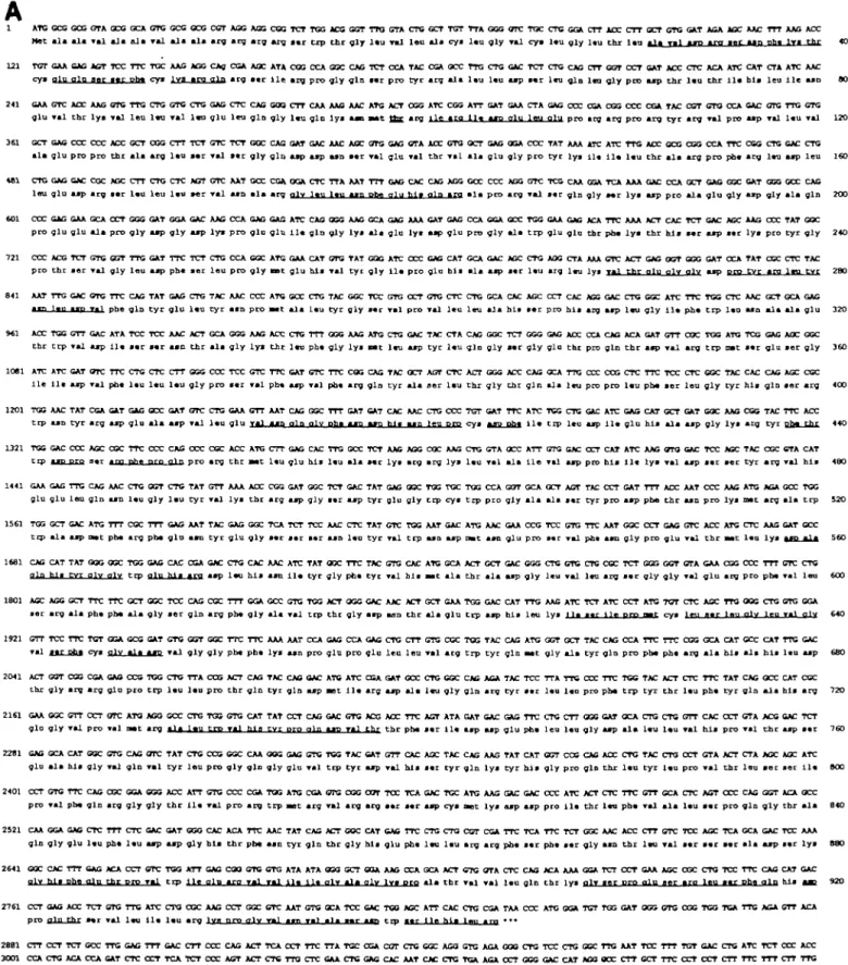
Analysis of the primary amino acid sequence
Translation of the DNA sequence into a protein sequence
(Fig-ure 2A) predicts an acidic (pi 5.5) polypeptide of 106 kb. All
peptides obtained by protein sequencing were contained in this
sequence indicating its authenticity. The protein sequence
starts with a putative signal sequence of 32 amino acids. The
signal peptidase cleavage site precedes the N-terminal peptide
of glucosidase II subunit obtained by protein sequencing. The
hydrophilicity plot (Figure 3) indicates that glucosidase II is a
rather hydrophilic protein with no putative transmembrane
do-mains. Further, no double lysine motif (KKXX) was
detect-able. The primary sequence contains one potential
N-glycosylation site, Asn-Met-Th. The sequence at the
C-terminus does not contain any known ER retention signal such
as KDEL and its variants.
Glucosidase II is evolutionary conserved
A comparative sequence data analysis with the EMBL data
bank using the Wisconsin software package revealed
homol-ogy to a yeast (Z36O98) and human (D42041) cDNA of
un-known function. The yeast cDNA exhibited 59% similarity and
39% identity and the human cDNA 96% similarity and 92%
identity to the pig glucosidase II cDNA.
Glucosidase II exhibits amino acid homology to other
glucose-hydrolyzing enzymes but not to glucosidase I
A comparison of peptides 5 and 7 (see Figure 1) with protein
sequences deposited in data bases indicated homology with
other glucose-hydrolyzing enzymes. The primary amino acid
sequence of glucosidase II was of high homology to the
se-quence of lysosomal ot-glucosidase, sucrase-isomaltase, and
several yeast glucosidases (yeast family 31 glucosidase,
Can-dida tsukubaensis ot-glucosidase, Schwanniomyces
occiden-talis glucoamylase). From this analysis it appears that
gluco-sidase II is closer related to the yeast family 31 glucogluco-sidase
than to sucrase-isomaltase. The homologies occur more
fre-quently in the middle and the C-terminal part of the
polypep-tide than in N-terminal part (data not shown). Glucosidase II
shares apparently also the sequence around the active site
(DMNE) of the other glucose-hydrolyzing enzymes (Figure
2B).
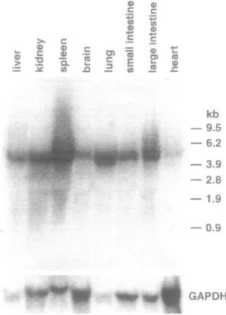
Expression of glucosidase II in pig and rat tissues
Northern blot analysis of total and mRNA from pig liver
re-vealed a message size of about 4.4 kb (Figure 4). A transcript
of the same size was found in other pig (Figure 5) and rat liver
as well as kidney, small and large intestine, heart, adrenal
gland, brain, submaxillary and parotid gland, thymus, lung,
ovary, and testis (data not shown). However, the levels of
glucosidase II expression varied between the tissues.
Glucosi-dase II mRNA was more abundant in pig liver than in brain and
heart (Figure 5). This was positively correlated with the
amount of enzyme protein in these tissues as detected by
West-ern blotting (Figure 6).
Mutant mouse lymphoma PHAR 2.7 cells are
transcriptionally deficient in glucosidase II
The mutant mouse lymphoma cell line PHAR 2.7 has been
previously shown to be deficient in glucosidase II activity
618
Cloning and expression of glucosidase II
A
1 ATC CCC GCG GTA CCC OCA CTC CCG CCC CCT ACG AGG CGQ TCT TGG ACG GGT TTO CTA CTC GCT TCT TTA GCC CTC TGC CTC GGA CTT JLOC CTT CCT CTC GAT ACA AGC AAC TTT AAG ACC Hot a l a a l a Tal a l a a l a Tal a l a a l a a r g arg a r g arg s a r t r p t h r g l y l a u Tml l e u a l a c y j l a u g l y Tal cy* l t u g l y l « u t h r l t u a l a T » 1 M P i m a«r « | n TTtil IT* f h r *> 121 TGT GAA GAG JUT TCC TTC TGC AAC AGG CAg CCA ACC ATA CGG CCA GGC CAG TCT CCA TAC CCA GCC TTG CTG GAC TCT CTG CAG CTT GGT CCT CAT ACC CTC ACA ATC CAT CTA ATC AAC
c y s g i n g i n aar s t r pb* c y s l v a ara g i n a r g s t r l i e a r a pro g l y g i n s a r p r o t y r arg a l a l « u l t u asp «*r l « u g i n l t n g l y p r o a s p t h r l « o t h r i l « b i s l e u l i e asn 00
241 GAA GTC ACC AAG GTC TTG CTG GTC CTG GAG CTC CAG GOO CTT CAA AAC AAC ATG ACT CCG ATC CGG ATT GAT GAA CTA GAG CCC CGA CGG CCC CGA TAC CGT CTO CCA GAC GTC TTG CTG g l u Tal thr l y « val l e u l t u Tal l « o y l u l e u g i n g l y l t u g i n l y * a s n * * t Qa_ arg i l e a r c 1 1 * U P QIU l e u o l u pro ary a r g p r o a r g t y r a r g Tal pro aap Tal l « u Tal 120 361 GCT GAG CCC CCC ACC GCT CGG CTT TCT GTC TCT GGC CAC OAT GAC AAC ACC GTG GAG GTA ACC GTC GCT CAG OGA CCC TAT AAA ATC ATC TTG ACC GCG CGG CCA TTC CGG CTG GAC CTG
a l a g l u pro pro thr a l a arg l t u s * r Tal i t r g l y g i n asp asp aso *«r v a l g l u Tal thr T « 1 a l a g l u g l y p r o t y r l y » i l t i l t l t u t h r a l a arg p r o ph* arg l*u asp l e u 160 481 CTG GAG GAC CGC AGC CTT CTG CTC ACT GTC AAT GCC CGA GGA CTC TTA AAT TTT GAG CAC CAG AGG GCC CCC AGG GTC TCG CAA GOA TCA AAA GAC CCA GCT GAC GGC CAT GGG GCC CAC
l e u g l u asp arg aar l a u l e u l « u « r Tal asn a l a arg a l v l t u \tu asn Pbe a l u h l i a l n ara a l a pro arg Tal *«r g i n g l y a«r l y * asp p r o a l a g l u g l y asp g l y a l a g i n 200
601 CCC GAG GAA GCA CCT GGG GAT GGA GAC AAC CCA GAG GAC ATC CAO GGG AAG GCA GAG AAA CAT GAG CCA GGA GCC TGG CAA GAG ACA TTC AAA ACT CAC TCT CAC AGC AAG CCC TAT GGC pro g l u g l u a l a pro g l y asp g l y asp l y s p r o g l u g l u 1 1 * g i n g l y l y a a l a g l u l y * asp g l u pro g l y a l a t r p g l u g l u thr ph« l y s t h r h i s s « r a s p s t r l y s pro t y r g l y 240
721 CCC ACG TCT GTC GGT TTG GAT TTC TCT CTG CCA GGC ATG GAA CAT GTG TAT GOG ATC CCC GAG CAT CCA GAC AGC CTG AGC CTA AAA CTC ACT GAG CGT GGG GAT CCA TAT CGC CTC TAC pro thr i « r Tal g l y l e u asp ph« s e r l e u pro g l y n e t g l u hla Tal tyr g l y i l « pro g l u h i * a l a asp «*r l « u arg l « u ly* f a l t h r a l u o l v a l v asp pro t v r ara \vn t v r 280
841 AAT TTG GAC GTG TTC CAG TAT GAG CTG TAC AAC CCC ATC GCC CTG TAC GGC TCC GTG CCT GTG CTC CTG GCA CAC AGC CCT CAC ACG GAC CTG GGC ATC TTC TGG CTC AAC GCT GCA GAG
mm U n t i n v«l ph« g i n t y r g l u l « u t y r asn pro Bat a l a l a u t y r g l y s*r Tal pro Tal l « u l e u a l a h i * i a r pro h i t arg asp l « u g l y i l * phe t r p l*u asn a l a a l a g l u 320
M l ACC TCC GTT GAC ATA TCC TCC AAC ACT GCA GGG AAG ACC CTG TTT GGG AAG ATG CTG GAC TAC CTA CAG GGC TCT GGG GAC ACC CCA CAG ACA GAT GTT CGC TGG ATG TCG GAC AGC GGC thr trp Tal asp 11« s*r s « r asn t h r *lm g l y l y a t h r l « u ph* g l y l y * nat I n asp t y r l*u g i n g l y s*r g l y g l u thr pro g i n t h r asp Tal arg t r p mat **r g l u aer g l y 360
1081 ATC ATC CAT GTC TTC CTC CTC CTT GGG CCC TCC CTC TTC CAT CTC TTC COG CAC TAC GCT AGT CTC ACT GGG ACC CAC CCA TTG CCC CCG CTC TTC TCC CTC GGC TAC CAC CAC AGC CGC l i e l i t asp Tal ph* l a u l a u l a u g l y pro *«r Tal ph* asp Tal pot arg g i n t y r a l a aer l a u t h r g l y thr g i n a l a l«u pro pro l « u ph« a e r l e u g l y t y r h i s g i n aer arg 400
1201 TGG AAC TAT CCA CAT GAG CCC GAT GTC CTG GAA GTT AAT CAC GGC TTT GAT CAT CAC AAC CTG CCC TGT GAT TTC ATC TCG CTG GAC ATC GAG CAT GCT GAT COC AAG CGG TAC TTC ACC trp a*D t y r arg asp g l u a l a a s p Tal l * u g l u Tal asq al,n a l v pfra asp, asr> h i s aan l e u pro cy» U P Pht i l * trp leu asp i l * g l u h i * a l a a s p g l y l y s arg t y r pb* thr 440
1321 TGC GAC CCC AGC COC TTC CCC CAC CCC CGC ACC ATG CTT GAG CAC TTC CCC TCT AAG AGC CGC AAG CTG CTA OCC ATT GTC GAC CCT CAT ATC AAG GTG GAC TCC ACC TAC CGC CTA CAT trp asp pro s«r *ng phf| pro glr? p r o arg t h r M t l*u g l u h i s lau a l a s*r l y s arg arg l y s l « u Tal a l a i l e Tal asp pro h i s l i t l y a Tal asp s t r s e r t y r arg Tal h i * 480
1441 GAA GAG TTC CAG AAC CTC 0CT CTG TAT GTT AAA ACC CGG GAT GGC TCT GAC TAT GAG OCC TGG TGC TGG CCA GOT GCA GCT AGT TAC CCT GAT TTT ACC AAT CCC AAC ATG AGA GCC TGG glu g l u l e a g i n asn l t u g l y l a u t y r Tal l y s t h r arg asp g l y s t r a s p t y r g l u g l y trp c y s t r p pro g l y a l a a l a s t r tyr pro a s p phe t h r asn pro l y s mat arg a l a t r p 520
1561 TGG GCT CAC ATG TTT CCC TTT GAG AAT TAC GAG GGC TCA TCT TCC AAC CTC TAT GTC TGC AAT GAC ATG AAC GAA CCG TCC GTG TTC AAT GGC CCT GAG GTC ACC ATC CTC AAG GAT GCC trp a l a asp mat pb* arg ph« g l o asn t y r g l u g l y a*r s a r s«r asn l*u t y r T « 1 t r p asn asp n a t asn g l u pro s t r Tal pht asn g l y pro g l u v a l t h r s a t l t u l y * *«** » 1 * 560
16B1 CAG CAT TAT GOG OGC TGG GAG CAC CGA GAC CTG CAC AAC ATC TAT OGC TTC TAC CTG CAC ATG GCA ACT GCT GAC GGG CTG GTG CTC CCC TCT GGG GGT CTA CAA CGG CCC TTT CTC CTG nin hia tvr a l v q l v t r p n l " ft< • %fp a s p l t u h i s asn i l a t y r g l y pht t y r Tal h i s a a t a l a t h r a l a asp g l y l e u v a l l«u arg s t r g l y g l y Tal g l u arg pro p b t Tal l e u 600
1801 AGC AGG GCT TTC TTC GCT GGC TCC CAG COC TTT GGA GCC CTC TGG ACT OGC GAC AAC ACT GCT GAA TGG GAC CAT TTG AAC ATC TCT ATC CCT ATC TOT CTC ACC TTC OCC CTG GTG GGA s e r arg a l a pht pba a l a g l y a e r g i n arg pht g l y a l a Tal trp thr g l y a s p asn t h r a l a g l u t r p asp h i s l e u l y * ,1 1* *«r 1 Iff ppc* Tt*" c y s l^ii m r l«n a l y l e u T*1 o l v 640
1921 CTT TCC TTC TCT GGA GCG GAT GTG GGT GGC TTC TTC AAA AAT CCA CAG CCA GAG CTG CTT GTC CGC TCG TAC CAG ATG GGT GCT TAC CAG CCA TTC TTC CGG GCA CAT GCC CAT TTC GAC •«1 f.«r Qbj cys a l v a l a asp Tal g l y g l y pbt p h t l y * asn pro g i n pro g l u l e u l e u Tal arg t r p t y r g i n w t g l y a l a t y r g i n pro ph* pht arg a l a b i s a l a h i s l t u asp 660
2041 ACT GCT CCG CGA GAG CCG TGG CTG TTA CCG ACT CAG TAC CAO OAC ATG ATC CGA GAT GCC CTG CGC CAC AGA TAC TCC TTA TTC CCC TTC TCC TAC ACT CTC TTC TAT CAG OCC CAT CGC thr g l y arg arg g l u pro t r p l t u l t u pro t h r g i n t y r g i n asp ant l i t arg asp a l a l t u g l y g i n arg t y r * t r l a u l t u pro pba t r p t y r t h r l a u p h t t y r g i n a l a h i s arg 720
2161 GAA OGC GTT CCT OTC ATO AGG CCC CTG TGC GTG CAT TAT CCT CAC GAC GTC ACG ACC TTC AGT ATA GAT GAC GAC TTC CTG CTT GGG GAT OCA CTG CTC GTT CAC CCT CTA ACC GAC TCT g l a g l y Tal pro Tal mat arg • ! * l*u t r p Tal h l a t v r o r o a l n asp Tal thr. t h r pba s t r i l t a s p asp g l u pht l e u l t u g l y asp a l a l t u l t u Tal h i s pro T S I t h r asp aar 760 2281 GAG GCA CAT COC GTG CAC OTC TAT CTG CCG GGC CAA OOC GAC CTC TGG TAC GAT GTT CAC ACC TAC CAG AAC TAT CAT CGT CCC CAG ACC CTG TAC CTG CCT GTA ACT CTA AGC AGC ATC
g l u a l a h i * g l y Tal g i n Tal t y r l e u pro g l y g i n g l y g l u Tal trp t y r asp Tal h i s sar t y r g i n l y * t y r h i s g l y pro g i n t h r l t u t y r l t u pro Tal t h r l e u s e r s e r i l t 8O0
2401 CCT GTG TTC CAC CGC GCA GGC ACC ATT CTC CCC CGA TOG ATC CGA CTG CCC CGT TCC TCA GAC TGC ATG AAC GAC GAC CCC ATC ACT CTC TTC GTT GCA CTC ACT CCC CAC GCT ACA GCC pro Tal pbt g i n arg g l y g l y t h r 1 1 * Tal pro arg t r p s e t arg Tal arg arg s a r s a r asp c y s n e t l y s asp a s p pro i l a thr l«u ph* Tal a l a l*u *«r pro g i n g l y thr a l a 640
2521 CAA GGA GAC CTC TTT CTC GAC GAT GGG CAC ACA TTC AAC TAT CAC ACT OOC CAT GAG TTC CTG CTG CGT CCA TTC TCA TTC TCT GGC AAC ACC CTT CTC TCC AGC TCA GCA GAC TCC AAA g i n g l y g l u l t u pht l t u a s p a s p g l y h i s t h r pbt asn t y r g i n t h r g l y h i * g l u pht l t u l t u arg arg ph* s * r ph* s a r g l y asn t h r l t u Tal s t r a t r s*r a l a a s p a*r l y s 880
2641 COC CAC TTT GAG ACA CCT GTC TOG ATT GAG COO GTG GTG ATA ATA OOO GCT GGA AAG CCA GCA ACT CTC OTA CTC CAG ACA AAA GGA TCT CCT CAA AGC CGC CTO TCC TTC CAC CAT GAC a l v h i s p h c a l n t h r p r o T « 1 t r p l i e a l p * m y»1 n l yi% t i t a l v a l a a l v l v * p r o a l a t h r v a l v a l l a u g i n t h r l y * a l v i a r P r o a l n e e r a r a l e u s a r pha a l n h i * I B 9 2 0 2761 CCT GAG ACC TCT GTG TTC ATC CTO CGC AAC CCT GGC CTC AAT GTG GCA TCC CAC TOO AGC ATT CAC CTG CGA TAA CCC ATC GGA TCT TGG GAT OGO GTC CGC TCG TCA TTC ASA CTT ACA
p r o p i n frhr s « r v a l l t u i l t l t u a r g l y q p,ro <y1y T » 1 j^y. y i l i l l — i- mr^ t r p s a r I l a h l a l m i a r g * * •
2881 CTT CCT TCT GCC TTC GAC TTT GAC CTT CCC CAG ACT TCA CCT TTC TTA TGC CGA CCT CTC CGC AGO CTG AGA GCC CTC TCC CTG GCC TTC AAT TCC TTT TGT GAC CTG ATC TCT CCC ACC 3001 CCA CTG ACA CCA CAT CTC CCT TCA TCT CCC AGT ACT CTG TTO CTC GAA CTC GAG CAC AAT CAC CTC TGA AGA CCT GCC GAC CAT AGO OOC CTT CCT TTC CCT CCT CTT TTC TTT CTT TTG 3121 GGO GCC CTC AAT CTC CTC CAC ACC CTC TCC ATT CAT CTC TCT TGT CTC TTC ATC CCA TTT CTT GGA AGA ACA TAA GCC CAC TGA GCT TTA GCC CTG CTT TTC TCC TTC CCC TTC CCT CCC 3241 CAC CGA ACT CCT CTC CCT CCT TTT ATT TCT TCC TCT GTC ACC CCT TCC CTT TTA ATO CCC CAT CCA TAC ACT OGG ACC ACC CCT TAC CTC ATO AGC CAT GAA TOO ATC ACA GGA CTC AGO 3361 TTG CTC GAA AAC CTC CTC TTC CCT CGC TCC CAA CTT TTC CTC TCC CCG CTT CTT TCT AGA CCT GCT GCA GTT CTC ACA GGG OCA OTT CTA CCT CCC CTC TCC TTT GGG GCA AGG AAC TTT 3481 CCA CTC CCT CAC AGO OGA TAA ACA AAA CTT CTC TTC CCT CCT AAA ATT TTG TCC CCT TCA GGG GCA TTC AAG ATC GAG AAA TCA CTT OTO GTT TCA TCO AAT CAC GGT CAT CTO TAT TTA 3*01 TTC CTC GGA GAA GGC TCA CCC TCC GGG ACA GAT CAT CAT CTC CGC CCA GGC CTG GCC CAA AOC CCT GGC TAG GGO GTC GGG TGG CCA AGG ACT AAC TGO GCG CGA GOG GGA ATA TTT GTG 3 7 2 1 GCA ATT TTT TTT ACT TCC TCT TGC CCT CCA CCC CTG ACA COT TTT GAT AAA ACC ACA AAC AAT AAA AGA CAT AAA CCA TAA AAA AAA AAA AAA AAA AAA AAA AAA AAA AAA A
Fig, Z (A) Nucleotide sequence of pig liver glucosidase II and the deduced amino acid sequence. Numbers on the right site show the amino acid residues from 1 to 944 in the ORF, numbers on the left the nucleotide sequence. All sequenced peptides are underlined (dotted line). The possible N-glycosylation site (bold letters) and the polyadenylation signal AAT AAA (double underlined) are marked. The GenBank accession number for the pig liver glucosidase II is U71273.
T.Flura et al
B
pig liver glucosidase II
human cDNA(alpha-glucosidase-related) H. sapiens lysosomal glucosidase 3. cerevlsiae glucosidase S. pombe glucosidase S. tsukurxbacnsis glucosidase 3. occidentalis glucoamylase H. sapiens sucrase-isomaltase 0. cuninculus sucrase-isomaltase R. norvegicus sucrase-isomaltase - - - N Y E G S S 3 N L Y V W N N Y E G S A P N L F V W N - V A E F H D Q V P F D G H W I D L P A D L T N L F I W N G S N Y S Y D L - P F S G L C L - - - E I V D F 3 G I W L - - K D W Y E L T P F D G I W A - C S I F H Q E V Q Y D G L W I - C N I F H Q E V N Y D G L W I - C N L F H Q Q V E Y D G L W I D M V E D M N E D M M E D M N E D M N B D M N E D M N E D M N E D M N E D M N E P 3 V P 3 V P 3 N P 3 I P T 3 P S S V 3 S V 3 3 V 3 3 V 3 S - N G P E V T M L K 558 - _ _ _ - N C P E V T M L K 57 5 T R G S E D - - G C P N N E L E 539 D G P E T T A P K C I G S C G V I G N A A C V G 3 C G 553 514 539 483 I Q G S T K G C N V N K L N 526 V Q G S N K G C N D N T L N 526 F I Q G S L N L K G V L L I V L N 537
Fig. 2. (B) Comparison of the amino acid sequences of glucosidase II witii other glucose-hydrolyzing enzymes. Aligned pig liver glucosidase II, human glucosidase II (accession number D42041) H.sapiens lysosomal glucosidase (Y0O839), S.cerevisiae glucosidase (Z36098), S.pombe glucosidase (Z67961, Z69728), S.tsukunbaensis glucosidase (X56024), S.occidentalis glycoamylase (M60207), H.sapiens sucrase-isomaltase (X63597, M22616), O.cuninculus sucrase-isomaltase (M14046), R.norvegicus sucrase-isomaltase (L25926, M62889) around the active site. Boxes are placed around the amino acids identical in all 10 sequences.
(Reitman, 1982). Western blot analysis of electrophoretically
resolved protein extracts from the parental BW5147 line and
the mutant PHAR 2.7 cells showed absence of an
immunore-active band in the mutant cells (Figure 7A). Moreover, by
Northern blot analysis of total RNA, mRNA encoding
gluco-sidase II could not be detected in the mutant cells (Figure 7B).
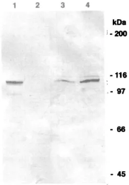
Expression of glucosidase II in bacteria, CHO cells and
mutant mouse lymphoma PHAR2.7 cells
Extracts of transformed bacteria and of stably transfected CHO
cells were assayed for glucosidase n activity. In bacteria, no
enzymatic activity could be measured although glucosidase n
immunoreactivity was detectable by Western blotting (not
shown). In CHO cells transfected with pcDNA3-glu n , the
enzymatic activity was increased up to threefold compared to
untransfected and mock-transfected cells (Table I) and this was
associated with enzyme protein amounts (Figure 8).
Transfec-tion of mutant PHAR2.7 cells with a vector containing the
cDNA for glucosidase II failed.
Discussion
Glucosidase n plays a key role in the processing of N-linked
oligosaccharide chains of glycoproteins and seems to be
in-volved in the ER quality control mechanism of glycoproteins.
Using degenerated oligonucleotides based on the amino acid
sequences from purified pig liver glucosidase n, a 1.2 kb
cDNA fragment could be amplified by RT-PCR, which was
used for the screening of a pig liver cDNA library to obtain a
full-length clone. Six different clones were identified, and two
of them were used for the construction of a full-length cDNA
consisting of a single open reading frame of about 2.9 kb which
coded for the entire glucosidase n enzyme. Several lines of
evidence indicated the authenticity of the cDNA clone. All
peptides obtained by protein sequencing were encoded in the
open reading frame. Further, expression of the cDNA in CHO
cells resulted in threefold overexpression of an active enzyme.
Western blot analysis showed an immunoreactive band at
about 100 kDa, which is in agreement with our earlier
bio-chemical data (Brada and Dubach, 1984). Notably, expression
in bacteria resulted in the synthesis of enzymarically inactive,
glucosidase II immunoreactive protein.
The open reading frame encodes a polypeptide of 944 amino
acids with a pi of 5.5 and a deduced molecular mass of 106
kDa. Further, the potential N-glycosylation site is apparently
used in vivo since glucosidase II was shown to carry a single
N-linked oligosaccharide of the high mannose-type (Strous et
al., 1987). The N-terminus contains an arginine-rich, cleavable
signal sequence of 32 amino acid residues. This signal
se-quence is unusual in its length but contains the recognition site
for the signal peptidase. Kyte and Doolittle hydropathy plot
analysis of the sequence did not reveal any hydrophobic region
which could serve as transmembrane domain. Further, the
ab-sence of a double lysine motif at the C-terminus (Jackson et
al., 1990) and of a double arginine motif at the N-terminus
(Schulze et aL, 1994) characteristic of ER transmembrane
pro-teins, strongly indicates that the purified glucosidase II protein
is not a proteolytic fragment (Brada and Dubach, 1984). On the
other hand, the C-terminal part of the deduced amino acid
sequence did not contain the KDEL ER retention signal or
1
•o100
200 300
400
4.00-
2.00-
o.oo-
-2.00-
-4.00-i -4.00-i L l !''
1 i i
M .11} li
J
• i i
liMliliiiJj
I " 1 ' " 1 ' I
F
f
l i l i
l . . I
j liJlLU. IJM
¥ «
iiiil i
1
y
h
r
500 600
700
800
900
Fig. 3. Hydrophilicity plot of the amino acid sequence of glucosidase IL The hydrophobicity profile was calculated by the Kyte-Doolittle method with a window size of seven amino acids (Kyte and Doolittle, 1982).
Cloning and expression of giucosJdase II
S a.
2 E
<Den n .2
I 2
g S>
2
<5
•4.4 kb— 9.5
kb
— 6.2
— 3.9
— 2.8
— 1.9
— 0.9
Fig. 4. Northern blot analysis of pig liver. Northern blots containing 50 (j.g of total and 1 jjtg mRNA from pig liver were hybridized with a radiolabeled glucosidase II cDNA fragment revealing a message size of approximately 4.4 kb.
variants thereof characteristic of soluble ER glycoproteins
(Munro and Pelham, 1987; Pelham, 1995). This is in good
accordance with our earlier biochemical results indicating that
glucosidase II is a loosely membrane-associated, luminally
ori-ented glycoprotein (Brada and Dubach, 1984). The recent
suc-cessful cloning of glucosidase I clearly demonstrated tiiis first
acting trimming enzyme to be a transmembrane type II
glyco-protein (Kalz et ai, 1995). In contrast, glucosidase II, the
sec-ond acting trimming enzyme, seemingly belongs to another
class of ER resident proteins and may be retained by a different
mechanism. Another example for such a type of glycoprotein is
lysyl hydroxylase (Kellokumpu et ai, 1994) an enzyme
in-volved in collagen processing, which neither contains a
C-terminal KDEL sequence nor a double lysine motif (Hautala et
ai, 1992). The mechanism by which glucosidase II and lysyl
hydroxylase are retained in the ER is unknown. It is tempting
to speculate that this is achieved upon interaction with another
ER protein and experiments will be performed to clarify this
important aspect. When our manuscript was under review, we
became aware of the work of Trombetta et al. (1996) on
glu-cosidase II purified from rat liver. These authors reported
pres-ence of two tightly bound proteins in the glucosidase II activity
containing fractions obtained from MonoQ column with
ap-parent molecular weights of 110 kDa and 80 kDa, respectively.
They proposed that glucosidase II is an heterooligomer with
the larger protein being catalytically active glucosidase II. The
smaller, noncatalytic protein contained the HDEL sequence
and was proposed to be responsible for glucosidase II ER
retention. Under our experimental conditions, we never
ob-served copurification of such an 80 kDa protein from pig
kid-ney and liver (Brada and Dubach, 1984; M.Ziak, unpublished
observations). All our evidence points to glucosidase II
puri-fied from these tissues as being composed of identical subunits
of 100 kDa. It remains to be demonstrated if the 80 kDa HDEL
containing rat liver protein represents truly a glucosidase II
subunit (Trombetta et al., 1996) or rather a protein generally
GAPOH
Fig. 5. Northern blot analysis of pig tissues. Northern blots containing 50 (i.g of total RNA from various pig tissues were hybridized with a radiolabeled glucosidase II cDNA fragment and showed a transcript of the same size as in liver. The expression levels varied in the different tissues.
=* 5
Si.-kDa
-200
-116
- 97
- 66
- 45
Fig. 6. Western blot analysis of pig tissues. Western blot analysis of electrophoretically resolved extracts (100 u,g) from liver, heart, and brain revealed differences in glucosidase II protein amounts.
T.Flura et al I O 00 I*;
ni
DC X Q.B
m
oi
CO < Q.kDa
-200
-116
- 97
- 66
kb
-9.5
-6.2
-3.9
-2.8
-1.9
-0.9
kDa
-200
-116
- 97
- 66
- 45
Actin
Fig. 7. Mutant lymphoma cell line PHAR 2.7 is deficient in glucosidase II protein and message. The mutant mouse lymphoma cell line PHAR 2.7 has been previously shown to be deficient in glucosidase II activity. (A) Western blot analysis of electrophoretically resolved extracts from the parental BW 5147 cells shows a glucosidase II immunoreactive band that is undetectable in the mutant PHAR 2.7 cells. (B) Northern blot analysis of total RNA reveals the presence of mRNA encoding glucosidase D in the parental BW 5147 cells which is absent in the mutant PHAR 2.7 cells.
functioning in the ER retention of glucosidase n , lysyl
hydrox-ylase, and related yet unknown ER proteins.
Computer assisted comparison of neutral trimming
glucosi-dase II amino acid sequence with other deposited sequences
revealed that this enzymes is highly conserved from yeast to
Table L Glucosidase II activity in cell lysate
CHO-K1 wt" CHO-pcDNA4* CHO-pcDNA3-GluIP Clone 23 Clone 24 Gone 25 Clone 26 Clone 27 Clone 2 Clone 4 Clone 6 Clone 8 Clone 10 Clone 12 Clone 14 Clone 16 Clone 18 Clone 20 GluII-activity mU/mg protein 21.3 24.1 20.1 22.0 21.2 17.5 24.6 31.9 38.6 36.9 32.7 50.4 28.3 31.0 35.9 34.1
*CHO-K1 wt represents a polyclonal cell population and the transfected cell lines are clonal.
- 45
Fig. 8. Overexpression of glucosidase n in CHO cells. Extracts of CHO cells transfected with pcDNA3-gluII (lane 1, 10 u.g protein) or pcDNA3 (lane 2, 10 p.g protein) and extracts from pig liver (lane 3, 10 p.g protein; lane 4, 50 jig protein) were analyzed by Western blotting.
mammals. Furthermore, it revealed a striking homology to
ly-sosomal acidic a-glucosidase, sucrase-isomaltase, and several
yeast glucosidases. The sequence similarity between lysosomal
a-glucosidase and both subunits of the intestinal
sucrase-isomaltase enzyme complex was demonstrated previously
(Hoefsloot et al., 1988). Apparently, the enzymes comprise a
group whose members contain conserved single amino acids or
clusters throughout the sequence. Homologous amino acids are
present most frequently in the middle and C-terminal parts of
the sequences. The homology is low in the N-terminal part
apparently reflecting the fact that the enzymes are located in
different cellular organelles. Comparison of the deduced amino
acid sequence of glucosidase I (Kalz et al., 1995) with that of
glucosidase II failed to reveal sequence similarities. Likewise,
the mammalian a-mannosidases exhibited no sequence
simi-larities (Bischoff et al., 1990; Moremen and Robbins, 1991;
Bause et al, 1993). Thus, the two trimming glucosidases,
which differ in their substrate specificity, are apparently coded
for by evolutionary unrelated genes. Currently, nothing is
known about the sequence of the active site of glucosidase II.
The active sites of the lysosomal a-glucosidase and the
su-crase-isomaltase have been shown to consist of the sequence
DMNE. Glucosidase II contains this amino acid sequence, and
it is very likely that it represents part of the active site. This can
now be tested by in vitro site-directed mutagenesis.
Glucosidase II has been implicated in the ER quality control
for newly synthesized glycoproteins but thus far the evidence
is indirect. The availability of the full length cDNA will permit
investigations to directly prove that the removal of a single
glucose residue by glucosidase II is an essential element of the
ER quality control system for glycoproteins.
Nonetheless, the availability of a glucosidase II cDNA has
already allowed to define the molecular basis of the enzyme
deficiency in the mutant mouse lymphoma PHAR2.7 cells. It is
622
Cloning and expression of gtucosidase II
expected that it will assist in the clarification of the
cell-type-specific variation in glucosidase II subcellular distribution
(Brada et al, 1987). In contrast to liver hepatocytes (Lucocq et
al., 1986), various kidney tubular epithelia exhibited
immuno-labeling for glucosidase II additionally in the Golgi apparatus,
the plasma membrane and a system of vesicular structures
involved in exo- and endocytosis (Brada et al, 1987).
Enzy-matically active and sialylated glucosidase II was detected in
plasma membrane (brush border) fractions, and evidence could
be obtained for a ligand for glucosidase II present in this
lo-cation.
Materials and methods
Preparation of protein sequence data
Glucosidase II from pig liver was purified to homogeneity as described by (Brada and Dubach, 1984) with modifications and using an FPLC system (Pharmacia, Uppsala, Sweden). The purified enzyme was digested by trypsin according to the manufacturer's instructions (Boehringer, Mannheim, Ger-many) and the Cryptic fragments separated by reverse-phase HPLC. In addition, partially purified glucosidase D was resolved by SDS^PAGE (4-15% gradient gels), transferred to PVDF-membrane and digested with endoprotease glu C (Boehringer, Mannheim, Germany). The purified peptides were subjected to automated Edman degradation.
Cloning of the cDNA
Standard techniques were performed as described (Sambrook, 1989). Protein sequences of the N-terminus of glucosidase II subunit and of peptide 4 were chosen for preparation of the sense and antisense degenerate oligonucleotides 5' -GTIGAT/CaAGIA/TG/CIAAT/CTTT/CAAA/G ACIC/TTCGAA/GGA-3' and 5' -GGIAA/GA/GTTA/GTGA/GTCA/GTCA/GAAICCT/CTGA/GTT-3', respectively). The degenerate primers were used in PCR amplification with a first-strand cDNA template obtained from pig liver poly(A) mRNA using Superscript II reverse transcriptase (GibcoBRL). A 1.2 kb PCR product was subcloned into pBluescript KS (Stratagene, La Jolla, CA) and sequenced using an automated sequencer from ABI. A (32P) labeled Pstl-PstI fragment of the PCR-product (760 bp) was then used to screen an unamplified pig liver cDNA library. The library prepared in Uni-ZAP XR vector (Zap-cDNA Synthesis Kit, Stratagene, La Jolla, CA) was ohgo(dT) primed; 10* pfu were screened and four independent clones of different sizes isolated. The clones were charac-terized by restriction mapping. Overlapping cDNA fragments were sequenced on both strands. Additional 5' end sequences upstream from the already re-ceived cDNA sequence were obtained by screening 2 x 10* pfu of a random primed Uni-ZAP library. The sequence data were analyzed and compared with the EMBL and the Swissprot data banks using the Wisconsin software package (Genetics Computer Group, Madison, WI).
Northern blot analyses
Total RNA was isolated by the single step method (Chomczynski and Sacchi, 1987) or with TRI Reagent (Chomczynski, 1993). Isolation of mRNA, prepa-ration of formaldehyde gels, labeling of cDNA fragments, and hybridizations in 50% formamide were carried out according to standard protocols (Sam-brook, 1989). "Oligolabeling" was performed according to Feinberg and Vo-gelstein (1983).
Western blot analysis and glucosidase II activity measurement
SDS-PAGE, Western blotting and enzyme activity measurements were per-formed as described previously (Brada and Dubach, 1984). For Western blot-ting, rabbit polyclonal antibodies raised against the denatured enzyme subunit were used (Brada and Dubach, 1984; Lucocoq et al, 1986).
Expression of the cDNA coding for glucosidase II in E.coli
The full-length cDNA was subcloned into EcoRI site of the expression vector pGEX-4T-l (Pharmacia, Uppsala, Sweden). Expression of the fusion protein, purification and digestion with thrombin were performed according to the manufacturer's instructions.
Cell culture and transfection of CHO and PHAR2.7 cells
The cell lines BW5147 and PHAR2.7 were kindly provided by Dr. L Trow-bridge (Salk Institute, San Diego, CA) and were grown in Dulbecco's modified
Eagle's medium containing 10% fetal bovine serum, penicillin (100 U/ml), and streptomycin (100 ng/ml) at 37°C and 10% CO2. CHO-K1 cells were obtained from ATCC.
The full-length cDNA coding for glucosidase II was constructed as follows. The cDNA fragment starting at the single PvuII site and containing the whole 3' end region inclusive poly(A) tail was ligated together with a cDNA frag-ment lying upstream of the PvuH site into the expression vector pcDNA3 (pcDNA3-gluIT). The cDNA fragment containing this PvuII site and the up-stream 5' end region was generated by RT-PCR. Both the mutant PHAR2.7 cells and CHO-K1 cells were transfected with pcDNA3-glu II using the lipo-fectamine method (Hawley-Nelson, 1993).
CHO cells were transfected with 5p.g of either pcDNA3 or pcDNA3-gluII using 50 pJ of lipofectamine according to standard protocol. Selection on G418 (1.5 u.g/u.1) was started 3 days after transfection. Clones were isolated using cloning rings (Sigma)
Acknowledgments
We arc grateful to Dr. ICMoremen for suggestions on designing the degenerate oligonucleotides and thank Dr. D.Zimmennann for DNA sequencing and Caroline Bornmann for performing transfection experiments. This study was supported by the Swiss National Science Foundation Grant 31-40754.94 and the Hartmann MUller-Stiftung filr Medizinische Forschung Zurich.
References
Bause.E., SchwedenJ., GrossA and Orthen.B. (1989) Purification and char-acterization of trimming glucosidase I from pig liver. Eur. J. Biochem., 183, 661-669.
BauseJE., Biebench.E., Rolfs,A., Volker.C. and Schmidt,B. (1993) Molecular cloning and primary structure of Man9-mannosidase from human kidney.
Eur. J. Biochem., 217, 535-540.
BergeronJJ., Brenner,M.B., ThomasJ).Y. and Williams.D.B. (1994) Caln-exin: a membrane-bound chaperone of the endoplasmic reticulum. Trends
Biochem. Set, 19, 124-128.
BischoffJ., Moremen.K. and Lodish.H.F. (1990) Isolation, characterization, and expression of cDNA encoding a rat liver endoplasmic reticulum alpha-mannosidase. / Biol Chem., 265, 17110-17117.
BradaJ). and Dubach.U.C. (1984) Isolation of a homogeneous glucosidase II from pig kidney microsorrjes. Eur. J. Biochem., 141, 149-156.
Brada,D., Kerjaschki.D. and RothJ. (1990) Cell type-specific post-Golgi ap-paratus localization of a "resident" endoplasmic reticulum glycoprotein, glucosidase U. J. CeU Biol, 110, 309-318.
BumsJD.M. and Touster.O. (1982) Purification and characterization of gluco-sidase n, an endoplasmic reticulum hydrolase involved in glycoprotein bio-synthesis. J. Biol Chem., 257, 9990-10000.
Chomczynski,P. (1993) A reagent for the single-step simultaneous isolation of RNA, DNA and proteins from cell and tissue samples. BwTechniques, 15, 532-537.
Chomczynski J>. and Sacchi,N. (1987) Single-step method of RNA isolation by acid guanidinium thiocyanate phenol-chloroform extraction. Anal
Bio-chem., 162, 156-159.
Feinberg.A.P. and Vogelstein3- (1983) A technique for radiolabeling DNA restriction endonuclease fragments to high specific activity. Anal Biochem., 132, 6-13.
Fernandezes., Trombetta,S.E., Hellman.U. and Parodi,AJ. (1994) Purifica-tion to homogeneity of UDP-glucose:glycoprotein glucosyltransferase from
Schizosaccharomyces pombe and apparent absence of the enzyme from Sac-charomyces cerevisiae. J. Biol Chem., 269, 30701-30706.
GrinnaX- and Robbins.P. (1979) Glycoprotein biosynthesis. Rat liver rrricro-somal glucosidases which process oligosaccharides. J. Biol Chem., 254, 8814-8818.
Hammond,C. and Helenius.A. (1994) Quality control in the secretory pathway: retention of a misfolded viral membrane glycoprotein involves cycling be-tween the ER, intermediate compartment, and Golgi apparatus. /. Cell Biol, 126, 41-52.
Hammond,C. and Helenius^A. (1995) Quality control in the secretory pathway.
Curr. Opin. CeU Biol, 7, 523-529.
Hammond,C, Braakman.L and Helenius^A. (1994) Role of N-linked oligosac-charide recognition, glucose trimming, and calnexin in glycoprotein folding and quality control- Proc. Nad. Acad. Set USA, 91, 913-917.
Hantala,T., Byers,M.G., Eddy,R.L., Shows.T.B., Kivirikko.K.L and Mylly-la,R. (1992) Cloning of human lysyl hydroxylase: complete cDNA-derived amino acid sequence and assignment of the gene (PLOD) to chromosome Ip36.3—p36.2.Genomics, 13, 62-69.
T.Flura et at
Helenius,A. (1994) How N-linked oligosaccharides affect glycoprotcin folding in the endoplasmic reticulum. MoL Biol. Cell, 5, 253—265.
Hertkamp.H., Legler.G. and BauseE. (1984) Purification by affinity chroma-tography of glucosidase I, an endoplasmic reticulum hydrolase involved in the processing of asparagine-linked oligosaccharides. Eur. J. Biochem., 142, 85-90.
Hoefsloot.L.H., Hoogeveen-Westerveld,M., Kroos,M.A., vanBeeumenJ., Reuser,AJ. and Oostra,B.A. (1988) Primary structure and processing of lysosomal alpha-glucosidase; homology with the intestinal sucrase-isomaltase complex. EMBO J., 7, 1697-1704.
Jackson,M.R., Nilsson.T. and PetersonJ'.A. (1990) Identification of a consen-sus motif for retention of transmembrane proteins in the eDdoplasmic re-ticulum. EMBO J., 9, 3153-3162.
Kalz,F.B., BieberichJE. and Bause,E. (1995) Qoning and expression of glu-cosidase I from human hippocampus. Eur. J. Biochem., 231, 344—351. Kaushal.G.P., PastuszakJ., Hatanaka,K. and Elbein,A.D. (1990) Purification
to homogeneity and properties of glucosidase II from mung bean seedlings and suspension-cultured soybean cells. J. Biol Chem., 265, 16271-16279. Kellokumpu.S., Sormunenji., HeikkinenJ. and Myllyla,R. (1994) Lysyl hy-droxylase, a collagen processing enzyme, exemplifies a novel class of lu-minally-oriented peripheral membrane proteins in the endoplasmic reticu-lum. J. Biol Chem., 269, 30524-30529.
Kornfeld,R. and Komfeld,S. (1985) Assembly of asparagine-linked oligosac-charides. Annu. Rev. Biochem., 54, 631-664.
KyteJ. and Doolittle,R.F. (1982) A simple method for displaying the hydro-pathic character of a protein. J. MoL Biol, 157, 105-132.
Labriola,C, CazzuloJ.J. and Parodi,AJ. (1995) Retention of glucose units added by the UDP-GLC:glycoprotein glucosyltransferase delays exit of gly-coproteins from the endoplasmic reticulum. J. Cell Biol, 130, 771-779. LucocqJ.M., Brada,D. and RothJ. (1986) Immunolocalization of the
oligo-saccharide trimming enzyme glucosidase n. / Cell Biol, 102, 2137-2146. MartiniukJ., Ellenbogen,A. and Hirschhom.R. (1985) Identity of neutral al-pha-glucosidase AB and the glycoprotein processing enzyme glucosidase IL Biochemical and genetic studies. J. Biol Chem., 260, 1238-1242. Moremen.K. and TousteT.O. (1988) Mannosidases in mammalian glycoprotem
processing. In Das,R. and Robbins J". (eds.), Protein Transfer and Organelle
Biosynthesis. Academic Press, San Diego, pp. 209-240.
Moremen.K.W. and Robbins,P.W. (1991) Isolation, characterization, and ex-pression of cDNAs encoding murine alpha-mannosidase n, a Golgi enzyme that controls conversion of high mannose to complex N-glycans. J. Cell
Biol, 115, 1521-1534.
Munro.S. and Pelham.R. (1987) A C-terminal signal prevents secretion of luminal ER proteins. Cell, 48, 889-907.
Parker.C.G., Fessler.L.I., Nelson,R.E. and FesslerJ.H. (1995) Drosophila UDP-glucose:glycoprotein glucosyltransferase: sequence and characteriza-tion of an enzyme that distinguishes between denatured and native proteins.
EMBO J., 14, 1294-1303.
Parodi.A.J., Mendelzon.D.H. and Lederkrcmer.G.Z. (1983) Transient gluco-sylation of protein-bound Man9GlcNAc2, Man8GlcNAc2, and Man7GlcNAc2 in calf thyroid cells. A possible recognition signal in the processing of glycoproteins. J. Biol Chem., 258, 8260-8265.
PelhamrH.R.B. (1995) Sorting and retrieval between the endoplasmic reticu-lum and Golgi apparatus. Curr. Opin. Cell Biol, 7, 530-535.
Reirman^M.L., Trowbridge.I.S. and Kornfeld.S. (1982) A lectin-resistant mouse lymphoma cell line is deficient in glucosidase n, a glycoprotein-processing enzyme. / Biol Chem., 257, 10357-10363.
Roth.J. (1995) Compartmentation of glycoprotcin biosynthesis. In Mon-treuilj., VliegenhartJ.F.G. and Schachter.H. (eds.), New Comprehensive
Biochemistry. Glycoproteins. Elsevier Press, Amsterdam, pp. 287-312.
SambrookJ., FritschJEJ. and Maniatis.T. (1989) Molecular cloning: A
Labo-ratory Manual Cold Spring Harbor LaboLabo-ratory Press, Cold Spring Harbor,
NY.
Saunier.B., Kilker^R.D., TkaczJ.S., Quaroni^A. and Herscovics^A (1982) In-hibition of N-linked complex ougosaccharide formation by 1-deoxynojiri-mycin, an inhibitor of processing glucosidases. /. BioL Chem., 257,
14155-14161.
SchulzeJ^.P., Peterson,P.A. and Jackson,M.R. (1994) An N-terminal double-arginine motif maintains type II membrane proteins in the eodoplasmic reticulum. EMBO J., 13, 1696-1705.
SchwedeaJ., Borgmann,C, Legler.G. and Bause,E. (1986) Characterization of calf liver glucosidase I and its inhibition by basic sugar analogs. Arch.
Biochem. Biophys., 248, 335-340.
Shailubhai.K., PrattaJtf.A. and VijayJ.K. (1987) Purification and character-ization of glucosidase I involved in N-linked glycoprotein processing in bovine mammary gland. Biochem. J., 247, 555-562.
Sousa>4. and Parodi^AJ. (1995) The molecular basis for the recognition of
misfolded glycoproteins by the UDP-Glc:glycoprotein glucosyltransferase.
EMBO J., 14, 4196-^203.
SousaJU.C, Ferrero,G.M.A. and Parodi^J. (1992) Recognition of the oligo-saccharide and protein moieties of glycoproteins by the UDP-Glc:glycoprotein glucosyltransferase. Biochemistry, 31, 97-105.
Spiro,R.G., Zhu.Q., Bhoyroo.V. and SohngJI.D. (1996) Definition of the lec-tin-like properties of the molecular chaperone, calreticulin, and demonstra-tion of its copurificademonstra-tion with endomannosidase from rat liver Golgi. J. BioL
Chem., 271, 11588-11594.
Strous.GJ., vanKerkhofJ1., Brok,R., RothJ. and Brada,D. (1987) Glucosidase D, a protein of the endoplasmic reticulum with high mannose oligosaccha-ride chains and a rapid turnover. J. Biol. Chem., 262, 3620-3625. Trombetta,S.E. and Parodi.AJ. (1992) Purification to apparent homogeneity
and partial characterization of rat liver UDP-glucose:glycoprotein glucos-yltransferase. /. BioL Chem., 267, 9236-9240.
Trombetta,S.E., Bosch,M., and Parodi^AJ. (1989) Glucosylation of glycopro-teins by mammalian, plant, fungal, and trypanosomatid protozoa micro-somal membranes. Biochemistry, 28, 8108—8116.
Trombetta.S.E., Ganan.S.A. and Parodi.A.J. (1991) The UDP-Glc:glycoprotein glucosyltransferase is a soluble protein of the endoplasmic reticulum. Gtycobiology, 1, 155—161.
Trombetta,S.E., SimonsJ.F. and Helenius,A. (1996) Endoplasmic reticulum glucosidase II is composed of a catalytic subunit, conserved from yeast to mammals, and a tightly bound non-catalytic HDEL-containing subunit. J.
BioL Chem., Ill, 27509-27516.
Ugalde.R.A., StaneloniJU. and Leloir.L.F. (1980) Microsomal glucosidases of rat liver. Partial purification and inhibition by disaccharides. Eur. J.
Biochem., 113, 97-103.
Ware,F.E., VassilakosA, PetersonJ'.A., JacksonM-R-, Lehrman>l.A. and Williams,D.B. (1995) The molecular chaperone calnexin binds Glc(l)Man(9)GlcNAc(2) oligosaccharide as an initial step in recognizing unfolded glycoproteins. /. Biol. Chem., 270, 4697^704.
Received on October 25, 1996 revised on December 3, 1996; accepted on January 7, 1997