Radiology
L A B O R A T O R Y
I N V E S T I G A T I O N S
A New Rotational Thrombectomy Catheter:
System Design and First Clinical Experiences
Hans-Erich Schmitt, 1 Kurt A. J iger, 2 Augustinus L. Jacob, 1 Helmuth Mohr, 3
Karl-Heinz Labs, 2 Wolfgang Steinbrich 1
JDepartment of Diagnostic Radiology, University Hospital Basel, Kantonsspital, CH-4031 Basel, Switzerland
2Department of Medicine, Division of Angiology, University Hospital Basel, Kantonsspital, CH-4031 Basel, Switzerland 3Straub Medical, Straubstrasse, CH-7323 Wangs, Switzerland
Abstract
Purpose: To describe a new catheter for the percutaneous mechanical removal of fresh and organized thrombi, and to assess its efficacy and safety in vitro and in vivo.
Methods: The catheter consists of a coated stainless steel spiral that rotates at 40,000 rpm over a guidewire inside the whole length of an 8 Fr, single-lumen, polyurethane catheter, driving a dual-blade cutting crown. Abraded occlusion ma- terial is sucked into the catheter head through distal side holes and transported by the spiral into a reservoir at the proximal end. The efficacy of the device was tested in arterial models and fresh bovine carotid arteries (n = 72). In a clinical pilot study 10 patients (8 women, 2 men; mean age 70.6 ___ 10.1 years) with occlusions of the superficial femoral artery (2-12 cm, mean 5.8 cm), not older than 4 weeks, underwent thrombectomy with the new catheter.
Results: In arterial models and bovine cadaver arteries the catheter completely removed fresh thrombi. Occlusion ma- terial of higher consistency was cut into particles of 100-500 /xm and transported outside. Thrombectomy was successful and vessel patency restored in all 10 patients. The ankle/ brachial pressure index significantly (p < 0.0005) in- creased from 0.41 _+ 0.18 before intervention to 0.88 _+ 0.15 after 48 hr and to 0.84 ___ 0.20 after 3 months. Two reocclu- sions occurred within 14 days after the intervention. Conclusion: Thrombectomy with the new device appears to be feasible and safe in patients with acute and subacute occlusions of the femoropopliteal artery.
Key words: T h r o m b e c t o m y - - C a t h e t e r - - T h r o m b o s i s - - Femoropopliteal artery
Correspondence to: H.E. Schmitt, M.D., Rottmannsbodenstrasse 90, CH- 4102 Binningen, Switzerland
The drawbacks of conventional nonsurgical treatment of thrombotic arterial occlusions, such as long procedure time, bleeding, vessel wall injury and peripheral embolization, have stimulated the development of devices for mechanical removal of thrombi in the last decade [1-3]. Several systems have entered clinical practice, while others were described as prototypes. Each device has its own limitations, including restriction to fresh thrombi only, incomplete clot removal, vessel walt damage, or design complexity. The ideal instru- ment should be wire-guided to avoid vessel perforation, remove fresh and organized thrombi, and transport them to the outside without risk of peripheral embolization. With this aim in view a rotational catheter was constructed and is described here for the first time.
Materials and M e t h o d s
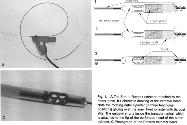
The system has three components: the Rotarex catheter (Straub Rotarex, patent pending, Straub Medical, Wangs, Switzerland), a 40 W DC electric motor drive, and an electronic control unit (Fig. 1A). Inside the whole length of the 8 Fr polyurethane catheter rotates a coated stainless steel spiral, which glides over a 0.020-inch guidewire (Schneider Europe, Btilach, Switzerland). The catheter head consists of two cylinders that fit over each other (Fig. 1B, C). The outer rotating cylinder is fixed to the spiral, the inner one is attached to the catheter shaft. Each cylinder has two oval slits. The blunt tip of the outer cylinder is perforated for the guidewire. Catheter and motor drive are connected by a magnetic clutch. The motor rotates the spiral at 40,000 rpm, resulting in 80,000 cuts/min. The high fi'equency of revolution creates a negative pressure at the catheter head of 5.8 kPa (=43.5 mmHg). When the catheter is activated, soft and solid occlusion material is caught in the slits, transported by the spiral to the proximal sideport, and discharged into a plastic bag. No additional suction is required. The transport of the occlusion material is done exclusively by the rotating spiral. The catheter is for one-time use, while the motor drive and the connecting cable to the electronic control unit can be sterilized.
A
/
/
1 Guide wire
Rotating cyhnder e dec
Catheter shaft / /
3 B
Spwat
Fig. 1. A The Straub Rotarex catheter attached to the motor drive. B Schematic drawing of the catheter head. Note the rotating outer cylinder (in three functional positions) gliding over the inner fixed cylinder with its oval slits. The guidewire runs inside the transport spiral, which is attached to the tip of the perforated head of the outer cylinder. C Photograph of the Rotarex catheter head.
Preclinical Studies
The catheter was tested in an arterial model made of silicon tubing and in fresh bovine carotid arteries. Translucent silicon tubing of 4, 6, and 8-mm inner diameter and length 15-30 cm served as an arterial model, allowing the observation and the video documenta- tion of the catheter function. The tubes were bendable to test the behavior of the catheter at various angles. The tubing was filled with occlusion material of different consistencies: (a) bovine blood that was allowed to coagulate for 48 hr or 2 weeks and stored at 4~ (b) stamp cylinders of "black pudding", a mixture of throm- bus, muscle, fat, and connective tissue, 4, 6, and 8 mm thick, and 15 cm long, simulating organized thrombus; (c) strips of bovine arteries (3 cm long, 1 mm broad) sutured intratuminally to the vessel wall, imitating intimal flaps. The tubing was clamped at one end and connected to an infusion line on the other, where a mixture of saline and glycerine could be infused under a continuous pres- sure of 120 mmHg. In this infusion tube an 8 Fr sheath (Cook, Bjaerverskov, Denmark) was introduced, a 0.020-inch Teflon coated guidewire threaded through the occlusion material, and the catheter advanced.
In a second test series (n = 72) the silicon tubing was replaced by fresh bovine carotid arteries with an average length of 20 cm and a diameter of 6 - 7 mm. The side branches were ligated and the arteries filled with the above-mentioned occlusion material. During catheter activation tubes and arteries were either kept straight or bent in different angles up to 50 ~ . Occlusion of the arteries was achieved by tightening a 5-mm-wide rubber band around the vessel, completely interrupting the flow of perfusate. The occlusion was
passed with the catheter over the wire. All tests were recorded on video. The time of catheter activation was recorded, the volume of the aspirated fluid measured, and the fluid passed through different filters for analysis of particle size. The arteries were cut open and examined visually and histologically for remnants of occlusion material and possible intimal damage.
Clinical Studies
Based on the results of the preclinical tests the institutional review board of the Department of Internal Medicine, University Hospital Basel, accepted the protocol of a pilot study for the evaluation of the device for the treatment of thrombotic occlusions in femoro- popliteal arteries in humans. Ten patients (8 women, 2 men; age 5 8 - 8 7 years, mean 70.6 _+ 10.1 years) with acute or subacute occlusion of the femoropopliteal artery with an estimated age of less than 4 weeks and patent proximal segments of lower leg vessels, were included in the study. All patients were informed in detail about the procedure and gave their written consent. Patients with aneurysms of the popliteal artery, severe coagulation distur- bances or a history of adverse reactions to contrast media were excluded.
Seven patients suffered from critical ischemia (rest pain) and three from peripheral arterial occlusive disease (PAOD) stage II (intermittent claudication). The estimated age of the lesions was between 2 and 28 days. The mean length of the occluded segments was 5.8 cm (range 2-15 cm). The diagnosis was established by clinical examination, oscillography, Doppler pressure recordings,
duplex sonography [4], and digital subtraction arteriography. Lab- oratory examinations included hemoglobin, free plasma arteriogra- phy, hematocrit, and coagulation parameters, both before, immediately after, and 24 hr after thrombectomy. Noninvasive examinations (oscillography, Doppler pressure recordings, duplex sonography) were performed after 48 hr and 3 months. Antegrade arteriography was performed via the common femoral artery to determine the length of occlusion, and to document the collateral circulation and the peripheral runoff.
Using the road map technique the occlusion was passed with a Teflon-coated 0.020-inch guidewire and its flexible tip placed in the distal popliteal artery. An 8 Fr sheath was introduced and 5000 U of heparin injected. The thrombectomy catheter was threaded over the guidewire. One centimeter proximal to the upper end of the occlusion the catheter was activated by a foot switch and advanced through the occlusion in gentle forward and back movements under fluoroscopic control. The continuous suction of blood and occlusion material into the reservoir bag was observed. After the catheter had passed the occlusion it was withdrawn in the proximal femoral artery and angiography was repeated via the side port of the sheath. Depending on the result, thrombectomy was either terminated, repeated, or completed by percutaneous transluminal angioplasty (PTA). The number and the duration of catheter passes and the volume of aspirated fluid were recorded. Extracted occlusion ma- terial was examined histologically.
Results
Preclinical Studies
The catheter removed 48-hr-old thrombi in silicon tubing of 4 and 6 m m diameter completely and regularly. The maxi- mum working diameter was 8 mm. On average 1 cm of thrombus was aspirated within 2 sec. Thrombi stored in the refrigerator for 2 weeks often showed adherence to the wall of the tubing, but could also easily be aspirated, leaving occasionally a thin residual layer attached to the inner cur- vature if the tube was bent. Occlusion material of higher and inhomogeneous consistency, such as muscle, fat, and con- nective tissue, was caught readily in the catheter head and transported to the outside by the spiral. Fluid was aspirated at a rate of 1.5 ml/sec. While fresh thrombus was completely homogenized, more solid material was cut into particles of 100-500 /xm. Strips of bovine arteries, sutured intralumi- nally to the vessel wall to imitate intimal flaps, were cut and fragmented, leaving only small stumps at the suture site. Bovine arteries filled with thrombi or "black pudding" could be cleared of the occlusion material without remnants. No
intimal damage was noticed on visual examination or after staining. When the arteries were compressed to complete occlusion by a rubber band, the catheter drilled a lumen corresponding to its shaft size. When tubing or arteries were bent, the catheter followed the guidewire smoothly. No per- foration was noticed.
The catheter needs fluid (blood) for lubrication. Under experimental conditions the temperature of the catheter head rose by t.5~ after rotating for 4 min at 40,000 rpm in a tube of 3-mm diameter perfused with a saline-glycerine solution (80 ml/min) at room temperature.
Clinical Studies
Thrombectomy was technically successful in all patients (Figs. 2, 3). A mean of 2.4 catheter passes (range 2-3) were required to recanalize the occluded segment, and 2.8 sec on average were needed to open 1 cm of occlusion. Blood and detritus were aspirated at 1.1 ml/sec; the total aspirated volume ranged from 20 to 90 ml, depending on the length and consistency of the lesion. In nine patients an underlying residual stenosis was treated by PTA after the thrombec- tomy. Forty-eight hours after the intervention no patient suffered from critical ischemia: eight patients were classified as PAOD stage I, and 2 patients as stage II. Ankle/brachial index (ABI) improved from 0.41 ___ 0.18 (range 0.13-0.65) preinterventionally to 0.88 __- 0.15 (range 0.64-1.11) after the intervention (p < 0.0005). Three months after the intervention the mean ABI was 0.84 + 0.20 (range 0.52- 1.07).
All patients received either oral anticoagulation or anti- platelet drugs and were assessed by duplex sonography 48 hr and 3 months after the intervention. The treated arterial segments remained patent in eight patients. Two patients had a reocclusion within 2 weeks after the intervention. In a 60-year-old woman with an occlusion 3 cm long in a smoothly outlined superficial femoral artery of 5-mm diam- eter and with three patent lower leg vessels, fresh thrombus was removed with the Rotarex catheter in two passes of 7 sec. Complete vessel patency was restored, no peripheral embolism occurred, and additional PTA was not considered to be indicated. As usual the patient received 5000 U of heparin during the intervention and oral anticoagulation af- terwards. After 48 hr duplex sonography revealed a reocclu- sion. At that time coagulation parameters were not in the
y
Fig. 2. A Angiogram of a 63-year-old woman with a history of percutaneous transluminal angioplasty (PTA) of a short subtotal occlusion of the superficial femoral artery (SFA) 3 months previously. She had been exhibiting signs of critical ischemia for 8 days, the ankle/brachial index (ABI) was 0,14 and there was a 6-cm-long occlusion of the distal SFA. B After two passes with the thrombectomy device (2 x 10 sec) vessel patency has been restored; there is residual stenosis at 16 cm. C After additional PTA the stenosis was removed. The ABI was 0.91.
Fig. 3. A Angiogram of a 78-year-old diabetic woman with rest pain. She had a 12-cm-long occlusion of the popliteal artery and of the tibiofibular trunk. B The artery was patent after one pass with the Rotarex catheter. C After additional PTA of the tibiofibular trunk and the anterior tibial artery.
therapeutic range, which might have contributed to the re- thrombosis. The ABI, which improved from 0.61 to 0.71 after the intervention, remained at this level. The patient was classified as PAOD stage II without the need for a reinter- vention. The second patient was a 78-year-old diabetic woman with a history of claudication of several years, rest pain for 12 days and a 15-cm-long occlusion of the popliteal artery with poor lower leg outflow. Thrombectomy was primarily successful and complete vessel patency was re- stored after additional PTA. The ABI rose from 0.65 to 0.95 and duplex sonography showed a residual stenosis of less than 50% diameter reduction. One week after hospital dis- charge the patient complained of recurrence of intermittent claudication and a reocclusion was documented. Since the patient was clinically in PAOD stage II no further interven- tion was performed. Reduced outflow due to pre-existing lower leg artery occlusions was considered as a possible cause for rethrombosis.
No patient had signs of mechanical damage to red blood cells. There was no increase in free plasma hemoglobin, nor relevant changes in hemoglobin and hematocrit after throm- bectomy.
Histologic examination of the aspirate showed mostly fresh thrombotic material and small components of fibrotic intima with sclerotic fragments. No serious complications occurred. In one patient a small embolus was detected an- giographically within the proximal posterior tibial artery and lysed successfully by 100,000 units of urokinase.
Discussion
In the last decade great efforts have been made to provide an alternative to time-consuming local fibrinolysis and surgical thrombectomy in the treatment of intravascular thrombosis. Percutaneous aspiration thrombectomy through thin-walled, straight catheters is still a common, readily available, simple and cost-effective technique for the removal of fresh thrombi [5]. Its restriction to loose, not adherent thrombi, the neces- sity of usually several passes, and the risk of plaque avulsion and antegrade dissection have, however, led to the construc- tion of more sophisticated devices [6]. The experience with the first catheter equipped with a fast-rotating, coaxially driven cam (Trac-Wright, formerly Kensey) was disappoint- ing. The device was, however, the predecessor of several generations of rotational catheters [7, 8]. These can roughly be classified into devices using recirculation, e.g., pulveri- zation of thrombi by a hydrodynamic vortex created either by a high-speed impeller [9-12] or the Venturi effect [13, 14], and nonrecirculation catheters with concomitant suc- tion, using either a rotating recessed propeller [15, 16] or rotating cutting blades [17, 18] and aspiration via a roller pump or any other suction modality [19]. These devices are effective in clearing fresh thrombi, especially in arterial and hemodialysis grafts; however, their effectiviness decreases with the age of thrombi and their adherence to the vessel wall. Since many occlusions are "acute on chronic" throm-
boses, a thrombectomy device should have the potential to remove not only fresh but also underlying organized throm- bus. Furthermore it should track over a guidewire in order to prevent vessel perforation. Finally it should transport the occlusion material to the outside without the risk of periph- eral embolization.
The new catheter described here meets many of these requirements. Its construction is relatively simple, using only one lumen for spiral and coaxial guidewire. No extra chan- nels for suction, lubrication, or cooling are needed. The transport of the occlusion material is done exclusively by the spiral. Some devices are easily obstructed by sticky occlu- sion material, especially by fibrin, which has to be trans- ported through narrow catheters over a distance of 80-100 cm. In the new catheter this drawback was eliminated by a special coating of the spiral and by adjusting the rotational speed to 40,000 rpm. As with all rotating devices heat caused by friction can be a problem [20]. The new device needs blood for lubrication. Therefore it should not be advanced continuously in an occlusion, but in gentle forward and back movements, allowing the uninterrupted aspiration of blood and avoidance of running dry. Provided these precautions were observed, no undue warming of the catheter was noted. Average time for thrombectomy was short and did not ex- ceed 90 sec for occlusions up to 15 cm in length. Blood loss during thrombectomy is generally low, amounting to 80-90 ml/min depending on the composition of the occlusion ma- terial that has to be transported and the blood available in the artery.
A useful parameter for the assessment of the efficacy of a thrombectomy catheter is the radial expansion coefficient, i.e., the ratio of the lumen recanalized by thrombectomy to the catheter diameter. For the new catheter the coefficient is 2 in vitro, meaning that the catheter with a diameter of approximately 3 mm clears a thrombus of 6 mm. In vivo the radial expansion coefficient depends on the composition of the occlusion material. It is about 2 in fresh thrombi but drops to 1 in solid material, which means that the reopened lumen corresponds to the diameter of the catheter head. Experimental data suggest the use of the catheter for the treatment of obstructed vessels with a diameter up to 8 mm. This study showed its feasibility and efficacy in superficial femoral arteries of 4 - 7 mm diameter. Its applicability in larger vessels, such as pelvic arteries, vena cava, or pulmo- nary arteries, needs to be validated in further trials.
Compared with other thrombectomy devices the potential of the Rotarex catheter to remove not only loose thrombus but also solid, organized occlusion material must be consid- ered an advantage. The catheter passes calcified plaques without abrading them. Therefore, adjunctive PTA after thrombectomy is often advisable to eliminate residual steno- sis. As shown in arterial models the wire-guided catheter removes thrombus without intimal abrasion if the original caliber of the artery is larger than the catheter head. Conse- quently the catheter in the 8 Fr version is not suitable for
thrombectomy in vessels distal to the popliteal artery. For use in small vessels a 5 Fr version is in preparation.
Hemolysis occurs in recirculation catheters without aspi- ration, but its clinical effect seems minimal [21 ]. In our study no change in free plasma hemoglobin was noticed after thrombectomy. Except for a small embolus in a posterior tibial artery, which was removed by local fibrinolysis, no relevant complication occurred. It can be assumed that the negative pressure built up by the catheter system is sufficient to avoid peripheral embolization. The majority of our pa- tients suffered from "acute on chronic PAOD" and thus from thrombotic and not embolic occlusions.
As expected from the preclinical tests, the catheter was easy to handle and helped to save considerable time by shortening thrombectomy to a few minutes and avoiding lengthy procedures such as local fibrinolysis or surgical intervention. To document the efficacy and safety of the device further, a larger-scale multicenter study is required and is in progress.
References
1. Sharafuddin MJA, Hicks ME (1997) Current status of percutaneous mechanical thrombectomy. I. General principles. J Vasc Interv Radiol 8:911-921
2. Sharafuddin MJA, Hicks ME (1998) Current status of percutaneous mechanical thrombectomy. II. Devices and mechanisms of action. J Vasc Interv Radiol 9:15-31
3. Sharafuddin MJA, Hicks ME (1998) Current status of percutaneous mechanical thrombectomy. III. Present and future applications, J Vasc Interv Radiol 9:209-224
4. J~ger K, Frauehiger B, Eichlisberger R (1996) Ultrasonographic inves- tigation of the peripheral arteries. In: Tooke JE, Lowe GDO (eds) A Textbook of Vascular Medicine. Oxford University Press, New York, pp 84-89
5. Wagner H-J, Starck E (1992) Acute embolic occlusions of the infrain- guinal arteries: Percutaneous aspiration embolectomy in 102 patients. Radiology 182:403-407
6. Brossmann J, Miiller-Htilsbeck S, Heller M (1998) Perkutane Throm- bektomie und mechanische Thrombolyse. Fortschr Rontgenstr 169: 344 -354
7. Kensey KR, Nash JE, Abrahams C, Zarins CK (1987) Recanalization of obstructed arteries with a flexible, rotating tip catheter. Radiology
165:387-389
8. Triller J, Do DD, Maddern G, Mahler F (1992) Femoropopliteal artery occlusion: Clinical experience with the Kensey catheter. Radiology 182:257-261
9. Bildsoe MC, Moradian GP, Hunter DW, Castafieda-Zdfiiga WR, Am- platz K (1989) Mechanical clot dissolution: A new concept. Radiology 171:231-233
10. Pozza CH, Gomes MR, Qian Z, Ambrozaitis R, Urness M, Amplatz K (1994) Evaluation of the newly developed Amplatz maceration and aspiration thrombectomy device using in vitro and in vivo models. AJR 162:139
11. Uftacker R (1997) Mechanical thrombectomy in acute and subacute thrombosis with use of the Amplatz device: Arterial and venous appli- cations. J Vasc Interv Radiol 8:923-932
12. Rilinger N, G6rich J, Scharrer-Pamler R, Vogel J, Tomczak R, Kr~imer S, Merkle E, Brambs H-J, Sokiranski R (1997) Short-term results with use of the Amplatz thrombectomy device in the treatment of acute lower limb occlusions. J Vasc Interv Radiol 8:343-348
13. Reekers JA, Kromhout JG, Van der Waal K (1993) Catheter for percutaneous thrombectomy: First clinical experience. Radiology 188: 871- 874
14. Bticker A, Schmitz-Rode T, Vorwerk D, Guenther RW (1996) Com- parative in vitro study of two percutaneous hydrodynamic thrombec- tomy systems. J Vasc Interv Radiol 7:445-449
15. Guenther RW, Vorwerk D (1990) A new aspiration thrombectomy catheter with propeller tipped rotating wire: In vitro study. J Interv Radiol 1 : 17-20
16. Guenther RW, Vorwerk D (1990) Aspiration catheter for percutaneous thrombectomy: Clinical results. Radiology 175:271-273
17. Yedlicka JW, Carlson JE, Hunter DW, Castafieda-Ztifiiga WR, Amplatz K (1991) Thrombectomy with the transluminal endarterectomy catheter (TEC) system. J Vasc Interv Radiol 2:343-347
18. Rilinger N, G6rich J, Scharrer-Palmer R, Vogel J, Tomczak R, Merkle E, Sokiranski R, Brambs H-J (1997) Percutaneous translu- minal rotational atherectomy in the treatment of peripheral vascular disease using transluminal endatherectomy catheter (TEC): Initial results and angiographic follow up. Cardiovasc Intervent Radiol 20:263-267
19. Miiller-Htilsbeck S, Schwarzenberg H, Bangard C, Steffens JC, Heller M (1998) Saugpumpenunterst0tzte Aspirationsthrombektomie: In vitro Vergleich mit einem Thrombusfragmentierungsverfahren. Fortschr Rontgenstr 162:191-194
20. Gehani AA, Rees MR (1998) Can rotational atherectomy cause thermal tissue damage? A study of the potential heating and thermal tissue effects of a rotational atherectomy device. Cardiovasc Intervent Radiol 21:481-486
21. Nazarian GK, Qian Z, Coleman CC, Rengel G, Castafieda-Zdfiiga WR, Hunter DW, Amplatz K (1994) Hemolytic effect of the Amplatz throm- bectomy device. J Vasc lnterv Radiol 55:155-160