The Pseudomonas aeruginosa Lectin LecB Causes Integrin
Internalization and Inhibits Epithelial Wound Healing
Roland Thuenauer,
a,b,c,dAlessia Landi,
a,bAnne Trefzer,
a,bSilke Altmann,
a,bSarah Wehrum,
a,bThorsten Eierhoff,
a,bBritta Diedrich,
e,fJörn Dengjel,
e,fAlexander Nyström,
fAnne Imberty,
gWinfried Römer
a,baFaculty of Biology, Albert Ludwigs University Freiburg, Freiburg, Germany
bSignalling Research Centers BIOSS and CIBSS, Albert Ludwigs University Freiburg, Freiburg, Germany
cAdvanced Light and Fluorescence Microscopy Facility, Center for Structural Systems Biology (CSSB), Hamburg, Germany dDepartment of Biology, University of Hamburg, Hamburg, Germany
eDepartment of Biology, University of Fribourg, Fribourg, Switzerland
fDepartment of Dermatology, Medical Center, Albert Ludwigs University Freiburg, Freiburg, Germany gUniv. Grenoble Alpes, CNRS, CERMAV, Grenoble, France
ABSTRACT
The opportunistic bacterium Pseudomonas aeruginosa produces the
fucose-specific lectin LecB, which has been identified as a virulence factor. LecB has
a tetrameric structure with four opposing binding sites and has been shown to act
as a cross-linker. Here, we demonstrate that LecB strongly binds to the glycosylated
moieties of
1-integrins on the basolateral plasma membrane of epithelial cells and
causes rapid integrin endocytosis. Whereas internalized integrins were degraded via
a lysosomal pathway, washout of LecB restored integrin cell surface localization, thus
indicating a specific and direct action of LecB on integrins to bring about their
en-docytosis. Interestingly, LecB was able to trigger uptake of active and inactive
1-integrins and also of complete ␣31-integrin–laminin complexes. We provide
a mechanistic explanation for this unique endocytic process by showing that LecB
has the additional ability to recognize fucose-bearing glycosphingolipids and causes
the formation of membrane invaginations on giant unilamellar vesicles. In cells, LecB
recruited integrins to these invaginations by cross-linking integrins and
glycosphin-golipids. In epithelial wound healing assays, LecB specifically cleared integrins from
the surface of cells located at the wound edge and blocked cell migration and
wound healing in a dose-dependent manner. Moreover, the wild-type P. aeruginosa
strain PAO1 was able to loosen cell-substrate adhesion in order to crawl underneath
exposed cells, whereas knockout of LecB significantly reduced crawling events.
Based on these results, we suggest that LecB has a role in disseminating bacteria
along the cell-basement membrane interface.
IMPORTANCE
Pseudomonas aeruginosa is a ubiquitous environmental bacterium
that is one of the leading causes of nosocomial infections. P. aeruginosa is able to
switch between planktonic, intracellular, and biofilm-based lifestyles, which allows it
to evade the immune system as well as antibiotic treatment. Hence, alternatives to
antibiotic treatment are urgently required to combat P. aeruginosa infections.
Lec-tins, like the fucose-specific LecB, are promising targets, because removal of LecB
re-sulted in decreased virulence in mouse models. Currently, several research groups
are developing LecB inhibitors. However, the role of LecB in host-pathogen
interac-tions is not well understood. The significance of our research is in identifying cellular
mechanisms of how LecB facilitates P. aeruginosa infection. We introduce LecB as a
new member of the list of bacterial molecules that bind integrins and show that P.
aeruginosa can move forward underneath attached epithelial cells by loosening
cell-basement membrane attachment in a LecB-dependent manner.
Citation Thuenauer R, Landi A, Trefzer A,
Altmann S, Wehrum S, Eierhoff T, Diedrich B, Dengjel J, Nyström A, Imberty A, Römer W. 2020. The Pseudomonas aeruginosa lectin LecB causes integrin internalization and inhibits epithelial wound healing. mBio 11:e03260-19.
https://doi.org/10.1128/mBio.03260-19.
Invited Editor Suzanne M. J. Fleiszig,
University of California, Berkeley
Editor Nina R. Salama, Fred Hutchinson Cancer
Research Center
Copyright © 2020 Thuenauer et al. This is an
open-access article distributed under the terms of theCreative Commons Attribution 4.0 International license.
Address correspondence to Roland Thuenauer, roland.thuenauer@cssb-hamburg.de, or Winfried Römer, winfried.roemer@bioss.uni-freiburg.de. Received 14 December 2019 Accepted 31 January 2020 Published
crossm
® 10 March 2020KEYWORDS
epithelial cells, fucose, glycosylation, integrins, laminin, membrane
invagination, bacterial infection, glycosphingolipids
P
seudomonas aeruginosa is a ubiquitous Gram-negative environmental bacterium.
For humans, it acts as an opportunistic pathogen and can cause severe infections,
predominantly in cystic fibrosis patients (1) and immunocompromised individuals, such
as HIV patients (2), patients receiving cancer treatment (3), patients with assisted
ventilation (4), and patients with burn wounds (5). P. aeruginosa infections are difficult
to treat because the bacterium has a high natural resistance to antibiotics and rapidly
acquires new antibiotic resistances (6). In fact, several outbreaks caused by
multidrug-resistant P. aeruginosa strains were recently reported (7, 8). In addition, the bacterium
is able to adopt various lifestyles that allow it to evade the immune system as well as
antibiotic treatment. In particular, P. aeruginosa can form biofilms (9) and invades and
proliferates in host cells (10). These properties make P. aeruginosa an imminent threat
for global health, and therefore, the World Health Organization (WHO) categorized P.
aeruginosa as priority 1 on its recently released WHO Priority Pathogens List for Research
and Development of New Antibiotics (11), which highlights the need to develop novel
treatment strategies for P. aeruginosa infections (12).
When infecting the human body, P. aeruginosa typically encounters polarized
epithelial cell layers, which function as protective barriers (10). As an opportunistic
bacterium, P. aeruginosa adapts its strategy according to the circumstances that it
encounters. It harnesses weak spots, for example, sites where cells divide or are
extruded, to proceed to the basolateral side of epithelia (13). P. aeruginosa has also
been shown to have a propensity to enter and colonize wounded epithelia (10), and
there is ample experimental evidence that loss of epithelial polarity increases
detri-mental effects of P. aeruginosa on host cells (10). In addition, P. aeruginosa has evolved
strategies to manipulate the polarity of host epithelial cells to facilitate infection (10,
14). When reaching the basolateral side, P. aeruginosa gets access to integrins, which
are typically restricted to the basolateral plasma membrane of epithelial cells. Although
integrins are well known as receptors for multiple pathogens (15–17), and previous
studies have shown that P. aeruginosa is able to bind to
␣51-integrins in nasal
epithelial cells (18) and to
␣v5-integrins in lung epithelial cells (19), the specific roles
for integrins for P. aeruginosa infection remain unclear.
P. aeruginosa produces two carbohydrate-binding proteins, so-called lectins, LecA
and LecB, which are also named PA-IL and PA-IIL, respectively (20). Whereas LecA is
galactophilic, LecB prefers fucose (20). LecB is transported to the outer bacterial
membrane, where it binds to the porin OprF, resulting in its presentation at the outer
surface of P. aeruginosa (21, 22). Several lines of evidence indicate that LecB is an
important virulence factor. LecB-deficient P. aeruginosa is less pathogenic (23) and
shows diminished biofilm formation (21). In addition, LecB was found to abrogate ciliary
beating in human airways (24) and to diminish tissue repair processes in lung epithelia
(25). These findings raised the prospect of establishing alternative treatment strategies
for P. aeruginosa infections by blocking LecB and stimulated ongoing efforts by several
research groups to develop LecB inhibitors (26–31).
However, the functions of LecB remain difficult to pin down, because as a lectin it
can bind to many different host cell receptors. Here, we demonstrate that integrins are
major receptors of LecB. Moreover, we observed that LecB binding to integrins resulted
in their rapid cellular uptake together with their basement membrane ligands. We
provide a mechanistic explanation for this distinctive endocytosis process by showing
that LecB binding to fucose-bearing lipids induces membrane invaginations and,
furthermore, that LecB positions integrins in these invaginations by cross-linking
integrins and lipids. As a functional consequence, purified LecB caused inhibition of cell
migration and abrogation of epithelial wound healing by specifically internalizing
exposed integrins in cells at the wound edge. Furthermore, we could demonstrate that
the wild-type (wt) P. aeruginosa strain PAO1 is able to locally disturb cell adhesion and
to crawl underneath epithelial cells. Importantly, knocking out LecB diminished the
number of P. aeruginosa bacteria found underneath epithelial cells, thus implicating
LecB as a virulence factor enabling bacteria to colonize host tissue along the interface
between cells and the basement membrane.
RESULTS
Differential effects of LecB at the apical and basolateral side of polarized
epithelial cells. When P. aeruginosa infects a human body, it typically encounters first
the apical pole of epithelial cells. Through induced or preexisting damages, the
bacterium can access the basolateral cell pole of epithelial cells. Since the apical and
basolateral plasma membranes of individual epithelial cells harbor distinct sets of
membrane proteins and lipids, we investigated if LecB causes different effects when
applied to the apical or basolateral side. We chose Madin-Darby canine kidney (MDCK)
cells as a model system because they reliably form polarized monolayers in vitro (32, 33)
and have been already successfully used in P. aeruginosa infection studies (14, 34).
Purified LecB was able to bind apical and basolateral plasma membranes of MDCK cells
(see Fig. S1A in the supplemental material). Interestingly, apical application of LecB
resulted in completely different responses of the host cells than did basolateral
application (Fig. 1A). After 6 h and 12 h of apical treatment with 50
g/ml (4.3 M) LecB,
the overall morphology of the cells was intact as evidenced by staining of
-catenin
(red) that remained basolateral and green fluorescent protein with a
glycophosphati-dylinositol anchor (GPI-GFP) (green) that remained apical. In addition, tight junction
integrity was not disturbed as demonstrated by the unchanged staining of ZO-1 (white
in Fig. 1A) and the preserved transepithelial electrical resistance (TEER) (Fig. 1B). In
contrast, 6 h and 12 h of basolateral treatment with 50
g/ml LecB resulted in rounded
cell morphologies and severely disturbed epithelial polarity. GPI-GFP became localized
all around the cells, and tight junctions almost disappeared (Fig. 1A), which was
corroborated by a drastic reduction of the TEER (Fig. 1B). Importantly, these effects
cannot be explained by potential LecB-mediated apoptosis or necrosis (Fig. S1B and C).
Yet, the observed consequences seem to be specific for LecB, because another
fucose-binding lectin, Ulex europaeus agglutinin I (UEA-I) (35), which also bound to apical and
basolateral plasma membranes of MDCK cells (Fig. S1D), did not cause apparent
changes in cell morphology (Fig. S1E), nor did it influence the TEER (Fig. S1F).
Taken together, basolateral application of LecB dissolves epithelial polarity in MDCK
cells, whereas another fucose-binding lectin, UEA-I, does not cause such effects.
Basolaterally applied LecB binds
1-integrin and causes its internalization. To
uncover the mechanisms of LecB-induced loss of epithelial polarity, we monitored the
localization of cell adhesion receptors upon basolateral LecB stimulation. This revealed
a rapid and efficient internalization of
1-integrins (Fig. 1C). Interestingly, this effect was
reversible after washout of LecB after 6 h (Fig. 1D), and the timing of
1-integrin
internalization and return to the cell surface correlated well with decrease and increase
of the TEER, respectively (Fig. 1E).
To elucidate LecB-triggered
1-integrin internalization, we first investigated if LecB
binds to
1-integrin. To this end, we used LecB-biotin to precipitate LecB-receptor
complexes with streptavidin beads. Western blot analysis of the precipitates showed
that LecB-biotin is able to bind to
1-integrins only when applied to the basolateral
side (Fig. 2A). The binding of LecB-biotin to
1-integrin appeared to be strong, since
LecB-biotin was able to extract approximately 75% of total
1-integrin when applied to
the basolateral side, as quantified from the band intensities of the Western blot. In
addition, fluorescently labeled LecB colocalized with internalized
1-integrins (Fig. 2B)
and was able to bind immunoprecipitated
1-integrin in a far-Western assay depending
on the
1-integrin glycosylation status (Fig. 2C), which provides complementary
evi-dence for the capacity of LecB to bind to glycosylated
1-integrin.
In summary, LecB recognizes
1-integrin at the basolateral cell surface and causes
its rapid internalization.
␣3-Integrin and laminin are also internalized and degraded upon basolateral
LecB application. From the far-Western assay in Fig. 2C, it can be seen that LecB
recognized not only glycosylated
1-integrin but also other receptors that were
presumably coprecipitated during
1-integrin immunoprecipitation (blue arrows).
Hence, in a next step, we identified basolateral interaction partners of LecB by
LecB-biotin coprecipitation followed by mass spectrometry (MS) analysis and found 65
profoundly enriched proteins (Table S1). This analysis revealed that LecB is able to pull
down virtually all integrins expressed by MDCK cells (36), and also, many proteins
known to interact with integrins, such as tetraspanins, basigin, and epidermal growth
factor receptor (EGFR), were detected. From this, it appears that integrins are major
cellular receptors of LecB. We focused our further analysis on
␣31-integrin and were
able to demonstrate that
␣3-integrins are cointernalized with 1-integrins upon
baso-FIG 1 Basolateral LecB application depolarizes MDCK cells and causes integrin internalization. (A) Polarized,filter-grown MDCK cells stably expressing the apical marker GPI-GFP (green) were left untreated (ctrl) or treated apically (AP) or basolaterally (BL) with 50g/ml LecB for the indicated time periods, fixed, and stained with antibodies recognizing the basolateral marker-catenin (red) and the tight junction marker ZO-1 (white); nuclei were stained with DAPI (blue). Representative sections along the apicobasal axis (x-z sections) extracted from confocal image stacks are shown. (B) Time course of the transepithelial electrical resistance (TEER) of MDCK monolayers treated AP or BL with LecB. The mean values from n⫽ 3 experiments are displayed. For evaluating statistical significance, a paired two-sided t test using GraphPad Prism 5 was applied;**, P⬍ 0.01. (C) LecB was applied BL to MDCK cells stably expressing PH-Akt-GFP (green) for the indicated time periods. Cells were fixed and stained for 1-integrin (red); nuclei were stained with DAPI (blue). Representative x-z sections extracted from confocal image stacks are depicted. (D) MDCK cells were treated with LecB as indicated, fixed, and stained for 1-integrin (red) and -catenin (green). Maximum-intensity projections of confocal image stacks covering total cell heights are shown. (E) The time course of the TEER of MDCK cells treated BL with LecB as indicated and after washout was measured.
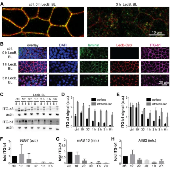
lateral LecB application (Fig. 3A). Surprisingly, also the major ligands of
␣31-integrin
expressed by MDCK cells, laminin-332 and/or -511 (37), were cointernalized (Fig. 3B).
This suggests that LecB is able to cause endocytosis of intact
␣31-integrin–laminin
complexes. To measure the dynamics of
␣31-integrin internalization, we carried out
surface biotinylation experiments (38). These experiments confirmed the rapid
LecB-triggered internalization of
␣3- and 1-integrin subunits (Fig. 3C to E). In addition, the
surface biotinylation experiments revealed that also the intracellular amount of
␣3- and
1-integrin subunits decreased upon LecB stimulation, suggesting a degradation of
internalized integrins. Consistently, LecB-mediated reduction of integrins was also
detected when whole-cell lysates were subjected to Western blot analysis (Fig. S2A).
Loss of integrins by degradation is further supported by our finding that internalized
integrins after basolateral LecB treatment showed a time-dependent increase in
colo-calization with the late endosome marker Rab9 (Fig. S2B and C) and the lysosome
marker Lamp1 (Fig. S2D and E).
In the surface biotinylation experiments, we were not able to distinguish between
active and inactive
1-integrins. Thus, we devised an alternative strategy in which we
applied activation-specific
1-integrin antibodies to the basolateral surface of live cells.
This approach revealed that LecB internalizes active and inactive
1-integrins at similar
kinetics (Fig. 3F to H and Fig. S3F), which indicates that the activation status of
1-integrins does not play an important role in LecB-mediated integrin internalization.
Taken together, LecB binds integrins, including
␣31-integrin, and causes their
internalization and degradation regardless of their activation status and bound
base-ment membrane ligands.
Membrane invagination by LecB and LecB-mediated cross-linking of
fucosy-lated lipids with
1-integrin can explain LecB-triggered integrin internalization.
Endogenous lectins, like galectin-3, were previously shown to be able to mediate
integrin internalization (39, 40). The proposed mechanism for galectin-3-mediated
integrin internalization is that galectin-3 is able to cause plasma membrane
invagina-FIG 2 LecB directly binds to1-integrin. (A) LecB-biotin was applied apically (AP) or basolaterally (BL)to polarized filter-grown MDCK cells, or cells were mock treated AP or BL. Cells were lysed, and LecB-biotin-receptor complexes were precipitated with streptavidin beads. Afterward, the presence of 1-integrin was probed by Western blotting in the precipitate and the remaining supernatant of the precipitation. (B) LecB-Cy3 (red) was applied basolaterally to MDCK cells for 6 h. Cells were fixed and stained for1-integrin (green). A confocal section (x-y section) crossing the cells in the subapical region is displayed, since most internalized vesicles were concentrated in this region. (C) MDCK cells were lysed, and1-integrins were immunoprecipitated and treated or left untreated with peptide-N-glycosidase F (PNGase F) to remove N-linked glycans. Western blot analysis of the immunoprecipitated1-integrins was performed, and1-integrin presence was proven by staining with anti-1-integrin antibodies (white arrows). Also, bands from the antibody used for 1-integrin precipitation (white arrowheads) and proteins that putatively coprecipitated with1-integrin (blue arrows) are visible. To probe the binding of LecB to1-integrin, LecB-Cy5 was incubated with membranes (far-Western assay).
tions by binding to glycolipids and also drags integrins into the invaginated membrane
regions by functioning as a cross-linker between glycolipids and integrins.
Since LecB is a tetramer with four opposing fucose-binding sites (41), which
repre-sents an ideal geometry for a potential cross-linker, we investigated if a galectin-like
mechanism could explain LecB-mediated
1-integrin internalization.
In a first step, we examined if binding of LecB to fucosylated glycosphingolipids is
sufficient to induce membrane invaginations in giant unilamellar vesicles (GUVs) (42).
Indeed, GUVs containing glycosphingolipids that bear the fucosylated Lewis a antigen
(Fig. 4A) or other fucosylated glycosphingolipids (Fig. S3) showed invaginations
imme-diately after LecB application, whereas control GUVs with the nonfucosylated
glyco-FIG 3 LecB internalizes␣31-integrin regardless of its activation status and together with laminins. (A) MDCK cells were
treated with LecB as indicated, fixed with methanol, and stained for␣3-integrin (red) and 1-integrin (green). Confocal sections (x-y sections) through the middle of the cells extracted from confocal image stacks are shown. (B) MDCK cells were treated with LecB-Cy3 (red) as indicated, fixed, and stained for panlaminin (green) and1-integrin (ITG-b1; magenta); nuclei were stained with DAPI (blue); x-y confocal sections through the middle of the cells are depicted. (C to E) MDCK cells were treated with LecB as indicated, and surface biotinylation from the basolateral side was performed. After precipitation of biotinylated proteins, the precipitates representing the surface fraction (S) and the supernatant representing the intracellular fraction (I) were subjected to Western blot analysis, and␣3-integrins and 1-integrins were probed, as well as actin to control for purity of the surface fractions. Quantification for␣3 (D) and 1 (E) subunit-composed integrins from n ⫽ 3 independent experiments. (F to H) LecB was applied basolaterally to MDCK cells for the indicated time periods followed by basolateral application of activation-specific anti-1-integrin antibodies to live cells. After fixation, the signal from bound anti-1-integrin antibodies in randomly chosen regions of interest was measured and normalized to the cell number in the regions (n⫽ 5 for one experiment). The graphs show the mean value from n⫽ 3 experiments with the activating anti-1-integrin antibody 9EG7 (F) and the inhibitory anti-1-integrin antibodies monoclonal antibody (MAb) 13 (G) and AIIB2 (H).
sphingolipid lactotetraosylceramide (Lc4cer) did not (Fig. 4A). To investigate the
rele-vance of this effect, we carried out experiments with energy-depleted cells, because
under these conditions cellular machineries cannot pinch off vesicles, which previously
led to easily visible membrane invaginations when other lipid-binding lectins like Shiga
toxin were applied (42, 43). Indeed, LecB was able to induce plasma membrane
invaginations in energy-depleted MDCK cells (Fig. 4B). Importantly,
1-integrin
colo-calized with fluorescently labeled LecB at invaginations (Fig. 4B, magnification), thus
implying that LecB can recruit integrins to invaginations. Furthermore, we observed
that basolateral LecB application led to marked clustering of endogenous galectin-3
(Fig. S4), which could suggest that LecB outcompetes galectin-3-integrin interaction.
In summary, LecB is able to cause membrane invaginations upon binding to
fucose-bearing glycosphingolipids. Since LecB is also able to bind integrins and thus to
cross-link lipids in membrane invaginations with integrins, this provides a mechanistic
explanation for LecB-mediated integrin internalization.
LecB inhibits cell migration and epithelial wound healing. As an opportunistic
pathogen, P. aeruginosa mainly relies on, and exploits, extrinsic circumstances—like a
wound—to gain access to the basolateral side of epithelia. In addition, integrin
blocking, e.g., through antibodies, has been previously shown to inhibit cell migration
in wound healing assays (44). These considerations motivated us to investigate the
effect of LecB on epithelial wound healing. Indeed, the presence of LecB strongly
inhibited collective cell migration and wound healing in MDCK monolayers (Fig. 5A).
Importantly, this effect was blockable with
L-fucose, demonstrating that LecB needs to
bind to host cells to cause migration defects. Moreover, we established that the
blockage of wound healing by LecB occurred in a dose-dependent manner, with
concentrations larger than 50
g/ml completely blocking cell migration (Fig. 5B and C),
FIG 4 Mechanism of LecB-mediated integrin internalization via cross-linking glycosphingolipids andintegrins. (A) LecB-Cy3 (15g/ml, red) was applied to GUVs containing fucosylated glycosphingolipids bearing the Lewis a antigen (Lewis a) or the nonfucosylated precursor lactotetraosylceramide (Lc4cer) and BODIPY FL C5HPC (bodipy; green) as a membrane marker. Confocal sections along equatorial planes of the GUVs are displayed; arrows point to membrane invaginations caused by LecB. (B) Subconfluent MDCK cells grown on glass coverslips were energy depleted (e.d.) or left untreated (no e.d.). LecB-Cy3 (red) was applied to the cells for 1 h, and cells were fixed and stained for1-integrin (green). Confocal
FIG 5 LecB inhibits epithelial wound healing. (A to C) Polarized monolayers of MDCK cells grown in 12-well plates were
wounded with a pipette tip and imaged with a wide-field microscope at the indicated time points to observe wound closure. In panel A, cells were treated with LecB and/orL-fucose (43 mM) to block LecB, whereas in panel B increasing concentrations
(Continued on next page)
whereas another fucose-binding lectin, UEA-I (50
g/ml), did not induce suppression of
wound healing (Fig. S5A). The inhibitory effect of LecB on wound healing was—like
other LecB-mediated effects before—reversible by washing out LecB (Fig. S5B).
To explain the abrogation of cell migration by LecB, we carried out live-cell imaging
experiments with MDCK cells stably expressing the plasma membrane marker ML-GFP
(GFP tagged with a Lyn-derived myristoylation motif) (45). ML-GFP allowed visualization
of lamellipodia formed by migrating MDCK cells (Fig. 5D, ctrl, arrows). Interestingly,
when cells were treated with LecB right after wounding, no lamellipodia formed
(Fig. 5D, LecB), whereas washout of LecB was sufficient to reinstate lamellipodium
formation and cell migration (Fig. S5C). When migrating cells were treated with LecB,
lamellipodia “froze” and LecB strongly bound to lamellipodia (Fig. S6). It is also
interesting that cells deeper within the monolayer, which expose only their apical
membranes to LecB in this assay, did still move (Fig. S6). In subsequent experiments, we
stained for
1-integrins in wound healing assays. This revealed that cells at the wound
edge take up large amounts of LecB, and in the same cells, pronounced
1-integrin
internalization was evident (Fig. 5E, arrows), which can explain why these cells are not
able to migrate any more.
Taken together, LecB inhibits epithelial wound healing in a reversible manner, which
is presumably caused by the fact that integrins in wound edge cells are accessible by
LecB and are internalized.
Depending on LecB expression, P. aeruginosa is able to crawl underneath cells.
The additional and probably dominant cytotoxic effects caused by the numerous toxins
produced by P. aeruginosa prevented us from directly quantifying an effect of LecB
knockdown in in vitro wound healing assays with live P. aeruginosa. However, we
observed that P. aeruginosa (PAO1-wt) is able to crawl underneath exposed cells
(Fig. 6A). We postulated that this requires at least local loosening of potentially
integrin-mediated cell-substrate adhesion. We tested this hypothesis by investigating
the influence of LecB on “P. aeruginosa crawling.” To this end, we used a LecB-deficient
PAO1 strain (PAO1-dLecB), which exhibited the same growth kinetics as PAO1-wt
(Fig. S7A). After overnight culture, the PAO1-wt strain showed clear expression of LecB,
whereas the LecB-deficient P. aeruginosa strain (PAO1-dLecB) did not (Fig. S7B and C).
In accordance with our hypothesis, PAO1-dLecB was visibly found in smaller numbers
underneath cells (Fig. 6A). To substantiate the experimental procedure, we established
that increasing the multiplicity of infection (MOI) (Fig. 6B and C) and increasing the
duration of incubation (Fig. 6B and D) also increased the number of bacteria crawling
underneath cells. Importantly, for all investigated conditions, the number of bacteria
crawling underneath per cell was lower for the PAO1-dLecB strain than the PAO1-wt
strain.
In summary, our study reveals a novel mechanism in which LecB, through impairing
cell-to-basement membrane attachment, allows for the creation of microniches
en-abling subepithelial colonization by P. aeruginosa.
DISCUSSION
Integrins are major receptors of LecB. Lectins bind to carbohydrates and can
therefore have multiple receptors that express the appropriate glycosylation. Hence, it
is interesting that our experiments suggest a favored binding of LecB to integrins. LecB
binding to
1-integrin appeared to be strong, because LecB was able to recover 75%
FIG 5 Legend (Continued)
of LecB were used. The quantification of the migration speeds of the wound edges from the latter experiment (C) shows that concentrations larger than 50g/ml LecB completely inhibit wound healing. n ⫽ 3. (D) Polarized monolayers of MDCK cells stably expressing the plasma membrane marker ML-GFP (green) grown on chambered cover glasses were wounded and left untreated (ctrl) or treated with LecB followed by live imaging of the wound edge by confocal microscopy. Lamellipodia are indicated with arrows. (E) Polarized MDCK monolayers grown on chambered cover glasses were wounded and treated with LecB-Alexa Fluor 488 (green) for 3 h. Cells were fixed and stained for1-integrin (red). An x-y confocal section at half height of the cells is shown. Arrows point to internalized1-integrins colocalizing with LecB-Alexa Fluor 488; the dashed line outlines the wound edge.
of total
1-integrin from cells when it was basolaterally applied and had access to
1-integrin only at the cell surface. As expected, LecB binding to 1-integrin solely
occurred through carbohydrates, since removal of
1-integrin glycosylation abolished
binding. Furthermore, mass spectrometry analysis of all basolateral LecB receptors
revealed that virtually all integrins expressed by MDCK cells were among the top hits,
including many integrin-associated proteins that were presumably recovered through
coprecipitation with integrins.
This introduces LecB as a new member of the list of bacterial molecules that bind
integrins. Another fucose-binding lectin, UEA-I, was not able to cause any of the cellular
effects that LecB caused. This shows that binding to fucose is not sufficient by itself and
suggests that LecB has additional features that bring about its specific capabilities. First,
FIG 6 LecB promotes crawling of P. aeruginosa underneath cells. (A) Sparsely seeded MDCK cells were incubated with GFP-tagged PAO1-wt or
LecB-deficient PAO1-dLecB (green) at an MOI of 50 for 1 h. After fixation,1-integrins were stained in red. For each condition, a confocal x-y section at the level of cell adhesion to the substrate, which was taken from a complete three-dimensional confocal stack, is displayed. Arrows indicate bacteria underneath cells. (B to D) After carrying out an experiment as described for panel A but with the indicated MOIs and incubation periods, the numbers of bacteria underneath cells were determined per cell. Each data point represents an individual cell, and the black lines indicate the mean and the SEM. For evaluating statistical significance, Mann-Whitney testing using GraphPad Prism 5 was applied;****, P⬍ 0.0001.
LecB and UEA-I prefer different fucose linkages in oligosaccharides (UEA-I binds
␣
1,2-linked fucose but only weakly binds
␣
1,3- and
␣
1,6-linked fucose [46, 47]; LecB shows a
slight preference for
␣
1,4-linked fucose but also binds to
␣
1,2- and
␣
1,3-linked fucose
[48–50]). In addition, LecB is a tetramer with four fucose-binding sites, but UEA-1 is a
dimer offering only two binding sites (35).
It is also worth noting that the sequence of LecB varies slightly between different
strains of P. aeruginosa (51, 52). Since ligand binding among LecB variants is conserved,
we expect that these LecB variants are utilized in similar ways as we reported here for
LecB in the P. aeruginosa strain PAO1.
Mechanism of LecB-mediated integrin internalization. Since LecB recognizes
only the carbohydrate moieties of integrins, it is able to manipulate integrins via unique
mechanisms. The structure of LecB with four opposing binding sites is ideal to cross-link
receptors. This has been demonstrated before in vitro by showing that LecB is able to
cross-link GUVs that contain LecB receptors (53).
We hypothesize that the cross-linking capacity of LecB is key for the observed rapid
internalization of integrins. In addition to integrins, LecB was also able to recognize
fucose-bearing glycosphingolipids and to cause membrane invaginations without
the need of additional energy input, which replicates effects caused by other
glycosphingolipid-binding lectins like Shiga toxin subunit B (StxB) (42), cholera toxin
subunit B 9 (CtxB) (54), or Ralstonia solanacearum lectin (RSL) (55). In addition,
1-integrin was also found on LecB-generated membrane invaginations on
energy-depleted cells. This suggests that LecB on the one hand causes membrane
invagina-tions and on the other hand is able to recruit integrins to these invaginainvagina-tions, thus
constituting a potent endocytic mechanism. A similar mechanism was suggested for
the host cell-endogenous protein galectin-3 (40). This means that bacteria have evolved
molecules like LecB that can hijack this endogenous uptake route. Importantly,
LecB-mediated lipid-integrin cross-linking for internalization represents a mechanism that
can explain the observed integrin internalization that occurred regardless of integrin
activation status and carried also basement membrane ligands like laminins with it.
LecB-triggered inhibition of wound healing. LecB binding to basolateral cell
surfaces caused cellular effects that could be causally linked to integrin internalization.
In fully polarized epithelial cells, binding of LecB to basolateral cell surfaces but not to
apical cell surfaces, which contain only minute amounts of integrins, led to loss of
apicobasal polarity. This indicates the need of integrin internalization for dissolution of
polarity. In addition, loss of polarity was reversible after washout of LecB and coincided
well with the return of
1-integrin to the basolateral plasma membrane. Our data also
suggest that LecB-mediated integrin internalization is responsible for the observed
block in cell migration in epithelial wound healing assays. First, integrins were
prefer-entially internalized in wound edge cells, and LecB prominently bound to lamellipodia
in edge cells. This makes sense, since edge cells offer more cellular surface area for LecB
binding and expose their integrins, in contrast to cells deeper within the intact
monolayer, which display only their apical surfaces to LecB. Second, edge cells rapidly
and efficiently stopped moving upon LecB treatment, whereas other cells deeper
within the monolayer preserved their capacity to move within the monolayer.
The effects that LecB exerts through integrin internalization require basolateral
access of LecB. In this context, it is interesting that P. aeruginosa possesses strategies to
convert apical into basolateral plasma membranes (34). However, the direct way for P.
aeruginosa and LecB to access integrins is through wounds in an epithelial cell layer.
P. aeruginosa preferentially colonizes wounds, and diminished healing of P.
aeruginosa-infected wounds was reported previously (10). Importantly, P. aeruginosa
possesses multiple mechanisms to manipulate and to intoxicate host cells. We
there-fore anticipate that, in order to inhibit wound healing, P. aeruginosa will apply different
combinations of its arsenal, including LecB-mediated epithelial depolarization and
inhibition of cell migration, depending on the host tissue (25).
LecB has a role in enabling bacteria to crawl underneath host cells. Our
experiments revealed a novel feature of P. aeruginosa behavior. We observed that
bacteria frequently crawled underneath host cells. For this, at least local loosening of
cell-substrate adhesion is required, which could be achieved by LecB-mediated integrin
internalization. We therefore investigated the contribution of LecB to crawling of the P.
aeruginosa strain PAO1 underneath cells. Indeed, knocking out LecB significantly
decreased crawling events. Based on our results, we suggest that P. aeruginosa uses
LecB to manipulate integrin-basement membrane interaction to proceed along the
interface between epithelial cells and the basement membrane.
In summary, our work brings integrins into focus as targets of P. aeruginosa and
provides additional rationales for the ongoing efforts to develop LecB inhibitors as a
treatment strategy in addition to antibiotics (26–31).
MATERIALS AND METHODS
Antibodies, plasmids, and reagents. Used antibodies are listed in Table S2 in the supplemental
material. The plasmid pPH-Akt-GFP encoding PH-Akt-GFP was a gift from Tamas Balla (Addgene plasmid no. 51465). The plasmid encoding GFP tagged with a glycosylphosphatidylinositol (GPI) anchor (pGPI-GFP) was described before (56). The plasmid encoding GFP tagged with a Lyn-derived myristoylation motif (pML-GFP) was a gift from Christian Wunder (Curie Institute, Paris, France).
Recombinant LecB was produced in Escherichia coli BL21(DE3) cells and purified with affinity columns as previously described (41). LecB and fluorophore-conjugated LecB were used at a concentration of 50g/ml (4.3 M) unless stated otherwise. UEA-I and UEA-I–fluorescein isothiocyanate (FITC) were from Vector Labs. Cycloheximide, andL-fucose (6-deoxy-L-galactose) were from Sigma-Aldrich.
Mammalian cell culture and creation of stable cell lines. MDCK strain II cells were cultured in
Dulbecco’s modified Eagle’s medium (DMEM) supplemented with 5% fetal calf serum (FCS) at 37°C and 5% CO2. Unless stated otherwise, 3⫻ 105MDCK cells were seeded per transwell filter (0.4-m pore size, polycarbonate membrane, catalog no. 3401 from Corning) and cultured for 4 days before experiments. For the creation of stable MDCK cell lines, plasmids encoding the proteins of interest and G418 resistance (pPH-Akt-GFP, pML-GFP, and pGPI-GFP) were transfected into cells with Lipofectamine 2000 (Thermo Fisher). After the cells were allowed to express the proteins overnight, they were trypsinized and plated sparsely in medium containing 1 mg/ml G418. After single colonies had formed, GFP-positive colonies were extracted with cloning rings. At least 6 colonies were extracted for each cell line, grown on transwell filters for 4 days, fixed, and stained against the basolateral marker protein-catenin and the tight junction marker protein ZO-1 to assay their polarized morphology. Based on these results, we chose one colony for each cell line for further experiments. TEER measurements were carried out using an EVOM2 equipped with chopstick electrodes (World Precision Instruments).
Immunofluorescence. Cells were washed two times with phosphate-buffered saline without Ca2⫹ and Mg2⫹(PBS) and then fixed with 4% (wt/vol) formaldehyde (FA) for 15 min at room temperature. Samples were treated with 50 mM ammonium chloride for 5 min to quench FA and then permeabilized with a SAPO medium (PBS supplemented with 0.2% [wt/vol] bovine serum albumin [BSA] and 0.02% [wt/vol] saponin) for 30 min. Primary antibodies were diluted in SAPO medium and applied on the samples for 60 min at room temperature. After three washes with PBS, secondary dye-labeled antibodies and, if required, 4=,6-diamidino-2-phenylindole (DAPI) and dye-labeled phalloidin were diluted in SAPO medium and applied to the cells for 30 min at room temperature (details for the antibodies used are listed in Table S2). After 5 washes with PBS, cells were mounted for microscopy. Since ␣3-integrin antibodies did not work in FA-fixed cells, methanol fixation was applied in this case. Briefly, cells were incubated with precooled methanol for 15 min at⫺20°C. After washing with PBS, cells were permeabil-ized with 0.05% (vol/vol) Triton X-100 for 10 min at room temperature and blocked with 3% (wt/vol) bovine serum albumin (BSA) for 1 h at room temperature. Staining with primary and secondary antibodies was then carried out as described before but with a 3% (wt/vol) BSA solution.
Microscopic imaging of fixed cells and live-cell experiments. For microscopic imaging, an A1R
confocal microscope (Nikon) equipped with a 60⫻ oil immersion objective (numerical aperture [NA]⫽ 1.49) and laser lines at 405 nm, 488 nm, 561 nm, and 641 nm was utilized. Image acquisition and analysis were performed with NIS-Elements 4.10.04 (Nikon).
Live-cell experiments were carried out at 37°C, and MDCK cells grown as polarized monolayers for 3 days on Lab-Tek II chambered cover glasses (8 well, 1.5 borosilicate glass) were used. The medium was changed to recording medium (Hanks’ balanced salt solution [HBSS] supplemented with 1% FCS, 4.5 g/liter glucose, and 20 mM HEPES).
Wound healing assays. MDCK cells were seeded on 12-well plates or, for live-cell microscopy of cell
migration, on 8-well Lab-Tek II chambered cover glasses and allowed to form confluent monolayers for 3 days. Then, cells were scratched with a 200-l pipette tip to inflict a wound. On 12-well plates, marker lines were drawn on the bottom to ensure that always the same position of the wound was imaged.
Western blot analysis. Cells were washed twice with PBS and lysed in RIPA buffer (20 mM Tris [pH
8], 0.1% [wt/vol] SDS, 10% [vol/vol] glycerol, 13.7 mM NaCl, 2 mM EDTA, and 0.5% [wt/vol] sodium deoxycholate in water), supplemented with protease inhibitors (0.8M aprotinin, 11 M leupeptin, 200M Pefabloc) and phosphatase inhibitor (1 mM sodium orthovanadate). Protein concentrations were analyzed using a bicinchoninic acid (BCA) assay kit (Pierce). Equal amounts of protein per lysate were
separated by SDS-PAGE and transferred to a nitrocellulose membrane. The membrane was blocked with Tris-buffered saline (TBS) supplemented with 0.1% (vol/vol) Tween 20 and 3% (wt/vol) BSA for 1 h and incubated with primary and horseradish peroxidase (HRP)-linked secondary antibodies diluted in the blocking solution. In some cases, TBS supplemented with 0.1% (vol/vol) Tween 20 and 5% (wt/vol) milk powder was used (details for the antibodies and conditions used are listed in Table S2). Detection was performed by a chemiluminescence reaction using the Fusion-FX7 Advance imaging system (Peqlab Biotechnologie GmbH).
Energy depletion. Energy depletion was carried out as described before (42). Briefly, MDCK cells
were washed 2 times with PBS supplemented with 100 mg/liter CaCl2and 100 mg/liter MgCl2·6H2O (PBS⫹⫹) and then treated with PBS⫹⫹supplemented with 10 mM 2-deoxy-D-glucose and 10 mM NaN3for 30 min at 37°C.
Bacterial culture and crawling experiments. GFP-tagged P. aeruginosa PAO1 wild-type (PAO1-wt)
and LecB-deficient (PAO1-dLecB) strains were provided by S. de Bentzmann (CNRS, Aix Marseille University, France). The generation of LecB-deficient PAO1 is described in reference 23, and GFP tagging was carried out according to the procedure described in reference 57. For experiments, bacteria were cultured overnight (approximately 16 h) in LB-Miller medium containing 60g/ml gentamicin in a shaker (Thriller; Peqlab) at 37°C and 650 rpm. The bacteria reached an optical density (OD) measured at 600 nm of approximately 5. Using these growth conditions, PAO1-wt and PAO1-dLecB strains showed compa-rable growth kinetics (Fig. S7A) and harvested PAO1-wt efficiently expressed LecB, whereas PAO1-dLecB did not (Fig. S7B and C).
For crawling experiments, MDCK cells were sparsely seeded on 8-well Lab-Tek II chambered cover glasses and cultured for 1 day, so that clusters of maximally 10 cells in diameter formed to ensure that all cells were exposed to P. aeruginosa similarly as at a wound edge. Then, PAO1-wt or PAO-dLecB was applied for the indicated MOI and duration. Bacteria crawling under cells were counted manually per cell from confocal image stacks of whole cells to ensure that only bacteria located directly underneath cells at the level of the glass coverslip were counted.
qPCR. PAO1-wt and PAO1-dLecB were cultured overnight as described before. RNA was extracted
using TRI reagent (Sigma-Aldrich). After DNase digest, 100 ng RNA was transcribed into cDNA using a first-strand cDNA synthesis kit (Thermo Fisher). Then, quantitative PCR (qPCR) was performed on a CFX384 qPCR cycler (Bio-Rad) using a SYBR Select master mix (Thermo Fisher) and the following primers: for LecB, forward 3=-AAGGAGTGTTCACCCTTCCC-5= and reverse 3=-GATGACGGCGTTATTGGTGC-5=; for
rpoD as reference, forward 3=-GGGATACCTGACTTACGCGG-5= and reverse 3=-GGGGCTGTCTCGAATACGT T-5=.
Labeling of lectins. LecB was labeled with fluorescent dyes bearing N-hydroxysuccinimide (NHS)
esters as reactive groups (Cy3 monoreactive NHS ester [GE Healthcare], Cy5 monoreactive NHS ester [GE Healthcare], Alexa Fluor 488 NHS ester [Thermo Fisher]) or with biotin using NHS-polyethylene glycol 12-biotin (Thermo Fisher) according to the instructions of the manufacturers and purified using PD-10 desalting columns (GE Healthcare).
Cell surface biotinylation and immunoprecipitation. For cell surface biotinylation, all following
steps were carried out in a cold room (4°C). Sulfo-NHS-biotin (Thermo Fisher) was freshly diluted in PBS⫹⫹(concentration, 0.3 mg/ml) and applied to the apical or basolateral plasma membrane of transwell filter-grown MDCK cells for 20 min. Afterward, the reaction was quenched for 20 min with PBS⫹⫹ supplemented with 50 mM ammonium chloride. Cells were lysed with RIPA buffer, and biotinylated proteins were precipitated with streptavidin-agarose beads (Thermo Fisher). Elution was carried out with Laemmli buffer (2% [wt/vol] SDS, 10% [vol/vol] glycerol, 60 mM Tris-Cl [pH 6.8] in water) and boiling at 98°C for 5 min.
For1-integrin immunoprecipitation (IP), MDCK cells were grown to confluence in 10-cm dishes and lysed in IP lysis buffer (50 mM Tris-HCl [pH 7.5], 150 mM sodium chloride, 1% [vol/vol] IGEPAL CA-630, 0.5% [wt/vol] sodium deoxycholate in water). The lysates were precleared with protein A-agarose beads (Roche) for 3 h and then incubated with anti-1-integrin antibodies (MAB2000 from Millipore) for 1 h. After addition of protein A-agarose beads overnight, beads were washed three times with IP-lysis buffer, and1-integrin was eluted with Laemmli buffer and boiling at 98°C for 5 min.
Mass spectrometry-based identification of LecB interaction partners. MDCK cells were cultured
in medium for stable-isotope labeling by amino acids in cell culture (SILAC medium) for 9 passages and then seeded on transwell filters and allowed to polarize for 4 days. For the first sample, biotinylated LecB was applied to the apical side of light-SILAC-labeled cells and on the basolateral side of medium-SILAC-labeled cells, whereas heavy-SILAC-medium-SILAC-labeled cells received no stimulation and served as control. For the second sample, the treatment conditions were permuted. After lysis with IP lysis buffer, the different SILAC lysates were combined and LecB-biotin-receptor complexes were precipitated using streptavidin-agarose beads as described before. Eluted LecB-biotin-receptor complexes were then prepared for mass spectrometry (MS) analysis using SDS-PAGE gel electrophoresis. Gels were cut into pieces, proteins therein were digested with trypsin, and resulting peptides were purified by stop-and-go-extraction (STAGE) tips.
For mass spectrometry analysis, samples were fractionated by nanoscale high-pressure liquid chro-matography (HPLC) on a 1200 HPLC (Agilent Technologies, Waldbronn, Germany) connected online to an LTQ Orbitrap XL mass spectrometer (Thermo Fisher Scientific, Bremen, Germany). Fused silica HPLC-column tips with a 75-m inner diameter were self-packed with ReproSil-Pur 120 ODS-3 (Dr. Maisch, Ammerbuch, Germany) to a length of 20 cm. Samples were directly injected into the mass spectrometer (for details see reference 58). The raw data files were uploaded into the MaxQuant software. Database searches were performed against a full-length dog database containing common
contaminants such as keratins and enzymes used for in-gel digestion. Carbamidomethylcysteine was set as fixed modification, and oxidation of methionine and protein amino-terminal acetylation were set as variable modifications. Triple SILAC was used as quantitation mode. The enzyme specificity was trypsin/ P⫹DP with three allowed miscleavages. The MS/MS tolerance was set to 0.5 Da, and the mass precision of identified peptides after recalibration was in general less than 1 ppm. For identification and quanti-tation, the following settings were used: peptide and protein false-discovery rates (FDR) were set to 0.01; maximum peptide posterior error probability (PEP) was set to 0.1; minimum peptide length was set to 7; minimum number peptides for identification and quantitation of proteins was set to two, of which one must be unique; minimum ratio count was set to two; and only unmodified peptides and the variable modification were used for protein quantification. The “match between run” option was used with a time window of 2 min.
From the generated list of MS-identified proteins, we defined those proteins as LecB interaction partners that showed more than 2-fold enrichment on a log2scale over controls in both SILAC samples (see Table S1 in the supplemental material).
GUV experiments. Giant unilamellar vesicles (GUVs) were composed of
1,2-dioleoyl-sn-glycero-3-phosphocholine (DOPC), spiked with 1 mol% 2-(4,4-difluoro-5,7-dimethyl-4-bora-3a,4a-diaza-s-indacene-3-pentanoyl)-1-hexadecanoyl-sn-glycero-3-phosphocholine (-BODIPY FL C5-HPC), 30 mol% cholesterol, and 5 mol% of the desired glycosphingolipid species. Blood group glycosphingolipids were provided by Göran Larson (Sahlgrenska University Hospital, Gothenburg, Sweden). GUVs were grown at room temperature using the electroformation technique on indium-tin oxide (ITO)-coated slides as described previously (42, 43). Briefly, lipid mixtures were dissolved in chloroform at a final concentration of 0.5 mg/ml, and 15l solution was spread on the conductive surface of ITO slides. After 2 h of drying under vacuum, GUVs were grown in a 290 mosM sucrose solution by applying an alternating electric field from 20 mV to 1.1 V for 3 h.
LecB-Cy3 (15g/ml) was incubated with GUVs at room temperature and examined under an inverted confocal fluorescence microscope (Nikon A1R) equipped with an oil immersion objective (60⫻; NA, 1.49).
Statistics. If not stated otherwise, data obtained from n⫽ 3 independent experiments were used to
calculate arithmetic means. Error bars represent standard errors of the means (SEM). Statistical signifi-cance analysis was carried out using GraphPad Prism 5.
SUPPLEMENTAL MATERIAL
Supplemental material is available online only.
FIG S1, PDF file, 1 MB.
FIG S2, PDF file, 2.1 MB.
FIG S3, PDF file, 0.8 MB.
FIG S4, PDF file, 1 MB.
FIG S5, PDF file, 0.7 MB.
FIG S6, PDF file, 0.5 MB.
FIG S7, PDF file, 0.2 MB.
TABLE S1, PDF file, 0.2 MB.
TABLE S2, PDF file, 0.2 MB.
ACKNOWLEDGMENTS
This work was supported by a German Research Foundation grants (RO 4341/2-1
and RTG 2202); the Excellence Initiative of the German Research Foundation (EXC 294);
the Ministry of Science, Research and the Arts of Baden-Württemberg (Az: 33-7532.20);
and a starting grant from the European Research Council (Programme “Ideas,”
ERC-2011-StG 282105). R.T. acknowledges support from the Ministry of Science, Research
and the Arts of Baden-Württemberg (Az: 7533-30-10/25/36).
REFERENCES
1. Lyczak JB, Cannon CL, Pier GB. 2002. Lung infections associated with cystic fibrosis. Clin Microbiol Rev 15:194 –222.https://doi.org/10.1128/ cmr.15.2.194-222.2002.
2. Franzetti F, Grassini A, Piazza M, Degl’innocenti M, Bandera A, Gazzola L, Marchetti G, Gori A. 2006. Nosocomial bacterial pneumonia in HIV-infected patients: risk factors for adverse outcome and implications for rational empiric antibiotic therapy. Infection 34:9 –16.https://doi.org/10 .1007/s15010-006-5007-x.
3. Markou P, Apidianakis Y. 2014. Pathogenesis of intestinal Pseudomonas aeruginosa infection in patients with cancer. Front Cell Infect Microbiol 3:115.https://doi.org/10.3389/fcimb.2013.00115.
4. Barbier F, Andremont A, Wolff M, Bouadma L. 2013. Hospital-acquired pneumonia and ventilator-associated pneumonia: recent advances in
epidemiology and management. Curr Opin Pulm Med 19:216 –228. https://doi.org/10.1097/MCP.0b013e32835f27be.
5. Azzopardi EA, Azzopardi E, Camilleri L, Villapalos J, Boyce DE, Dziewulski P, Dickson WA, Whitaker IS. 2014. Gram negative wound infection in hospitalised adult burn patients—systematic review and metanalysis. PLoS One 9:e95042.https://doi.org/10.1371/journal.pone.0095042. 6. Stover C, Pham X, Erwin A, Mizoguchi S, Warrener P, Hickey M, Brinkman
F, Hufnagle W, Kowalik D, Lagrou M, Garber R, Goltry L, Tolentino E, Westbrock-Wadman S, Yuan Y, Brody L, Coulter S, Folger K, Kas A, Larbig K, Lim R, Smith K, Spencer D, Wong G, Wu Z, Paulsen I, Reizer J, Saier M, Hancock R, Lory S, Olson M. 2000. Complete genome sequence of Pseudomonas aeruginosa PAO1, an opportunistic pathogen. Nature 406:959 –964.https://doi.org/10.1038/35023079.
7. Maillet M, Pelloux I, Forli A, Vancoetsem K, Cheong Sing JS, Marfaing S, Ducki S, Batailler P, Mallaret M-R. 2014. Nosocomial transmission of carbapenem-resistant Pseudomonas aeruginosa among burn patients. In-fect Control Hosp Epidemiol 35:597–599.https://doi.org/10.1086/675842. 8. Bicking Kinsey C, Koirala S, Solomon B, Rosenberg J, Robinson BF, Neri A,
Laufer Halpin A, Arduino MJ, Moulton-Meissner H, Noble-Wang J, Chea N, Gould CV. 2017. Pseudomonas aeruginosa outbreak in a neonatal intensive care unit attributed to hospital tap water. Infect Control Hosp Epidemiol 38:801– 808.https://doi.org/10.1017/ice.2017.87.
9. Mah T-F, Pitts B, Pellock B, Walker GC, Stewart PS, O’Toole GA. 2003. A genetic basis for Pseudomonas aeruginosa biofilm antibiotic resistance. Nature 426:306 –310.https://doi.org/10.1038/nature02122.
10. Engel J, Eran Y. 2011. Subversion of mucosal barrier polarity by Pseu-domonas aeruginosa. Front Microbiol 2:114. https://doi.org/10.3389/ fmicb.2011.00114.
11. WHO. 2017. WHO priority pathogens list for research and development of new antibiotics. WHO, Geneva, Switzerland. http://apps.who.int/ medicinedocs/documents/s23171en/s23171en.pdf.
12. Wagner S, Sommer R, Hinsberger S, Lu C, Hartmann RW, Empting M, Titz A. 2016. Novel strategies for the treatment of Pseudomonas aeruginosa infections. J Med Chem 59:5929 –5969.https://doi.org/10 .1021/acs.jmedchem.5b01698.
13. Golovkine G, Faudry E, Bouillot S, Elsen S, Attrée I, Huber P. 2016. Pseudomonas aeruginosa transmigrates at epithelial cell-cell junctions, exploiting sites of cell division and senescent cell extrusion. PLoS Pathog 12:e1005377.https://doi.org/10.1371/journal.ppat.1005377.
14. Tran CS, Eran Y, Ruch TR, Bryant DM, Datta A, Brakeman P, Kierbel A, Wittmann T, Metzger RJ, Mostov KE, Engel JN. 2014. Host cell polarity proteins participate in innate immunity to Pseudomonas aeruginosa infection. Cell Host Microbe 15:636 – 643.https://doi.org/10.1016/j.chom .2014.04.007.
15. Kwok T, Zabler D, Urman S, Rohde M, Hartig R, Wessler S, Misselwitz R, Berger J, Sewald N, Konig W, Backert S. 2007. Helicobacter exploits integrin for type IV secretion and kinase activation. Nature 449:862– 866. https://doi.org/10.1038/nature06187.
16. Stewart PL, Nemerow GR. 2007. Cell integrins: commonly used receptors for diverse viral pathogens. Trends Microbiol 15:500 –507.https://doi .org/10.1016/j.tim.2007.10.001.
17. Cossart P. 1997. Host/pathogen interactions. Subversion of the mamma-lian cell cytoskeleton by invasive bacteria. J Clin Invest 99:2307–2311. https://doi.org/10.1172/JCI119409.
18. Roger P, Puchelle E, Bajolet-Laudinat O, Tournier J-M, Debordeaux C, Plotkowski M-C, Cohen JHM, Sheppard D, De Bentzmann S. 1999. Fi-bronectin and␣51 integrin mediate binding of Pseudomonas aerugi-nosa to repairing airway epithelium. Eur Respir J 13:1301–1309.https:// doi.org/10.1034/j.1399-3003.1999.13f14.x.
19. Leroy-Dudal J, Gagnière H, Cossard E, Carreiras F, Di Martino P. 2004. Role of␣v5 integrins and vitronectin in Pseudomonas aeruginosa PAK interaction with A549 respiratory cells. Microbes Infect 6:875– 881. https://doi.org/10.1016/j.micinf.2004.05.004.
20. Imberty A, Wimmerová M, Mitchell EP, Gilboa-Garber N. 2004. Structures of the lectins from Pseudomonas aeruginosa: insights into the molecular basis for host glycan recognition. Microbes Infect 6:221–228.https://doi .org/10.1016/j.micinf.2003.10.016.
21. Tielker D, Hacker S, Loris R, Strathmann M, Wingender J, Wilhelm S, Rosenau F, Jaeger KE. 2005. Pseudomonas aeruginosa lectin LecB is located in the outer membrane and is involved in biofilm formation. Microbiology 151:1313–1323.https://doi.org/10.1099/mic.0.27701-0. 22. Funken H, Bartels KM, Wilhelm S, Brocker M, Bott M, Bains M, Hancock REW,
Rosenau F, Jaeger KE. 2012. Specific association of lectin LecB with the surface of Pseudomonas aeruginosa: role of outer membrane protein OprF. PLoS One 7:e46857.https://doi.org/10.1371/journal.pone.0046857. 23. Chemani C, Imberty A, De Bentzmann S, Pierre M, Wimmerová M, Guery
BP, Faure K. 2009. Role of LecA and LecB lectins in Pseudomonas aeruginosa-induced lung injury and effect of carbohydrate ligands. In-fect Immun 77:2065–2075.https://doi.org/10.1128/IAI.01204-08. 24. Adam EC, Mitchell BS, Schumacher DU, Grant G, Schumacher U. 1997.
Pseudomonas aeruginosa II lectin stops human ciliary beating: thera-peutic implications of fucose. Am J Respir Crit Care Med 155:2102–2104. https://doi.org/10.1164/ajrccm.155.6.9196121.
25. Cott C, Thuenauer R, Landi A, Kühn K, Juillot S, Imberty A, Madl J, Eierhoff T, Römer W. 2016. Pseudomonas aeruginosa lectin LecB inhibits tissue repair processes by triggering-catenin degradation. Biochim Biophys Acta 1863:1106 –1118.https://doi.org/10.1016/j.bbamcr.2016.02.004.
26. Sommer R, Wagner S, Rox K, Varrot A, Hauck D, Wamhoff E-C, Schreiber J, Ryckmans T, Brunner T, Rademacher C, Hartmann RW, Brönstrup M, Imberty A, Titz A. 2018. Glycomimetic, orally bioavailable LecB inhibitors block biofilm formation of Pseudomonas aeruginosa. J Am Chem Soc 140:2537–2545.https://doi.org/10.1021/jacs.7b11133.
27. Sommer R, Hauck D, Varrot A, Wagner S, Audfray A, Prestel A, Möller HM, Imberty A, Titz A. 2015. Cinnamide derivatives of d-mannose as inhibitors of the bacterial virulence factor LecB from Pseudomonas aeruginosa. Chem-istryOpen 4:756 –767.https://doi.org/10.1002/open.201500162.
28. Johansson EMV, Crusz SA, Kolomiets E, Buts L, Kadam RU, Cacciarini M, Bartels K-M, Diggle SP, Cámara M, Williams P, Loris R, Nativi C, Rosenau F, Jaeger K-E, Darbre T, Reymond J-L. 2008. Inhibition and dispersion of Pseudomonas aeruginosa biofilms by glycopeptide dendrimers target-ing the fucose-specific lectin LecB. Chem Biol 15:1249 –1257.https://doi .org/10.1016/j.chembiol.2008.10.009.
29. Donnier-Marechal M, Abdullayev S, Bauduin M, Pascal Y, Fu M-Q, He X-P, Gillon E, Imberty A, Kipnis E, Dessein R, Vidal S. 2018. Tetraphenylethylene-based glycoclusters with aggregation-induced emission (AIE) properties as high-affinity ligands of bacterial lectins. Org Biomol Chem 16:8804 – 8809. https://doi.org/10.1039/c8ob02035c.
30. Bucher KS, Babic N, Freichel T, Kovacic F, Hartmann L. 2018. Monodis-perse sequence-controlled alpha-L-fucosylated glycooligomers and their multivalent inhibitory effects on LecB. Macromol Biosci 18:e1800337. https://doi.org/10.1002/mabi.201800337.
31. Dupin L, Noel M, Bonnet S, Meyer A, Gehin T, Bastide L, Randriantsoa M, Souteyrand E, Cottin C, Vergoten G, Vasseur J-J, Morvan F, Chevolot Y, Darblade B. 2018. Screening of a library of oligosaccharides targeting lectin LecB of Pseudomonas aeruginosa and synthesis of high affinity oligoglyco-clusters. Molecules 23:3073.https://doi.org/10.3390/molecules23123073. 32. Rodriguez-Boulan E, Kreitzer G, Müsch A. 2005. Organization of vesicular
trafficking in epithelia. Nat Rev Mol Cell Biol 6:233–247.https://doi.org/ 10.1038/nrm1593.
33. Thuenauer R, Hsu YC, Carvajal-Gonzalez JM, Deborde S, Chuang J, Römer W, Sonnleitner A, Rodriguez-Boulan E, Sung C. 2014. Four-dimensional live imaging of apical biosynthetic trafficking reveals a post-Golgi sort-ing role of apical endosomal intermediates. Proc Natl Acad Sci U S A 111:4127– 4132.https://doi.org/10.1073/pnas.1304168111.
34. Kierbel A, Gassama-Diagne A, Rocha C, Radoshevich L, Olson J, Mostov K, Engel J. 2007. Pseudomonas aeruginosa exploits a PIP3-dependent pathway to transform apical into basolateral membrane. J Cell Biol 177:21–27.https://doi.org/10.1083/jcb.200605142.
35. Audette GF, Vandonselaar M, Delbaere L. 2000. The 2.2 Å resolution structure of the O (H) blood-group-specific lectin I from Ulex europaeus. J Mol Biol 304:423– 433.https://doi.org/10.1006/jmbi.2000.4214. 36. Myllymäki SM, Teräväinen TP, Manninen A. 2011. Two distinct
integrin-mediated mechanisms contribute to apical lumen formation in epithelial cells. PLoS One 6:e19453.https://doi.org/10.1371/journal.pone.0019453. 37. Greciano PG, Moyano JV, Buschmann MM, Tang J, Lu Y, Rudnicki J, Manni-nen A, Matlin KS. 2012. Laminin 511 partners with laminin 332 to mediate directional migration of Madin-Darby canine kidney epithelial cells. Mol Biol Cell 23:121–136.https://doi.org/10.1091/mbc.E11-08-0718.
38. Le Bivic A, Sambuy Y, Mostov K, Rodriguez-Boulan E. 1990. Vectorial targeting of an endogenous apical membrane sialoglycoprotein and uvomorulin in MDCK cells. J Cell Biol 110:1533–1539.https://doi.org/10 .1083/jcb.110.5.1533.
39. Furtak V, Hatcher F, Ochieng J. 2001. Galectin-3 mediates the endocy-tosis of-1 integrins by breast carcinoma cells. Biochem Biophys Res Commun 289:845– 850.https://doi.org/10.1006/bbrc.2001.6064. 40. Lakshminarayan R, Wunder C, Becken U, Howes MT, Benzing C, Arumugam
S, Sales S, Ariotti N, Chambon V, Lamaze C, Loew D, Shevchenko A, Gaus K, Parton RG, Johannes L. 2014. Galectin-3 drives glycosphingolipid-dependent biogenesis of clathrin-inglycosphingolipid-dependent carriers. Nat Cell Biol 16: 592– 603.https://doi.org/10.1038/ncb2970.
41. Mitchell EP, Sabin C, Šnajdrová L, Pokorná M, Perret S, Gautier C, Hofr C, Gilboa-Garber N, Kocˇa J, Wimmerová M, Imberty A. 2005. High affinity fucose binding of Pseudomonas aeruginosa lectin PA-IIL: 1.0 Å resolu-tion crystal structure of the complex combined with thermodynamics and computational chemistry approaches. Proteins Struct Funct Genet 58:735–746.https://doi.org/10.1002/prot.20330.
42. Römer W, Berland L, Chambon V, Gaus K, Windschiegl B, Tenza D, Aly MRE, Fraisier V, Florent J-C, Perrais D, Lamaze C, Raposo G, Steinem C, Sens P, Bassereau P, Johannes L. 2007. Shiga toxin induces tubular membrane invaginations for its uptake into cells. Nature 450:670 – 675. https://doi.org/10.1038/nature05996.
43. Römer W, Pontani LL, Sorre B, Rentero C, Berland L, Chambon V, Lamaze C, Bassereau P, Sykes C, Gaus K, Johannes L. 2010. Actin dynamics drive membrane reorganization and scission in clathrin-independent endocy-tosis. Cell 140:540 –553.https://doi.org/10.1016/j.cell.2010.01.010. 44. Wood S, Sivaramakrishnan G, Engel J, Shafikhani SH. 2011. Cell migration
regulates the kinetics of cytokinesis. Cell Cycle 10:648 – 654.https://doi .org/10.4161/cc.10.4.14813.
45. Thuenauer R, Nicklaus S, Frensch M, Troendle K, Madl J, Römer W. 2018. A microfluidic biochip for locally confined stimulation of cells within an epithelial monolayer. RSC Adv 8:7839 –7846.https://doi.org/10.1039/ c7ra11943g.
46. Baldus SE, Thiele J, Park Y-O, Hanisch F-G, Bara J, Fischer R. 1996. Characterization of the binding specificity of Anguilla anguilla agglutinin (AAA) in comparison to Ulex europaeus agglutinin I (UEA-I). Glycoconj J 13:585–590.https://doi.org/10.1007/bf00731446.
47. Sughii S, Kabat E, Baer HH. 1982. Further immunochemical studies on the combining sites of Lotus tetragonolobus and Ulex europaeus I and II lectins. Carbohydr Res 99:99 –101.https://doi.org/10.1016/S0008-6215(00)80982-9. 48. Marotte K, Sabin C, Préville C, Moumé-Pymbock M, Wimmerová M, Mitchell EP, Imberty A, Roy R. 2007. X-ray structures and thermodynam-ics of the interaction of PA-IIL from Pseudomonas aeruginosa with disaccharide derivatives. ChemMedChem 2:1328 –1338.https://doi.org/ 10.1002/cmdc.200700100.
49. Perret S, Sabin C, Dumon C, Pokorná M, Gautier C, Galanina O, Ilia S, Bovin N, Nicaise M, Desmadril M, Gilboa-Garber N, Wimmerová M, Mitchell EP, Imberty A. 2005. Structural basis for the interaction between human milk oligosaccharides and the bacterial lectin PA-IIL of Pseu-domonas aeruginosa. Biochem J 389:325–332.https://doi.org/10.1042/ BJ20050079.
50. Mitchell E, Houles C, Sudakevitz D, Wimmerova M, Gautier C, Pérez S, Wu AM, Gilboa-Garber N, Imberty A. 2002. Structural basis for oligosaccharide-mediated adhesion of Pseudomonas aeruginosa in the lungs of cystic fibrosis patients. Nat Struct Biol 9:918 –921.https://doi .org/10.1038/nsb865.
51. Boukerb AM, Decor A, Ribun S, Tabaroni R, Rousset A, Commin L, Buff S,
Doleans-Jordheim A, Vidal S, Varrot A, Imberty A, Cournoyer B. 2016. Genomic rearrangements and functional diversification of lecA and lecB lectin-coding regions impacting the efficacy of glycomimetics directed against Pseudomonas aeruginosa. Front Microbiol 7:811.https://doi.org/ 10.3389/fmicb.2016.00811.
52. Sommer R, Wagner S, Varrot A, Nycholat CM, Khaledi A, Haussler S, Paulson JC, Imberty A, Titz A. 2016. The virulence factor LecB varies in clinical isolates: consequences for ligand binding and drug discovery. Chem Sci 7:4990 –5001.https://doi.org/10.1039/c6sc00696e.
53. Villringer S, Madl J, Sych T, Manner C, Imberty A, Römer W. 2018. Lectin-mediated protocell crosslinking to mimic cell-cell junctions and adhesion. Sci Rep 8:1932.https://doi.org/10.1038/s41598-018-20230-6. 54. Ewers H, Römer W, Smith AE, Bacia K, Dmitrieff S, Chai W, Mancini R,
Kartenbeck J, Chambon V, Berland L, Oppenheim A, Schwarzmann G, Feizi T, Schwille P, Sens P, Helenius A, Johannes L. 2010. GM1 structure determines SV40-induced membrane invagination and infection. Nat Cell Biol 12:11–18.https://doi.org/10.1038/ncb1999.
55. Arnaud J, Tröndle K, Claudinon J, Audfray A, Varrot A, Römer W, Imberty A. 2014. Membrane deformation by neolectins with engineered glyco-lipid binding sites. Angew Chem Int Ed Engl 53:9267–9270.https://doi .org/10.1002/anie.201404568.
56. Thuenauer R, Juhasz K, Mayr R, Frühwirth T, Lipp A-M, Balogi Z, Sonn-leitner A. 2011. A PDMS-based biochip with integrated sub-micrometre position control for TIRF microscopy of the apical cell membrane. Lab Chip 11:3064 –3071.https://doi.org/10.1039/c1lc20458k.
57. Eierhoff T, Bastian B, Thuenauer R, Madl J, Audfray A, Aigal S, Juillot S, Rydell GE, Müller S, de Bentzmann S, Imberty A, Fleck C, Römer W. 2014. A lipid zipper triggers bacterial invasion. Proc Natl Acad Sci U S A 111:12895–12900.https://doi.org/10.1073/pnas.1402637111.
58. Thriene K, Gruning BA, Bornert O, Erxleben A, Leppert J, Athanasiou I, Weber E, Kiritsi D, Nystrom A, Reinheckel T, Backofen R, Has C, Bruckner-Tuderman L, Dengjel J. 2018. Combinatorial omics analysis reveals perturbed lyso-somal homeostasis in collagen VII-deficient keratinocytes. Mol Cell Pro-teomics 17:565–579.https://doi.org/10.1074/mcp.RA117.000437.