BIOTECHNOLOGICALLY RELEVANT ENZYMES AND PROTEINS
Listeria bacteriophage peptidoglycan hydrolases feature high
thermoresistance and reveal increased activity after divalent
metal cation substitution
Mathias Schmelcher&Florian Waldherr&
Martin J. Loessner
Received: 23 March 2011 / Revised: 4 May 2011 / Accepted: 5 May 2011 / Published online: 1 July 2011 # Springer-Verlag 2011
Abstract The ability of the bacteriophage-encoded pepti-doglycan hydrolases (endolysins) to destroy Gram-positive bacteria from without makes these enzymes promising antimicrobials. Recombinant endolysins from Listeria monocytogenes phages have been shown to rapidly lyse and kill the pathogen in all environments. To determine optimum conditions regarding application of recombinant Listeria phage endolysins in food or production equip-ments, properties of different Listeria endolysins were studied. Optimum NaCl concentration for the amidase HPL511 was 200 nM and 300 mM for the peptidases HPL118, HPL500, and HPLP35. Unlike most other peptidoglycan hydrolases, all four enzymes exhibited high-est activity at elevated pH values at around pH 8–9. Lytic activity was abolished by EDTA and could be restored by supplementation with various divalent metal cations, indicating their role in catalytic function. While substitution of the native Zn2+by Ca2+or Mn2+was most effective in case of HPL118, HPL500, and HPLP35, supplementation with Co2+ and Mn2+ resulted in an approximately 5-fold increase in HPL511 activity. Interestingly, the glutamate peptidases feature a conserved SxHxxGxAxD zinc-binding motif, which is not present in the amidases, although they also require centrally located divalent metals for activity.
The endolysins HPL118, HPL511, and HPLP35 revealed a surprisingly high thermostability, with up to 35% activity remaining after 30 min incubation at 90°C. The available data suggest that denaturation at elevated temperatures is reversible and may be followed by rapid refolding into a functional state.
Keywords Peptidoglycan hydrolase . Lytic enzyme . Listeria . Bacteriophage . Food safety
Introduction
Endolysins are bacteriophage-encoded murein hydrolases, which are produced inside an infected bacterial host cell at the end of the phage lytic multiplication cycle. Endolysin-mediated cell wall degradation results in disruption of the cell wall and release of the phage progeny. From within the bacterial cytoplasm, the enzymes gain access to the peptidoglycan with the aid of holins, hydrophobic proteins that oligomerize and form pores in the membrane, allowing the endolysins to pass through (reviewed in Young et al. 2000; Bernhardt et al. 2002). Based on the bonds targeted within the peptidoglycan network, endolysins can be classified into different categories (reviewed in Loessner 2005): (1) glucosaminidases, (2) muramidases, also known as “lysozymes,” (3) transglycosylases, which all cleave within the aminosugar backbone, (4) N-acetylmuramoyl-L -alanine amidases, and (5) endopeptidases, both of which hydrolyze bonds in the crosslinking peptide stems. Due to the absence of an outer membrane in Gram-positive bacteria, endolysins can also lyse these cells from without and are receiving increasing attention as alternative anti-microbials with potential applications in food, biotechnol-M. Schmelcher
:
F. Waldherr:
M. J. Loessner (*)Institute of Food, Nutrition and Health, ETH Zurich, Schmelzbergstrasse 7,
8092 Zurich, Switzerland e-mail: [email protected] Present Address:
M. Schmelcher
Animal Biosciences and Biotechnology Lab, ANRI, ARS, USDA, 10300 Baltimore Avenue,
ogy, and medicine (Hagens and Loessner 2007; Fischetti 2005; Loessner et al.1995a; Loessner2005; Hermoso et al. 2007; Fischetti2010).
Listeria monocytogenes is an invasive and intracellular pathogen, transmitted to humans via contaminated food. Among risk groups such as neonates, elderly, pregnant women, and immunocompromised individuals, it can cause severe infections with potentially fatal outcome (Vazquez-Boland et al.2001). Although the incidence of the disease known as listeriosis is relatively low compared to other food-borne pathogens, mortality rates of more than 30% make Listeria an important concern in the food industry.
Listeria cell wall peptidoglycan belongs to the A1γ type, featuring directly crosslinked peptidoglycan via meso-diaminopimelic acid (Schleifer and Kandler 1972). Cell wall-associated carbohydrates, in particular teichoic acids, are responsible for most of the antigenic variability within the genus, with five major serovar groups being distin-guished (Fiedler et al.1984; Fiedler and Ruhland1987).
Endolysins from Listeria phages have been shown to be highly active against these bacteria from without (Loessner et al.1995a,b,1996). Two different enzymatic specificities of Listeria phage endolysins have been reported: N-acetylmur-amoyl-L-alanine amidases and L-alanoyl-D-glutamate pepti-dases. The latter was first identified in Ply118 and Ply500 (Loessner et al.1995b) and has recently also been reported for a Bacillus subtilis enzyme (Fukushima et al.2007) and Escherichia coli phage T5 endolysin (Mikoulinskaia et al. 2009). Like most known endolysins, the Listeria phage enzymes show a modular organization: an N-terminal enzymatically active domain (EAD) is linked to a C-terminal cell wall binding domain (CBD), which directs the enzyme to a cell wall-associated ligand (Korndoerfer et al. 2006; Loessner et al. 2002; Schmelcher et al. 2010). The CBD domains of the peptidases Ply118 and Ply500 feature complementary binding properties, where CBD118 binds to Listeria cells of serovars 1/2 and 3, and CBD500 recognizes those of serovars 4–6 (Loessner et al. 2002). However, Ply118 was shown to also be able to lyse cells outside the CBD recognition pattern (Schmelcher et al.2011). Ply511 is an N-acetylmuramoyl-L-alanine amidase from the virulent
broad host range phage A511, recognizing and lysing strains from all serovars (Loessner et al.1995b; Schmelcher et al. 2010). The PlyP35 peptidase endolysin of phage P35 (Dorscht et al.2009) and its CBD were shown to recognize a majority of serovar 1/2 and 3 strains and some serovar 4–6 strains (Schmelcher et al.2010,2011). PlyPSA and Ply500 contain Zn2+ as an integral component of the catalytic domain (Korndoerfer et al.2006;2008).
Listeria phage endolysins are useful reagents in molec-ular biology (Loessner et al.1996), and their CBDs can be used for development of novel analytical procedures
(Loessner et al.2002; Hagens and Loessner 2007; Kretzer et al. 2007; Schmelcher et al. 2010). They also have potential as antimicrobials in food and have been cloned and modified for secretion in lactic acid bacteria (Gaeng et al.2000; Turner et al.2006). To further develop the various endolysin-based applications, better knowledge of their properties is desirable. Therefore, this study aimed to biochemically characterize Ply118, Ply500, Ply511, and the novel enzyme PlyP35 and study the effects of ionic strength, pH, divalent metal cations, and various temper-atures on lytic activity and enzyme stability.
Materials and methods
Bacteria, culture conditions, plasmids, and phages
E. coli strain JM109 (NEB) was used for cloning of the plyP35 gene and production of the His-tagged recombinant endolysin HPLP35. Strains JM109 pHPL118, JM109 pHPL500, and JM109 pHPL511 (Loessner et al. 1996) served for overexpression of genes encoding HPL118, HPL500, and HPL511, respectively. E. coli was cultivated in Luria–Bertani (LB) medium at 37°C, supplemented with 100 μg/ml ampicillin as needed. L. monocytogenes strains WSLC 1001 (serovar 1/2 c) and WSLC 1042 (serovar 4 b) were grown in tryptose broth at 30°C. Phage P35 DNA (Dorscht et al. 2009) served as template for PCR amplifi-cation of the plyP35 gene. Plasmid pQE-30 (Qiagen) was used for construction of pHPLP35.
Cloning of plyP35
The plyP35 gene was amplified by PCR with High Fidelity Taq Polymerase (Roche), using primers plyP35_BamHI_F (5′-ATCAGGATCCTTATTTCTTGATGTCAAACATG-TAACG-3′) and plyP35_SalI_R (5′-ATCAGTCGACT-TATTTCTTGATGTCAAACATGTAACG-3′), thereby introducing TAA as stop codon and adding BamHI and SalI restriction sites (underlined) to the 5′ and 3′ ends of the products, respectively. After endonuclease digestion, the DNA fragment was inserted into BamHI/SalI sites of pQE-30, the resulting plasmid pHPLP35 introduced into E. coli JM109 and the construct verified by nucleotide sequencing (GATC, Konstanz, Germany).
In silico analyses
The software AlignX (Vector NTI; Invitrogen) was used for creating multiple amino acid sequences alignments, employing the ClustalW algorithm (Thompson et al. 1994).
Production and purification of endolysins
This was performed essentially as previously described (Loessner et al.1996; Schmelcher et al 2011). In brief, E. coli strains harboring the desired constructs were grown in modified LB medium (15 g/l tryptose, 8 g/l yeast extract, and 5 g/l NaCl) with ampicillin, to an OD600nmof 0.5, when
gene expression and protein synthesis were induced by addition of 1 mM isopropyl β-D-1-thiogalactopyranoside. After 4 h incubation at 35°C with shaking, cells were harvested by centrifugation, resuspended in 20 ml buffer A (50 mM Na2HPO4, 500 mM NaCl, 5 mM imidazole, 0.1 %
Tween 20, pH 8.0) per 1 l of culture, and cells disrupted by two passages through a French Press 20K cell (SLM Aminco) at 100 MPa. Debris was removed by centrifuga-tion and the supernatant filtered (0.2μM PES membrane, Millipore). His-tagged proteins were purified from the supernatant by affinity chromatography, using Ni-NTA Superflow resin (Qiagen). Immobilized protein was eluted using buffer B (50 mM Na2HPO4, 500 mM NaCl, 250 mM
imidazole, 0.1% Tween 20, pH 8.0), dialyzed (50 mM NaH2PO4, 100 mM NaCl, 0.005% to 0.1% Tween 20,
pH 8.0), filtered (0.2μM PES membrane, Millipore), and stored at −20°C in 50% (v/v) glycerol. Purity and concentration of the purified enzyme samples was analyzed by gel electrophoresis and spectrophotometry (NanoDrop ND-1000).
Preparation of Listeria cells and purified cell walls as assay substrate
L. monocytogenes strains WSLC 1001 and WSLC 1042 were grown to an OD600nmof approximately 0.8, harvested
by centrifugation, washed, and resuspended in deionized water at approximately 50-fold concentration. Aliquots were stored at−80°C until use.
Preparation of purified cell walls was carried out as described earlier (Wendlinger et al.1996). Listeria cells in the late log phase were disrupted by use of a French press (40 K cell, SLM Aminco), and the remaining intact cells were removed by centrifugation at 1,400×g for 5 min. Raw cell walls were then harvested at 15,000×g for 30 min, washed with water, and resuspended in 7.0 ml SM buffer (Sambrook et al.1989) per 1 g pellet. Nucleic acids were removed by incubation with 0.1 mg DNAse and 0.1 mg RNAse (Sigma) per gram cell walls for 4 h, and proteins were degraded by Proteinase K (Sigma) digestion (0.1 mg/g pellet for 3 h). The preparations were then boiled at 100°C in 4% (w/v) sodium dodecyl sulfate (SDS) for 30 min, washed with water at least three times, and resuspended in deionized water. The purified cell walls were stored in aliquots at−80°C until use.
Photometric lysis assay
Lytic activity of purified recombinant endolysins was determined by photometric lysis assays as described previously (Loessner et al. 1995a,1996). The assays were carried out using polystyrene cuvettes (Greiner) in a total volume of 1 ml, in 10 mM Tris, 150 mM NaCl (300 mM in case of HPLP35), pH 8.0, at 30°C (the “standard conditions”). These conditions were modified for some experiments, as detailed below. Substrate was added to an initial OD600nm of approximately 1.0 in case of intact
Listeria cells and 0.2–0.3 with purified cell walls. Enzymes were added to the suspension at concentrations ranging from 2 to 5μg/ml (approximately 50–150 nM) in a volume of 15 μl, and the OD600nmwas recorded for up to 10 min.
As negative control, buffer was added. In tests using various pH values, these were measured directly in the cuvettes at the beginning and the end of each assay.
All experiments were carried out in triplicate, and data processing was performed as described before (Korndoerfer et al. 2006). Lysis data were normalized and corrected by subtraction of the corresponding control values [corrected value=normalized value+(1−normalized control value)]. The resulting curves were fitted to the sigmoid function f= y0+ a/{1 + exp[−(x−x0)/b]}c, employing Sigma-Plot 9.0
(Systat Software, Inc.). The steepest slope of the function was used to calculate lytic activity. Activities of all four recombinant endolysins were determined under standard conditions, and 1 U was defined as the amount of enzyme that decreases the OD600nmof Listeria cells in suspension
by 0.01/min (Loessner et al.1995a,1996). Biochemical characterization of endolysins
In order to determine the effect of pH on lytic activity, lysis assays were performed at different pH values between 3.8 and 10.1, with intact Listeria cells as substrate. Six different buffer systems (Good et al. 1966) at a concentration of 20 mM were used to cover the entire pH range: phosphate, citrate, MOPS, Tris, Tricine, and CHES (Sigma). The influence of salt concentration (i.e., ionic strength) on lytic activity was determined using various NaCl concentrations from 0 mM to 1.0 M. To assess the role of the divalent metal cations Mn2+, Mg2+, Ca2+, Cu2+, Co2+, Ni2+, and Zn2+ for enzyme activity, purified cell walls served as substrate. Both enzymes and purified cell walls were incubated with 250 mM EDTA for 30 min to sequester and remove any residual metal ions. Subsequently, enzymes were dialyzed against assay buffer, and cell walls were washed with deionized, highly purified water (Milli-Q, Millipore). Lysis of EDTA-treated cell walls by endolysins was monitored with and without addition of divalent metal ions at different
concentrations (1 and 10 mM). However, Zn2+could not be examined at 10 mM, due to precipitation effects. Lytic activities were compared to those recorded without EDTA treatment and cation substitution.
In order to test temperature stability of enzymatic activity, aliquots of the endolysins were incubated at different temperatures (40°C, 50°C, 60°C, 70°C, 80°C, and 90°C) for 5, 10, and 30 min, followed by cooling on ice for 10 min. Lytic activity on intact cells under standard conditions was then determined and compared to controls using non-heat treated enzymes.
Results
Expression, purification, and lytic activity of recombinant Listeria phage endolysins
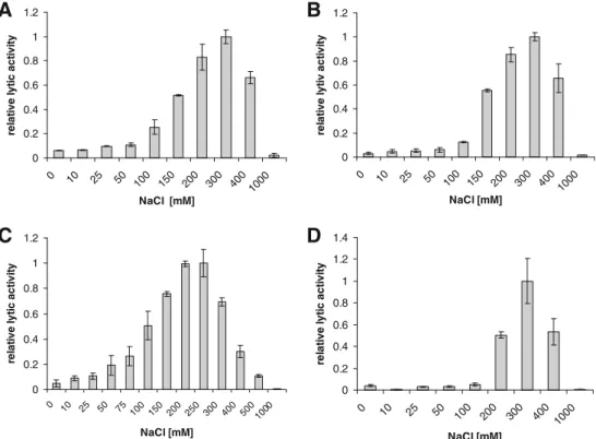
Listeria phage endolysins HPL118, HPL500, HPL511, and HPLP35 were produced in E. coli and purified by immobilized metal ion adsorption chromatography. Follow-ing dialysis and concentration, the proteins appeared as almost homogeneous bands (more than 90% pure) on SDS polyacrylamide gel electrophoresis gels (data not shown), with concentrations between 2.0 and 9.5 mg/ml (Table1). A linear relationship between enzyme concentration and the steepest slope of the lysis curve was observed in photo-metric lysis assays using intact Listeria cells as substrate. The results for HPL500 are shown in Fig. 1, and lytic activities of all four enzymes used here are listed in Table1. Endolysins show similar ionic strength requirements Lytic activities of all enzymes were determined at various NaCl concentrations up to 1,000 mM. Results were similar for peptidases HPL118 (Fig. 2a) and HPL 500 (Fig. 2b), which both showed optimum activity at 300 mM NaCl. The HPL511 amidase exhibited a slightly lower optimum (200– 250 mM), while its activity decreased to roughly 70% at 300 mM salt (Fig. 2c). At lower ionic strength, however, HPL511 was more active than the peptidases, displaying 50% activity at 100 mM NaCl, compared to 25% (HPL118) and 12% (HPL500). The peptidase HPLP35 showed best activity at 300 mM NaCl, albeit with a narrower range
compared to HPL118 and HPL500. It retained 50% lysis activity at 200 and 400 mM, and <5% at 100 mM and below (Fig. 2d). None of the four enzymes caused visible cell lysis and concomitant reduction of turbidity at 1,000 mM NaCl.
Influence of pH and buffer systems on lytic activity Endolysin activity was determined in a pH range of 3.8– 10.1, and optima for all enzymes were found to be at pH 8.0 and higher (Fig.3). HPL118 and HPL500 displayed best activity in a range from pH 9 to 10 (Fig. 3a, b), whereas HPL511 and HPLP35 were most active from pH 8 to 9 (Fig.3b, c). At more acidic conditions, activity of all tested enzymes dropped significantly. At neutral pH, enzymes exhibited between 30% (HPL500) and 70% (HPL511) activity, which dropped below 10% at pH<5.5. This decrease was most abrupt in case of HPL511, dropping from 70% at pH 7.0 to <20% at pH 6.4. Figure 3 also illustrates the effect of the different buffer systems used on lytic activity. Although the enzymes showed the same trends regarding their pH optima in different buffers, comparison of specific data obtained at similar pH values in the various buffers revealed considerable differences. For instance, the activity of HPL118 and HPL500 at pH 6.6 in citrate buffer was higher than at pH 6.4 and 7.0 in MOPS buffer. For all assays, pH was shown to be stable throughout the duration of the measurements (data not shown).
Removal of divalent metals and complementation by heterologous ions
Determination of the EAD500 structure disclosed the presence of a Zn2+ ion in the catalytic center of Ply500 (Korndoerfer et al. 2008), which is in perfect agreement with the zinc binding motif SxHxxGxAxD present in Ply118 (McCafferty et al.1997). Figure4 reveals the high conservation of this motif in sequence alignments of glutamate peptidases from different Listeria phages, includ-ing PlyP35 (Schmelcher et al. 2010). This suggested a general requirement of metal cations in the catalytic domain of these enzymes. In order to further investigate the role of divalent metals on lytic activity, both the enzymes and the Table 1 Listeria bacteriophage endolysin preparations used
Enzyme Concentration (mg/ml) Purity (% of total protein) Lytic activity (U/mg) Substrate cells used for lysis assays
HPL118 3.3 >90 9,370 WSLC 1001
HPL500 3.8 >90 25,259 WSLC 1042
HPL511 9.5 >90 22,871 WSLC 1001
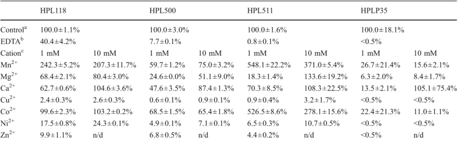
cell wall substrates were treated with 250 mM EDTA to sequester and remove residual divalent metals. This treatment drastically reduced or abolished activity of HPL118, HPL500, and HPL511, and HPLP35 (Table 2). We then used different divalent metal cations (Mn2+, Mg2+, Ca2+, Cu2+, Co2+, Ni2+, and Zn2+, at 1–10 mM) to supplement the EDTA-treated enzymes. We found Mn2+, Mg2+, Ca2+, and Co2+to be most effective in reconstituting the endolysin peptidoglycan hydrolase activity. HPL118 regained full activity after addition of Ca2+(10 mM) or Co2+ and up to 80% in the case of Mg2+. Surprisingly, 1 mM Mn2+ caused a more than 2.4-fold boost in activity compared to the control. With HPL500, the addition of 10 mM Ca2+resulted in close to 90% activity, while Mn2+, Mg2+, and Co2+ reconstituted activity to at least 50%. HPL511 regained its full activity with 10 mM Mg2+or Ca2+, and a more than 5-fold increase in activity compared to the non-EDTA treated control was observed following addition of 1 mM Mn2+ or Co2+. For HPLP35, Ca2+ at a
concentration of 10 mM appeared to be most effective, whereas Mn2+, Mg2+, and Co2+ only partly reconstituted enzyme activity. Generally speaking, Ca2+ and Mg2+ were more effective when added at 10 mM, whereas Mn2+ and Co2+enabled best enzyme function at 1 mM concentration. In contrast, substitution with Cu2+, Ni2+, and Zn2+was less effective or even inhibitory for endolysin activity.
HPL118, HPL511, and HPLP35 exhibit high temperature resistance
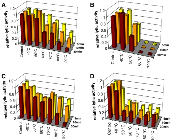
Temperature stability (defined as remaining enzyme activity after a heating step, not as activity at elevated temperature) of the four endolysins was determined following incubating the enzymes at temperatures of up to 90°C for maximum 30 min, and monitoring lytic activity by the standard photometric assay using intact Listeria substrate cells. Surprisingly, HPL118, HPL511, and HPLP35 revealed high temperature stability. After heating to 50°C for up to 30 min, HPL118 retained more than 90% of its activity (Fig. 5a). From 60°C upwards and increasing treatment time, activity was gradually reduced. After 5 min at 90°C, the enzyme still showed 40% activity compared to the non-heat-treated control. Only after 30 min at 90°C, lytic activity dropped below 10% of the control. The HPL511 amidase exhibited even higher stability (Fig. 5c). Even though incubation at 50°C reduced activity by approxi-mately 25%, this remained unchanged after incubation at up to 70°C. At higher temperatures and an incubation time of 30 min, its activity gradually decreased. Howev-er, the enzyme still retained more than 60% of its lytic activity following 5 min at 90°C. Similarly, HPLP35 showed a rapid decrease in leftover activity following treatment at 40°C (approximately 90%) and 50°C (35%), independent of the heating time (Fig.5d). However, using 50°C and 90°C as incubation temperatures, the enzyme retained activity. Even after 30 min at 90°C, it was able to quickly lyse substrate cells. Only boiling at 100°C was able to completely inactivate HPLP35 (data not shown). In contrast, HPL500 revealed only weak heat stability (Fig. 5b). While at 40°C lytic activity was not affected, treatment at 50°C reduced activity to approximately 60%, and temperatures of 60°C or higher completely inactivated the enzyme.
Discussion
We report the effects of different factors on lytic activity of four different Listeria bacteriophage peptidoglycan hydro-lases, including three L-alanoyl-D-glutamate peptidases
(HPL118, HPL500, and HPLP35) and one N-acetylmur-amoyl-L-alanine amidase (HPL511). Lytic activities of the
0 0.2 0.4 0.6 0.8 1 1.2 0 100 200 300 400 500 600 time [s] normalized OD 600nm -0.025 -0.02 -0.015 -0.01 -0.005 0 0 1 2 3 4 5 6 HPL500 [µg/ml] Slope [1/s]
A
B
Fig. 1 Linear relationship of HPL500 concentration used in a photometric lysis assay and steepest slope of the cell lysis curve. a Decrease in OD600nm of a suspension of WSLC 1042 cells after
addition of 0.2 μg/ml (diamonds), 0.5 μg/ml (circles), 1.6 μg/ml (triangles), and 4.8μg/ml (squares) of HPL500 (at t=0). All curves were normalized to an initial OD600nmof 1.0, and the control (addition
of buffer instead of enzyme) was subtracted. All assays were carried out in triplicate and curves represent mean values. b Steepest slopes of the lysis curves from a plotted against HPL500 concentration. The linear regression line has an R2of 0.9965. For all further lysis assays
performed in this work, HPL500 was used at a concentration of 2.87μg/ml
many different phage-encoded and bacterial cell wall lytic enzymes investigated to date range from 102 to 108 “units” per milligram (Vasala et al.1995; Loessner et al. 1995a, 2001; Nelson et al. 2001; Shida et al. 2001; Loeffler et al. 2003; Yoong et al. 2004; Fukushima et al. 2007; Kyomuhendo et al.2007). However, different assay types used and arbitrary unit definition makes comparison
difficult or impossible. Under the conditions employed here, concentrations as small as 2–5 μg/ml (approximately 100 nmol) of purified endolysin added to Listeria cells in suspension resulted in an instantaneous and rapid decrease in OD600nm of the substrate cells, reaching a baseline
within minutes or sometimes seconds. This highly efficient enzymatic lysis not only offers advantages for laboratory
B
A
D
C
0.0 0.2 0.4 0.6 0.8 1.0 1.2relative lytic activity
4 5 6 7 8 9 10 0.0 0.2 0.4 0.6 0.8 1.0 1.2
relative lytic activity
4 5 6 7 8 9 10 0.0 0.2 0.4 0.6 0.8 1.0 1.2
relative lytic activity
4 5 6 7 8 9 10 0.0 0.2 0.4 0.6 0.8 1.0 1.2
relative lytic activity
pH pH
pH
4 5 6 7 8 9
pH Fig. 3 Effect of pH and
different buffer systems on lytic activity of HPL118 (a), HPL500 (b), HPL511 (c), and HPLP35 (d) against Listeria cells in suspension. Results of following buffers are shown, covering a pH range of 3.8–10.1: citrate (yellow bars), MOPS (red bars), Tricine (green bars), and CHES (blue bars)
0 0.2 0.4 0.6 0.8 1 1.2 0 10 25 50 100 150 200 300 4001000 NaCl [mM] re la ti ve l y ti c act iv it y 0 0.2 0.4 0.6 0.8 1 1.2 0 10 25 50 100 150 200 300 400 1000 NaCl [mM] re la ti v e l y ti v a c ti v ity 0 0.2 0.4 0.6 0.8 1 1.2 0 10 25 50 75 100 150 200 250 300 400 5001000 NaCl [mM] re la ti ve l y ti c act ivi ty 0 0.2 0.4 0.6 0.8 1 1.2 1.4 0 10 25 50 100 200 300 400 1000 NaCl [mM] re la ti v e ly ti c a c tiv it y
B
A
D
C
Fig. 2 Influence of NaCl concentration on lytic activity of HPL118 (a), HPL500 (b), HPL511 (c), and HPLP35 (d) against Listeria cells in suspension
applications (Loessner et al.1995a) but also demonstrates the potential of these enzymes as novel antibacterial food additives (Hagens and Loessner2007).
The optimum ionic strength (200–300 mM NaCl) corresponds quite well to the salt concentration found in many food products associated with a high risk for Listeria contamination, such as soft-ripened cheeses and meat products. Our data are also in line with findings on other phage or bacterial cell wall hydrolases (Vasala et al.1995; Loeffler et al.2003; Cheng and Fischetti2006; Yoong et al. 2006; Becker et al. 2008), although some enzymes were shown to be more active at physiological conditions (approximately 130 mM NaCl) or even in the absence of NaCl (Yoong et al. 2004; Fukushima et al. 2007). Interestingly, the best binding of the CBDs of Ply118 and Ply500 to the Listeria cell wall occurs at lower ionic strength (100 mM) (Loessner et al. 2002). This indicates that the optima for binding (CBD) and enzymatic function (EAD) are different, and both contribute to the overall
activity of the enzymes. The somewhat lower NaCl concentration favored by the amidase HPL511 may also be due to the lower binding affinity of its CBD to the cell wall compared to the CBDs of the other three endolysins (Schmelcher et al. 2010). As CBD binding is required for full lytic activity and is based on ionic interactions between the binding domain and Listeria cell wall carbohydrates (Eugster et al., submitted for publication), higher binding affinity supports activity at higher ionic strength (Loessner et al. 2002; Cheng and Fischetti 2006; Schmelcher et al. 2010).
With respect to optimum pH, the data obtained for HPL118 and HPL500 in this study were slightly different from those reported earlier for their CBDs (Loessner et al. 2002). We found that the pH optima of HPL118 and HPL500 match well with predicted isoelectric points (pI) of 9.91 and 9.56, respectively. For HPL511 and HPLP35, the predicted pIs are in the same range (9.81 and 9.89, respectively), which in this case is higher than the
YP_001468860.1 CAA61519.1 YP_001468411.1 YP_001468664.1 CAA59362.1 NP_465802.1 YP_001468812.1 79 90 100 110 120 130 147 (79) GQSNHNYGVAVDLCLYTSDGKNVIWESTTSR-WKTVVSAMKAEGFEWGGDWKSFKDYPHFELYDAAGGE Ply006 (76) GQSNHNFGVAVDLCLYTSDGKDVIWESTTSR-WKKVVAAMKAEGFEWGGDWKSFKDYPHFELCDAASGE Ply2438 (76) GQSNHNYGVAVDLCLYTNDGKDVIWESTTSR-WKKVVAAMKAEGFKWGGDWKSFKDYPHFELCDAVSGE Ply500 (76) MRSYHIVGQAFDFVMAK--GKTVDWGGYKTAKAKKVIAKAKALGFSWGGDWSGFVDCPHMQYEYKGYGT Ply025 (62) MRSYHLVGQALDFVMAK--GKTVDWGAYRSDKGKKFVAKAKSLGFEWGGDWSGFVDNPHLQFNYKGYGT Ply118 (62) MRSYHLVGQALDFVMAK--GKTVDWGAYRSDKGKKFVAKAKSLGFEWGGDWSGFVDNPHLQFNFKGYGT PlyEGDe (62) GQSNHNFGVAVDFAIDLIDDGKIDSWQPSAT----IVNMMKRRGFKWGGDWKSFTDLPHFEACDWYRGE PlyP35 (77)
GQSNHNFGVAVDF L DGK VDW S TS KKVVA MKA GFEWGGDWKSF D PHFE D G GE
Consensus (79)
* * *
Fig. 4 Amino acid sequence alignment of the zinc binding central regions of peptidase endolysins from different Listeria phages. Residues conserved in all seven enzymes are highlighted in yellow. The zinc binding motif SxHxxGxAxD is boxed in red, and residues
required for coordination of the central Zn2+ion in Ply500 are marked
by asterisks (see “Discussion”). Database accession numbers of all proteins are indicated on the right
Table 2 Effect of different divalent metal cations on lytic activity of Listeria phage endolysins
HPL118 HPL500 HPL511 HPLP35 Controla 100.0±1.1% 100.0±3.0% 100.0±1.6% 100.0±18.1% EDTAb 40.4±4.2% 7.7±0.1% 0.8±0.1% <0.5% Cationc 1 mM 10 mM 1 mM 10 mM 1 mM 10 mM 1 mM 10 mM Mn2+ 242.3±5.2% 207.3±11.7% 59.7±1.2% 75.0±3.2% 548.1±22.2% 371.0±5.4% 26.7±21.4% 15.6±2.1% Mg2+ 68.4±2.1% 80.4±3.0% 24.6±0.0% 51.1±9.0% 18.3±1.4% 133.6±19.2% 6.3±2.0% 8.4±1.7% Ca2+ 62.7±0.6% 104.6±3.6% 47.6±3.5% 87.4±1.3% 70.3±8.5% 108.3±22.5% 13.5±2.1% 105.1±75.4% Cu2+ 2.4±0.3% 2.6±0.3% 0.6±0.1% 0.9±0.1% 0.9±0.4% 3.2±1.7% <0.5% <0.5% Co2+ 99.6±2.3% 103.2±0.2% 68.5±1.5% 65.4±1.8% 526.5±8.6% 278.1±15.6% 22.4±21.3% 11.0±1.1% Ni2+ 17.5±0.8% 24.3±0.1% 4.9±0.1% 7.1±0.1% 6.5±0.3% 10.7±0.5% <0.5% <0.5% Zn2+ 9.9±1.1% n/d 6.8±0.5% n/d 4.4±0.2% n/d <0.5% n/d
Cell walls of Listeria monocytogenes WSLC 1042 were used for HPL500, those of strain WSLC 1001 for all other enzymes. Activities are expressed as percentage in relation to the non-treated enzyme controls. Same enzyme concentration was used before and after EDTA treatment n/d not determined (see text)
a
Lytic activity under standard conditions before EDTA treatment of cell walls and enzyme (see text)
b
Lytic activity of EDTA treated and dialyzed enzyme against EDTA-treated cell walls under standard conditions
c
Lytic activities of EDTA treated and dialyzed enzyme against EDTA-treated cell walls supplemented with different divalent metal ions at two different concentrations
experimentally determined pH optima, suggesting that these enzymes require a positive net charge for full activity. In general, pH optima above 8.0 seem uncommon for cell wall hydrolases. In fact, most other phage and bacterial lysins studied require neutral or slightly acidic conditions, with activity rapidly decreasing above pH 7.5 or 8 (Vasala et al. 1995; Morita et al. 2001; Loeffler et al.2003; Pritchard et al. 2004; Yoong et al. 2004; Cheng and Fischetti 2006; Donovan et al.2006; Fukushima et al.2007; Sugahara et al. 2007). However, another L-alanoyl-D-glutamate peptidase
from E. coli phage T5 revealed a pH profile similar to the enzymes reported here, with highest activity at pH 8.5 (Mikoulinskaia et al. 2009). It is interesting to note that these properties correspond well to the optimum range for Listeria, which also grows best at a pH of around 8.0 (Vanderzant and Splittstoesser1992). At this point, it is not clear whether the endolysins would be fully active in complex food matrices such as cheese, which often feature low pH values. However, on surface ripened soft cheeses, which are particularly prone to Listeria contamination, growth of the pathogens is paralleled by an increase in pH during ripening from about 4.5 to above 8.0 (Farber and Peterkin 1991; Guenther and Loessner 2011), which also supports activity of the Listeria phage endolysins.
The fact that EDTA treatment reduces or abolishes endolysin function, which can be reconstituted by supple-mentation with several divalent metal cations, confirms
their crucial role for catalytic activity. The differences in inactivation observed by EDTA treatment might be explained by different affinities of the metal ions to their coordination sites or varying accessibility within the proteins. The zinc binding site SxHxxGxAxD was first identified in the Enterococcus dipeptidase VanX, which cleaves the terminalD-Ala–D-Ala bond in the peptidoglycan peptide stem and was shown to contain a central Zn2+ required for catalytic function (McCafferty et al.1997). In Ply500, the Zn2+ ion is also coordinated by His and Asp residues within an identical motif, in addition to another His at position 133 (Korndoerfer et al. 2008). The latter is located within a unique region highly conserved among Listeria phage peptidases, featuring the consensus sequence KxxGFxWGGDWxxFxDxPH (Fig. 4). These similarities suggest an identical catalytic mechanism for these enzymes, employing Zn2+ as integral component required for func-tion. Corresponding sites are also present in the murine sonic hedgehog protein Shh-N and carboxypeptidase Ddp from Streptomyces albus (Murzin 1996). Even though EAD500 is a peptidase and its sequence differs from the EADPSA amidase (Korndoerfer et al.2006; Schmelcher et al. 2010), the latter also features a catalytic zinc, which is, however, positioned by two distinct His residues. Consider-ing that Zn2+ obviously assumes a crucial role in catalytic function of these enzymes, it was surprising to find that Ca2+ and Mn2+were more effective in restoring lytic activity after
Cont rol 40°C 50°C 60°C 70°C 80°C 90° C 30min 10min 5min 0 0.2 0.4 0.6 0.8 1 1.2 Co n tr o l 40° C 50° C 60° C 70° C 30min 10min 5min 0 0.2 0.4 0.6 0.8 1 1.2 Co n tr o l 40° C 50° C 60° C 70° C 80° C 90° C 30min 10min 5min 0 0.2 0.4 0.6 0.8 1 1.2 Co n tr o l 40 ° C 50 °C 60 ° C 70 ° C 80 ° C 90 ° C 30min 10min 5min 0 0.2 0.4 0.6 0.8 1 1.2
B
A
D
C
rel a ti v e ly ti c a c ti v it y re la ti v e ly ti c ac ti vi ty rel a ti v e ly ti c ac ti v it y rel at ive lyt ic a cti vi tyFig. 5 Temperature stability of HPL118 (a), HPL500 (b), HPL511 (c), and HPLP35 (d). The enzymes were incubated at different temperatures for 5, 10, or 30 min, cooled down on ice, and then photometric lysis assays were performed under standard conditions. Lytic activity values are expressed relative to the control (non-heat treated enzyme)
removal of the Zn2+. Similar observations regarding the effects of divalent metals on activity of cell wall hydrolases were reported earlier (Vasala et al.1995; Shida et al.2001; Pritchard et al.2004; Donovan et al. 2006; Sugahara et al. 2007; Mikoulinskaia et al. 2009; Garcia et al. 2010). For aminopeptidase A, a zinc metalloendopeptidase that can be reactivated by Ca2+ or Mn2+ after exposure to chelating agents (Danielsen et al.1980), it was recently shown that the Ca2+ ion increases substrate affinity by binding to two aspartate residues of the enzyme and interacting with an acidic side chain of the substrate (Claperon et al.2008). We believe that this might also be the case for the peptidases described here and the interaction of the catalytic domain with the D-glutamic acid residue of the peptidoglycan
peptide stem.
An entirely unexpected finding was the unusual temper-ature stability of HPL511, HPL118, and HPLP35, mostly because there is no obvious selection pressure for phages infecting psychrotrophic bacteria such as Listeria to evolve and maintain heat-stable proteins. Peptidoglycan hydrolases may still function after heating up to 60°C (Vasala et al. 1995; Fischetti 2005; Yoong et al. 2006; Schmitz et al. 2011); however, lytic activity after treatment at 90°C has not been reported before. Possible reasons for this phe-nomenon may include occurrence of cysteine residues (Cys), which are able to stabilize proteins through forma-tion of disulfide (cystine) bonds. However, this mechanism is unlikely to be responsible for the effects seen here, since HPL118, HPL511, and HPLP35 feature only one or two Cys residues each, whereas the least thermostable enzyme HPL500 contains four Cys residues (data not shown). SalG, a goose-type lysozyme from salmon fish, also revealed high heat stability and does not contain Cys, unlike other less heat stable lysozymes of the same type (Kyomuhendo et al. 2007). This suggests that not disulfide bonds, but unfolding of the protein with increasing temperatures, followed by rapid reactivation due to instant refolding upon cooling may reconstitute enzyme activity after prolonged high tempera-ture exposure. The latter assumption receives support from a study on thermoresistant lytic structural proteins of Pseudomonas aeruginosa phages (Lavigne et al. 2004; Briers et al.2008). It was found that Gp36C, a part of the putative injection needle of phage ΦKMV, retained 50% and 21% of its activity after 1-h incubation at 100°C or 121°C, respectively (Lavigne et al. 2004) and also lacks Cys residues. Further investigations revealed that the enzyme starts to unfold at 50°C and, therefore, features only low thermodynamic stability. However, it shows high kinetic stability, i.e., resistance to irreversible denaturation, which allows the protein to refold at appropriate temper-atures (Briers et al. 2006). With respect to the enzymes studied here, available evidence suggests that HPL118 and HPL511 have similar properties. Regarding HPLP35,
however, the prominent drop in activity after incubation at 40–50°C suggests a significant degree of irreversible denaturation. Here, it is possible that the CBD is more prone to irreversible inactivation than the EAD, which would reflect and explain the observed reduction in lytic activity of the enzyme to 30–40%.
Thermoresistance of phage endolysins can be considered a very useful property regarding their potential application as antimicrobials in food products that undergo heat treatment. The potential of using purified recombinant Listeria phage endolysins in food was recently assessed in a pilot study and yielded promising results. Application of HPL118, HPL500, or HPL511 to leaves of iceberg lettuce artificially contaminated with L. monocytogenes reduced viable counts of the pathogen by up to 2.4 logs after storage for 6 days at two different temperatures (6°C and 12°C) (Guenther et al., unpublished data). In general, the extraordinary lytic specificity of bacteriophage endolysins represents an important advantage since they specifically act on only their target cells, leaving the endogenous and often desired background flora untouched. Furthermore, as they cleave bonds in highly conserved portions of the peptidoglycan, formation of resistance is unlikely and has not yet been reported (Loessner2005; Fischetti2005; Low et al.2005). These favorable properties, in conjunction with the high stability and robustness of the lysins studied here, offer promising perspectives for their application in foods and associated production and packaging environments.
Acknowledgments We are grateful to Sabina Anklin and Sylvia Pruksa for their contribution to this work and thank Monique Herensperger and the late Ursula Schuler for their excellent technical assistance. This study was partially supported by the WWTF, Vienna, Austria.
References
Becker SC, Foster-Frey J, Donovan DM (2008) The phage K lytic enzyme LysK and lysostaphin act synergistically to kill MRSA. FEMS Microbiol Lett 287:185–191
Bernhardt TG, Wang IN, Struck DK, Young R (2002) Breaking free: “protein antibiotics” and phage lysis. Res Microbiol 153:493–501 Briers Y, Lavigne R, Plessers P, Hertveldt K, Hanssens I, Engelborghs
Y, Volckaert G (2006) Stability analysis of the bacteriophage phiKMV lysin gp36C and its putative role during infection. Cell Mol Life Sci 63:1899–1905
Briers Y, Miroshnikov K, Chertkov O, Nekrasov A, Mesyanzhinov V, Volckaert G, Lavigne R (2008) The structural peptidoglycan hydrolase gp181 of bacteriophage phiKZ. Biochem Biophys Res Commun 374:747–751
Cheng Q, Fischetti VA (2006) Mutagenesis of a bacteriophage lytic enzyme PlyGBS significantly increases its antibacterial activity against group B streptococci. Appl Microbiol Biotechnol 74:1284–1291
Claperon C, Rozenfeld R, Iturrioz X, Inguimbert N, Okada M, Roques B, Maigret B, Llorens-Cortes C (2008) Asp218participates with
Asp213to bind a Ca2+atom into the S1 subsite of aminopeptidase A: a key element for substrate specificity. Biochem J 416:37–46 Danielsen EM, Noren O, Sjostrom H, Ingram J, Kenny AJ (1980) Proteins of the kidney microvillar membrane. Aspartate amino-peptidase: purification by immunoadsorbent chromatography and properties of the detergent- and proteinase-solubilized forms. Biochem J 189:591–603
Donovan DM, Lardeo M, Foster-Frey J (2006) Lysis of staphylococcal mastitis pathogens by bacteriophage phi11 endolysin. FEMS Microbiol Lett 265:133–139
Dorscht J, Klumpp J, Bielmann R, Schmelcher M, Born Y, Zimmer M, Calendar R, Loessner MJ (2009) Comparative genome analysis of Listeria bacteriophages reveals extensive mosaicism, programmed translational frameshifting, and a novel prophage insertion site. J Bacteriol 191:7206–7215
Farber JM, Peterkin PI (1991) Listeria monocytogenes, a food-borne pathogen. Microbiol Rev 55:476–511
Fiedler F, Ruhland G (1987) Structure of Listeria monocytogenes cell walls. Bull Inst Pasteur 85:287–300
Fiedler F, Seger J, Schrettenbrunner A, Seeliger HPR (1984) The biochemistry of murein and cell wall teichoic acids in the genus Listeria. Syst Appl Microbiol 5:360–376
Fischetti VA (2005) Bacteriophage lytic enzymes: novel anti-infectives. Trends Microbiol 13:491–496
Fischetti VA (2010) Bacteriophage endolysins: a novel anti-infective to control Gram-positive pathogens. Int J Med Microbiol 300:357–362
Fukushima T, Yao Y, Kitajima T, Yamamoto H, Sekiguchi J (2007) Characterization of new L, D-endopeptidase gene product CwlK (previous YcdD) that hydrolyzes peptidoglycan in Bacillus subtilis. Mol Genet Genomics 278:371–383
Gaeng S, Scherer S, Neve H, Loessner MJ (2000) Gene cloning and expression and secretion of Listeria monocytogenes bacteriophage-lytic enzymes in Lactococcus lactis. Appl Environ Microbiol 66:2951–2958
Garcia P, Martinez B, Rodriguez L, Rodriguez A (2010) Synergy between the phage endolysin LysH5 and nisin to kill Staphylococ-cus aureus in pasteurized milk. Int J Food Microbiol 141:151–155 Good NE, Winget GD, Winter W, Connolly TN, Izawa S, Singh RM (1966) Hydrogen ion buffers for biological research. Biochem-istry 5:467–477
Guenther S, Loessner MJ (2011) Bacteriophage biocontrol of Listeria monocytogenes on soft ripened white mold and red-smear cheeses. Bacteriophage 1:94–100
Hagens S, Loessner MJ (2007) Application of bacteriophages for detection and control of foodborne pathogens. Appl Microbiol Biotechnol 76:513–519
Hermoso JA, Garcia JL, Garcia P (2007) Taking aim on bacterial pathogens: from phage therapy to enzybiotics. Curr Opin Micro-biol 10:461–472
Korndoerfer IP, Danzer J, Schmelcher M, Zimmer M, Skerra A, Loessner MJ (2006) The crystal structure of the bacteriophage PSA endolysin reveals a unique fold responsible for specific recognition of Listeria cell walls. J Mol Biol 364:678–689 Korndoerfer IP, Kanitz A, Danzer J, Zimmer M, Loessner MJ, Skerra
A (2008) Structural analysis of the L-alanoyl-D-glutamate endopeptidase domain of Listeria bacteriophage endolysin Ply500 reveals a new member of the LAS peptidase family. Acta Crystallogr D Biol Crystallogr 64:644–650
Kretzer JW, Lehmann R, Schmelcher M, Banz M, Kim KP, Korn C, Loessner MJ (2007) High affinity cell wall-binding domains of bacteriophage endolysins for immobilization and separation of bacterial cells. Appl Environ Microbiol 73:1992–2000
Kyomuhendo P, Myrnes B, Nilsen IW (2007) A cold-active salmon goose-type lysozyme with high heat tolerance. Cell Mol Life Sci 64:2841–2847
Lavigne R, Briers Y, Hertveldt K, Robben J, Volckaert G (2004) Identification and characterization of a highly thermostable bacteriophage lysozyme. Cell Mol Life Sci 61:2753–2759 Loeffler JM, Nelson D, Fischetti VA (2001) Rapid killing of
Streptococcus pneumoniae with a bacteriophage cell wall hydrolase. Science 294:2170–2172
Loeffler JM, Djurkovic S, Fischetti VA (2003) Phage lytic enzyme Cpl-1 as a novel antimicrobial for pneumococcal bacteremia. Infect Immun 71:6199–6204
Loessner MJ (2005) Bacteriophage endolysins—current state of research and applications. Curr Opin Microbiol 8:480–487 Loessner MJ, Schneider A, Scherer S (1995a) A new procedure for
efficient recovery of DNA, RNA, and proteins from Listeria cells by rapid lysis with a recombinant bacteriophage endolysin. Appl Environ Microbiol 61:1150–1152
Loessner MJ, Wendlinger G, Scherer S (1995b) Heterogeneous endolysins in Listeria monocytogenes bacteriophages: a new class of enzymes and evidence for conserved holin genes within the siphoviral lysis cassettes. Mol Microbiol 16:1231–1241 Loessner MJ, Schneider A, Scherer S (1996) Modified Listeria
bacteriophage lysin genes (ply) allow efficient overexpression and one-step purification of biochemically active fusion proteins. Appl Environ Microbiol 62:3057–3060
Loessner MJ, Kramer K, Ebel F, Scherer S (2002) C-terminal domains of Listeria monocytogenes bacteriophage murein hydrolases determine specific recognition and high-affinity binding to bacterial cell wall carbohydrates. Mol Microbiol 44:335–349 Low LY, Yang C, Perego M, Osterman A, Liddington RC (2005)
Structure and lytic activity of a Bacillus anthracis prophage endolysin. J Biol Chem 280:35433–35439
McCafferty DG, Lessard IA, Walsh CT (1997) Mutational analysis of potential zinc-binding residues in the active site of the enterococcal D-Ala-D-Ala dipeptidase VanX. Biochemistry 36:10498–10505 Mikoulinskaia GV, Odinokova IV, Zimin AA, Lysanskaya VY,
Feofanov SA, Stepnaya OA (2009) Identification and character-ization of the metal ion-dependent L-alanoyl-D-glutamate pepti-dase encoded by bacteriophage T5. FEBS J 276:7329–7342 Morita M, Tanji Y, Mizoguchi K, Soejima A, Orito Y, Unno H (2001)
Antibacterial activity of Bacillus amyloliquefaciens phage endo-lysin without holin conjugation. J Biosci Bioeng 91:469–473 Murzin AG (1996) Structural classification of proteins: new
super-families. Curr Opin Struct Biol 6:386–394
Nelson D, Loomis L, Fischetti VA (2001) Prevention and elimination of upper respiratory colonization of mice by group A streptococci by using a bacteriophage lytic enzyme. Proc Natl Acad Sci USA 98:4107–4112
Pritchard DG, Dong S, Baker JR, Engler JA (2004) The bifunctional peptidoglycan lysin of Streptococcus agalactiae bacteriophage B30. Microbiology 150:2079–2087
Sambrook J, Maniatis J, Fritsch EF (1989) Molecular cloning: a laboratory manual. Cold Spring Harbor Laboratory Press, New York Schleifer KH, Kandler O (1972) Peptidoglycan types of bacterial cell walls and their taxonomic implications. Bacteriol Rev 36:407– 477
Schmelcher M, Shabarova T, Eugster MR, Eichenseher F, Tchang VS, Banz M, Loessner MJ (2010) Rapid multiplex detection and differentiation of Listeria cells by use of fluorescent phage endolysin cell wall binding domains. Appl Environ Microbiol 76:5745–5756 Schmelcher M, Tchang VS, Loessner MJ (2011) Domain shuffling and module engineering of Listeria phage endolysins for enhanced lytic activity and binding affinity. Microb Biotechnol. doi:10.1111/j.1751-7915.2011.00263.x
Schmitz JE, Ossiprandi MC, Rumah KR, Fischetti VA (2011) Lytic enzyme discovery through multigenomic sequence analysis in Clostridium perfringens. Appl Microbiol Biotechnol 89:1783– 1795. doi:10.1007/s00253-010-2982-8
Shida T, Hattori H, Ise F, Sekiguchi J (2001) Mutational analysis of catalytic sites of the cell wall lytic N-acetylmuramoyl-L-alanine amidases CwlC and CwlV. J Biol Chem 276:28140–28146 Sugahara K, Yokoi KJ, Nakamura Y, Nishino T, Yamakawa A, Taketo
A, Kodaira KI (2007) Mutational and biochemical analyses of the endolysin Lys(gaY) encoded by the Lactobacillus gasseri JCM 1131(T) phage phigaY. Gene 404:41–52
Thompson JD, Higgins DG, Gibson TJ (1994) CLUSTAL W: improving the sensitivity of progressive multiple sequence alignment through sequence weighting, position-specific gap penalties and weight matrix choice. Nucleic Acids Res 22:4673–4680
Turner MS, Waldherr F, Loessner MJ, Giffard PM (2006) Antimicro-bial activity of lysostaphin and a Listeria monocytogenes bacteriophage endolysin produced and secreted by lactic acid bacteria. Syst Appl Microbiol 30:58–67
Vanderzant C, Splittstoesser DF (1992) Compendium of methods for the microbiological examination of foods, 3rd edn. APHA, Washington Vasala A, Valkkila M, Caldentey J, Alatossava T (1995) Genetic and biochemical characterization of the Lactobacillus delbrueckii
subsp. lactis bacteriophage LL-H lysin. Appl Environ Microbiol 61:4004–4011
Vazquez-Boland JA, Kuhn M, Berche P, Chakraborty T, Dominguez-Bernal G, Goebel W, Gonzalez-Zorn B, Wehland J, Kreft J (2001) Listeria pathogenesis and molecular virulence determinants. Clin Microbiol Rev 14:584–640
Wendlinger G, Loessner MJ, Scherer S (1996) Bacteriophage receptors on Listeria monocytogenes cells are the N-acetylglucosamine and rhamnose substituents of teichoic acids or the peptidoglycan itself. Microbiology 142:985–992 Yoong P, Schuch R, Nelson D, Fischetti VA (2004) Identification of a
broadly active phage lytic enzyme with lethal activity against antibiotic-resistant Enterococcus faecalis and Enterococcus fae-cium. J Bacteriol 186:4808–4812
Yoong P, Schuch R, Nelson D, Fischetti VA (2006) PlyPH, a bacteriolytic enzyme with a broad pH range of activity and lytic action against Bacillus anthracis. J Bacteriol 188:2711–2714
Young I, Wang I, Roof WD (2000) Phages will out: strategies of host cell lysis. Trends Microbiol 8:120–128