ESCRT-III-driven piecemeal micro-ER-phagy
remodels the ER during recovery from ER stress
Marisa Loi
1,2
, Andrea Raimondi
3
, Diego Morone
1
& Maurizio Molinari
1,4
*
The endoplasmic reticulum (ER) produces about 40% of the nucleated cell
’s proteome. ER
size and content in molecular chaperones increase upon physiologic and pathologic stresses
on activation of unfolded protein responses (UPR). On stress resolution, the mammalian ER is
remodeled to pre-stress, physiologic size and function on activation of the LC3-binding
activity of the translocon component SEC62. This elicits recov-ER-phagy, i.e., the delivery of
the excess ER generated during the phase of stress to endolysosomes (EL) for clearance.
Here, ultrastructural and genetic analyses reveal that recov-ER-phagy entails the LC3
lipi-dation machinery and proceeds via piecemeal micro-ER-phagy, where RAB7/LAMP1-positive
EL directly engulf excess ER in processes that rely on the Endosomal Sorting Complex
Required for Transport (ESCRT)-III component CHMP4B and the accessory AAA
+ATPase
VPS4A. Thus, ESCRT-III-driven micro-ER-phagy emerges as a key catabolic pathway
acti-vated to remodel the mammalian ER on recovery from ER stress.
https://doi.org/10.1038/s41467-019-12991-z
OPEN
1Faculty of Biomedical Sciences, Institute for Research in Biomedicine, Università della Svizzera italiana (USI), Bellinzona, Switzerland.2Department of
Biology, Swiss Federal Institute of Technology, 8093 Zurich, Switzerland.3Experimental Imaging Center, San Raffaele Scientific Institute, 20132 Milan, Italy. 4School of Life Sciences, École Polytechnique Fédérale de Lausanne, 1015 Lausanne, Switzerland. *email:maurizio.molinari@irb.usi.ch
123456789
T
he endoplasmic reticulum (ER) is the site of protein, lipid,
and oligosaccharide synthesis, calcium storage, and drugs
detoxification. Its size (and functions) is maintained at
steady state and is adapted to environmental and developmental
conditions by a homeostatic equation that comprises the
ana-bolic transcriptional and translational programs of the unfolded
protein response (UPR) and the catabolic programs relying on
receptor-mediated, lysosomal clearance of select ER
sub-domains. The UPR increases the size of the ER and its content in
resident proteins
1,2. In contrast, the lysosomal-regulated ER
turnover maintains ER size at steady state, prevents excessive ER
expansion during ER stress, and, as we recently discovered,
regulates ER return at physiologic size during recovery from ER
stresses
3,4. Selective clearance of the ER by endolysosomes (ELs)
has originally been observed, in the mid-1960s, during butterfly
pupation
5and remained confined to morphological analyses for
over 50 years. It is only with the discovery that lysosomal
turnover of the ER is controlled by dedicated ER-resident
pro-teins
(FAM134B
6,
SEC62
7,
RTN3
8,
ATL3
9,
CCPG1
10,
TEX264
11,12in mammalian cells, Atg39 and Atg40 in yeast
13)
that the studies entered a phase of mechanistic dissection
(reviewed in refs.
4,14–18). All these proteins engage components
of the autophagic machinery via their cytosolic domains, which
display FIP200-
10and/or LC3-
6–8,10–12and/or
GABARAP-interacting
9regions in mammals, and Atg11- and/or
Atg8-interacting motifs in yeast
13. Nutrient deprivation
indis-criminately enhances several autophagic pathways to rapidly
mobilize amino acids and other cellular building blocks and it
has extensively been used to activate and investigate the
mechanisms of receptor-mediated ER clearance by mammalian
and yeast macro-ER-phagy pathways
6,8–13. Starvation-induced
mammalian macro-ER-phagy relies on engulfment of ER
sub-domains decorated with FAM134B
6, RTN3
8, ATL3
9, CCPG1
10,
and TEX264
11,12by double-membrane autophagosomes. These
eventually fuse with EL for cargo clearance. However,
ER-centric signals do exist that trigger clearance of select ER
sub-domains on activation of individual receptors as reported for
CCPG1-mediated control of ER expansion during ER stress
10,
for SEC62-controlled reduction of the ER volume to physiologic
state after conclusions of acute ER stresses
7, and for
ER-to-lysosome-associated degradation (ERLAD) pathways activated
to deliver proteasome-resistant misfolded proteins from the ER
to EL for destruction
19–21(and reviewed in refs.
15,17).
Mechanistic dissection of all these pathways is in its infancy and
characterization of signal-specific (ER-centric) activation of
individual LC3 receptors at the ER membrane awaits further
studies and is assessed here in the case of SEC62-regulated
recov-ER-phagy. SEC62 is an essential component of the SEC61
protein translocation machinery, where it acts in a functional
complex with SEC63 to promote the post-translational entrance
of newly synthesized polypeptides in the ER
22,23. Notably, the
function of SEC62 in selective delivery of ER subdomains to EL
for clearance is not activated by nutrient deprivation
12, nor at
steady state or during ER stress
7. Our studies revealed that
SEC62 controls delivery of excess ER to RAB7/LAMP1-positive
EL for clearance during the recovery phase that follows the
conclusion of acute ER stresses. In our experiments, acute ER
stresses were triggered on transient perturbation of calcium or
redox homeostasis to mimic original observation in liver cells
showing lysosomal removal of excess ER after cessation of
treatments with antiepileptic drugs such as phenobarbital
24,25(please refer to the detailed description of the protocols for
reversible and non-toxic induction of ER stress in ref.
7and in
the Methods section). SEC62-controlled ER turnover during
recovery from ER stress, recov-ER-phagy, can also be induced
on SEC62 overexpression or on silencing of SEC63, which
participates in SEC62-containing heterodimers
7,26. Here we
report that in contrast to starvation-induced, receptor-mediated
ER-clearance
6,8–13, SEC62-driven ER turnover, which is
acti-vated in response to an ER-centric signal, that is, the conclusion
of an acute ER stress, does not rely on engagement of the
macro-autophagy pathway. Rather, resolution of ER stress activates
catabolic processes where RAB7/LAMP1-positive EL directly
engulf excess ER subdomains via ESCRT-III-mediated
piece-meal micro-ER-phagy.
Results
ER subdomains delivery within EL on ER stress resolution. To
characterize the mechanisms of mammalian ER remodeling that
re-establish physiologic (pre-stress) condition on resolution of ER
stresses, we made use of a previously established protocol for
acute induction of ER stress on transient exposure of mouse
embryonic
fibroblasts (MEFs) to cyclopiazonic acid (CPA)
7, a
reversible inhibitor of the sarco/ER calcium ATPase
27. Western
blot analyses show that the level of ER stress marker proteins
increases in wild-type (WT) MEFs exposed to CPA (Fig.
1
a, lane
1 vs. 2, upper and middle panels for BiP and HERP, respectively)
and decreases after interruption of the pharmacologic treatment
(lane 3)
7. For ERAD factors like HERP, return at the pre-stress
level relies on the activity of cytosolic proteasomes and other
ERAD tuning mechanisms
7,28–30. For conventional ER-resident
chaperones and members of the protein disulfide isomerase
superfamily, it relies on SEC62-controlled delivery of excess ER
within EL for clearance in catabolic processes collectively defined
as recov-ER-phagy
7. Inactivation of lysosomal hydrolases with
bafilomycin A1 (BafA1) delays return of these ER-resident
cha-perones to the pre-stress level
7and causes their accumulation
within EL displaying RAB7 and LAMP1 at the limiting
mem-brane
7(Fig.
1
b, o, Supplementary Fig. 1a). Recov-ER-phagy is
faithfully recapitulated on SEC62 overexpression (Supplementary
Fig. 1b) and on release of orphan endogenous SEC62 upon
silencing of SEC63 expression
7.
LC3 lipidation in resolution of ER stress. LC3 lipidation is
required for FAM134B-, RTN3-, CCPG1-, ATL3-, and
TEX264-dependent macro-ER-phagy
3,6,8–12,14,18. Not surprisingly,
indi-vidual ablation of Atg4B, Atg7, or Atg16L1, three components of
the LC3 lipidation machinery (Supplementary Fig. 2)
31, inhibits
return of ER stress-induced marker proteins to the pre-stress level
(BiP in Fig.
1
c, e, g, upper panels, lanes 2 vs. 3). Return of HERP
at the pre-stress level, which relies on cytosolic proteasomes
7,28,
remains unaffected (Fig.
1
c, e, g, middle panels). Consistently,
ablation of LC3 lipidation abolishes delivery of ER subdomains
within EL during recovery from ER stress (Fig.
1
d, f, h, o) and
when recov-ER-phagy is recapitulated by overexpression of
HA-tagged SEC62 (Supplementary Fig. 1d).
Autophagosome dispensability to resolve ER stress. To
corro-borate the notion that SEC62-driven clearance of excess ER on
UPR resolution occurs via macro-ER-phagy, we verified the
involvement of the autophagosome biogenesis machinery in
recov-ER-phagy. Surprisingly, individual ablation of Ulk1, Ulk2,
Atg13, and Atg14, which are dispensable for LC3 lipidation
(Supplementary Fig. 2) but are required for biogenesis of
double-membrane autophagosomes
31–34, does not prevent
return of BiP to pre-stress level on ER stress resolution (Fig.
1
i,
k, m, upper panels, lanes 1 vs. 3). Consistently, inactivation of
autophagosome biogenesis does not affect delivery of excess ER
within EL during recovery from stress (Fig.
1
j, l, n, o) and when
recov-ER-phagy is recapitulated by SEC62 overexpression
(Supplementary Fig. 1e–g).
SNAREs dispensability to resolve ER stress. To investigate
lysosomal delivery of ER subdomains with ultrastructural
resolu-tion, we turned to immuno electron microscopy (IEM) and
invariably found that ER-derived vesicles (EVs) displaying SEC62
at their limiting membrane (red arrows, Fig.
2
a, b) are sequestered
by a SEC62-negative membrane (blue arrows, Fig.
2
a, b), within
the EL (green arrows, Fig.
2
a, b). This topology is consistent with
macro-ER-phagy, where ER subdomains are captured by
double-membrane autophagosomes that eventually fuse with EL to clear
their cargo (Fig.
2
c), or with micro-ER-phagy, where ER
sub-domains are directly engulfed by EL (Fig.
2
d). A major difference
between macro- and micro-autophagy is the requirement for the
former of an heterotypic membrane fusion event (arrow 2, Fig.
2
c,
fusion vs. Fig.
2
d, engulfment), which is substantially impaired on
ablation of the SNARE proteins STX17 and VAMP8
35,36. Ablation
of STX17 (Fig.
3
a, d, f) or of VAMP8 (Fig.
3
b, e, f) does not affect
delivery of excess ER within EL and does not delay return of
chaperones at their pre-stress level during recovery from ER stress
(Fig.
3
g, h). In agreement with our genetic analyses showing
dispensability
of
autophagosomes
and
macro-ER-phagy
(Figs.
1
i–o,
3
a–h), IEM analyses do not reveal SEC62-labeled ER
fragments within double-membrane autophagosomes when cells
are recovering from ER stress. Rather, they show EL caught in the
act of capturing SEC62-positive EV by inward invagination of
their membranes (Fig.
4
a–d and Supplementary Movie 1). Thus,
recov-ER-phagy is topologically equivalent to micro-autophagy, a
LAMP1 Merge Inset
SEC62
b
WT 0 2 4 * BiP r.iMock ER stressRecovery BiP GAPDH 66 -45 - HERP 31 -WT Atg4B KO Atg16L1 KO Ulk1/2 DKO Atg13 KO Atg14 KO Atg7 KO 0 20 40 60 80 100 ******** ns **** ns ns SEC62-positive EL (%)
o
a
WT 1 2 3 1 2 3Autophagosome biogenesis machinery
LAMP1 Inset SEC62 0 1 2 3 4 5 ** BiP r.i
j
Ulk1/2DKO BiP HERP GAPDH 66 45 31-i
Ulk1/2DKOLAMP1 Merge Inset
SEC62 0 2 4 6 * BiP r.i
l
Atg13KO BiP HERP GAPDH 66 45 31-k
Atg13KOLAMP1 Merge Inset
SEC62 0 1 2 3 * BiP r.i
n
Atg14KO 1 2 3 BiP HERP GAPDH 66 45 31 -1 2 3m
Atg14KOMock ER stressRecovery
LC3 lipidation machinery
LAMP1 Merge Inset
SEC62 0 1 2 3 ns BiP r.i
d
Atg4BKO BiP HERP GAPDH 66 45 31-c
Atg4BKOLAMP1 Merge Inset
SEC62 BiP r.i
f
Atg7KO BiP HERP GAPDH 66 45 31-e
Atg7KOLAMP1 Merge Inset
SEC62 0 1 2 3 ns BiP r.i
h
Atg16L1KO BiP HERP GAPDH 66 45 31-g
Atg16L1KOMock ER stressRecovery
1 2 3 1 2 3 0 2 4 6 8 ns Merge
poorly characterized type of autophagy involved in clearance of
organelles including large ER whorls in yeast
37–40.
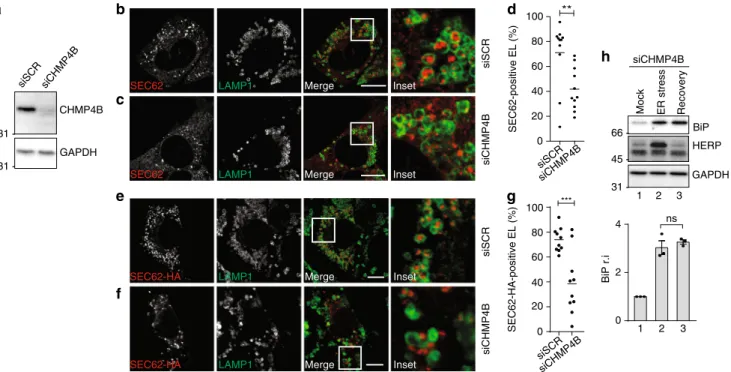
CHMP4B and VPS4A intervention during ER stress
resolu-tion. Inward membrane invagination (i.e., reverse-topology
mem-brane remodeling and scission) is driven by ESCRT-III
41–44.
Consistently, silencing of the charged multivesicular body
pro-tein 4B (CHMP4B), an essential ESCRT-III subunit, prevents
capture of cytosolic proteins by inward endolysosomal
mem-brane budding
45. In our experimental setup, silencing of
CHMP4B expression (Fig.
5
a) substantially inhibits delivery of
EV within LAMP1-positive EL during recovery from ER stress
(Fig.
5
b–d) and when recov-ER-phagy is recapitulated by SEC62
induction (Fig.
5
e–g). Consistently, CHMP4B silencing delays
return of ER stress-induced chaperones at their pre-stress level
(Fig.
5
h, upper panel), without affecting return of HERP (middle
panel).
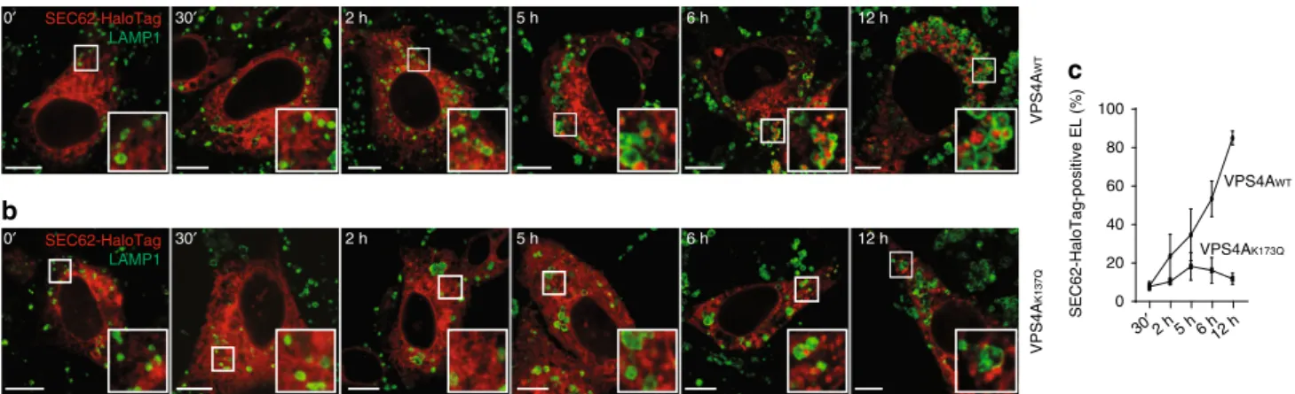
ESCRT-III-driven membrane remodeling and scission relies on
the energy delivered on ATP hydrolysis by the auxiliary AAA
+ATPase VPS4A
41–44. In our experiments, engulfment of
SEC62-positive EV by EL during recovery from ER stress was normal in
cells expressing
fluorescently labeled VPS4A
WT(Fig.
6
a, merge 1,
inset 1, Fig.
6
c), but it was substantially inhibited in cells
expressing VPS4A
K173Q, a dominant-negative mutant that cannot
bind and hydrolyze ATP
46(Fig.
6
b, merge 1, inset 1, Fig.
6
c). The
Fig. 1 Delivery of endogenous SEC62-labeled EV within EL during recovery from ER stress. a Upper panel, WB analysis showing steady-state level of BiP in WT MEF (Mock), BiP induction on cell exposure to CPA (ER stress, lane 2), and return of BiP to the pre-stress level after CPA wash-out (Recovery, lane 3); middle panel, same for HERP; lower panel, GAPDH as a loading control. Quantification of BiP levels in WB, n = 3 independent experiments, mean ± SEM, unpaired, two-tailed t test, *P= 0.0037. b Delivery of SEC62-decorated EV within LAMP1-positive EL in WT MEF during 12 h recovery from an ER stress in the presence of 50 nM BafA1.c Same as a in Atg4BKO MEF; n= 3 independent experiments, mean ± SEM, unpaired, two-tailed t test, P = 0.4001. d Same asb in Atg4BKO MEF. e Same as a in Atg7KO MEF, n= 3 independent experiments, mean ± SEM; unpaired, two-tailed t test, P = 0.3159. f Same as b in Atg7KO MEF. g Same as a in Atg16L1KO MEF; n = 4 independent experiments, mean ± SEM; unpaired, two-tailed t test, P = 0.6909. h Same as b in Atg16L1KO MEF. i Same as a in Ulk1/2 double-KO MEF; n = 3 independent experiments, mean ± SEM; unpaired, two-tailed t test, **P = 0.0083. j Same as b in Ulk1/2 double-KO MEF. k Same as a in Atg13KO MEF; n= 3 independent experiments, mean ± SEM; unpaired, two-tailed t test, *P = 0.0232. l Same as b in Atg13KO MEF. m Same as a in Atg14KO MEF; n= 3 independent experiments, mean ± SEM; unpaired, two-tailed t test, *P = 0.0497. n Same as b in Atg14KO MEF. See Supplementary Fig. 1 for recov-ER-phagy recapitulated on overexpression of SEC62-HA. o Quantification of SEC62-positive EV delivery within LAMP1-positive EL (n= 10, 11, 10, 10, 9, 10, 11 cells, respectively). One-way analysis of variance (ANOVA) and Dunnett’s multiple comparisons test,n.s.P > 0.05, ****P < 0.0001. Molecular weight markers in WB are in kDa. Scale bars for CLSM: 10 μm. WB and IF panels are representative of at least three
independent experiments EL EV EV
a
EV Inset SEC62-negative membrane SEC62-positive EV membrane EL membraneb
c
Macro-ER-phagyd
Micro-ER-phagy EV ER EL EL 1 2 3 1 2 3 EL EL Fusion Engulfment BafA1 BafA1 ER EV SEC62 LIR LIR EV EV EV SEC62 EV EV EV EL ELFig. 2 Distribution of gold-labeled, endogenous SEC62 by IEM. a WT MEFs are exposed 12 h to 10μM CPA. Recovery is initiated on CPA wash-out and progresses for 12 h in the presence of 50 nM BafA1 to inhibit clearance of EV delivered within EL.b Inset of a. IEM images are representative of two independent experiments with similar results.c Macro-ER-phagy pathway. d Micro-ER-phagy pathway. EV, ER-derived vesicle; EL, endolysosome; LIR, LC3-interacting region. Color code of arrows is given in the panels. Scale bar for IEM: 1μm
CRISPRWT
LAMP1 Merge Inset
SEC62
LAMP1 Merge Inset
SEC62
LAMP1 Merge Inset
SEC62 CRISPR17 CRISPR8 0 20 40 60 80 100
CRISPRWTCRISPR17CRISPR8 ns ns SEC62-positive EL (%)
f
c
d
e
45 45 CRISPRWTCRISPR17 STX17 GAPDHa
b
15 45 VAMP8 GAPDH CRISPRWT CRISPR8 CRISPR17Mock ER stress Recovery
1 BiP HERP GAPDH 66 45 -CRISPR8
Mock ER stress Recovery BiP HERP GAPDH 66 45
-g
h
0 1 2 3 ** 0 1 2 3 **BiP r.i BiP r.i
1 2 3 1 2 3
31 - 31
-2 3 1 2 3
Fig. 3 SNAREs dispensability for delivery of endogenous SEC62-labeled EV within EL. a WB analysis showing the efficiency of STX17 knockout in CRISPR17 MEF (please also refer to ref.19).b Same as a for VAMP8 in CRISPR8 MEF (please also refer to ref.19).c Delivery of endogenous SEC62-labeled EV within
LAMP1-positive EL during recovery from CPA-induced ER stress in WT MEF exposed to 50 nM BafA1 for 12 h.d Same as c in MEF lacking STX17. e Same as c in MEF lacking VAMP8. Scale bars: 10μm. f Quantification of EV delivery within EL in c–e (n = 15, 12, 10 cells, respectively). One-way ANOVA and Dunnett’s multiple comparisons test,n.s.P > 0.05. IF panels are representative of three independent experiments. g Same as Fig.1a in cells lacking STX17,
n = 4 independent experiments, mean ± SEM, unpaired, two-tailed t test, **P = 0.0083. h Same as Fig.1a in cells lacking VAMP8; n= 3 independent experiments, mean ± SEM, unpaired, two-tailed t test, **P= 0.0043. Molecular weight markers in WB are in kDa
a
EV EV EV EV EL EV EV EV EV ELd
LAMP1 Inset SEC62b
c
SEC62 (gold) EV EL 200 nmFig. 4 Engulfment of endogenous SEC62-labeled EV by EL. a WT MEF after 12 h recovery from an ER stress in the presence of 50 nM BafA1. IF panel is representative of >10 independent experiments.b Inset from a. c Single slice of an electron tomogram showing the distribution of gold-labeled endogenous SEC62 during recovery from ER stress. IEM image is representative of two independent experiments with similar results. Color code of arrows inb, c as in Fig.2a–d. d 3D visualization by electron tomography of EL containing EV that display gold-labeled, endogenous SEC62 at the limiting membrane. See also Supplementary Movie 1. Scale bar for CLSM: 10μm; scale bar for IEM: 200 nm
role of VPS4A in EV engulfment by LAMP1-positive EL was
confirmed on overexpression of SEC62-HA to faithfully
recapi-tulate recov-ER-phagy (Fig.
6
d–g). Ultrastructural analyses of
cells where the engulfment of excess ER by LAMP1-positive EL is
inhibited on inactivation of the ESCRT-III machinery show that
SEC62-positive EV remain in close proximity of the EL and are
not delivered within the degradative organelles (Fig.
6
h–k,
Supplementary Movie 2). Notably, VPS4A
WT, which drives EV
engulfment, accumulates within LAMP1-positive EL on
inactiva-tion of proteolytic enzymes with BafA1 (Fig.
6
a, merge 2, inset 2,
Fig.
6
e, merge 2, white arrows in inset 2 and in inset VPS4A). The
inactive VPS4A
K173Qis not found within EL (Fig.
6
b, merge 2,
inset 2, Fig.
6
f, merge 2, red arrows in inset 2 and in inset
VPS4A). This is consistent with partitioning of VPS4A within the
inward budding structure after the ESCRT-III mediated scission
event
44. Finally, we confirm VPS4A-dependent engulfment of
excess ER by LAMP1-positive EL by HaloTag pulse chase (Fig.
7
),
a protocol for time-resolved analyses of EV segregation recently
developed in our lab
19. Briefly, to monitor by time-resolved
fluorescence microscopy the sequential steps of ER subdomains
delivery within EL for clearance, WT MEF were transfected with
SEC62-HaloTag (Supplementary Fig. 3) and with VPS4A
WT(Fig.
7
a, c) or VPS4A
K173Q(Fig.
7
b, c). The fate of newly
synthesized SEC62-HaloTag is followed by pulsing cells, for
15 min, with the
fluorescent HaloTag ligand PBI 5030 (ref.
19legend of Fig.
7
and Materials and methods). Initially (0–2 h
chase), SEC62-HaloTag is not visible within LAMP1-positive EL.
Only in cells expressing VPS4A
WT, it is eventually delivered
within the EL, where it progressively accumulates on EL
inactivation with BafA1 (Fig.
7
a, c, 5–12 h chase). In cells
expressing inactive VPS4A
K173Q, SEC62-HaloTag remains
vir-tually excluded from the EL throughout the chase (Fig.
7
b, c).
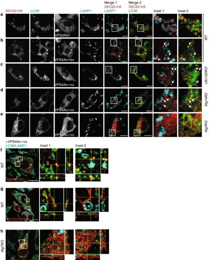
Endogenous LC3B decorates EV. In macro-autophagy, LC3 is
lipidated on the phagophore membrane and eventually recruits
cargo within double-membrane autophagosomes
31. Since
autop-hagosome biogenesis is dispensable for ER turnover at the end of
acute ER stresses, we assessed LC3 localization during
recov-ER-phagy. Our analyses reveal that endogenous LC3 co-localizes with
SEC62-labeled ER accumulating within LAMP1-positive EL on
inhibition of lysosomal activity with BafA1 (Fig.
8
a). The
inhi-bition of EV engulfment by LAMP1-positive EL on expression of
the inactive variant of VPS4A reveals the presence of endogenous
LC3B on SEC62-labeled structures both in WT MEF (Fig.
8
b) and
in MEF with defective autophagosome biogenesis (Fig.
8
c, d). The
vesicular nature of the SEC62/LC3-positive ER-derived structures
that deliver excess ER within EL during recovery from ER stress is
confirmed by orthogonal sections of deconvoluted images
(Fig.
8
f). The formation of EV is abolished in cells expressing a
SEC62LIR variant that cannot bind LC3
7(Fig.
8
g) and in Atg7KO
MEF with defective LC3 lipidation (Fig.
8
e, h). All in all, these
results support the notion that the ER-resident LC3-binding
protein SEC62, LC3 lipidation and the VPS4A-powered
ESCRT-III machinery, but not autophagosome biogenesis, regulate ER
fragmentation and piecemeal micro-ER-phagy characterizing the
ER turnover on ER stress resolution.
Discussion
The ER is a plastic organelle of eukaryotic cells, whose volume
and activities are adapted in response to intra- and extracellular
signals. The anabolic pathways that enlarge the ER and increase
its content in resident proteins and enzymes are collectively
defined as UPR and have been characterized in molecular
details
1,2. The catabolic pathways that reduce ER volume and
Merge Merge
a
SEC62-HA-positive EL (%) 0 20 40 60 80 100 siCHMP4B siSCR *** 31 -CHMP4B GAPDH siSCR siCHMP4B Mergee
LAMP1 SEC62-HA Inset siSCR siCHMP4B LAMP1SEC62-HA Merge Inset
f
g
LAMP1 SEC62 Inset LAMP1 SEC62 Inset siSCR siCHMP4B SEC62-positive EL (%) 31 - siSCR siCHMP4B 0 20 40 60 80 100 **b
c
d
66 45 -BiP HERP GAPDH siCHMP4BMock ER stress Recovery
1 0 4 2 BiP r.i
h
31 -2 3 1 2 3 nsFig. 5 A role of CHMP4B in recov-ER-phagy. a WB showing knockdown efficiency for siCHMP4B. b Same as Fig.1b in MEF transfected with a scrambled siRNA.c Same as b in cells transfected with a siRNA silencing CHMP4B expression. d Quantification of EV delivery within EL in b, c (n = 11, 11 cells, respectively). Unpaired, two-tailed t test, **P= 0.0052. e Same as b in cells where recov-ER-phagy is recapitulated on SEC62 overexpression. f Same as c in cells where recov-ER-phagy is recapitulated on SEC62 overexpression. g Quantification of EV delivery within EL in e, f (n = 10, 10 cells, respectively). Unpaired t test, ***P= 0.0006. Data are representative of at least two independent experiments. Scale bars: 10 μm. h Same as Fig.1a in cells transfected with a siRNA silencing CHMP4B expression. Quantification of BiP levels in WB, n = 3 independent experiments, mean ± SEM; unpaired, two-tailed t test, P = 0.4776. Molecular weight markers in WB are in kDa; n.s. not significant
content by delivering ER subdomains to acidic compartment for
destruction are much less understood. The identification of
sev-eral ER-resident proteins that may engage, upon appropriate
stimuli, cytosolic factors such as LC3s/Atg8, GABARAPs, and
FIP200/Atg11 to promote delivery of ER fragments within EL/
vacuole for clearance has opened the run to dissect
receptor-mediated, lysosomal-controlled, ER turnover pathways. At the
end of an ER stress, the reduction of the ER volume and of the ER
content in resident proteins to pre-stress, physiologic status is
crucial to re-establish ER homeostasis. In this phase, cells must,
among other things, also re-gain the capacity to produce the
secretOME (i.e., the 40% of the proteOME destined to the
organelles of the secretory and endocytic compartments,
the plasma membrane, and the extracellular space), whose
pro-duction is temporarily halted during the ER stress phase
47. The
secretOME enters the ER co-translationally via the SEC61 protein
translocation machinery that is endowed with a SEC62:SEC63
functional complex to facilitate access of those polypeptides that
are synthesized in the cytosol and imported only after completion
of the polypeptide chain via post-translational translocation
22,23.
We recently reported that conclusion of ER stresses activates the
LC3-binding function of SEC62 and triggers lysosomal turnover
of ER subdomains containing folding factors, but lacking ERAD
factors via catabolic pathways collectively defined as
recov-ER-phagy
7. The
finding that selective ER delivery to EL for clearance
is recapitulated by SEC62 overexpression and by SEC63 silencing
7hint at possible mutually exclusive role of SEC62 in ER protein
import vs. receptor-mediated, selective ER turnover. Notably, the
LC3-binding region is dispensable for the former and required for
the latter function of SEC62 (ref.
7and Supplementary Fig. 3c). It
will be of interest to verify whether the changes in newly
syn-thesized proteins getting access to the ER during steady state
e
d
f
SEC62-HA-positive EL (%) 0 20 40 60 80 100 VPS4A WT VPS4A K173Q VPS4A WT VPS4A K173Q Mock **** nsj
SEC62-HaloTag LAMP1 Inset EV EL EL EV EL EVk
h
VPS4A K137Q SEC62-HA (gold) EL EV EL EVi
SEC62 SEC62 SEC62-HA SEC62-HA SEC62-HA LAMP1 LAMP1 LAMP1 LAMP1 LAMP1 0 20 40 60 80 100 SEC62-positive (EL%) ***a
b
c
g
Inset 1Inset 1 Inset VPS4A
pcDNA3 VPS4AWT VPS4AK173Q VPS4AWT VPS4AK173Q Merge 1 SEC62 LAMP1 1 1 1 1 1 Merge 1 SEC62 LAMP1 2 Inset Inset Inset 2 Inset 2 Merge 2 VPS4A LAMP1 2 2 2 Merge 2 VPS4A LAMP1 2
Fig. 6 A role of VPS4A in recov-ER-phagy. a Same as Fig.1b in MEF transfected with a plasmid for expression of GFP-VPS4A (VPS4AWT).b Same as a in
cells transfected with a plasmid for expression of GFP-VPS4AK137Q(VPS4AK137Q).c Quantification of EV delivery within EL in a, b (n = 8, 8 cells,
respectively). Unpaired, two-tailed t test, ***P= 0.0002. Data are representative of three independent experiments with similar results. d Delivery of SEC62-HA within LAMP1-positive EL in MEF transfected with an empty plasmid exposed to BafA1 for 12 h.e Same as d in cells transfected with a plasmid for expression of GFP-VPS4AWT.f Same as d in cells transfected with a plasmid for expression of GFP-VPS4AK137Q.g Quantification of EV delivery within
EL ind–f (n = 10, 10, 9 cells, respectively); ANOVA and Dunnett’s multiple comparisons test,n.s.P > 0.05, ****P < 0.0001. Data are representative of at
least three independent experiments.h SEC62-HaloTag-positive EV outside LAMP1-positive EL in cells expressing VPS4AK137Q. See also Supplementary
Fig. 3.i inset of h. Scale bars: 10μm. j Single slice of an electron tomogram. k 3D visualization by electron tomography of SEC62-positive EV adjacent to EL. Forj, k, IEM images are representative of two independent experiments with similar results. Scale bar: 250 nm. See Supplementary Movie 2
(presumably more cargo) vs. ER stress (presumably more
ER-resident proteins) vs. recovery from ER stress (back to more cargo
proteins import?) phases results in variations in the SEC61:
SEC62:SEC63 oligomeric composition
22,23and if this in turn
generates orphan SEC62 to promote ER turnover. Alternatively,
the triggering signal for a switch in SEC62 function from protein
translocation to ER turnover could be a change in the luminal
fraction of free BiP (which is regulated in complex manner
48–50).
In fact, SEC63 is a BiP-binding protein and BiP association could
set SEC62 free for its role in recov-ER-phagy. All these issues will
be assessed in future work.
Here, we report that lipidation of LC3 is required, but the
autophagosome biogenesis machinery and macro-ER-phagy are
dispensable for clearance of excess ER characterizing the recovery
phase from acute ER stresses. As such, recov-ER-phagy, which is
triggered by an ER-centric signal, is mechanistically different from
starvation-induced turnover of the mammalian ER, which
acti-vates (unselective?) macro-autophagic clearance of ER
sub-domains decorated with FAM134B
6, RTN3
8, ATL3
9, CCPG1
10,
TEX264
11,12. Rather, SEC62-driven return of ER size and content
at pre-stress, physiologic status, entails the LC3 lipidation
machinery, the ESCRT-III component CHMP4B and the
acces-sory AAA
+ATPase VPS4A. During recovery from ER stress,
CHMP4B and VPS4A, whose involvement in physiologic and
pathogen-induced membrane repair, remodeling and
fission
events has been reported
43,44,51, ensure the inward budding of the
EL membrane required for the engulfment and clearance of
pre-formed ER vesicles that display SEC62 and LC3 at the limiting
membrane and contain excess ER chaperones and membranes
generated during the phase of ER stress. The function of LC3 in
recov-ER-phagy (and in other types of receptor-mediated ER
turnover by lysosomes/vacuole) as well as the mode of recruitment
of the ESCRT-III machinery that welcomes the incoming
ER-derived vesicle containing material to be cleared from cells
remains a matter for further studies. However, these data highlight
the variety of pathways that cells can activate in response to
pleiotropic and to ER-centric signals to ensure lysosomal turnover
of the ER. For the mechanistic dissection of catabolic regulation of
ER function via lysosomal-controlled ER turnover, it seems crucial
to examine in detail cellular responses to ER-centric signals
that may activate individual ER-resident LC3-binding proteins.
ER-centric signals activate client-specific autophagic and not
autophagic pathways that deliver fragmented ER subdomains
containing defective material (certainly misfolded proteins
15,17,
possibly aberrant lipids or ER-resident proteins, whose activity is
determined post-translationally by regulated turnover
29) to the
lysosomal/vacuolar compartments. Significant on this line, is the
characterization of ERLAD pathways that ensure disposal of ER or
of ER-exit site subdomains containing misfolded proteins that
cannot be dislocated across the membrane for proteasomal
degradation
15,17. Emerging evidence reveal the involvement in
ERLAD of mammalian ER-resident LC3-binding proteins such as
FAM134B
19,20and CCPG1
10and the yeast ER-resident
Atg8-binding protein Atg40
52. These receptors operate conventionally
to engage autophagosomes in macro-ER-phagy-like processes
20,52,
or unconventionally by ensuring direct delivery of the ER
sub-domain to be cleared from cells to EL via vesicular trafficking
19or
via poorly characterized micro-autophagy-like processes
21.
Back to recov-ER-phagy, the broader, possible implications of
our
findings relate to the fact that amplification of the SEC62 gene
and the consequent enhanced ER turnover confers a stress
tol-erance that correlates with resistance to anti-cancer therapies to a
number of carcinomas, including breast, prostate, thyroid, lung
adenocarcinoma,
and
head
and
neck
squamous
cell
carcinoma
53–55. Our characterization of the catabolic pathways
activated by cells recovering from ER stress to remodel the ER
paves the way for identification of therapeutic targets to treat
diseases caused by impaired proteostatic control and to unravel in
more detail the novel function of the ESCRT-III/VPS4A
machinery in membrane remodeling to better define molecular
aspects of organelle and membrane dynamics.
Materials and methods
Antibodies, expression plasmids, and chemicals. Antibodies against CNX (Western blot (WB) 1:3000, immunofluorescence (IF) 1:100), CHMP4B (1:1000), SEC62 (WB 1:1000, IF 1:100), and ERj3 (1:250) are kind gifts from A. Helenius, H. Stenmark, and R. Zimmermann. Plasmid encoding GFP-RAB7 is a kind gift from T. Johansen. Commercial antibodies used in this study are from Stressgen (BiP, 1:1000), Chondrex (HERP, 1:1500), Developmental Studies Hybridoma Bank (LAMP1, 1:50), Sigma and Santa Cruz (HA, WB 1:3000, IF 1:100), Merck Millipore (GAPDH, 1:30,000), Sigma (STX17, 1:1000), Abcam (VAMP8, 1:1000), Santa Cruz (Actin 1:500), Novus and Sigma (LC3B, WB 1:1000, IF 1:50), Promega (HaloTag, 1:1000), and MBL (p62, 1:1500). The secondary horse radish peroxidase (HRP)-conjugated antibodies were from Jackson Immunoresearch (rabbit, 1:10,000), Santa Cruz (goat, 1:20,000), and SouthernBiotech (mouse, 1:20,000). Alexa Fluor-conjugated secondary antibodies (1:300) from Invitrogen, Jackson
a
b
VPS4A WT VPS4A K137Qc
SEC62-HaloTag-positive EL (%) 0 20 40 60 80 100 30′2 h 5 h 6 h12 h VPS4AWT VPS4AK173Q SEC62-HaloTag LAMP1 SEC62-HaloTag LAMP1 30′ 2 h 5 h 6 h 0′ 30′ 2 h 5 h 6 h 12 h 0′ 12 hFig. 7 Time-resolved analyses of VPS4A-dependent delivery of EV within EL. a Cells expressing SEC62-HaloTag and VPS4AWTare incubated for 30 min
with 6-chlorohexanol, a non-fluorescent HaloTag ligand that quenches fluorescent detection of the SEC62-HaloTag chimera. Cells are subsequently incubated with a cell-permeablefluorescent HaloTag ligand (PBI 5030) to label, for 15 min, newly synthesized SEC62-HaloTag (fluorescent pulse). Cells arefixed at different times after interruption of the fluorescent pulse on PBI 5030 replacement with 6-chlorohexanol (0 to 12 h19) to monitor the
progressive delivery of SEC62-HaloTag-labeled EV within LAMP1-positive EL.b Same as a in MEF expressing SEC62-HaloTag and VPS4AK137Q, where EV
delivery within EL is virtually abolished.c Quantification of SEC62-HaloTag positive EL (VPS4AWTn = 3, 3, 3, 4, 4; VPS4AK173Qn = 3, 3, 4, 4, 4 cells for the
SEC62-HA LC3B VPS4AWT LAMP1 VPS4AK173Q VPS4AK173Q VPS4AK173Q Merge 1 SEC62-HA LAMP1 Merge 2 SEC62-HA LC3B Inset 1 Inset 2 WT Atg14K O Ulk1/2DKO Inset 1 Inset 2 1 2
c
a
b
d
e
1 2 1 2 1 2 1 2 1 2 1 2 WT WT Atg7 KO SEC62-HA SEC62-HA SEC62LIR-HA +VPS4AK173Q LC3B/LAMP1 Atg7 KO VPS4AK173Q 1 2f
g
h
Fig. 8 LC3B localization on SEC62-positive EV. a SEC62-HA co-localizes with endogenous LC3B inside LAMP1-positive EL in WT MEF-expressing VPS4AWTexposed to BafA1 for 12 h.b SEC62-HA/LC3B-positive EV (white arrows) fails to be delivered inside the EL in WT MEF-expressing VPS4AK173Q.
c Same as b in Ulk1/2 double-KO MEF. d Same as b in Atg14KO MEF. e Same as b in Atg7KO MEF. f Max projection of a WT cell expressing SEC62-HA and VPS4AK173Qafter deconvolution. Insets show orthogonal sections of selected region.g Same as in f with SEC62LIR-HA. h Same as f in Atg7KO MEF cells.
Immunoresearch, Thermo Fisher, and BioLegend. Plasmids encoding GFP-VPS4AWTand GFP-VPS4AK173Qwere a kind gift from John McCullough. SEC62
was subcloned in a pcDNA3.1 expression plasmid with the addition of a C-terminal HA-tag or HaloTag7. CPA (Sigma) and BafA1 (Sigma) were used for 12 h,
if not otherwise specified, at final concentrations of 10 μM and 50 nM, respectively. PS341 (Millennium Pharmaceuticals) was used for 8 h at afinal concentration of 10μM.
Induction of transient ER stress. To induce a mild and reversible ER stress, cultured cells were exposed for up to 12 h to CPA, a reversible inhibitor of the sarco/ER calcium ATPase7,27. An alternative protocol implies cell exposure to
dithiothreitol7(DTT, a reversible perturbator of redox homeostasis56), whereas
other ER stress-inducing drugs such as tunicamycin and thapsigargin, which are irreversible inhibitors of GlcNAc phosphotransferase57and of the sarco/ER
cal-cium ATPase58, respectively, are toxic7. Consistent with UPR induction, CPA
treatment caused splicing of XBP1 transcripts, 25% attenuation of global protein synthesis, and induction of ER stress marker transcripts and proteins7. The
cata-bolic events characterizing recovery from ER stress (i.e., the delivery of ER sub-domains within EL for clearance) were investigated after CPA wash-out as specified in thefigure legends and in ref.7.
Cell culture, transient transfection, and RNA interference. MEF and HEK cells were grown at 37 °C and 5% CO2in Dulbecco’s modified Eagle’s medium (DMEM)
medium supplemented with 10% fetal calf serum (FCS). WT and Atg7KO MEF cells were gifts from M. Komatsu. Atg4BKO, Atg13KO, Atg14 and Atg16L1KO, and Ulk1/2DKO MEF cells were kindly provided by G. Marino, F. Reggiori, T. Saitoh, and S. Tooze, respectively, and have been characterized in ref.19and in
Supple-mentary Fig. 2. To induce autophagy, cells were washed three times with Earle’s balanced salt solution (EBSS, Thermo Fisher) and then incubated in EBSS for 2 h in the presence or absence of 100 nM BafA1.
Transient transfections were performed using JetPrime transfection reagent (PolyPlus) according to the manufacturer’s instructions. RNA interferences were performed in MEF cells plated at 50–60% confluence. Cells were transfected with scrambled small interfering RNA or small interfering RNA (siRNA) against CHMP4B (5′-AAACAGUCCCUCUACCAAAtt-3′, 50 nM per dish, Silencer Select Pre-designed, Ambion). Cells were processed for immunofluorescence or for biochemical analyses 48 h after transfection (see below).
Preparation of KO cells. STX17- and VAMP8-KO MEF and SEC62-KO HEK cells were generated by CRISPR/Cas9 genome editing. For the generation of the guideRNA‐Cas9 plasmids, lentiCRISPRv2‐puro system (Addgene52961) was obtained from Addgene (http://www.addgene.org). Guide sequences were obtained from the Cas9 target design tools (crispr.mit.edu:8079 and/orwww.addgene.org/ pooled-library). All protocols and information can be found at the websitehttps:// www.addgene.org/crispr. The target sequences for guide RNA were synthesized by Microsynth. Two annealed oligonucleotides (5′-CACCGCTGTGGTTGACTACTG CAAC-3′, 5′-AAACGTTGCAGTAGTCAACCACAGC-3′ for human SEC62; 5′‐G CGCTCCAATATCCGAGAAA‐3′, 5′‐TTTCTCGGATATTGGAGCGC‐3′ for murine STX17; 5′‐CCACCTCCGAAACAAGACAG‐3′, 5′‐CTGTCTTGTTTCGG AGGTGG‐3′ for murine VAMP8) were inserted into the lentiCRISPRv2‐puro vector using the BsmBI restriction site. Vectors were transfected in HEK and MEF cells with JetPrime (Polyplus) according to the manufacturer’s instructions7,19.
Cells were cultured in DMEM supplemented with 10% FBS. Two days after transfection, the medium was supplemented with 2μg/ml puromycin. After 10 days, puromycin‐resistant clones were picked and gene KO was verified by WB (ref.7for SEC62; Fig.3a, b and ref.19for STX17 and VAMP8) and with a
translocation assay for SEC62 (ref.7and Supplementary Fig. 3c).
Cell lysis, Western blot. Cells were washed in ice cold phosphate-buffered saline (PBS) containing 20 mM N-ethylmaleimide (NEM) and lysed in 1% sodium dodecyl sulfate (SDS) in HEPES-buffered saline, pH 6.8, or with RIPA buffer (1% Triton X-100, 0.1% SDS, 0.5% sodium deoxycholate in HBS, pH 7.4) containing 20 mM NEM and protease inhibitor cocktail (1 mM PMSF, chymostatin, leupeptin, antipain, and pepstatin, 10μg/ml each). Post-nuclear supernatants were collected by centrifugation at 10,000 × g for 10 min at 4 °C. Samples were denatured and reduced in DTT-containing sample buffer for 5 min at 95 °C and separated by SDS-PAGE (polyacrylamide gel electrophoresis). Proteins were transferred to poly-vinylidenefluoride membranes with the Trans-Blot Turbo Transfer System (Bio-Rad). Membranes were blocked 10 min with 10% (w/v) non-fat dry milk (Bio-Rad) and stained with the above-mentioned primary antibodies for 90 min and for 45 min with HRP-conjugated secondary antibodies. Membranes were developed using the Luminata Forte ECL detection system (Millipore) and signals were acquired with the ImageQuant LAS 4000 system (GE Healthcare Life Sciences) or with the Amersham Imager 680 system. Image quantifications were performed with the Multi Gauge Analysis tool (Fujifilm). Membrane stripping was performed using Re-Blot Plus Strong Solution (Millipore) following the manufacturer’s instructions. Uncropped blots can be found as Supplementary Fig. 4.
Confocal laser scanning microscopy. MEF cells plated on alcian blue-coated glass coverslips were treated according to the experimental setup. Cells were washed twice in PBS andfixed at room temperature for 20 min with 3.7% formaldehyde in PBS or 5 min in 100% methanol at−20 °C for endogenous LC3B detection. Cells were then incubated for 15 min with permeabilization solution (PS, 0.05% saponin, 10% goat serum, 10 mM HEPES, 15 mM glycine, pH 7.4) to improve antigen accessibility. Cells were incubated with the primary antibodies diluted 1:50–1:200 in PS for 90 min, washed for 15 min in PS, and then incubated with Alexa Fluor-conjugated secondary antibodies diluted 1:300 in PS for 30 min. Cells were rinsed with PS and water and mounted with Vectashield (Vector Laboratories) supple-mented with 4′,6-diamidino-2-phenylindole. Confocal pictures were acquired using a Leica TCS SP5 microscope with a 63.0 × 1.40 Oil UV objective. FIJI was used for image analysis and processing. Figure8f–h were acquired with LEICA HCX PL APO CS 100.0 × 1.44 Oil UV objective with a XY pixel size of 50 nm and Z step of 125 nm and pinhole 0.8 AU. Images were deconvoluted with Autoquant 3.1.1 (Media Cybernetics) with a spherical aberration correction.
Immunogold electron microscopy. Cells were plated on alcian blue-coated glass coverslips andfixed 10 min with 0.05% glutaraldehyde in 4% paraformaldehyde (PFA) EM grade and 0.2 M HEPES buffer and 50 min in 4% PFA EM grade in 0.2 M HEPES buffer. After three washes in PBS, cells were incubated 10 min with 50 mM glycine and blocked 1 h in blocking buffer (0.2% bovine serum albumin, 5% goat serum, 50 mM NH4Cl, 0.1% saponin, 20 mM PO4buffer, 150 mM NaCl).
Staining with primary antibodies and nanogold-labeled secondary antibodies (Nanoprobes) were performed in blocking buffer at room temperature. Cells were fixed 30 min in 1% glutaraldehyde and nanogold was enlarged with gold enhancement solution (Nanoprobes) according to the manufacturer’s instructions. Cells were postfixed with osmium tetroxide, embedded in epon, and processed into ultrathin slices. After contrasting with uranyl acetate and lead citrate, the sections were analyzed with Zeiss LEO 512 electron microscope. Images were acquired by 2k × 2k bottom-mounted slow-scan Proscan camera controlled by the EsivisionPro 3.2 software.
HaloTag pulse-chase analyses. MEF cells were plated on alcian blue-coated glass coverslip and transfected with SEC62-HaloTag and VPS4WTor VPS4K173Q.
Seventeen hours after transfection, cells were incubated with 15μM 6-chlorohexanol (Sigma) in DMEM 10% FCS for 30 min. 6-Chlorohexanol is cell-permeable black ligand that irreversibly binds the SEC62-HaloTag-binding pocket. After three washes in DMEM 10% FCS, cells are pulsed 15 min with 1μM of the fluorescent ligand PBI 5030 (Promega), which exclusively enters the HaloTag ligand binding pocket of newly synthetized SEC62-HaloTag, and 100 nM BafA1 in DMEM 10% FCS. Thefluorescent ligand is removed and after three washes in DMEM 10% FCS, cells are incubated with 15μM of the black ligand to block incorporation of thefluorescent ligand in the newly synthetized SEC62-HaloTag, and 100 nM BafA1 in DMEM 10% FCS. Cells werefixed at increasing time points (0, 30 min, 2, 5, 6, and 12 h) and processed for confocal laser scanning microscopy as described above. HaloTag pulse chase has been described in ref.19.
Statistical analyses and reproducibility. In panels showing WB or IF, unless stated otherwise, images are representative of three independent experiments with similar results. The number of independent experiments or cell numbers is given in thefigure’s legend. Statistical analyses were performed only if sample size was ≥3 using GraphPad Prism 7 software. One-way analysis of variance with Dunnett’s multiple comparisons test or unpaired, two-tailed t test were used to asses statistical significance. P values are given in the figure legends;n.sP < 0.05; *P < 0.05; **P < 0.01; ***P < 0.001; ****P < 0.0001.
Reporting summary. Further information on research design is available in the Nature Research Reporting Summary linked to this article.
Data availability
The data that support thefindings of this study are available from the authors on reasonable request. The source data underlying thefigures can be found in the Source Datafile.
Received: 3 June 2019; Accepted: 10 October 2019;
References
1. Walter, P. & Ron, D. The unfolded protein response: from stress pathway to homeostatic regulation. Science 334, 1081–1086 (2011).
2. Wang, M. & Kaufman, R. J. Protein misfolding in the endoplasmic reticulum as a conduit to human disease. Nature 529, 326–335 (2016).
3. Smith, M. & Wilkinson, S. ER homeostasis and autophagy. Essays Biochem. 61, 625–635 (2017).
4. Fregno, I. & Molinari, M. Endoplasmic reticulum turnover: ER-phagy and otherflavors in selective and non-selective ER clearance. F1000Res 7, 454 (2018).
5. Locke, M. & Collins, J. V. The structure and formation of protein granules in the fat body of an insect. J. Cell Biol. 26, 857–884 (1965).
6. Khaminets, A. et al. Regulation of endoplasmic reticulum turnover by selective autophagy. Nature 522, 354–358 (2015).
7. Fumagalli, F. et al. Translocon component Sec62 acts in endoplasmic reticulum turnover during stress recovery. Nat. Cell Biol. 18, 1173–1184 (2016).
8. Grumati, P. et al. Full length RTN3 regulates turnover of tubular endoplasmic reticulum via selective autophagy. Elife 6, e25555 (2017).
9. Chen, Q. et al. ATL3 is a tubular ER-phagy receptor for GABARAP-mediated selective autophagy. Curr. Biol. 29, 846–855 (2019).
10. Smith, M. D. et al. CCPG1 is a non-canonical autophagy cargo receptor essential for ER-phagy and pancreatic ER proteostasis. Dev. Cell 44, 217–232 (2018).
11. An, H. et al. Tex264 is an endoplasmic reticulum-resident ATG8-interacting protein critical for ER remodeling during nutrient stress. Mol. Cell 74, 891–908 (2019).
12. Chino, H., Hatta, T., Natsume, T. & Mizushima, N. Intrinsically disordered protein TEX264 mediates ER-phagy. Mol. Cell 74, 909–921 (2019). 13. Mochida, K. et al. Receptor-mediated selective autophagy degrades the
endoplasmic reticulum and the nucleus. Nature 522, 359–362 (2015). 14. Loi, M., Fregno, I., Guerra, C. & Molinari, M. Eat it right: ER-phagy and
RecovER-phagy. Biochem. Soc. Trans. 46, 699–706 (2018).
15. Wilkinson, S. ER-phagy: shaping up and destressing the endoplasmic reticulum. FEBS J. 286, 2645–2663 (2019).
16. Wilkinson, S. Emerging principles of selective ER autophagy. J. Mol. Biol. S0022-2836, 30279–7 (2019).
17. Fregno, I. & Molinari, M. Proteasomal and lysosomal clearance of faulty secretory proteins: ER-associated degradation (ERAD) and ER-to-lysosome-associated degradation (ERLAD) pathways. Crit. Rev. Biochem. Mol. Biol. 54, 153–163 (2019).
18. Grumati, P., Dikic, I. & Stolz, A. ER-phagy at a glance. J. Cell Sci. 131, 1–6 (2018).
19. Fregno, I. et al. ER-to-lysosome-associated degradation of proteasome-resistant ATZ polymers occurs via receptor-mediated vesicular transport. EMBO J. 37, e99259 (2018).
20. Forrester, A. et al. A selective ER-phagy exerts procollagen quality control via a Calnexin-FAM134B complex. EMBO J. 38, e99847 (2019).
21. Omari, S. et al. Noncanonical autophagy at ER exit sites regulates procollagen turnover. Proc. Natl Acad. Sci. USA 115, E10099–E10108 (2018).
22. Conti, B. J., Devaraneni, P. K., Yang, Z. Y., David, L. L. & Skach, W. R. Cotranslational stabilization of Sec62/63 within the ER Sec61 translocon is controlled by distinct substrate-driven translocation events. Mol. Cell 58, 269–283 (2015).
23. Jadhav, B. et al. Mammalian SRP receptor switches the Sec61 translocase from Sec62 to SRP-dependent translocation. Nat. Commun. 6, 1–11 (2015). 24. Feldman, D., Swarm, R. L. & Becker, J. Elimination of excess smooth
endoplasmic reticulum after phenobarbital administration. J. Histochem. Cytochem. 28, 997–1006 (1980).
25. Bolender, R. P. & Weibel, E. R. A morphometric study of the removal of phenobarbital-induced membranes from hepatocytes after cessation of treatment. J. Cell Biol. 56, 746–761 (1973).
26. Meyer, H. A. et al. Mammalian Sec61 is associated with Sec62 and Sec63. J. Biol. Chem. 275, 14550–14557 (2000).
27. Pirot, P. et al. Global profiling of genes modified by endoplasmic reticulum stress in pancreatic beta cells reveals the early degradation of insulin mRNAs. Diabetologia 50, 1006–1014 (2007).
28. Yan, L. et al. Ube2g2-gp78-mediated HERP polyubiquitylation is involved in ER stress recovery. J. Cell Sci. 127, 1417–1427 (2014).
29. Bernasconi, R. & Molinari, M. ERAD and ERAD tuning: disposal of cargo and of ERAD regulators from the mammalian ER. Curr. Opin. Cell Biol. 23, 176–183 (2011).
30. Hagiwara, M., Ling, J. J., Koenig, P. A. & Ploegh, H. L. Posttranscriptional regulation of glycoprotein quality control in the endoplasmic reticulum is controlled by the E2 Ub-conjugating enzyme UBC6e. Mol. Cell 63, 753–767 (2016).
31. Bento, C. F. et al. Mammalian autophagy: how does it work? Annu. Rev. Biochem. 85, 685–713 (2016).
32. McAlpine, F., Williamson, L. E., Tooze, S. A. & Chan, E. Y. W. Regulation of nutrient-sensitive autophagy by uncoordinated 51-like kinases 1 and 2. Autophagy 9, 361–373 (2013).
33. Kaizuka, T. & Mizushima, N. Atg13 is essential for autophagy and cardiac development in mice. Mol. Cell. Biol. 36, 585–595 (2016).
34. Kageyama, S. et al. The LC3 recruitment mechanism is separate from Atg9L1-dependent membrane formation in the autophagic response against Salmonella. Mol. Biol. Cell 22, 2290–2300 (2011).
35. Itakura, E., Kishi-Itakura, C. & Mizushima, N. The hairpin-type tail-anchored SNARE syntaxin 17 targets to autophagosomes for fusion with endosomes/ lysosomes. Cell 151, 1256–1269 (2012).
36. Matsui, T. et al. Autophagosomal YKT6 is required for fusion with lysosomes independently of syntaxin 17. J. Cell Biol. 217, 2633–2645 (2018). 37. Tekirdag, K. & Cuervo, A. M. Chaperone-mediated autophagy and endosomal
microautophagy: jointed by a chaperone. J. Biol. Chem. 293, 5414–5424 (2018).
38. Lefebvre, C., Legouis, R. & Culetto, E. ESCRT and autophagies: endosomal functions and beyond. Semin. Cell Dev. Biol. 74, 21–28 (2018).
39. Schuck, S., Gallagher, C. M. & Walter, P. ER-phagy mediates selective degradation of endoplasmic reticulum independently of the core autophagy machinery. J. Cell Sci. 127, 4078–4088 (2014).
40. Lipatova, Z. & Segev, N. in Autophagy: Cancer, Other Pathologies, Inflammation, Immunity, Infection, and Aging (ed. Hayat, M. A.) (Elsevier, 2018).
41. Schoneberg, J. et al. ATP-dependent force generation and membrane scission by ESCRT-III and Vps4. Science 362, 1423–1428 (2018).
42. Raiborg, C. & Stenmark, H. The ESCRT machinery in endosomal sorting of ubiquitylated membrane proteins. Nature 458, 445–452 (2009).
43. McCullough, J., Frost, A. & Sundquist, W. I. Structures, functions, and dynamics of ESCRT-III/Vps4 membrane remodeling andfission complexes. Annu. Rev. Cell Dev. Biol. 34, 85–109 (2018).
44. Schoneberg, J., Lee, I. H., Iwasa, J. H. & Hurley, J. H. Reverse-topology membrane scission by the ESCRT proteins. Nat. Rev. Mol. Cell. Biol. 18, 5–17 (2017).
45. Mejlvang, J. et al. Starvation induces rapid degradation of selective autophagy receptors by endosomal microautophagy. J. Cell Biol. 217, 3640–3655 (2018).
46. Bishop, N. & Woodman, P. ATPase-defective mammalian VPS4 localizes to aberrant endosomes and impairs cholesterol trafficking. Mol. Biol. Cell 11, 227–239 (2000).
47. Uhlen, M. et al. Tissue-based map of the human proteome. Science 347, 1260419 (2015).
48. Preissler, S. et al. AMPylation matches BiP activity to client protein load in the endoplasmic reticulum. Elife 4, e12621 (2015).
49. Preissler, S. & Ron, D. Early events in the endoplasmic reticulum unfolded protein response. Cold Spring Harb. Perspect. Biol. 11, 1–18 (2019). 50. Chambers, J. E., Petrova, K., Tomba, G., Vendruscolo, M. & Ron, D. ADP
ribosylation adapts an ER chaperone response to short-termfluctuations in unfolded protein load. J. Cell Biol. 198, 371–385 (2012).
51. Radulovic, M. & Stenmark, H. ESCRTs in membrane sealing. Biochem. Soc. Trans. 46, 773–778 (2018).
52. Cui, Y. et al. A COPII subunit acts with an autophagy receptor to target endoplasmic reticulum for degradation. Science 365, 53–60 (2019). 53. Linxweiler, M., Schick, B. & Zimmermann, R. Let’s talk about Secs: Sec61,
Sec62 and Sec63 in signal transduction, oncology and personalized medicine. Signal Transduct. Target Ther. 2, 1–10 (2017).
54. Bergmann, T. J., Fumagalli, F., Loi, M. & Molinari, M. Role of SEC62 in ER maintenance: a link with ER stress tolerance in SEC62-overexpressing tumors? Mol. Cell Oncol. 4, e1264351 (2017).
55. Takacs, F. Z. et al. Identification of 3q oncogene SEC62 as a marker for distant metastasis and poor clinical outcome in invasive ductal breast cancer. Arch. Gynecol. Obstet. 299, 1405–1413 (2019).
56. Braakman, I., Helenius, J. & Helenius, A. Manipulating disulfide bond formation and protein folding in the endoplasmic reticulum. EMBO J. 11, 1717–1722 (1992).
57. Takatsuki, A. & Tamura, G. Effect of tunicamycin on synthesis of macromolecules in cultures of chick embryofibroblasts infected with Newcastle Disease Virus. J. Antibiot. 24, 785–794 (1971).
58. Lytton, J., Westlin, M. & Hanley, M. R. Thapsigargin inhibits the sarcoplasmic or endoplasmic reticulum Ca-ATPase family of calcium pumps. J. Biol. Chem. 266, 17067–17071 (1991).
Acknowledgements
We thank G. Brambilla Pisoni, F. Fumagalli, A. Helenius, M. Komatsu, G. Marino, N. Mizushima, J. Noack, P. Paganetti, F. Reggiori, T. Saitoh, C. Settembre, S. Tooze, X. Wang, and the members of Molinari’s lab for gift of reagents, discussions, and critical reading of the manuscript. We are grateful to the ALEMBIC facility at San Raffaele Scientific Institute, Milan, Italy for the help in electron microscopy analyses. M.M. is supported by Signora Alessandra, AlphaONE Foundation, Foundation for Research on Neurodegenerative Diseases, Swiss National Science Foundation, and Comel and Gelu Foundations.
Author contributions
Conceptualization: M.M. together with M.L.; methodology: M.L., A.R., D.M., and M.M.; investigation: M.L., A.R., D.M., and M.M.; writing original draft: M.M.; supervision: M.M.
Competing interests
The authors declare no competing interests.
Additional information
Supplementary informationis available for this paper at https://doi.org/10.1038/s41467-019-12991-z.
Correspondenceand requests for materials should be addressed to M.M.
Peer review informationNature Communication thanks Simon Wilkinson and other, anonymous, reviewers for their contributions to the peer review of this work. Peer review reports are available.
Reprints and permission informationis available athttp://www.nature.com/reprints
Publisher’s note Springer Nature remains neutral with regard to jurisdictional claims in published maps and institutional affiliations.
Open Access This article is licensed under a Creative Commons Attribution 4.0 International License, which permits use, sharing, adaptation, distribution and reproduction in any medium or format, as long as you give appropriate credit to the original author(s) and the source, provide a link to the Creative Commons license, and indicate if changes were made. The images or other third party material in this article are included in the article’s Creative Commons license, unless indicated otherwise in a credit line to the material. If material is not included in the article’s Creative Commons license and your intended use is not permitted by statutory regulation or exceeds the permitted use, you will need to obtain permission directly from the copyright holder. To view a copy of this license, visithttp://creativecommons.org/ licenses/by/4.0/.