CASE MANAGEMENT AND CLINICAL CONSEQUENCES
Sudden death after open gastric bypass surgery
Bettina Karin Wölnerhanssen&Igor Langer&Urs Eriksson&Markus Schneider
Received: 19 March 2008 / Accepted: 10 June 2008 / Published online: 3 July 2008
# Springer-Verlag 2008
Abstract
Purpose Gastric bypass surgery has become a relatively low-risk bariatric surgical intervention in a high-risk patient population (Nguyen et al., Arch Surg, 141:445–449,2006; Buchwald et al. JAMA, 13:1724–1737, 2004). Surgical interventions in patients suffering from morbid obesity are typically associated with excess morbidity (Parikh et al., Am Surg, 73:959–962, 2007). Though overall mortality after bariatric surgery is <1% is low (Mason et al., Obes Surg, 17:9–14, 2007), some surgical complications such as anastomotic leaks, staple line disruption and bowel ob-struction may still impact on postoperative outcome (Parikh et al., Am Surg, 73:959–962, 2007; Mason et al., Obes Surg, 17:9–14,2007). Early symptoms are often missed, as clinical presentation may be discreet, inexistent or falsely attributed to obesity.
Methods This case report refers to a patient in whom discomfort and agitation associated with a rise in temper-ature heralded a fulminant septic shock syndrome precip-itating his death. Literature on early complications and management after gastric bypass is reviewed.
Conclusion A high level of suspicion should be present in the case of an unexpected postoperative deterioration of the patient’s general condition. Time to treat may be very short (Mason et al., Obes Surg, 17:9–14, 2007). Computed tomography is mandatory to rule out pulmonary embolism and bypass obstruction.
Keywords Morbid obesity . Bariatric surgery . Mortality . Complication . Septic shock
Case report
After many years of unsuccessful conservative treatment attempts, a 40-year-old morbidly obese male patient with a BMI of 69 kg·m−2 (220 kg, 179 cm) was scheduled for open gastric bypass surgery. The patient suffered from obstructive sleep apnoea, hypertension and venous insufficiency with chronic crural ulcer. He fulfilled the high-risk criteria of the Obesity Surgery Mortality Risk Score (OS-MRS) [5]. Medication consisted of 5 mg torasemide/day. Preoperative diagnostics included gastros-copy, which showed normal findings; whereas pulmonary function testing revealed a restrictive pattern of respiratory compromise.
Laboratory findings were all normal and a chest X-ray showed no abnormality.
Anaesthesia Only ranitidine and metoclopramide were given as premedication, and a second generation cephalo-sporin was administered 60 min prior to the start of the operation. Electrocardiography and pulse oximetry were completed by continuous blood pressure monitoring via a radial artery line, which was also used for intermittent blood gas control. Before anaesthesia, a subclavian central venous access was established without complication. In addition, a 20 G multiport epidural catheter was inserted at midthoracic level (Th7) after having identified the epidural space at a depth of 14 cm using a 15 cm Tuohy needle. After a test dose of 0.5% bupivacaine with epinephrine (1:200 000), epidural anaesthesia was based on an initial bolus of 0.5% bupivacaine followed by a continuous infusion of 0.25% bupivacaine at 3–4 ml·h−1. General anaesthesia was induced with thiopental and fentanyl, and was maintained with isoflurane. Tracheal intubation was facilitated by succinylcholine; and intraoperative muscle
B. K. Wölnerhanssen (*)
:
I. Langer:
U. Eriksson:
M. Schneider University Hospital of Basel,Spitalstrasse 21, 4031, Basel, Switzerland e-mail: [email protected]
relaxation was achieved by bolus administration of atracu-rium. Anaesthetic management was uneventful. Intraoper-ative fluid losses including a urine output of 525 ml and a blood loss of less than 500 ml were made up for by administration of 5,700 ml Ringer’s lactate and 500 ml 6% hydroxyethyl starch over the 210 min of surgery. For haemodynamic stability a total dose of 20 mg intravenous ephedrine was administered. At the end of surgery, the patient’s trachea was extubated as soon as there was evidence of full recovery of consciousness and spontaneous breathing.
Surgery After laparotomy by bicostal incision and chole-cystectomy, a 30 ml gastric pouch was formed. A jejunal loop was prepared and dissected 50 cm distal to the ligament of Treitz; and the distal staple line of the jejunum was covered with a running suture and brought up to the gastric pouch as alimentary limb retrocolically and ante-gastrically. A manual side-to-side gastrojejunostomy was performed with interrupted Maxon 4.0 sutures. Finally, an end-to-side jejuno-jejunostomy was performed 150 cm distal to the gastrojejunostomy. All mesenteric defects were closed to prevent from potential internal hernias. No suction drains were placed.
Postoperative course The patient spent the first uneventful postoperative night in the intensive care unit before being transferred to the ward. On the first postoperative day, however, a rise in temperature to 38.5°C and an elevated white blood cell count were interpreted as an inflammatory response (postaggression syndrome) frequently encountered during the first 48 h after surgery (Table1). There was no clinical evidence of a surgical complication; a control contrast swallow performed on the following day showed neither signs of contrast media leakage nor any indication of functional obstruction of the surgical anastomoses (Table 2). Therefore, the patient was allowed to drink up to 500 ml·24 h−1. On postoperative day 2, there was a further increase in temperature, peaking at 40.4°C, which was associated with weakness and dyspnoea. Upon phys-ical examination, the abdomen was soft. In particular, there
was neither abdominal distension nor abnormal tenderness. Blood samples were taken for culture only (no blood gas analysis was carried out, Table3) and the patient was given oxygen, antipyretics and amoxicillin/clavulanic acid. Addi-tional intravenous fluids and a single bolus of 10 mg furosemide were given, resulting in a urinary output of 70 ml·h−1. Within a few hours, progressive haemodynamic instability developed in absence of overt tachycardia. The rapidly worsening clinical condition was accompanied by increasing hypoxemia, continuing restlessness and breath-lessness culminating in acute cardiorespiratory arrest during the transfer of the patient to the intensive care unit. ECG monitoring revealed that sinus tachycardia had turned into an idioventricular escape rhythm followed by asystole. Echocardiography and ultrasound during resuscitation excluded pericardial effusion and showed no signs of acute right ventricular pressure overload. No intraabdominal or retroperitoneal fluid was detectable. Despite aggressive fluid replacement and high dosages of epinephrine the patient died after almost 30 min of unsuccessful cardiopul-monary resuscitation.
Autopsy Septic shock was diagnosed as the primary cause of death. While E. coli was identified in the blood samples taken shortly before cardiorespiratory arrest, Enterobacter-iaceae were found in the swabs taken from the spleen during the autopsy. Two litres of feculent gastric content, fibrinous peritonitis of the small bowel and some ascites at the site of the gastric anastomosis were found. However, there was no evidence of an anastomotic leak. In addition, there were signs of an acute tracheobronchitis, whereas pulmonary thromboembolism could be ruled out. Cardiac findings showed a mild biventricular eccentric hypertrophy in the absence of any other pathology. In particular, no atherosclerosis was found.
Discussion
By the year 2040, one in two adults in the United States is expected to fulfil the criteria of obesity, defined by a body
Table 1 Signs of infection and clinical findings CRP (mg·l−1) WBC (109·l−1) Neutrophils(109·l−1) Temperature(°C) Normal values <10 <10 <6.7 <37 Admission 7 9.3 6.9 37.0 1st POD 8 15.2 13.8 37.0 2nd POD 95 10.6 9.0 38.7 3rd POD – – – 40.4
Legend: POD: postoperative day; WBC: white blood cell count
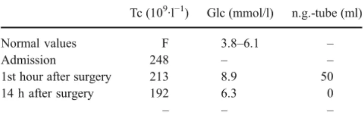
Table 2 Thrombocytes, glucose levels and drainage via naso-gastric tube in the first 24 hours after surgery
Tc (109·l−1) Glc (mmol/l) n.g.-tube (ml) Normal values F 3.8–6.1 –
Admission 248 – –
1st hour after surgery 213 8.9 50 14 h after surgery 192 6.3 0
– – –
mass index (BMI) above 30 kg·m−2, assuming that the present global trend of increasing prevalence of obesity persists over the coming decades [6]. Obesity is a disease which is associated with numerous comorbidities that reduce life expectancy if left untreated [7–9]. As a consequence, this has important economic implications for health care systems worldwide [10]. The results of surgical treatment of morbid obesity are by far more promising than those of conservative therapeutic approaches [11]. Bariatric surgery reduces not only morbidity but also mortality, as has been recently shown [7, 8]. Data provided by the American Society for Metabolic and Bariatric Surgery indicate that approximately 150,000 patients in the US underwent some form of surgery for obesity in 2004 [12]. This figure corresponds to a nearly six-fold increase since 1990. In a review of more than 18,000 gastric bypass operations, overall mortality has been reported to be as low as 0.24% [4]. Nevertheless, some surgical complications such as anastomotic leaks, staple line disruption and bowel obstruction may still impact on postoperative outcome [3, 4]. Early symptoms of these complications are often missed, as clinical presentation may be discreet, inexistent or falsely attributed to obesity, as in our patient. Furthermore, time to treat (TTT) is very short in some typical causes of death after gastric bypass surgery [4]. In our case, in the absence of ominous radiographic findings in the postoper-ative control images and showing no symptoms suggestive of peritonitis, the rise in temperature accompanied by tachypnea and restlessness were attributed to a pulmonary rather than an abdominal complication. Thus, misinterpre-tation of the worsening clinical condition and failure to recognise tachypnea as a criterion of systemic inflammatory response syndrome (SIRS) potentially caused a delay in transfer to intensive care. Furthermore, clinical signs of peritonitis may be overlooked in morbidly obese patients and even more so in the presence of epidural analgesia.
Pulmonary embolism associated with an undiagnosed deep venous thrombosis was considered to be the most probable pathophysiology underlying the acute cardiovas-cular collapse. In fact, at a rate of 30%, pulmonary embolism was the leading cause in a multicenter series of 93 cases of death after gastric bypass surgery, while a gastrointestinal leak accounted for 14%, peritonitis without
a leak for 11%, heart failure for 12%, haemorrhage for 7%, and bypass obstruction for 5%, respectively [4]. Even in patients receiving low molecular weight heparin prophy-laxis, lethal thromboembolism can never be ruled out with certainty [13]. In addition, a number of mediators released from metabolically active adipose tissue are apparently linked to a prothrombotic and proinflammatory state [14, 15]. Of note, the production and release of cytokines such as TNF-alpha and interleukin-6 from adipose tissue appears to be relevant for the pathophysiology underlying a broad array of comorbidities. As of now, the roles of these and other secretory products of adipose tissue and their relationship to inflammation in obesity are poorly under-stood and are the focus of ongoing research [16].
In our case, autopsy did not reveal a clear cause for the septic shock syndrome. Despite the absence of a typical anastomotic leak, the presence of feculent fluid in the gastric remnant in association with localized fibrinous peritonitis and ascites is suggestive of a microscopic leak as most probable cause of death in presence or absence of an underlying bypass obstruction. Bypass obstruction is one of the most urgent surgical complications, and the time to treat may be extremely short. The risk of fatal outcome within a lag of only 2 days after gastric bypass surgery is a matter of concern [4]. Even peritonitis in absence of a leak (with similar symptoms as in a microscopic leak) has been described as cause of death [4]. The issue should be raised as to whether and to what degree obesity contributed to the magnitude of sepsis triggered by a minor surgical compli-cation. Whenever a patient deteriorates after gastric bypass surgery and the cause is not clear, clinical evaluation should be supplemented by computed tomography of the thorax and abdomen in order to rule out pulmonary embolism and bypass obstruction.
A multidisciplinary approach is paramount for preoper-ative risk stratification [5,17]. Risk factors that may modify the perioperative course should be identified during preoperative assessment and include laboratory screening, a pulmonary function test and echocardiography, if appro-priate. Low-molecular-weight heparin prophylaxis for thromboembolism should be administered before surgery [18]. Compression stockings with or without sequential dynamic function may confer an additional benefit [19].
Table 3 Blood gas analysis
Normal i.o. 1st hour i.o. 4 hours p.o. 1st hour p.o. 14 hours pH 7.38–7.42 7.36 7.36 7.36 7.42 Bicarbonate 21–26 mmol/l 22.4 20.9 20.5 24.7 Base Excess −2.4–2.3 mmol/l −2.5 −3.7 −3.9 0.8 pCO2 5.0–5.5 KPA 5.4 5.0 4.9 5.2
pO2 >10.7 KPA 16.1 20.5 9.3 9.6
For perioperative blood pressure and blood gas monitor-ing, a radial arterial line should be used in extremely obese patients in whom non-invasive blood pressure monitoring— even if extra wide cuffs are used—is often unreliable. A central venous catheter should be used in patients whose peripheral veins are difficult to access. Blood volume status should be checked by hourly urine output monitoring and careful assessment of perioperative volume changes; since central venous pressure monitoring is unreliable in the presence of pneumoperitoneum and anti-Trendelenburg positioning. Bispectral index (BIS) monitoring of depth of anaesthesia is recommended as a means to tailor drug administration to the patient’s needs [20].
Both general anaesthesia and obesity have a direct bearing on the occurrence of postoperative respiratory impairment. The magnitude of which has been shown to be directly related to the BMI of patients undergoing laparotomy [21]. There-fore, administration of perioperative epidural anaesthesia is recommended as an effective means to reduce the impact of laparotomy on postoperative pulmonary function and to enhance postoperative recovery [22]. Obesity is a known risk factor for aspiration pneumonia resulting from regurgitation, therefore, administration of an H2 receptor antagonists and/ or metoclopramide is recommended [23]. As obesity is a risk factor for difficult intubation, equipment for difficult airway management should be readily available in the operating room. An awake fiberoptic intubation of the trachea should be considered whenever preoperative airway assessment suggests a high probability of a difficult airway [24]. Preoxygenation in a 25° head-up position rather than in supine position before induction of anaesthesia results in a 23% higher oxygen tension, thus, allowing more time for intubation and airway control [25, 26]. Similarly, positive end-expiratory pressure during induction increases the duration of non-hypoxic apnoea [27]. The volatile anaesthe-tic desflurane and the opioid remifentanil are recommended for maintenance of general anaesthesia since both agents have a very short half-life and are rapidly eliminated [24].
High quality postoperative care is the centrepiece of successful management after bariatric surgery. High-risk obese patients with a history of obstructive sleep apnoea syndrome warrant admission to an intensive care unit [28]. Severe obesity predisposes patients to postoperative respi-ratory complications such as atelectasis, particularly after open abdominal surgery [29]. Even though laparoscopic techniques decrease the severity of postoperative pain experience, the need for effective postoperative analgesia remains. Therefore, thoracic epidural analgesia is instru-mental in allowing early mobilisation, unhindered deep breathing and the use of incentive spirometry.
In conclusion, a high level of suspicion should be present in the case of an unexpected postoperative deterioration of the patient’s general condition. The
lag-time between onset of subtle symptoms and life-threatening exacerbation (TTT) may be very short [4], as evidenced again by this case. Such complications are best managed by a multidisciplinary skilled team approach. Computed tomography is mandatory to rule out pulmonary embolism and bypass obstruction.
Acknowledgements The authors wish to thank Dr. Allison Dwileski and Dr. Ralph Peterli for proof-reading and discussion.
References
1. Nguyen NT, Silver M, Robinson M et al (2006) Results of a national audit of bariatric surgery performed at academic centres: A 2004 University Health System Consortium Benchmarking Project. Arch Surg 141:445–449 doi:10.1001/archsurg.141.5.445
2. Buchwald H, Avidor Y, Braunwald E et al (2004) Bariatric surgery: a systematic review and meta-analysis. JAMA 13 (292):1724–1737 doi:10.1001/jama.292.14.1724
3. Parikh J, Yermilov I, McGory M, Jain S, Ko CY, Maggard M (2007) Is high BMI associated with specific complications after laparoscopic Roux-en-Y gastric bypass. Am Surg 73:959–962 4. Mason E, Renquist K, Huang YH, Jamal M, Samuel I (2007)
Causes of 30-day bariatric surgery mortality: with emphasis on bypass obstruction. Obes Surg 17:9–14 doi: 10.1007/s11695-007-9021-6
5. De Maria EJ, Portenier D, Wolfe L (2007) Obesity surgery mortality risk score: proposal for a clinically useful score to predict mortality risk in patients undergoing gastric bypass. Surg Obes Relat Dis 3:134–140 doi:10.1016/j.soard.2007.01.005
6. World Health Organization (1997) Obesity: preventing and managing the global epidemic. Report of a WHO Consultation of Obesity. Geneva, 3–5 June 1997
7. Sjöström L, Narbro K, Sjöström D et al (2007) Effects of bariatric surgery on mortality in Swedish obese subjects. N Engl J Med 357:741–752 doi:10.1056/NEJMoa066254
8. Adams TD, Gress RE, Smith SC et al (2007) Long-term mortality after gastric bypass surgery. N Engl J Med 357:753–761 doi:10.1056/NEJMoa066603
9. Adams KF, Schatzkin A, Harris TB et al (2006) Overweight, obesity, and mortality in a large prospective cohort of persons 50 to 71 years old. N Engl J Med 355:763–778 doi:10.1056/NEJ Moa055643
10. Nguyen NT, Varela JE, Sabio A, Naim J, Stamos M, Wilson SE (2006) Reduction in prescription medication costs after laparo-scopic gastric bypass. Am Surg 72:853–856
11. Sjöström L, Lindroos AK, Peltonen M et al (2004) Lifestyle, diabetes, and cardiovascular risk factors 10 years after bariatric surgery. N Engl J Med 351:2683–2693 doi:10.1056/NEJMoa 035622
12. Buchwald H, Williams SE (2004) Bariatric surgery worldwide 2004. Obes Surg 14:1157–1164 doi:10.1381/0960892042387057
13. Melinek J, Livingston F, Cortina G, Fishbein MC (2002) Autopsy findings following gastric bypass surgery for morbid obesity. Arch Pathol Lab Med 126:1091–1095
14. Calabro P, Yeh ET (2007) Obesity, inflammation, and vascular disease: the role of the adipose tissue as an endocrine organ. Subcell Biochem 42:63–91
15. Van Gaal LF, Mertens IL, De Block CE (2006) Mechanisms linking obesity with cardiovascular disease. Nature 444:875–880 doi:10.1038/nature05487
16. Schäffler A, Müller-Ladner U, Schölmerich J, Büchler C (2006) Role of adipose tissue as an inflammatory organ in human diseases. Endocr Rev 27:449–467 doi:10.1210/er.2005-0022
17. Abir F, Bell R (2004) Assessment and management of the obese patient. Am J Resp Crit Care Med 32(4 Suppl):S87–S91 18. Wu EC, Barba CA (2000) Current practices in the prophylaxis of
venous thromboembolism in bariatric surgery. Obes Surg 10:7–13 doi:10.1381/09608920060674021
19. Cotter SA, Cantrell W, Fisher B, Shopnick R (2005) Efficacy of venous thromboembolism prophylaxis in morbidly obese patients undergoing gastric bypass surgery. Obes Surg 15:1316–1320 doi:10.1381/096089205774512690
20. Myles PS, Leslie K, McNeil J, Forbes A, Chan NT (2004) Bispectral index monitoring to prevent awareness during anaes-thesia; the B-aware randomised controlled trial. Lancet 363:1757– 1763 doi:10.1016/S0140-6736(04)16300-9
21. Von Ungern-Sternberg BS, Regli A, Schneider MC (2004) Effect of obesity and site of surgery on perioperative lung volumes. Br J Anaesth 92:202–207 doi:10.1093/bja/aeh046
22. Von Ungern-Sternberg BS, Regli A, Reber A, Schneider MC (2005) Effect of obesity and thoracic epidural analgesia on perioperative spirometry. Br J Anaesth 94:121–127 doi:10.1093/ bja/aeh295
23. Smith G, Ng A (2003) Gastric reflux and pulmonary aspiration in anaesthesia. Minerva Anestesiol 69:402–406
24. Gaszynski T, Gaszynski W, Strzelczyk J (2003) General anaesthesia with remifentanil and cisatracurium for a superobese patient. Eur J Anaesthesiol 20:77–78 doi:10.1017/S0265021503250134
25. Dixon BJ, Dixon JB, Carden JR et al (2005) Preoxygenation is more effective in the 25 degrees head-up position than in the supine position in severely obese patients: a randomized controlled study. Anesthesiology 102:1110–1115 doi: 10.1097/00000542-200506000-00009
26. Boyce J, Ness T, Castroman P, Gleysteen JJ (2003) A preliminary study of the optimal anaesthesia positioning of the morbidly obese patient. Obes Surg 13:4–9 doi:10.1381/096089203321136511
27. Gander S, Frascarolo P, Suter M, Spahn DR, Magnusson L (2005) Positive end-expiratory pressure during induction of general anaesthesia increases duration of non-hypoxic apnea in morbidly obese patients. Anesth Analg 100:580–584 doi:10.1213/01. ANE.0000143339.40385.1B
28. Wilson AT, Reilly CS (1993) Anaesthesia and the obese patient. Int J Obes Relat Metab Disord 17:427–435
29. Taylor RR, Kelly TM, Elliott CG, Jensen RL, Jones SB (1985) Hypoxemia after gastric bypass surgery for morbid obesity. Arch Surg 120:1298–1302