Springer Semin lmmunopathol (2000) 22:219-238
Springer Seminars
in Immunopathology 9 Springer-Vertag 2000Tissue distribution, antigen specificity
and effector functions of 7~ T cells in human diseases
Gennaro De LiberoExperimental Immunology, University Hospital, Basel, Switzerland
Introd uction
Since the discovery of T cells bearing the 7~i TCR [1], a large number of studies have described the molecular characteristics of this receptor and examined the possible functions of these cells in a wide range of vertebrate species. However, although we know the genetics of y and ~5 genes in great detail, the function of 5'8 T cells in vivo is only in part defined. In man, 5'5 T cells constitute - 5 % of circulating CD3 + cells and are most often CD4-8- or CD4-8 + [2, 3]. Although 5'8 T cells represent a small popu- lation when compared to o~[3 T cells, they show peculiar functional characteristics that make them unique and important. Rather than discussing studies on the 78 T cells in general, this chapter will focus on the physiology of human 5'8 T lym- phocytes and their possible role in diseases.
Gene organization of'y and ~ loci
In the human genome the TCR 5' locus spans 160 kb of DNA, and maps to chromo- some 7. The 7 locus comprises two constant gene segments and five joining elements, J t, JR and JP1 located upstream of C5'1 and JP2 J2, which are upstream of C'{2. Four- teen vaaiable 5' (V 7) genes have been identified, including eight pseudogenes and six active genes (reviewed in [4]). These genes fall into four subgroups designated V,{I-VS'IV. Five functional genes belong to the VS'I family (VT2,3,4,5,8), whereas family II consists of only one gene, designated V,/9 (V72 in another nomenclature). Structural differences exist between C71 and C5'2 genes: Cy1 is made of three exons, with e~on 2 encoding the cysteine residue forming a disulfide bridge with the ~5 chain. C,{2 has an allelic polymorphism with either two or three copies of an exon homolo- gous to C71 exon 2 without codons for this cysteine residue. Thus, 5'5 receptors using C5'2 are not disulfide linked to the 8 chain. On the cell surface at least four forms of y Correspondence to: Gennaro De Libero, Experimental Immunology. University Hospital, Hebelstrasse 20, 4031 Basel, Switzerland,
220 G, De Libero chain can be found: the 40-kDa 5'1 chain, the differently glycosylated 72 chains (of 40 or 44 kDa) encoded by the Cy2 gene with the duplication of the exon 2, and the 55- kDa form encoded by the C72 gene with the triplication of exon 2.
The human ~5 locus is within the TCR c~ locus on chromosome 14. One C6 gene segment is located in front of the J8 segment cluster and is preceded by four different J8 segments. Three diversity (D) elements are also identified in front of the J8 cluster [5-7]. The number of V8 chains found expressed on the cell surface is limited to six [8]. A molecular map of the relative localization of V8 genes indicates V82 as the most 3' V8 gene, followed in a 3 ' - 5 ' direction by V~5 genes 8, 7, 5, l, 6 and 4, while V~53 is located at 3' of C~5 [8].
The different TCR y8 which can be potentially expressed approximates to the large number of 1018. The 8 genes show an unprecedented junctional diversity with up to three D8 segments used in tandem, together with imprecise joining and the ex- tensive incorporation of N nucteotides [9]. Despite the small number of V8 genes, these characteristics generate TCR 8 chains with an extremely high variability in the CDR3 region, thus leading to a potential TCR 78 repertoire which is at least three or- ders of magnitude higher than the TCR c~[3 repertoire. This property may have im- portant implications in the antigen recognition by y8 T cells.
Another striking feature of the TCR y6 is the preferential association of different V8 and Vy chains. V62 mostly associates with the VSO chain, while VS1 and V83 chains mostly associate with Vy chains other than V3,9. This preferential association seems to be generated by some kind of antigen selection, since mixed combinations are found in y8 T cell clones isolated from thymus [10].
Maturation and selection in the thymus
Ordered and coordinated rearrangement of Vy and V8 loci in the human fetal thymus has been reported. The initial rearrangements join V152 to D53, and downstream Vy genes (V5'8 and V79) to upstream Jy gene segments. These rearrangements are char- acterized by minimal junctional diversity. At later developmental stages there is a switch such that VSI is joined to upstream D8 gene segments and Vy genes are joined to downstream Jy gene segments, These rearrangements are characterized by
extensive junctional diversity [ t 0, l 1 ].
How y8 T cells differentiate in human thymus is not as clear as in the mouse. Mouse thymic y8 T cells are positively selected [12] and autoreactive "/8 T cells are anergized [t3] or deleted [14]. Positive selection is driven by cortical epithelial cells [15] and for some TCR y8 by MHC class I-like TL molecules [16]. The p56 lck pro- tein tyrosine kinase, the CD45 protein tyrosine phosphatase [17] and the adapter SLP-76 [ 18] are necessary for intrathymic y~5 T cell differentiation. MAP kinase acti- vation is required for positive selection of c~, but not y5 T ceils [191. lk-7 [20], IL-7 receptor [21] as well as IL-2 receptor beta chain [22] are required for maturation of 3/8 thymocytes. CD30 is required for negative selection of 5'8 T cells [23].
Maturation and selection in other organs
Mouse 5'8 T cells may also mature extrathymically. Intestinal maturation is inferred by the absence of Thy 1 antigen on mouse 5'8 T cells [24], and by the presence of ira-
7~3 T cells in human diseases 221 mature T cells in the gut of normal [25] and athymic mice (reviewed in [26]). Like- wise in the human gut a minor population of cells has a phenotype of T cell progeni- tors. These precursor cells are CD7 +, CD2-, CD3-, CD4-, CD5-, and CD8- [27], and give rise to phenotypicalty mature o~t3 and T8 T cells.
TCR 3'8 transgenic or deficient mice have been instrumental in further under- standing extrathymic maturation of these cells. CD3 ~ chain-deficient animals dis- play highly reduced numbers of thymocytes, but normal numbers of intraepithelial lymphocytes (IELs), which however constitute a new population. These cells express normal levels of TCR-CD3 complexes associated with FcaRI3' homodimers. In con- trast to CD3 ~-containing IELs, these cells do not proliferate when triggered with an- ti-TCR mAbs or mitogens, suggesting that they mature in the intestine independently fl'om TCR engagement [28]. In the intestine, precursor cells are Thy-1 +, CD3-, CD44 +, CD25 § CD45R-, CD24 +. They mature into the 3'8 T cell lineage after ex- pression of CD3e chain and IL-2 receptor [29] and in the presence of locally pro- duced IL-7 [30]. Expression of CD45 is also required for normal numbers of IEL 3'8 T cells. Indeed, in CD45 null mice apparently normal intestinal T cell maturation is followed by increased intestinal apoptosis and reduced numbers of IEL 3'8 T cells [3 t]. After maturation in the gut, y8 T cells become CD3 bright but are still function- ally immature. They acquire a potent cytolytic activity only upon interaction with an- tigens and continuous stimulation [32]. Most likely antigen recognition induces an oligoclonal expansion of IELs, as suggested by the clonality of TCR sequences of in- traepithelial, but not intra lamina propria T cells [33]. Extrathymic selection of 3'8 T cells has also been reported in the lung of mice [34]. These 3'8 T cells are positively selected by strain-specific polymorphic ligands that are encoded outside of the clas- sical H-2 region. Selection can take place in the absence of thymus and requires IL-7 [35]. Maturation of 3'8 T cells may also occur in decidual tissue [36].
Distribution of~t3 T ceils in h u m a n tissues
A detailed analysis of the distribution of different V 7 and V8 chains in various hu- man tissues has been partially performed. Within the lymphoid organs, 3'8 T cells ap- pear to be evenly distributed [2]. At variance with mouse, 78 T cells are rare in hu- man skin, and form a minority of IELs in the gut. Nevertheless, human 3'~5 T cells ap- pear preferentially associated with epithelial cells, as seen in the mouse. In the tonsil they m-e mainly located in the interfollicular area, often around the small vessels and in the intraepithelial and sub epithelial zones. In these areas about 30% of cells ex- press the TCR 3'8 [37], while very few @ T cells are found in liver, kidney, salivary glands 1138], and the bronchial tree [39]. [n the thymus, which is the organ where most lymphocytes develop, 3'8 T cells are a very minor population, constituting less than 1% of total thymocytes.
The small number of available V3' and V8 segments are not used randomly and the TCR T5 repertoire is markedly restricted. The intestinal 3'8 T cells mostly use the VS1 and V83 chains paired with members of V3'I gene family [40]. Nearly all the re- maining 3'8 T cells in peripheral blood use VSI. Only a fraction of total thymocytes (tess than 0.05%) express the V~,/9/V82 heterodimer. On the contrary, V~9/V82 T cetts are very frequent in peripheral blood, in the tonsil and in the spleen, where they represent 5-10% of total lymphocytes and 50-80% of 3'8 T cells [10, 41]. VT9/V82 cells are rare also in the placenta and in the peripheral blood of neonates [42]. Dur-
222 G. De Libero ing the first years of life these cells expand and become the predominant circulating ~'8 T cell population. This finding has suggested the possibility that environmental antigens stimulate and increase the number of these cells during the first years of life [421.
Antigen specificities of human ~ T cells
Despite intensive efforts, the antigens stimulating human 7~5 T cells remain in part unknown. Several types of antigen specificities have been reported. Here the most conunon ones, which are likely the most important, will be discussed first.
Recognition of non-peptidic phosphorylated antigens
Peripheral ~,~i T ceils react with ligands present in many different microbes. Lysates from Mycobacterium tuberculosis, M. bov'is, M. leprae, Listeria monocytogenes, Sta- phylococcus aureus, group A streptococci, Escherichia coli, Salmonella o, phi, Yer- sinia enterocolitica, Frcmcisella tularensis and Plasmodiunz falciparum induce pro- liferation of human T~5 T cells (reviewed in [43]). In all these cases proliferating ceils express the V79/V82 heterodimer.
The mycobacterial ligands have been extensively studied. They are protease~re- sistant non-peptidic compounds with a tow molecular weight (<3 kDa) [44]. Con- stant et al. [45] isolated four different active compounds from M. tuberculosis cell extracts. One of these substances contains triphosphorylated thymidine linked to an unknown compound. Tanaka et al. [46] found that monoalkylpbosphates may induce activation of VT9/V~52 cells and not of other yfi T cell clones bearing different V T and V5 chains. In a third publication, it was reported that M. tuberculosis lysates contain active compounds with phosphate and an unidentified carbohydrate [47]. In later studies a ligand was purified from culture supernatants of M. scrojhlaceum and M. fortuitum and characterized as a phosphorylated molecule without nucleotide resi- dues [48]. This compound was identified as isopentenylpyrophosphate (IPP), which is an intermediate of prenyl and cholesterol biosynthesis and is present in both pro- karyotic and eukaryotic cells [48]. At the same time we reported that IPP, together with five other naturally occurring non-peptidic metabolites, stimulate the same pop- ulation of'[~ T cells [491. In addition, we isolated and characterized from M. ulber- culosis cell extracts a non-peptidic molecule with a molecular mass of 262 daltons, containing a pyrophosphate but no nitrogen or cheton residues [43]. This molecule has an activity which is ~1000 times higher than IPP and could represent an interme- diate in IPP synthesis in bacteria. By analysis of all active compounds reported so far it appears that the length and structure of the alkyl chain arc important for immuno- genicity. For example, while methylphosphate is active, dimethylphosphate and tri- methylphosphate are not stimulatory [46]. We found that substitutions of IPP with a cheton in position 2 or with a hydroxyl in position 3 destroy immunogenicity. More- over, the number and position of the phosphate groups also play an important role: while 2,3-diphosphoglyceric acid is active, neither 2-diphosphoglyceric acid nor 3- diphosphoglyceric acid are immunogenic (our unpublished data).
Recently, two new antigen reactivities have been reported for VT9/V~52 cells. Bukowski et al. [50] have shown that this population specifically recognizes several
78 T cells ia human diseases
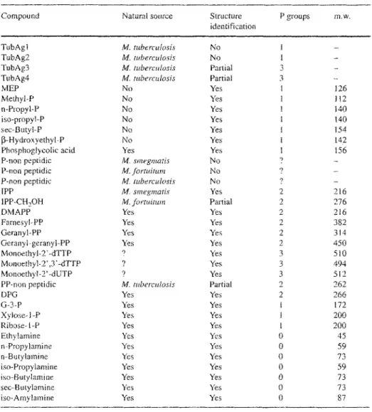
Table 1. Ligands stimulating Vy9/V82 cells
223
Compound Natural source Structure P groups m,w.
identification
TubAgt M. tuberculosis No 1 -
TubAg2 M. tuberculosis No 1 -
TubAg3 M. tuberculosis Partial 3 -
TubAg4 M. tube~rulosis Partial 3 -
NIEP No Yes 1 126
Methyl@ No Yes 1 1 ! 2
n-Propyt-P No Yes t 140
iso-propyl-P No Yes 1 140
sec-gutyl-P No Yes 1 t 54
[3-Hydroxyethyl-P No Yes 1 I42
Phosphoglycolic acid Yes Yes I 156
P-non peptidic M. smegmatis No ? -
P-non peptidic M. formitum No ? -
P-non peptidic M tuberculosis No 9 _
tPP M. smegmatis Yes 2 216
IPP-CH2OH M. fortuimm Partial 2 276
DMAPP Yes Yes 2 216
Farnesyl-PP Yes Yes 2 382
Geranyl-PP Yes Yes 2 314
Geranyl-geranyl-PP Yes Yes 2 450
Monoethyl-2'-dTTP ? Yes 3 510
Monoethyl-2',3'-dTFP ? Yes 3 494
Monoethyl-2'-dUTP 9 Yes 3 512
PP-non peptidic M. tuberculosis Partial 2 262
DPG Yes Yes 2 266
G-3-P Yes Yes t 172
Xylose- 1 -P Yes Yes 1 200
Ribose- t-P Yes Yes 1 200
Ethylamine Yes Yes 0 45
n-Propylamine Yes Yes 0 59
n-Butylamine Yes Yes 0 73
iso-Propylamine Yes Yes 0 59
iso-Butylamiae Yes Yes 0 73
sec-Butylamine Yes Yes 0 73
iso-Amylamine Yes Yes 0 87
p, Phosphate; MER monoethylphosphate; P-non peptidic, phosphorylated non peptide; IPP, isopentenylpy- rophosphale; DMAPP, dymethylallylpyrophosphate; PP, pyrophosphate; 2'-dTTP, 2'-deoxythymidine tri- phosphate; 2',3'-d'FFR 2',3'-dideoxythymidine rriphosphate; 2'-dUTR 2'-deoxyuridine triphosphate; DPG, diphosphoglyceric acid; G-3-P, glycerol-3-phosphate
a l k y l a m i n e s r e l e a s e d in m i l l i m o l a r a m o u n t s by different bacteria. R e c o g n i t i o n o f these c o m p o u n d s is m e d i a t e d by the VT9/V82 T C R and is a f f e c t e d by the type o f the alkyl chain. Thus, a l k y l a m i n e s r e p r e s e n t a n e w class o f 78 l i g a n d s w i d e l y p r e s e n t in nature and c a p a b l e o f r e c r u i t i n g a large n u m b e r o f 78 T cells. A s e c o n d r e a c t i v i t y is d i r e c t e d against b i s p h o s p h o n a t e s ( p a m i d r o n a t e and alendronate), drugs usually u s e d to contrast b o n e resorption. O n e o f t h e m o s t f r e q u e n t side effects o f this therapy is an a c u t e - p h a s e r e a c t i o n , w h o s e intensity appears c o r r e l a t e d w i t h e x p a n s i o n o f the V y 9 / V 6 2 T cell p o p u l a t i o n [51 ]. T h e m e c h a n i s m b y which these c o m p o u n d s i n d u c e p r o l i f e r a t i o n o f "[8 T cells is not clear: they c o u l d d i r e c t l y m i m i c IPP and o t h e r pre-
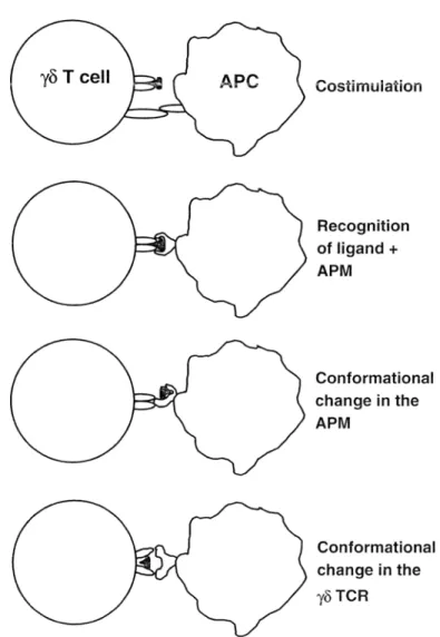
224 G. De Libem Costirnulation Recognition of ligand + APM Conformational change in the APM Conformational change in the
~(5 TCR
Fig. 1. Four possible models of interaction between 7~ T cell receptor, stimulatory ligand and putative an- tigen-presenting molecule (APt!4") are shown (APC antigen-presenting cell)
nylphosphates, due to a structural homology, or instead induce accumulation of IPP and other metabolic intermediates since they block the mevalonate pathway [52]. Ta- ble 1 reports the active ligands described so far.
Analysis of several 5'5 clones pointed out that only cells expressing both VT9 and V32 chains react to non-peptidic ligands [46]. In addition, the junctional regions of the V79 or V~2 chains are important to confer reactivity to non-peptidic ligands [53], implying a reactivity different from that to protein superantigens and MHC-peptide complexes. However, as T cell clones with different junctional sequences cross-react with the same collection of ligands [49, 54], it has to be argued that many different ~5 T C R recognize a structural motif c o m m o n to all these compounds. An additional important observation is that a consistent fraction of V,/9/V82 thymocytes [55] or V3,9/V52 clones isolated from postnatal thymus [10] also react to this class of li-
y8 T cells in human diseases 225 gands, suggesting the possibility that this receptor has intrinsic structural characteris- tics which confer reactivity to non-peptidic ligands and their putative antigen-pre- senting molecules (APM). Whether dedicated molecules exist which present non- peptidic ligands to V79/V82 TCR is still an open issue. Filler cells and membrane- membrane interactions are definitely required to optimally stimulate y~5 T cells [54, 56, 57] and only cells of human origin have this capacity (our unpublished results). These filler cells might provide necessary co-stimulation or, alternatively, they could associate non-peptidic ligands with species-specific APM, thus forming new com- plexes recognized by the Vy9/V82 TCR. Another possible way of interaction is ini- tial binding of ligands to APC and induction of conformational changes of surface APM, which then make cognate interaction with the TCR. A speculative possibility is that non-peptidic ligands first bind to conserved regions of the Vy9/V~2 TCR, which acquires a new confo~vnation and the capacity to interact with APC. These four possible models of interaction are illnstrated in Figure 1.
Recognition of cell surface molecules
Antigen reactivities of human y8 T cells against allo-MHC molecules [58-60], CDlc [61,621, and CD48 [631 have been described. However, only a few 78 clones react to these molecules, suggesting that they are rare specificities. Also in the mouse a few alloreactive y~ clones were isolated and characterized. The reactivities of these cells are quite different from those of cr.[3 T cells. One clone reacted to an unknown pep- tide presented by the TL 27b molecule [64], a second to the MHC class I-like TL 10b molecule [65], and a third to the MHC class II l-Ek surface glycoprotein [66], independently of associated peptides and antigen processing [67]. Site-directed mu- tagenesis of MHC molecules showed that the topology of y5 TCR interaction with the MHC is distinct from that of o~[~ T cells [67]. An intriguing finding is that mouse [68] and human [69] y8 T cells accumulate in the decidua during normal pregnancy. Mouse y8 T cells recognize non-MHC-encoded molecules present on both mouse and human trophoblast cell lines, but not other tumor cells [70]. The recognized sur- face structures have not been identified.
Recognition of peptides and c a r b o h y d r a t e s
Rare human yg T cell clones have been isolated which recognize peptide-MHC com- plexes [59, 71-73]. In mice there are also examples of these infrequent reactivities. Polyclonal y6 T cells specific for ovalbumin were induced after antigen inhalation [74]. It was not analyzed whether they were activated by the intact protein or by small peptides. A series of mouse hybridomas were reported to be stimulated by pep- tides derived from bacterial heat shock proteins (HSPs) [75]. Recognition of these peptides required presentation by unknown non-MHC molecules, and was affected either by amino acid substitutions in the peptide or by the polymorphism of Vy chain.
Mouse T~ T cells may also recognize di- and trisaccharides coupled to class I- binding peptides [76]. The crystal structure analysis of these glycosylated peptide- MHC complexes has shown that the carbohydrates are located in the central region of the putative TCR binding site without altering the overall MHC structure [77].
226 G. De Libero These findings support a model of antigen recognition by these 78 TCR characterized by cognate interaction with the sugar residues and not with MHC molecules.
Recognition of heat shock proteins
A few studies have reported the possible recognition of HSPs by human yfi T cells [78-80]. In two of these studies a rabbit antiserum or an mAb specific for an un- known human 58-kDa HSP partially inhibited reactivity of freshly isolated, but not of cloned, 3'8 T cells to a tumor line stained by these antibodies. The HSP stimulat- ing 78 cells was not identified and solid data supporting cognate interaction of the 3'8 TCR with members of this protein family have not been published. Taken togeth- er, all these findings show that antigen recognition by 78 T cells is different from that of ~ T cells. Additional evidence supporting this conclusion comes from the different length of CDR3 loops which are often critical for antigen binding in Ig and appear to provide the principal peptide binding residues in c~13 TCRs. Comparison of the CDR3 regions of Ig H and L chains with TCR ~, ~3, y and 5 chains, showed that Ig H and TCR 5 CDR3 are the most variable in size and are significantly longer than Ig L and TCR y chains, respectively. In contrast, TCR o~ and 13 chains pairing is constrained by nearly identical average CDR3 lengths [81]. These TCR structural differences have been related to 78 TCR recognition of molecules other than pep- tide-MHC complexes and, in general, more to a type of antigen recognition similar to that of Ig [821.
Effector functions of'~8 T cells
Like o~{3 T cells, y8 T cells can be classified in different functional subsets. 78 T cell clones are potent cytotoxic cells which lyse different tumor cell lines [1] and secrete different lymphokines irrespective of the expressed V chains [3]. y8 T cells isolated from leprosy patients release soluble factors that positively influence development of granulomas [83]. In contrast, an inhibitory role in granuloma formation during my- cobacterial infection has been attributed to y8 T cells in mice lacking the 8 gene [84] or treated with anti-TCR y8 mAbs [85]. Another important function is protection during infections. By releasing proinftammatory lymphokines, ')'8 T cells might facil- itate activation of macrophages in the early phases of infection, This has been de- scribed in mice infected with Candida albicans, in which 5'8 T cells facilitate nitric oxide production by macrophages and, thus, enhance resistance to mucosal candidia- sis [86]. In addition, by killing the infected target cells, y5 T cells might contribute to microbial burden, as shown with cytotoxic CD8 + T cells in mycobacterial and listeri- al infections in mice [87-89] or with human CD8 + TCR o~[3 § T cells [90, 91].
y8 T cells may also regulate the effector phases of the immune response. After in- halation of soluble antigens, Y8 T cells potently inhibit induction of IgE secretion in the mouse, possibly by blocking maturation of Th2 cells [74]. Thus, 78 T cells might have a prominent role in protection against primary allergic sensitization to environ- mental antigens.
78 T cells may also influence the functional type of T helper cells which develop during immune responses. In mice infected with Listeria monocytogenes, y6 T cells produce Thl-type cytokines, while they produce Th2-type cytokines in Nippostron-
75 T ceils in human diseases 227
gyIus brasiliensis-infected animals [92]. In both infections y5 T cells are among the first type of lymphocytes activated. As the effector phase of the immune response is influenced by the cytokine milieu in which the initial antigen priming occurs, 3'8 T cells may have the important regulatory role of determining the later response [92]. Furthermore, a possible role during viral infection has been proposed based on the findings that Vy9/V82 T cells recognize and kill target cells infected with different types of viruses [93].
An additional important function attributed to human 3'5 T cells is help to B ceils. Indeed, it has been shown that, analogously to c~13 T cells, human 3'5 T may facilitate B cell maturation, isotype switching and IgG production [94].
Role of'~3 T cells in animal disease models
Studies conducted in mice deficient in 3'5 T cells by homologous recombination or treated with anti-TCR y8 mAbs have provided convincing evidence of the impor- tance of 75 T cells in different disease models. 3'5 T cells provide protection during lethal encephalitis with HSV-1 [95], in malaria after immunization with inactivated sporozoites [96], and in the early phases of listeriosis [84, 97]. In the absence of 3'fi T cells, mice are more susceptible to high M. tuberculosis inocula [98] and have more severe tissue inflammatory injury after low inocula of the same bacteria [99]. In in- fection with Nocardia asteroides, y8 cells induce local recruitment of neutrophits re- sponsible for microbial clearance [I00]. In autoimmune disease models, 3'8 T cells appear to have an important regulatory role on c~[3 T cells and other effector cells. Elimination of 76 T cells worsens clinical manifestations in adjuvant arthritis [101] and lupus nephritis [102], leads to the aggravation and disease recurrence in experi- mental allergic encephalomyelitis [103], and mediates prevention of diabetes follow- ing mucosal insulin aerosolization in NOD mice [104].
Evidence for participation ol'3~3 T cells in h u m a n diseases Infections
Expansion of human 75 T cells has been described in bacterial, viral and parasite in- fections. The number of y8 T cells has been found increased in infections of bacterial origin such as brucellosis [105], salmonellosis [106], tuberculosis [107] and tulare- mia [108]. Increase of yg T ceils in hospital workers who are in close contact with tu- berculosis patients has been also reported [109]. Increased numbers of circulating y5 T cells have been found in parasite infections such as malaria [106, l l0, 1ll], ehr- lichosis [112], leishmaniasis [113, 114], toxoplasmosis [115], and trypanosomiasis [116]. In most of these infections the expanded cells display a VT9]V82 TCR.
The number of Y5 T cells is also altered in some viral infections. In patients with AIDS a large number of oligoclonal VS1 + cells are found in the peripheral blood [117] and of V82 + cells in the lung [118}. These 7~5 populations might contribute to the immune defense against opportunistic microorganisms frequently present in HIV- infected patients. Expansion of y8 T cells has also been found after cytomegalovirus (CMV) infection in kidney transplant patients [119]. The expanded cells express oli- goclonal VS1 and V83 chains and proliferate when challenged with CMV-infected
228 G. De Libero fibroblasts. These findings strongly suggest that an antigen-driven proliferation oc- curs during this viral infection.
The exact role of the expanded ?'8 populations in infections is not clear. 3'8 T cells might recognize microbial ligands and contribute to protection by reducing microbial load or by killing cells infected with intracellular pathogens. 3'5 T cells might also exert the important function of signaling the microbial presence in the early phases of infection, when a small load of microbial cells is present in tissues and other lym- phocyte populations have not yet been recruited.
Tumors
One of the first identified functions of human 78 T cells was the "nonspecific" cyto- toxicity toward tumor targets [1]. Moreover, some human Y8 T cells kill Burkitt's lymphomas, thymic lymphomas and erythroleukemia cells [54, 120, 121]. Other studies identified y8 T cell clones reacting with unknown surface molecules ex- pressed by activated B cells or EBV-transformed cells [122]. In all these cases no an- tigens interacting with the 3'8 TCR have been identified. Only a single y8 T cell clone that recognizes a peptide derived from the Ig expressed by a myeloma from the same patient has been described [123]. In some CNS neoplasms, including primary malig- nancies, metastatic cancers, and meningiomas, oligoclonal expansion of y8 popula- tions was detected [124]. y8 T cells were found infiltrating cutaneous melanocytic tu- mors and capable of killing solid tumors [125]. 75 T cells may also accumulate around epithelial [1261, renal [127] and lung [1281 carcinomas.
A recent finding is that many tumors of epithelial origin express MICA [129]. Since this MHC-re]ated molecule stimulates VS1 + T cells [130], recognition of this surface antigen may allow 3'8 T ceil response against tumor cells.
Autoimmune and inflammatory diseases
A possible involvement of 78 T cells in the pathogenesis of autoimmune diseases has been claimed. However, clear evidence of direct participation of 5'8 T cells in autoim- mune reactions is not available. Most of the studies have analyzed the relative num- ber of T8 T cells in peripheral blood or in other tissues, their phenotype and TCR rep- ertoire. In rheumatoid arthritis the number of y8 T cells is increased in the affected joints [131], mainly in those with active synovitis [132]. In multiple sclerosis 78 T cells accumulate around brain lesions during the exacerbation phases [133] and in the cerebrospinal fluid where they display polyclonal VS1 chains [134, 135]. Expan- sion of y8 T cells has been reported in cases of autoimmune thrombocytopenia [136] and autoimmune neutropenia [137]. In this latter study it was reported that five af- fected patients showed monoclonal expansion of 78 T cells, thus suggesting a direct correlation with the development of neutropenia. Interestingly, clonal expansion of VS1 + T cells was observed in an HTLV-I carrier patient with chronic neutropenia [ 138]. Taken together, these findings suggest that in some circumstances neutrophils might express surface molecules recognized by unique subsets of 3'8 TCR.
The number of circulating 78 T cells is increased in several inflammatory diseas- es. Coeliac disease (CD), an immune-mediated disorder arising from an hyper-re- sponsiveness to gluten with histological alterations in the small intestine, is charac-
78 T cells in human diseases 229 terized by increased numbers of intraepitheliat and lamina propria 75 T cells showing a phenotype of activated and memory cells [139, 140]. However, increased numbers of 3'8 T cells are found also in patients with normal jejunal morphology or with latent disease [14l]. Furthermore, the number of `/8 T cells does not directly correlate with the presence of gluten in the diet [ 142]. According to all these observations, it is like- ly that y8 T cells do not react against gluten components. Interestingly, most `/6 T cells in duodenum of CD patients express polyclonal TCR with predominant use of VSI, V83 and V88 chain [40], and the rare J53 segment [143]. It is tempting to spec- ulate that recognition of polymorphic molecules expressed by normal epithelial cells drives expansion of `/8 T cells expressing these particular V8 and J5 gene segments. Candidate "{~ T cell ligands m'e the MICA and MICB molecules, which activate VS1 + T cells [130]. In CD, 78 T cells might have an anti-inflammatory rote facilitating the repair of damaged tissue. The local protective role might be exerted by release of epithelial cell growth factors such as keratinocyte growth factor [144].
In inflammatory bowel disease (IBD), the number of 78 T cells has also been found to be increased in blood [145] and intestine [1461. VSI-D53-JSt-bearing cells were found to be expanded in patients either with severe disease or in those with re- cently diagnosed or less severe forms of IBD.
Other inflammatory diseases showing increased numbers of ,/8 T cells are Still's disease [ t 47], hypertrophic obstructive adenoids [ 148], Lyme arthritis [ 149], derma- titis herpetiformis [150], primary Sjogren's syndrome and untreated patients with systemic lupus erythematosus [151]. In chronic cutaneous lupus erythematosus V,/9/V82 T cells were observed in close vicinity to the damaged basal keratinocyte layer, suggesting their participation in the inflammatory reaction [152]. In lupus ne- phritis, `/8 T cells help production of anti-DNA immunoglobulins [153]. in patients undergoing surgical interventions, there is infiltration of T8 T cells immediately after blood circulation is reestablished in tigated ischemic arteries, suggesting specific and immediate recruitment at the site of injury [154]. 75 T cells are also present in the transition zone between normal intima and fatty streaks in atherosclerotic plaques, suggesting a role in initiating the inflammatory process in atherosclerosis [155].
All these studies suggest that 78 T cells have multiple roles in autoimmune and inflammatory diseases. It is likely that in some cases `/8 T cells participate in the pathogenesis of autoimmune diseases as they may recognize self antigens. However, it is conceivable that in other instances they may also limit inflammatory reactions and facilitate tissue repair.
Regulation of ~ T cells
The property of recognizing a large variety of non-peptidic ligands present both in eukaryotic and prokaryotic cells makes VT9/V82 cells a readily activated cell popu- Iation. Their overactivation may sometimes be responsible for dangerous acute in- flammatory reactions, as reported in malaria infection [l i0] and following therapy with bisphosphonates [51]. Therefore, this type of antigen reactivity necessitates a tight control, which is likely provided by different mechanisms, leading to a fine bal- ance between activation and inhibition of 3'5 T cells.
One regulatory mechanism is provided by weak agonist non-peptidic tigands which induce a state of transient anergy in 78 T cells after repeated stimulation. Nat- ural compounds such as 2,3-diphosphoglyceric acid, which is present in huge
230 G. De Libero amounts (5 mM) in erythrocytes, induce unresponsiveness in y8 T cells to the most active ligands [156]. This state of anergy lasts for a few days and is associated with partial tyrosine phosphorylation of the CD3-TCR complex. All the V79/V82 T cells are affected by this inhibitory TCR engagement [156]. Thus, this mechanism may ef- ficiently and simultaneously shut off the response of the whole Vy9/V82 T cell popu- lation, preventing its massive activation. Such a regulation may occur in patients constantly exposed to P.
falciparum,
e.g., individuals living in malaria-endemic areas who do not expand Vy9/V52 cells in vivo [157] and do not suffer from the pathogno- monic acute inflammatory reaction concomitant to the blood stage of the disease.A second regulatory mechanism is represented by expression of inhibitory recep- tors (IR) shared with NK cells. These receptors recognize MHC class I molecules and inhibit the response of 5'8 T cells more effectively when low rather than high an- tigen doses are present [t58]. Engagement of IR facilitates recruitment of SHP-1 pbosphatase to TCR-CD3 complex and affects phosphorylation of Lck and ZAP-70 kinase, but not of CD3 ~ chain upon TCR triggering [158]. These events may cause abortion of proximal TCR-mediated signaling. The role of IR is to set a higher TCR 75 activation threshold and therefore to focus the response of y8 T cells against APC loaded with high amounts of antigen.
In some cases, activation of y8 T cells requires facilitatory mechanisms. Optimi- zation of "/8 T cell response is important in tissues where a small number of y8 T cells is present and their effector functions have to be maximized to be effective. An enhancing mechanism is provided by expression of the CD66a molecule. This sur- face glycoprotein belongs to the CD66/CEA family and makes homotypic interac- tions with different CD66/CEA members. Engagement of CD66a with its physiologi- cal ligands enhances the amounts of released lymphokines, while it does not induce a shift of the dose-response curve (our unpublished data). Thus, it increases the poten- cy of y8 response. As CD66 molecules are frequently expressed by epithelial cells, this interaction facilitates release of large amounts of lymphokines in epithelial tis- sues when small numbers of responding ,/8 T cells are present.
The function of ~5 T cells in i m m u n e response
The enormous body of information derived from the reviewed studies allows us to discuss the role of y8 T cells in immune response on the basis of solid experimental data. A series of hypotheses can be made based on non-peptidic antigen specificity, on effector functions and also on cell number and distribution in human diseases.
The first function attributed to y8 T lymphocytes was that of sentinel cells accord- ing to their tissue distribution [ 159]. As 3'5 T cells in the mouse have the tendency to accumulate in the epithelia, it was suggested that this population may participate in the early host response against invading pathogens. A sentinel function has been at- tributed also to human VT9/V82 lymphocytes according to their unique antigen rec- ognition characteristics, rather than tissue distribution [43]. Vy9/V82 cells may fulfill the function of readily recruited and alerted sentinel cells, capable of immediately signaling the presence of danger. Their activation would have important consequenc- es such as prompt release of pro-inflammatory cytokines and chemokines capable of facilitating the onset of local inflammation.
A second important function of 78 T cells may be to fill the gap between innate and acquired immunity, providing the response during the time when antigen-specif-
78 T cells in human diseases 231
ic c~[3 T cells have not yet been recruited and expanded [43, 50]. The capacity of a series o f ligands to activate a large number o f u T cells, all sharing the same VT/V8 pair, in a cross-reactive manner is unique for a lymphocyte population. This type of T cell activation is different from activation induced by peptides or bacterial and vi- ral superantigens and, to some extent, it resembles ligand recognition by receptors expressed on the surface of innate immunity cells (e.g., CD14, CR1 and mannose re- ceptor). In other words, VT9/V82 receptors recognize patterns of molecules and do not make fine discriminations among them. In this respect, this subset o f human 3,8 T cells exploits an antigen-recognition strategy typical of innate immunity cells, while at the same time retaining the functions o f other lymphocytes.
A third important fnnction is to drive maturation o f antigen-specific T cells into different effector subsets. As shown during the early phases of infection in mice, the lymphokine secretion pattern of 78 T lymphocytes controls the subsequent functional maturation of cq3 T cells [92]. Although there is no clear evidence that similar mech- anisms also occur in human infections, it is likely that soluble factors released by hu- man T8 T cells influence local tymphokine milieu. Therefore, it is conceivable that human y8 T cells also participate in the T h l versus Th2 functional maturation of c~13 T cells.
A fourth function of human ~'~5 T cells is recognition of damaged or transformed epithelial cells expressing M I C A and MICB proteins, whose gene transcription is regulated by promoter heat shock elements similar to those of HSP genes. As M I C A and MICB are recognized by VSl-bearing cells, this subset may patrol the presence of recently altered cells in epithelial tissues.
Conclusions
In conclusion, the large number of studies on human ~'8 T cells have shown that these lymphocytes share several characteristics in c o m m o n with 0~3 T cells, and also em- body many unique properties. Some investigations have perhaps suffered a constant analogy with the r T cell population, which has precluded new and original experi- mental approaches. Nevertheless, this gigantic amount o f work has provided solid clues for defining the role of human 75 T cells in diseases. In addition, we have also learnt more about the extreme plasticity o f the immune system and its polymorphic capacity to adapt and recognize foreign molecules.
Acknowledgement. This work was supported by grant no. 3 1-045518.95 from Swiss National Fund, and by grants from the Velux Foundation, the Krebsliga beider Basel and Schweizerische Krebsliga. 1 thank Dr. L. Mort for reading the manuscript.
References
1. Brenner MB, McLean I, Dialyaas DP, Strominger JL, Smith IA, Owen FL, Seidman JG, Ip S, Rosen F, Kranget MS (1986) Identification of a putative second T-cell receptor. Nature 322:145
2. Groh V. Porcelli S, Fabbi M, Lanier LL, Picker LJ, Anderson T, Warnke RA, Bhan AK, Strominger JL, Brenner MB (1989) Human tymphocytes bearing T cell receptor Y5 are phenotypically diverse and evenly distributed throughout the lymphoid system. J Exp Med 169:1277
3. Modta CT, Verma S, Aparicio P, Martinez C. Spits H, Brenner MB (1991) Functionally distinct sub- sets of human "~8 T cells. Eur J hnmunol 21:2999
232 G. De Libero 4. Leffanc MP, Rabbitts TH (1990) Genetic organization of the human T-cell receptor 3' and ~ loci. Res
lmmunol 14l :565
5. Chien YH, Iwashima M, Kaplan KB, Elliott JF, Davis MM (1987) A new T-cell receptor gene located within the ft, locus and expressed early in T-cell differentiation. Nature 327:677
6. Hata S, Brenner MB, Krangel MS (1987) Identification of putative human T cell receptor 8 comple- mentary DNA clones. Science 238:678
7. Takihara Y, Reimann J, Michaloponlos E, Ciccone E, Moretta L, Mak TW (1989) Diversity and struc- ture of human T cell receptor 5 chain genes in peripheral blood y~-bearing T lymphocytes. J Exp Med
169:393
8. Migone N, Padovan S, Zappador C~ Giachino C, Bottaro M, Matullo G, Carbonara C, Libero GD, Casorali G (t995) Restriction of the T-cell receptor V5 gene repertoire is due to preferential rearrange- ment and is independent of antigen selection. Immunogenetics 42:323
9. Hata S, Satyanarayana K, Devlin R Band H, McLean J, Strominger JL, Brenner MB, Krangel MS (1988) Extensive junctional diversity of rearranged human T cell receptor 5 genes. Science 240:1541
10. Casorati G, De Libero G, Lanzavecchia A, Migone N (1989) Molecular analysis of human y8 § clones from thymus and peripheral blood. J Exp Med 170:1521
11. Krangel MS, Yssel H, Brocklehurst C, Spits H (1990) A distinct wave of human T cell receptor y~5 tymphocytes in the early fetal thymus: evidence for controlled gene rearrangement and cytokine pro- duction. J Exp Med 172:847
12. Lafaille Jl, Haas W, Coutinho A, Tonegawa S (1990) Positive selection of y8 T cells. Immunol Today 11:75
13. Bonneville M, tshida [, Itohara S, Verbeek S, Berns A, Kanagawa O, Haas ~ ; Tonegawa S (1990) Self-tolerance to n'ansgenic 3'8 T cells by intrathymic inactivation. Nature 344:163
14. Dent AL, Matis LA, Hooshmand F, Widacki SM, Bluestone JA, Hedrick SM (1990) Self-reactive y~i T cells are eliminated in the thymus. Nature 343:714
15. De Koning J, Di Molfetto L, Reilly C, Wei Q, Havran WL, Lo D (1997) Thymic cortical epithelium is sufficient for the development of mature T cells in relB-deficient mice. J hnmunol 158:2558
16. Tsujimura K, Takahashi T, Morita A, Hasegawa NH, lwase S, Obata Y (1996) Positive selection of 7~5 CTL by TL antigen expressed in the thymus. J Exp Med 184:2175
17. Kawai K, Kishihara K, Molina TJ, Wallace VA, Mak TW, Ohashi PS (1995) Impaired development of V3`3 dendritic epidermal T cells in p56tck protein tyrosine kinase-deficient and CD45 protein tyrosine phosphatase-deficient mice. J Exp Med 181:345
18. Pivnionk V, Tsitsikov E, Swinton E Rathbun G, Ah FW, Geha RS (1998) Impaired viability and pro- found block in thymocyte development in mice lacking the adaptor protein. SLP-76. Cell 94:229 19. Alberola IJ, Hogquist KA, Swan KA, Bevan M.I, Perhnutter RM (1996) Positive and negative selec-
tion invoke distinct signaling pathways. J Exp Med 184:9
20. Moore TA, yon Freedcn, Jeffry U, Murray R, Zlotnik A (1996) Inhibition of y8 T celt development and early thymocyte maturation in IL-7 -1- mice. J hnmunol 157:2366
21. Maki K, Sunaga S, Ikuta K (1996) The V-J recombination of T cell receptor-3` genes is blocked in in- terleukin-7 receptor-deficient mice. J E• Med 184:2423
22. Kawai K, Suzuki H, Tomiyama K, Minagawa M, Mak TW, Ohashi PS (1998) Requirement of the IL-2 receptor ~3 chain for the development of Vy3 dendritic epidermal T cells. J Invest Dcrmatol 110:96t 23. Amakawa R, tlakem A, Kundig TM, Matsuyama T, Simard JJ, Timms E. Wakeham A. Mittruecker
HW, Griesser H, Takimoto H, Schmits R, Shahinian A, Ohashi P, Penninger JM, Mak TW (1996) Impaired negative selection of T celts in Hodgkin's disease antigen CD30-deficient mice. Cell 84:55 t24
24. Guy-Grand D, Cerf-Bensussan N, Matissen B, Malassis-Seris M, Briottet C, Vassalli P (1991) Two gut intraepithelial CDS+ lymphocyte populations with diffierent T cell receptors: a role for the gut epi- thelium in T cell differentiation. J Exp Med 173:471
25. Bandeira A, Mota ST, Itohara S, Degermann S, Heusser C, Tonegawa S, Coutinho A (1990) Localiza- tion of 25 T cells to the intestinal epithelium is independent of normal microbial colonization. J Exp Med 172:239
26. Haas W, Pereira P, Tonegawa S (1993) 3'5 celts. Annu Rev Immunol t 1:637
27. Jarry A, Cerf-Bensussan N, Brousse N, Selz F, Guy GD (1990) Subsets of CD3 + (T cell receptor o~ or 3'8) and CD3- lymphocytes isolated from normal human gut epithelium display phenotypical features different from their counterparls in peripheral blood. Eur J Immunol 20:}097
q,8 T cells in human diseases 233 28. Liu CR Ueda R, She J, Sancho J, Wang B, Weddell G, Loring J, Kurahara C, Dudley EC, Hayday A, Terhorst C, Huang M (1993) Abhorrent T cell development in CD3 ~-/- mutant mice and identifica- tion of a novel T cell population in the intestine. EMBO J 12:4863
2q. Page ST, Bogatzki LY, Hamerman JA, Sweenie CH, Hogarth PJ, Malissen M, Perlmutter RM, Pullen AM (t998) Intestinal intraepitheliat lymphocytes include precursors committed to the T cell receptor cz~ lineage. Proc Natl Acad Sci USA 95:9459
30. kaky K, Lefrancois L, Freeden-Jeffry U yon, Murray R, Puddington L (1998) The role o f I L 7 in thy- mic and extrathymic development of TCR 3'5 cells. J Immunol 161:707
3 l. Yada S, Kishihara K, Kong YY, Nomoto K (1998) Differential requirements of CD45 protein tyrosine phosphatase for eytolytic activities and intrathymic and extrathymic development of intestinal intra- epithelial lymphocytes. J Immunol t 61:2208
32. Lefrancois L, Goodman T (1989) In vivo modulation of cytolytic activity and Thy-I expression in TCR-y8 + intraepithelial lymphocytes. Science 243:1716
33. Van Kerekhove C, Russet GJ, Deusch K, Reich K, Bahn AK, DerSimonian H, Brenner MB (1992) Oligoclonatity of human ir~testinal intraepithelial T cells. J Exp Med 175:57
34. Sire GK, Augustin A (1991) Extrathymic positive selection of 7,5 T cells Vy4171 rearrangements with "'GxYS" junctions. J [mmunol 146:2439
35. Hayes SM, Sin" A, Jacob S, Sire GK, Augustin A (1996) Role of IL-7 in the shaping of the pulmonary ~'6 T cell repertoire. J Immunol 156:2723
36. Hayakawa S, Shiraishi II, Saitoh S, Satoh K (l 996) Decidua as a site of extrathymic VyI T-cell differ- entiation. Am J Reprod lmmunol 35:233
37. Graeme CP, Bhan AK, Harris NL (1993) Immunohistochemical characterization of intraepithelial and subepithelial mononuelear cells of the upper airways. Am J Pathol 143:1416
38. Falini B, Ftenghi L, Piled S. Pelicci P, Fagioli M, Martelli MF, Moretta L, Ciccoue g (t989) Distribu- tion of T cells bearing different forms of the T cell receptor 'y8 in normal and pathological human tis- sues. J lmmunot 143:2480
39. Richmond I, Pritchard GE, Ashcroft T, Corris PA, Waiters EH (1993) Distribution of "r T cells in the bronchial tree of smokers and non-smokers. J Clin Pathol 46:926
40. De Libero G, Roeci MP, Casorati G, Giachino C, Oderda G, Tavassoli K, Migone N (1993) T cell re- ceptor heterogeneity in ~8 T cell clones from intestinal biopsies of patients with celiac disease. Eur J Immunol 23:499
41. Triebei F, Faure F, Mami-Chouaib F, Jitsukawa S. Griscelti A, Genevee C, Roman RS, Hercend T (1988) A novel human V8 gene expressed predominantly in the TiTA fraction of y,3+ peripheral lym- phocytes. Eur J lmmunot 18:2021
42. Parker CM, Groh V, Band H, Porcelli SA, Morita C. Fabbi M, Glass D, Strominger JL, Brenner MB (1990) Evidence for extrathymic changes in the T cell receptor u repertoire. J Exp Med 171 : 1597 43. De Libero G (1997) Sentinel function of broadly reactive human T~3 T cells, lmmuuol Today l 8:22 44. Pt?.ffer K, Schoet B, Gulle H, Kaufntann SH, V'r H (1990) Primary responses of human T cells to
mycobacteria: a frequent set of ~{~ T cells are stimulated by protease-resistant ligands. Eur I lmmunol 20:1175
45, Constant P, Davodeau F, Peyrat MA, Poquet Y, Puzo G, Bonneville M, Foumie 11 (1994) Stimulation of human 5'6 T cells by nonpeptidic mycobacteriat tigands. Science 264:267
46. Tanaka Y, Sano S, Nieves E, De Libero G, Rosa D, Modlin RL, Brenner MB, Bloom BR, Morita CT ( t 994) Nonpeptide ligands for human "{6 T cells. Proc Natl Acad Sci USA 91:8175
47. Schoel B, Sprenger S, Kaufmann SHE (1994) Phosphate is essential for stimulation of V~tYV~32 T lym- phocytes by mycobacteria[ low molecular weight tigand. Eur J Immunot 24:1886
48 Tanaka Y, Morita CT, Tanaka Y, Nieves E, Brenner MB, Bloom BR (1995) Natural and synthetic non- peptide antigens recognized by human ~/6 3' cells. Nature 375:155
49. Btirk MR, Mori L, De Libero G (1995) Human V'~-)-V62 cells are stimulated in a cross-reactive fash- ion by a variety of phosphorylated metabolites. Eur J immunol 25:2052
50. Bukowski JF. Morita CT, Brenner MB (1999) Human '~8 T cells recognize atkytamines derived from microbes, edible plants, and tea: implications for innate immunity. Immunity 1l :57
51 Kunzmann V. Bauer E, Wilhelm M (1999) u T-cell stimulation by pamidronate. N Engl J Meal 340:737 52. Fisher JE, Rogers MJ, Hatasy JM, Luckman SP, Hughes DE, Masarachia PJ, Wesolowski G, Russell
RG, Rodan GA, Reszka AA (1999) Alendronate mechanism of action: geranylgeraniol, an intermedi- ate in the mevalonate pathway, prevents inhibition of osteoclast formation, bone resorption, and kirtase activation in vitro. Proc Natl Acad Sci USA 96:t33
234 G. De Libero 53. Bukowski IF, Morita CT, Band H, Brenner MB (1998) Crucial role of TCR y chain junctional region
in prenyl pyrophosphate antigen recognition by y~ T cells. J Immunol t6t:286
54. De Libero G, Casorati G, Giachino C, Carbonara C, Migone N. Matzinger P, Lanzavecchia A (1991) Selection by two powerful antigens may account for the presence of the major population of human peripheral u T cells. J E• Med 173:131 t
55. Kabelitz D, Bender A, Prospero T, Wesselborg S, Janssen O, Pechhold K (1991) The primary response of human y5 ~ T cells to ~'(vcobacterium tubercutasis is restricted to V~O-bearing cells. J Exp Meal 173:1331
56. Morita CT, Beckman EM, Bukowski IF, Tanaka Y, Band H, Bloom BR, Golan DE, Brenner MB (t995) Direct presentation of nonpeptide prenyl pyrophosphate antigens to human 75 T ceils. Immuni- ty 3:495
57. Lung F, Peyrat MA, Constant P, Davodeau E David AJ, Poquet Y, Vie H, Fournie J J, Bonneville M (1995) Early activation of human V~/9 V82 T cell broad cytotoxicity and TNF production by nonpepti- dic mycobacterial tigands. J Immunol 1.54:5986
58. Ciccone E, Viale O, Peude D, Matnati M, Battista FG, Barocci S, Moretta A, Moretta L (1989) Speci- ficity of human T lymphocytes expressing a y~ T cell antigen receptor. Recognition of a polymorphic determinant of HLA class I molecules by a y~5 clone. Eur J Immunol 19:1267
59. Spits H, Paliard X, gngelhard VH, de Vries J (1990) Cytotoxic activity and lymphokine production of T cell receptor (TCR)-cq3 + and TCR-T~i ~" cytotoxic T lymphocyte (CTL) clones recognizing HLA-A2 and HLA-A2 mutants. Recognition of TCR-5,~ -~ CTL clones is affected by mutations at positions t 52 and 156. J Immunol 144:4156
60. Rivas A, Koide J, Cleary ML, Engleman EG (1989) Evidence for involvement of the "/, fi T cell anti- gen receptor in cytotoxicity mediated by human alloantigeu-specific T cell clones. J lmmunol 142:1840
61. Porcelli S, Brenner MB, Greenstein JL~ Balk SP, Terhorst C, Bleicher PA (1989) Recognition of clus- ter of differentiation 1 antigens by human CD4-CDS- cytolytic T lymphocytes. Nature 341:447 62. Faure F, Jitsukawa S, Miossec C, Hercend T (1990) CDlc as a target recognition structure for human
T lymphocytes: analysis with peripheral blood g~ cells. Eur J lmmunol 20:703
63. Del Porto R Mami Chouaib F, Bruneau JM, Jitsukawa S, Dumas J, Harnois M, Hercend T (1991) TCT 1, a target molecule for y5 T cells, is encoded by an immnnoglobulin superfamily gone (Blast-l) locat- ed in the CD1 region of human chromosome 1. J Exp Med 173:1339
64. Van Kaer L, Wu M, lchikawa Y, Ito K, Bonneville M, Ostrand-Rosenberg S, Murphy DB, Tonegawa S (1991) Recognition of MHC TL gene producls by "t'8 T cells. Immunol Rev 120:89
65. Bluestone J-A, Cron RQ, Cottennan M, Houtdeu BA, Matis LA (t988) Structure and specificity of T cell receptor ')'~ on major histocompatibility complex antigen-specific CD3 +, CD4-, CDS:- T lympho- cytes. J Exp Med t68:1899
66. Marls LA, Fry AM, Cron RQ, Cotterman MM, Dick RF, Bluestone JA (1989) Strnctnre and specificity of a class II MHC alloreactive @ T cell receptor heterodimer. Science 245:746
67. Schild H, Mavaddat N, Litzenberger C, Ehrich EW, Davis MM, Bluestone JA, Marls L, Draper RK, Chien YH (1994) The nature of major histocompatibility complex recognition by y6 T cells. Cell 76:29
68. Heyborne KD, Cranfill RL, Carding SR, Born WK, O'Brien RL (1992) Characterization of~'~ T lym- phocytes at the maternal-fetal interface. J tmmunol 149:2872
69. Mincheva-Nilsson L, Baranov V, Young MM, Hammarstrom S, Hammarstrom ML (1994) Immnno- morphologic studies of human decidua-associated lymphoid cells in normal early pregnancy. J lmmu- nol 152:2020
70. Heyborne K, Fu YX, Nelson A, Farr A, O'Brien R, Born W (1994) Recognition of trophoblasts by Ti5 T cells. J lmmunol 153:2918
7l. Kozbor D, Trinchieri G, Monos DS, lsobe M, Russo G, Haney JA, Zmijewski C, Croce CM (1989) Human TCR-T+/5 § CD8 § T lymphocytes recognize tetanus toxoid in an MHC-restricted fashion. J Exp Med 169:1847
72. Holoshitz J, Vila LM, Keroack BJ, McKinley DR, Bayne NK (t992) Dual antigenic recognition by cloned human y~ T cells, l Clin Invest 89:308
73. Del Porto P, D'Amato M, Fiorillo MT, Tuosto L, Piccolelta E, Sorrentino R (1994) Identification of a novel HLA-B27 subtype by restriction analysis of a cytotoxic y5 T cell clone. J Immunol 153:3093 74. McMenamin C, Pimm C, McKersey M, Holt PG (1994) Regulation of IgE responses to inhaled anti-
28 T ceils in human diseases 235 75. Born W, Hall L, Dallas A, Boymel J, Shinnick T, Young D, Brennan R O'Brien R (1990) Recognition
of a peptide antigen by heat shock-reactive "/8 T lymphocytes. Science 249:67
76. Abdel-Motat UM, Berg L, Rosen A, Bengtsson M, Thorpe CJ, Kihlberg J, Dahmen J, Magnusson G, Karlsson KA, Jondal M (1996) immunization with glycosylated Kb-binding pep[ides generates carbo- hydrate-specific, unrestricted cytotoxic T cells. Eur J lmmunol 26:544
77. Spelt JA, Abdel-Motat UM, Jondal M, Wilson IA (1999) Crystal structure of an MHC class 1 present- ed glycopeptide that generates carbohydrate-specific CTL. Immunity 10:51
78. Haregewoin A. Soman G, Horn RC. Finberg RW (1989) Human 78 + T ceils respond to mycobacterial heat-shock protein. Nature 340:309
79. Fisch P, Malkovsky M, Kovats S. Sturm E, Braakman E, Klein BS, Voss SD, Morrissey LW, DeMars R, Welch WJ, Bolhius RLH, Sonde[ PM (1990) Recognition by human VT9/V82 T cells of a GroEL homolog on Daudi Burkitt's lymphoma cells. Science 250:1269
80. Kaur 1, Voss SD, Gupta RS, Schell K, Fisch P, Sondel PM (1993) Human peripheral 78 T cells recog- nize hsp60 molecules on Daudi Burkitt's tymphoma cells. J lmmunol 150:2046
81. Rock ER Sibbald PR, Davis MM, Chien YH (1994) CDR3 length in antigen-specific immune recep- tors. J Exp Meal 179:323
82. Davis MM, Chien Y (1995) Issues concerning the nature of antigen recognition by oq3 and 78 T-cell receptors. Immunol Today 16:316
83. Modtin RL, Pirmez C, Hofinan FM, Torigian V, Uyemura K, Rea TH, Bloom BR, Brenner MB (1989) Lymphocytes bearing antigen-specific 28 T-cell receptors accumulate in human infectious disease le- sions. Nature 339:544
84, Mombaerts P, Arnoldi J, Russ F, Tonegawa S, Kaufmana SH (t993) Different roles of oc~3 and "t'8 T cells in immunity against an intracellular bacterial pathogen. Nature 365:53
85. Fu Y-X, Roark CE, Kelly K, Drevets D, Campbell P, O'Brien R, Born W (1994) Immune protection and control of inflammatory tissue necrosis by 3'8 T cells. I Immunol 153:3 t01
86. Jones-Carson J, Vazquez-Torres A, van der Heyde HC, Warner 11, Wagner RD, Balish E (1995) "/8 T celt induced nitric oxide production enhances resistance to mucosal candidiasis. Nat Meal 1:552 87, De Libero G, Flesch [, Kaufmann SH (1988) Mycobacteria-reactive Lyt-2 + T celt lines. Eur J Immu-
nol 18:59
88. Flynn JL, Goldstein M, Triebold KJ, Koller B, Bloom BR (1992) MHC class I restricted T cells are re- quired for resistance to M tlLberculosis infection. Proc Nail Acad Sci USA 89:12013
89. K~gi D, Lederman B, B/Jrki K, Hengartner H, Zinkernagel R M (1994) CD8 + T celt-mediated protec- tion against an intracel~ular bacterium by perforin-dependent cytomxicity Eur J Immunol 24:3068 90. Stenger S, Mazzaccaro RJ, Uyemura K, Cho S, Barnes PF, Rosat JP, Sette A, Brenner MB, Porcelli
SA, Bloom BR, Modlin RE (1997) Differential effects of cytolytic T cell subsets on intracellular in- fection. Science 276:1684
91. Stenger S, Hanson DA, Teitelbaum R, Dewan R Niazi KR, Froelich CJ, Ganz T, Thoma-Uszynski S. Metian A, BoTSan C, Porcelli SA, Bloom BR, Krensky AM, Modlin RL (199g) An antimicrobial ac- tivity of cytolytic T cells mediated by granulysin. Science 282:12 l
92_ Ferrick DA, Schrenzel MD, Mulvania T, Hsieh B, Ferlin WG, Lepper H (1995) Differential produc- tion of interferon-'~ and interleukin-4 in response to Thl- and Th2-stimulating pathogens by 28 T cells in vivo. Nature 373:255
93. Bukowski IF, Morita CT, Brenner MB (1994) Recognition and destruction of virus-infected cells by human 28 CTL. J tmmunol 153:5133
94. Hornet AA, Jabara H, Ramesh N, Geha RS (1995,~ ~8 T lymphocytes express CD40 ligand and induce isotype switching in B lymphocytes. J Exp Med 181 : 1239
95. Shammas R, Kodukula P, Tang Q, Hendricks RL, Bluestone IA (1997) T cell 28 cells protect mice from herpes simplex virus type l-induced lethal encephalitis. J Exp Med 185:1969
96. Tsuji M, Mombaerts P, Lefrancois L, Nussenzweig RS, Zavala F, Tonegawa S (1994) u T cells con- tribute to immunity against the liver stages of malaria in ~ T-cell-deficient mice. Proc Natl Acad Sci USA 91:345
97. Hiromatsu K, Yoshikai Y, Matsazaki G, Ohga S, Muramori K, Matsumoto K, Bluestone JA, Nomoto K (1992) A protective role of 28 T cells in primary infection with Listeria monocytogepws in mice. J Exp Meal 175:49
98. Ladel CH, Hess J, IDaugetat S, Mombaerts P, Tonegawa S, Kaufmann Sft (I995) Contribution of ct~ and 28 T lymphocytes to immunity against M.vcobacterium bo~,is bacillus Calmette Guerin: studies with T celt receptor-deficient mutant mice. Eur J Immunol 25:838
236 G. De Libero 99. D'Sonza CD, Cooper AM, Frank AA, Mazzaccaro R J, Bloom BR, Orme IM (1997) An anti-inflam- matory role for u T lymphocytes in acquired immnnity to Mycobacrerimn tuberculosis. J Immunol
158:12t7
100. King DP, Hyde DM, Jackson KA, Novosad DM, Ellis TN, Pumey L, Stovalt MY, Van Winkle LSI Beaman BL, Ferrick DA (1999) Cutting edge: protective response to puhnonary injury requires 78 T lymphocytes. J Immunol 162:5033
101. Pelegri C, Kuhnlein P, Bachner E, Schmidt CB, Franch A, Castell M, Hunig T, Emmrich F, Kinne RW (1996) Depletion of',/5 T cells does not prevent or ameliorate, but rather aggravates, rat adjuvant arthritis. Arthritis Rheum 39:204
102. Peng SL, Madaio MP, Hayday AC, Craft J (1996) Propagation and regulation of systemic autoimmu- nity by "18 T cells. J Immunot 157:5689
103. Kobayashi Y, Kawai K, Ito K, Honda H, Sobue G, Yoshikai Y (1997) Aggravation of routine experi- mental allergic encephalomyelitis by administration of T-cell receptor yS-specific antibody. J Neuro- immunoI 73:t 69
104. Harrison LC, Dempsey-Collier M, Kramer DR, Takahashi K (1996) Aerosol insulin induces regula- tory CD8 "/8 T cells that prevent murine insulin-dependent diabetes. J Exp Med 184:2t67
105. Bertotto A, Gerli R, Spinozzi F, Muscat C, Scalise F, Castellucci G, Sposito M, Candio F, Vaccaro R (1993) Lymphocytes bearing the "/8 T cell receptor in acute Brucetla melitensis infection. Eur J Im- munol 23:1177
I06. Hara T. Mizuno Y, Takaki K. Takada H, Akeda H, Aoki T, Nagata M, Ueda K. Matsuzaki G, Yoshikai Y, Nomoto K (1992) Predominant activation and expansion of V~)-bearing "/8 T cells in vi- vo as well as in vitro in Salmonella infection. J Clin Invest 90:204
107. Balbi t3, Valle MT, Oddera S, Giunti D, Manca F, Rossi GA, Allegra L (1993) T-lymphocytes with "/8 + V82 + antigen receptors are present in increased proportions in a fraction of patients with tubercu- losis or with sarcoidosis. Am Rev Respir Dis 148:1685
108. Sumida T, Maeda T, Takahashi H. Yoshida S, Yonaka F, Sakamoto A, Tomioka H. Koike T, Yoshida S (1992) Predominant expansion of V'I,9/V82 cells in a tularemia patient. Infect lmmun 60:2554 109. Ueta C. Tsuyuguchi I, Kawasumi H, Takashima T, Toba H, Kishimoto S (1994) Increase of @ T cells
in hospital workers who are in close contact with tuberculosis patients. Infect lmmun 62:5434 110. Ho M, Webster HK, Tongtawe P, Pattanapanyasat K, Weidanz WP (1990) Increased ,/8 T ceils in
acute Plasmodium[cdciparum malaria. Immunot Lett 25:139
I t 1. Nakazawa S, Brown AE, Macho 5:\ Smith CD, Aikawa M (1994) Malaria-induced increase of splenic '/8 T cells in humans, monkeys, and mice. Exp Parasitol 79:391
112. Caldwell CW, Everett ED, McDonald G, Yesus YW. Roland WE (1995) Lymphocytosis of y8 T cells in human ehrlichiosis. Am J Clin Pathol 103:761
113. Raziuddin S, Tetmasani AW, EI-Awad E, A1-Amari O, At-Yanadi M (1992) "1"8 T cells and the im- mune response in visceral leishmaniasis. Eur J 1mmutlol 22:t 143
t 14. Alaibac M, Daga A, Harms G, Morris J, Yu RC, Zwingerberger K, Chu AC (1993) Molecular analy- sis of the "/5 "I-cell receptor repertoire in normal human skin and in Oriental cutaneous leishmaniasis. Exp Dermatol 2:106
115. Scalise F, Gerli R, Castetlucci G, Spinozzi F, Fabietti G M, Cmpi S, Sensi L, Britta R, Vaccaro R, Bertotto A (1992) Lymphocytes bearing the y8 T-cell receptor in acute toxoplasmosis. Immunology 76:668
116. Ftynn IN, Sileghem M (1994) Invotvemem of ~/85 T cells in immunity to trypanosomiasis. Immunolo- gy 83:86
117. Hinz T. Wesch D, Friese K, Reckziegel A, Arden B, Kabelitz D (1994) T cell receptor "/8 repertoire in HIV-l-infected individuals. Eur J Immunol 24:3044
118. Agostini C, Zambello R, Trentin L, Cerutti A, Bulian P, Crivellaro C, Cipriani A, Semenzato G (1994) "/5 T cell receptor subsets in the lung of patients with HIV-1 infection. Cell lmmunol 153:194 119. Dechanet J, Merville R Berge F, Bone-Mane G, Taupin JL, Michel P, Joly P, Bonneville M, Potaux
L, Moreau JF (1999) Major expansion of 78 T lymphocytes following cytomegalovirus infection in kidney allograft recipients. J lnfect Dis 179:1
120. Fisch P, Malkovsky M, Braakman E, Sturm E, Bolhuis R L, Prieve A, Sosman J A, Lain VA, Sondel PM (1990) "/5 T cell clones and natural killer cell clones mediate distinct patterns of nort-major histo- compatibility complex-restricted cytolysis. J Exp Med l 71 : 1567
121. Di Fabrizio L, Kimura Y, Ware R, Rogozinski L, Chess L (1991) Specific triggering of"/8 receptors by K562 cells activates the '/8 TCR and may regulate natural kilter-like function. J lmmanoI 146:2495