Long-term results of a multicenter SAKK trial on
high-dose ifosfamide and doxorubicin in advanced
or metastatic gynecologic sarcomas
S. Leyvraz
1*, M. Zweifel
1, G. Jundt
2, A. Lissoni
3, T. Cerny
4, C. Sessa
5, M. Fey
6, D. Dietrich
7&
H. P. Honegger
8For the Swiss Group for Clinical Cancer Research (SAKK)
1
Centre Pluridisciplinaire d’Oncologie, University Hospital, Lausanne;2
Pathology Institute, Kantonsspital, Basel, Switzerland;3
Clinica Ostetrica e Ginecologica, Universita degli Studi Milano-Bicocca, Ospedale S. Gerardo dei Tintori, Monza, Italy;4
Oncology/Hematology, Kantonsspital, St. Gallen;5
Istituto Oncologico della Svizzera italiana, Bellinzona;6
Institute of Medical Oncology, Inselspital, Bern;7
Coordinating Center SAKK, Bern;8
City Hospital Triemli, Zurich, Switzerland
Received 21 October 2005; revised 10 January 2006; accepted 11 January 2006
Background:Dose intensive chemotherapy has not been tested prospectively for the treatment of gynecologic sarcomas. We investigated the antitumor activity and toxicity of high-dose ifosfamide and doxorubicin, in the context of a multidisciplinary strategy for the treatment of advanced and metastatic, not pretreated, gynecologic sarcomas.
Patients and methods:Thirty-nine patients were enrolled onto a phase I–II multicenter trial of ifosfamide, 10 g/m2 as a continuous infusion over 5 days, plus doxorubicin intravenously, 25 mg/m2/day for 3 days with Mesna and granulocyte-colony-stimulating factor every 21 days. Salvage therapy was allowed after chemotherapy.
Results:Among the 37 evaluable patients, the tumor was locally advanced (n = 11), with concomitant distant metastases (n = 5) or with distant metastases only (n = 21). After a median of three (range 1–7) chemotherapy cycles, six patients experienced a complete response and 12 a partial response for an overall response rate of 49% (95% CI 32% to 66%). The response rate was higher in poorly differentiated tumors (62%) compared with moderately well differentiated ones (18%), but was not different according to histology subtypes. Eleven patients had salvage therapy, either immediately following chemotherapy (n = 7) or at time of progression (n = 4). With a median follow-up time of 5 years, the median overall survival was 30.5 months. Hematological toxicity was as expected neutropenia, thrombopenia and anemia‡grade 3 at 50%, 34% and 33% of cycles respectively. No toxic death occurred.
Conclusions:High-dose ifosfamide plus doxorubicin is an active regimen for all subtypes of gynecological sarcomas. Its toxicity was manageable in a multicentric setting. The prolonged survival might be due to the multidisciplinary strategy that was possible in one-third of the patients.
Key words:gynecologic sarcomas, leiomyosarcomas, endometrial stromal sarcomas, mixed mesodermal tumors, ifosfamide, doxorubicin
introduction
Gynecologic sarcomas are a rare and heterogeneous group of tumors accounting for only up to 5% of all female genital tract malignancies. They include (i) pure sarcomas such as
leiomyosarcomas and endometrial stromal sarcomas, as well as (ii) mixed sarcomas commonly referred to as mixed
mesodermal tumors or carcinosarcomas. Although tissue cultures and immunohistological studies have suggested that this latter group are metaplastic carcinomas rather than true mixed tumors, and thus should be classified as sarcomatoid carcinomas [1], all these subtypes are still characterized by
similar prognosis and recurrence patterns, involving high rates of local regrowth and dissemination to the liver and the lungs, suggesting a hematogenous spread [2].
Patients with recurrent disease are candidates for systemic chemotherapy, although there is little evidence of cure [3]. Doxorubicin and ifosfamide are the most active single agents offering variable response rates depending on histological tumor types. Standard-dose doxorubicin has been shown to be more active in leiomyosarcomas than in mixed mesodermal tumors, while the reverse can be said for standard-dose ifosfamide [4–6]. Standard combined doses of doxorubicin and ifosfamide yielded good anti-tumor activity in advanced or metastatic leiomyosarcomas (response rate 30%) but showed no advantage over single agents in terms of survival (median survival 6–12 months; long-term survivors <5%) [7]. Other combination regimens, including cisplatin, darcabazine, etoposide,
original
article
*Correspondence to: Dr S. Leyvraz, Centre Pluridisciplinaire d’Oncologie, University Hospital – CHUV BH06, Rue du Bugnon 46, 1011 Lausanne, Switzerland.
gemcitabine or taxanes, yielded similar results (median survival £18 months) [8–10].
There is evidence for a dose–response relationship of doxorubicin and 4-epidoxorubicin in advanced soft-tissue sarcomas [11]. Several reports have suggested the same for ifosfamide [12, 13]. Recently it was shown that relapse-free survival may be improved when high-dose ifosfamide or high-dose doxorubicin are used, although an effect on overall survival was not demonstrated in those studies [14, 15].
No trials have yet been published for high-dose doxorubicin combined with high-dose ifosfamide in the treatment of gynecologic sarcomas. A pilot study based on incremental doses of 4-epidoxorubicin and fixed high doses of ifosfamide in advanced soft-tissue sarcomas yielded objective responses in four of six patients with uterine sarcomas, which appeared to be a dose-dependent effect as well [11].
In a multicenter phase I trial of advanced sarcomas, including 12 gynecologic sarcomas, our study group
demonstrated that chemotherapy with high-dose doxorubicin (up to 90 mg/m2) combined with high-dose ifosfamide (10 g/m2) and supported by hematopoietic growth factors is a feasible treatment option [16]. Although this regimen involved severe myelosuppression, those events resolved quickly. Encouraged by the high response rates obtained in that study, our next step was to perform a regular phase II trial of ifosfamide 10 g/m2combined with doxorubicin at 75 mg/m2in patients with advanced or metastatic gynecologic sarcomas who had not previously received chemotherapy. The purpose of this trial was to verify the high response rate and to evaluate toxicity, time to progression and overall survival, as well as the effect of salvage surgery on the treatment outcomes.
Although multidisciplinary management with an essential role for surgery has been widely accepted in the management of primary soft tissue or gynecologic sarcoma [17], in handling recurrent or metastatic disease, its place has not yet been settled even though it is recognized as critical for the outcome of some patients [3]. It has often been left to the judgment of the investigators, and in the published chemotherapy trials it has been either discouraged or not reported. In the present trial, salvage therapy was allowed whenever feasible after chemotherapy and should be accounted for in the long-term results.
patients and methods
The trial protocol was in accordance with the Declaration of Helsinki principles and was approved by the competent ethics commission. All patients gave their written informed consent.
patient selection
Patients with histologically proven advanced or metastatic gynecologic sarcoma were eligible for the trial. Tissue specimens were reviewed by an independent group of pathologists (lead by G.J.) to confirm diagnosis and differentiation scores.
Additional inclusion criteria were: measurable disease in one or two dimensions as verified by physical examination or imaging techniques (ascitis or pleural effusions were not considered measurable); ECOG performance status £2; age 18–70 years; no previous chemotherapy;
no previous radiotherapy on the bladder; no CNS metastases; as well as adequate hematological, renal, hepatic and cardiac (assessed by echocardiography or multigated nuclear scanning) functions.
study design and treatment plan
This was a multicenter, non-randomized phase II trial conducted after the completion of the phase I trial that identified the proper doses and in which patients with gynecologic sarcomas had been treated. For the phase II trial, ifosfamide was administered intravenously at a dose of 10 g/m2as a continuous infusion over 5 days. Mesna was given
intravenously at a dose of 2 g/m2/day as a continuous infusion over 6 days. Doxorubicin was administered intravenously at a dose of 25 mg/m2as
a bolus for 3 days (total dose 75 mg/m2), starting 4 h after the beginning of ifosfamide on day 1 of each 3-week cycle. Granulocyte colony stimulating factor (G-CSF, Filgrastim) was administered once daily at a dose of 5 lg/kg s.c., starting 24 h after the end of ifosfamide infusion for a total of 10 days, or 14 days if the neutrophile count did not reach at least 500/ll after 10 days. The ifosfamide and doxorubicin doses for 11 patients in the phase I trial were: ifosfamide 12 g/m2and doxorubicin
50 mg/m2(two patients), ifosfamide 10 g/m2and doxorubicin 60 mg/m2 (two patients), ifosfamide 10 g/m2and doxorubicin 75 mg/m2(three
patients), ifosfamide 10 g/m2and doxorubicin 90 mg/m2(four patients). Each chemotherapy cycle was given every 3 weeks. For this report those 11 patients were pooled together with the patients included in the phase II trial. Indeed, when analyzed separately for all major end points, the results of both trials were comparable.
Salvage therapy with surgery and/or radiotherapy aiming at removing all residual tumor tissue was performed in all patients who were considered to potentially benefit from such an approach, either immediately following chemotherapy or at the time of progression.
response, toxicity assessment, dose modification
All tumors were evaluated based on standard WHO criteria to assess response to chemotherapy [18] and responses had to be confirmed at least 4 weeks later. CT scans were obtained at baseline, after every other chemotherapy cycle and at the end of treatment. Up to three uni- or bi-dimension measurable lesions had to be documented.
Toxicity was assessed based on WHO grading. Cardiotoxicity was monitored by assessing cardiac ejection fraction every two to three cycles and prior to each cycle exceeding doxorubicin cumulative doses of <450 mg/ m2. Chemotherapy was discontinued if cardiac ejection fraction dropped
to <40% and was not administered in the presence of serum creatinine >150 lmol/l or creatinine clearance <60 ml/min.
Neurotoxicity was assessed based on M. D. Anderson scores [19]. On obtaining scores of ‡2, an attempt was made not to decrease the dose of ifosfamide. Any neurotoxicity was managed by administering methylene blue as an intravenous bolus of 50 mg repeated at the same dose every 2 h until the neurotoxic event resolved, or as continuous infusion at 200 mg/day diluted in 5% dextrose [20].
Drug administration was delayed by 1 or 2 weeks if white blood cell count was <3500/ll or platelet count was <100 000/ll at day 22. When treatment with intravenous antibiotics was necessary, or when bleeding due to thrombocytopenia occurred, the total dose of ifosfamide could be decreased from 10 to 8 g/m2. In the case of normal recovery from myelosuppression
and if no febrile episodes requiring administration of intravenous antibiotics occurred, ifosfamide dose could be increased up to a total dose of 12 g/m2during subsequent cycles. Doxorubicin dose could be decreased
from 75 to 60 mg/m2in case of WHO mucositis grade 3 or higher. In the absence of mucositis, it was proposed to increase the dose of doxorubicin to 30 mg/m2/day for a total of 90 mg/m2.
statistical methods
The primary end point was the tumor response rate (WHO criteria). The Simon’s optimal two-stage design was used to calculate sample size for the phase II trial. The treatment would be considered uninteresting if the response rate were less than 50% and promising if the response rate were at least 70%. For a significance level of 5% and a power of 80%, a total of 43 patients was needed with 15 patients in stage I and a further 28 patients in stage II. Based on the evaluation of the first 15 patients, it was decided to continue the trial up to the planned target number of 43 patients. However, the trial was closed after including 27 patients due to low accrual. It was planned to analyze the data from this phase II trial together with those from phase I, for which the results were published in 1998 by our group [16], in order to preserve the statistical power of 80% to detect a promising response, instead of 63% if only 27 patients had been included in the analysis.
Confidence intervals for response rates were calculated by the Clopper– Pearson method. Time to progression was calculated for all patients from registration to the day of evidence of progressive disease or death, with censoring at last follow-up. Survival time was calculated from registration to death or last follow-up evaluation and was analyzed using the Kaplan–Meier method. Differences in survival were tested by the log-rank test. Exploratory multivariate analysis of survival using Cox regression was also performed. Variables represented by appropriate binary indicators were the following: histology (leiomyosarcoma versus other), differentiation (poorly
differentiated versus other), response (CR/PR versus other), tumor location (presence of distant metastases versus loco-regional alone) and salvage therapy (yes versus no).
results
patients characteristics
Between March 1993 and June 1999, 39 patients from six SAKK (Swiss Group for Clinical Cancer Research) centers with advanced gynecologic sarcomas were enrolled: 12 in the phase I and 27 in the phase II study. Two patients were not evaluable and were excluded from analysis: one patient in phase I never started treatment due to a rapidly worsening performance status and died 1 month after enrollment; another patient in phase II presented with an undifferentiated carcinoma, which was only detected after histopathological review and showed a complete remission upon six cycles of treatment.
Characteristics of the 37 evaluable patients are shown in Table 1. The median age of the patients was 53 years and ranged from 34 to 69 years. They had a good performance status, with ECOG performance status 0 or 1 in 92% of cases. The tumor was confined to the pelvis and to the local lymph nodes in 11 patients and with concomitant distant metastases in five patients. Twenty-one patients had only distant metastases, mainly within the lung (n = 18), and some as distant lymph nodes (n = 11), liver metastases (n = 5) or peritoneal seeding (n = 4). Other tumor locations were infrequent, with pleura, bone and pancreas in one patient each.
Confirmed by the pathology reviews, the most common diagnosis was leiomyosarcomas in 25 patients. Endometrial stromal sarcoma and mixed mesodermal tumors were diagnosed in five patients each. One patient had a rhabdoid tumor of the pudendal great lip with lymph node metastasis and another had a pleomorphic spindle cell sarcoma of the uterus. The tumor was classified according to the differentiation status. Seventy per cent had poorly differentiated and 30% moderately or well differentiated tumors.
toxicity profile and dose intensity
A total of 130 chemotherapy cycles were analyzed for toxicity. A median of three (range 1–7) chemotherapy cycles were administered per patient. As expected, myelosuppression was the predominant type of toxicity. Severe neutropenia of WHO ‡grade 3 was observed in half of the cycles and 73% of patients, and developed into febrile neutropenia in 29% of cycles with proven infection in 23%. No toxic death occurred.
Thrombocytopenia and anemia of WHO ‡grade 3 were found in 34% and 33% of cycles, respectively, and involved more than half of the patients.
Neurological toxicity of grade 1 according to the M. D. Anderson score was observed during 10 cycles and of grade 2 or higher in four cycles; all were manageable by methylene blue administration and/or stopping ifosfamide. No severe renal toxicity was noted, but transient microscopic hematuria was observed during 15 cycles with minimal creatinine elevation in four of them. Mucositis occurred in 45% of cycles but was moderate, except in 12 cycles with WHO grade 3. No clinical cardiac failure was reported. Cardiac ejection fractions measured repeatedly in 28 patients did not show any relevant decrease, except for one patient in which it dropped from 62% to 48%.
Doses were modified in 28% of cycles because of toxicity. A total of 27% chemotherapy cycles were delayed because of toxicity or because the patients so requested. However, the median dose intensity (calculated in mg/m2/week) could be
Table 1. Patients characteristics
Number of patients 37
Age (years)
Median (range) 53 (34–69)
ECOG performance status
0 2
1 12
2 3
Histologic diagnosis
Leiomyosarcoma (LMS) 25
Endometrial stromal sarcoma (ESS) 5
Mixed Mullerian tumor (MMT) 5
Othera 2 Histologic grade Well differentiated 6 Moderately differentiated 5 Poorly differentiated 26 Tumor location Loco-regional 11
Loco-regional and distant 5
Distant 21 Lung 18 Lymph node 11 Liver 5 Peritoneum 4 Pleura 1 Bone 1 Pancreas 1
maintained at ‡75% of the planned dose for ifosfamide and at ‡88% for the planned dose of doxorubicin. Dose escalation in either drug has never been performed in any patient.
response to chemotherapy and survival
Responses were assessed in all 37 patients. Six patients (16%) showed a complete response to chemotherapy and 12 (32%) experienced a partial remission, resulting in an overall response rate of 49% (95% CI 32% to 66%). Eleven patients (30%) had stable disease and eight (21%) progressive disease. Stratified by histology, the overall response rate was 12 of 25 (48%) for patients with leiomyosarcomas, two of five (40%) for patients with endometrial stromal sarcomas and three of five (60%) for patients with mixed Mullerian tumors. The response rate was higher in poorly differentiated tumors (16 of 26 = 62%, 95% CI 41% to 80%) than in well or moderately differentiated ones (2 of 11 = 18%, 95% CI 2% to 52%).
With a median follow-up time of 5 years, 22 patients have progressed and 18 have died of their disease. The median time to progression was 27.7 months (95% CI 8.9–45.9) and the median overall survival was 30.5 months (95% CI 15.7 to upper limit not reached) (Figure 1).
In seven patients, chemotherapy was immediately followed by salvage therapy aimed at removing all residual disease (surgery, n = 4; surgery and radiotherapy, n = 2; radiotherapy, n = 1). In four more patients, salvage surgery was performed at the time of progression. These 11 patients included eight responders to chemotherapy. Additional palliative treatment was applied in 16 patients (chemotherapy, n = 14; radiotherapy, n = 2) at the time of disease progression.
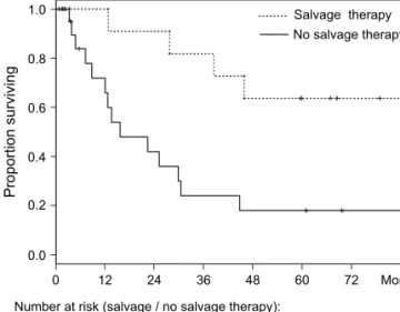
Salvage therapy improved survival, with 64% of the patients surviving at 5 years (95% CI 41% to 99%) and the median survival time not yet reached. For patients in whom it was not possible to perform major surgery and/or radiotherapy, the median survival remained at 15.7 months (95% CI 12.1–44.8) (Figure 2), with 18% survival at 5 years (95% CI 6% to 50%). Multivariate analysis confirmed that salvage therapy was a significant prognostic factor for better outcomes (hazard ratio 0.19, 95% CI 0.05–0.73). Distant metastasis, by contrast, was an unfavorable prognostic factor (hazard ratio 7.29, 95% CI 1.03–51.5).
discussion
The present trial demonstrated that high-dose ifosfamide in combination with doxorubicin and hematopoietic growth factors, is an effective treatment modality for the treatment of advanced and metastatic gynecologic sarcomas. In our patient population it yielded a high response rate of 49%. Compared with the uninteresting response rate of 50% and the
promising response rate of 70% considered in the trial design, this observed activity was, however, at the lower end of the expected response rate. Moreover, the data of the 26 and 11 patients from the phase II and I studies had to be pooled to increase the statistical power to 80% with a 5% significance level. The poorly differentiated tumor in our trial had the highest response. The response rate was 48% in
leiomyosarcoma, 40% in endometrial stromal sarcoma and
60% in mixed mesodermal. Only a few active agents are known to be effective in the treatment of gynecologic sarcomas [10]. A 30% response rate has been reported for standard-dose ifosfamide plus doxorubicin in leiomyosarcomas [7]. A 19% response rate was obtained with doxorubicin plus cyclophosphamide in mixed mesodermal tumors [21]. Ifosfamide 7.5 g/m2administered as a single agent over 5 days yielded response rates of 32%–36% in mixed mesodermal tumors, 17% in leiomyosarcomas and 33% in endometrial stromal sarcomas [4, 5, 22]. In metastatic endometrial carcinoma, the addition of hormonal therapy to chemotherapy might be beneficial [23]. The most active regimen has
combined gemcitabine and docetaxel. It has produced a 45% response rate in 29 uterine leiomyosarcomas, half of them having received previous anthracyclines [9]. Up to now, no prospective studies have been available on dose-intensive chemotherapies for gynecologic sarcomas. Only one report
0.0 0.2 0.4 0.6 0.8 1.0 0 12 24 36 48 60 72 Proportion surviving Months Number at risk: 37 23 17 13 10 8 4
Figure 1. Kaplan–Meier curve for overall survival (broken line: 95% CI).
0.0 0.2 0.4 0.6 0.8 1.0 0 12 24 36 48 60 72 Salvage therapy No salvage therapy Proportion surviving Months Number at risk (salvage / no salvage therapy):
11 11 10 9 7 5 3
26 12 7 4 3 3 1
Figure 2. Overall survival of patients subjected to salvage therapy aimed at eliminating all residual disease after chemotherapy, compared with patients receiving chemotherapy only.
indicated a high response rate of 67% in six patients treated with ifosfamide 9 g/m2plus epidoxorubicin 100–140 mg/m2[11].
Myelosuppression was the anticipated toxicity. Although dose modifications or treatment delays due to toxicity were necessary in 28% and 27% of the cycles, respectively, the dose intensity could be maintained at 75% of the planned dose for ifosfamide and almost 90% for doxorubicin. WHO ‡3 neutropenia was reported in 49% of the cycles, thrombopenia in 34% and anemia in 33%. There was no patient death that could be attributed to toxicity and this might also explain the good results of the study. This toxicity profile was not different from similar single-agent or combination regimen for the treatment of sarcomas, even if in some studies the doses were much lower than ours and not supported by hematopoietic growth factors [24, 25]. Myelosuppression was thus severe with febrile neutropenia in a third of the cycles, but should probably be accepted as inherent to these combination regimens [26] and should be administered only in experienced centers. Transient neurological and renal toxicities were infrequent. Clinical heart failure was not observed.
While the overall survival times obtained in previous studies usually ranged from 6 to a maximum of 18 months [10], our prolonged median follow-up period of 5 years strikingly revealed a median survival time of 30 months. Most of the available data on high-dose ifosfamide and doxorubicin were collected in adult patients with soft-tissue sarcomas [16, 27–29]. Some trials suggested a dose–effect relationship; however, their results concerning the effect of high-dose ifosfamide and doxorubicin on survival remain controversial.
In our trial, the Cox multivariate analysis suggested that salvage treatment with the intention to remove all residual disease had a major positive prognostic influence. Indeed, the patients who did not receive any treatment except
chemotherapy had a median overall survival of 15.7 months and a 5-year survival rate of 18%, while the selected patients for whom complementary therapy was judged possible had a 5-year survival rate of 65%. The concept of multidisciplinary management, with an essential role being reserved for surgical intervention, has been widely accepted for soft-tissue or gynecologic primary sarcomas [17]. The precise role of such strategies in handling recurrent or metastatic disease remains to be defined, even though they are known to make a critical difference in some patients [3]. In the published chemotherapy trials, the use of salvage treatment modalities was either discouraged, not reported or left to the investigators’ discretion [30]. In the present trial, 29% of patients were subjected to surgery. Previous phase II chemotherapy trials in soft-tissue sarcomas have included similar percentages of surgical intervention. In accordance with our own results, those subgroups reached extended survival times of 30–39 months after complete resection of liver or lung metastases [31, 32]. Likewise, salvage surgery for pulmonary or extra-pulmonary recurrences of uterine leiomyosarcoma increased the median overall survival to 3.9 years irrespective of the site of recurrences [33, 34].
These improved outcomes in patients undergoing salvage treatments after chemotherapy might conceivably be due mainly to a patient selection bias. The true benefit of surgery needs further clarification in prospective studies.
acknowledgement
This trial was partially supported by Baxter Oncology GmbH, successor in right to ASTA Medica Oncologie, Deutschland. We are indebted to Marianne Gonin for editorial assistance.
references
1. Amant F, Vloeberghs V, Woestenborghs H et al. Transition of epithelial toward mesenchymal differentiation during ovarian carcinosarcoma tumorigenesis. Gynecol Oncol 2003; 90: 372–377.
2. Rose PG, Piver MS, Tsukada Y et al. Patterns of metastasis in uterine sarcoma. An autopsy study. Cancer 1989; 63: 935–938.
3. Blay JY, van Glabbeke M, Verweij J et al. Advanced soft-tissue sarcoma: a disease that is potentially curable for a subset of patients treated with chemotherapy. Eur J Cancer 2003; 39: 64–69.
4. Sutton GP, Blessing JA, Barrett RJ et al. Phase II trial of ifosfamide and mesna in leiomyosarcoma of the uterus: a Gynecologic Oncology Group study. Am J Obstet Gynecol 1992; 166: 556–559.
5. Sutton G, Blessing JA, Park R et al. Ifosfamide treatment of recurrent or metastatic endometrial stromal sarcomas previously unexposed to chemotherapy: a study of the Gynecologic Oncology Group. Obstet Gynecol 1996; 87: 747–750.
6. Omura GA, Major FJ, Blessing JA et al. A randomized study of adriamycin with and without dimethyl triazenoimidazole carboxamide in advanced uterine sarcomas. Cancer 1983; 52: 626–632.
7. Sutton G, Blessing JA, Malfetano JH. Ifosfamide and doxorubicin in the treatment of advanced leiomyosarcomas of the uterus: a Gynecologic Oncology Group study. Gynecol Oncol 1996; 62: 226–229.
8. Campos SM, Matulonis UA, Penson RT et al. Phase II study of liposomal doxorubicin and weekly paclitaxel for recurrent Mullerian tumors. Gynecol Oncol 2003; 90: 610–618.
9. Hensley ML, Maki R, Venkatraman E et al. Gemcitabine and docetaxel in patients with unresectable leiomyosarcoma: results of a phase II trial. J Clin Oncol 2002; 20: 2824–2831.
10. Kanjeekal S, Chambers A, Fung Kee Fung M et al. Systemic therapy for advanced uterine sarcoma: A systematic review of the literature. Gynecol Oncol 2005; 97: 624–637.
11. Frustaci S, Buonadonna A, Galligioni E et al. Increasing 49-epidoxorubicin and fixed ifosfamide doses plus granulocyte-macrophage colony-stimulating factor in advanced soft tissue sarcomas: a pilot study. J Clin Oncol 1997; 15: 1418–1426.
12. Cerny T, Leyvraz S, von Briel T et al. Saturable metabolism of continuous high-dose ifosfamide with mesna and GM-CSF: a pharmacokinetic study in advanced sarcoma patients. Swiss Group for Clinical Cancer Research (SAKK). Ann Oncol 1999; 10: 1087–94.
13. Patel SR, Vadhan-Raj S, Papadopolous N et al. High-dose ifosfamide in bone and soft tissue sarcomas: results of phase II and pilot studies—dose-response and schedule dependence. J Clin Oncol 1997; 15: 2378–2384.
14. Worden FP, Taylor JM, Biermann JS et al. Randomized phase II evaluation of 6 g/m2of ifosfamide plus doxorubicin and granulocyte colony-stimulating factor (G-CSF) compared with 12 g/m2of ifosfamide plus doxorubicin and G-CSF in the treatment of poor-prognosis soft tissue sarcoma. J Clin Oncol 2005; 23: 105–112.
15. Le Cesne A, Judson I, Crowther D et al. Randomized phase III study comparing conventional-dose doxorubicin plus ifosfamide versus high-dose doxorubicin plus ifosfamide plus recombinant human granulocyte-macrophage colony-stimulating factor in advanced soft tissue sarcomas: A trial of the European Organization for Research and Treatment of Cancer/Soft Tissue and Bone Sarcoma Group. J Clin Oncol 2000; 18: 2676–2684.
16. Leyvraz S, Bacchi M, Cerny T et al. Phase I multicenter study of combined high-dose ifosfamide and doxorubicin in the treatment of advanced sarcomas. Swiss Group for Clinical Research (SAKK). Ann Oncol 1998; 9: 877–884. 17. Maki RG. Multidisciplinary management of soft-tissue sarcomas. Cancer Invest
18. Miller AB, Hoogstraten B, Staquet M et al. Reporting results of cancer treatment. Cancer 1981; 47: 207–214.
19. Castellanos AM, Fields WS. Grading of neurotoxicity in cancer therapy. J Clin Oncol 1986; 4: 1277–1278.
20. Kupfer A, Aeschlimann C, Wermuth B et al. Prophylaxis and reversal of ifosfamide encephalopathy with methylene-blue. Lancet 1994; 343: 763–764. 21. Sutton GP, Blessing JA, Rosenshein N et al. Phase II trial of ifosfamide and
mesna in mixed mesodermal tumors of the uterus (a Gynecologic Oncology Group study). Am J Obstet Gynecol 1989; 161: 309–312.
22. Sutton GP, Blessing JA, Homesley HD et al. A phase II trial of ifosfamide and mesna in patients with advanced or recurrent mixed mesodermal tumors of the ovary previously treated with platinum-based chemotherapy: a Gynecologic Oncology Group study. Gynecol Oncol 1994; 53: 24–26.
23. Piver MS, Lele SB, Patsner B et al. Melphalan, 5-fluorouracil, and medroxyprogesterone acetate in metastatic endometrial carcinoma. Obstet Gynecol 1986; 67: 261–264.
24. Blum RH, Edmonson J, Ryan L et al. Efficacy of ifosfamide in combination with doxorubicin for the treatment of metastatic soft-tissue sarcoma. The Eastern Cooperative Oncology Group. Cancer Chemother Pharmacol 1993; 31 (Suppl 2): S238–S240.
25. Schutte J, Mouridsen HT, Stewart W et al. Ifosfamide plus doxorubicin in previously untreated patients with advanced soft tissue sarcoma. The EORTC Soft Tissue and Bone Sarcoma Group. Eur J Cancer 1990; 26: 558–561. 26. Benjamin RS. Grade 3 nausea, vomiting, and myelosuppression or
progressive, metastatic sarcoma? J Clin Oncol 1987; 5: 833–835.
27. Patel SR, Vadhan-Raj S, Burgess MA et al. Results of two consecutive trials of dose-intensive chemotherapy with doxorubicin and ifosfamide in patients with sarcomas. Am J Clin Oncol 1998; 21: 317–321.
28. Reichardt P, Tilgner J, Hohenberger P et al. Dose-intensive chemotherapy with ifosfamide, epirubicin, and filgrastim for adult patients with metastatic or locally advanced soft tissue sarcoma: a phase II study. J Clin Oncol 1998; 16: 1438–1443.
29. Maurel J, Fra J, Lopez-Pousa A et al. Sequential dose-dense doxorubicin and ifosfamide for advanced soft tissue sarcomas: a Phase II trial by the Spanish Group for Research on Sarcomas (GEIS). Cancer 2004; 100: 1498–1506. 30. Billingsley KG, Lewis JJ, Leung DH et al. Multifactorial analysis of the survival
of patients with distant metastasis arising from primary extremity sarcoma. Cancer 1999; 85: 389–395.
31. Maurel J, Buesa J, Lopez-Pousa A et al. Salvage surgical resection after high-dose ifosfamide (HDIF) based regimens in advanced soft tissue sarcoma (ASTS): a potential positive selection bias—a study of the Spanish group for research on sarcomas (GEIS). J Surg Oncol 2004; 88: 44–49.
32. Billingsley KG, Burt ME, Jara E et al. Pulmonary metastases from soft tissue sarcoma: analysis of patterns of diseases and postmetastasis survival. Ann Surg 1999; 229: 602–612.
33. Levenback C, Rubin SC, McCormack PM et al. Resection of pulmonary metastases from uterine sarcomas. Gynecol Oncol 1992; 45: 202–205. 34. Leitao MM, Brennan MF, Hensley M et al. Surgical resection of pulmonary
and extrapulmonary recurrences of uterine leiomyosarcoma. Gynecol Oncol 2002; 87: 287–294.