The Effects of Malignant Transformation on
Susceptibility of Human Urothelial Cells to
CD40-Mediated Apoptosis
Urszula Bugajska, Nikolaos T. Georgopoulos, Jennifer Southgate, Peter W. M.
Johnson, Pierre Graber, John Gordon, Peter J. Selby, Ludwik K. Trejdosiewicz
Background: The tumor necrosis factor (TNF) superfamily
of ligands and receptors mediates immune cell survival. Some members possess a death domain, a protein motif that functions to transmit apoptotic signals, whereas others, such as CD40, do not. CD40 is expressed by both normal and malignant epithelial cells. To investigate the functional sig-nificance of this expression, we studied the effects of ligation of CD40, Fas, and TNF receptors (TNFRs) on the prolifera-tion and survival of normal and malignant human urothelial cells and urothelial cells with disabled p53 function.
Meth-ods: Normal and malignant human urothelial cells were
cul-tured with soluble TNF family agonists (CD40 ligand [CD40L], TNF-␣, anti-Fas antibody, or cocultured with mouse fibroblasts stably transfected with plasmids that caused the cells to constitutively express CD40L or CD32; cell proliferation was estimated by an [3H]thymidine incor-poration assay, and apoptosis was determined by Annexin V staining and by a DNA fragmentation assay. Messenger RNA levels for CD40 and potential downstream effector molecules were quantified by polymerase chain reaction-based and ribonuclease protection assays, respectively, and nuclear factor (NF) B nuclear translocation was detected by immunofluorescence. All statistical tests were two-sided.
Results: Soluble trimeric CD40L inhibited the growth of
nor-mal and nor-malignant urothelial cells but did not induce ptosis. Cell surface-presented CD40L induced massive apo-ptosis in CD40-positive transitional cell carcinoma cells but not in normal urothelial cells. Normal cells underwent CD40L-mediated apoptosis only in the presence of other TNFR agonists. An agonistic anti-CD40 antibody presented on the surface of CD32-transfected fibroblasts also induced apoptosis in transitional cell carcinoma cells and in normal urothelial cells. Apoptotic responses of tumor (but not nor-mal) cells to soluble agonists were enhanced by blocking protein synthesis. Karyotypically normal urothelial cells with disabled p53 function underwent apoptosis during co-culture with CD40L-expressing fibroblasts alone but were not additionally sensitive to additional TNFR agonists.
Con-clusions: Susceptibility to CD40 ligation-induced apoptosis
may be a novel mechanism for eliminating neoplastically transformed urothelial cells. Loss of CD40 expression may be an important adaptive mechanism for transitional cell carcinoma development and progression. [J Natl Cancer Inst 2002;94:1381–95]
CD40 and its cognate ligand, CD40L (CD154), are central to the efficient functioning of many aspects of the adaptive im-mune system (1–9). Along with other members of the tumor necrosis factor receptor (TNFR) superfamily, CD40 regulates lymphoid cell death and survival by transmitting either apoptotic
or rescue signals to B cells, according to their state of differen-tiation (9–12). However, unlike other members of the TNFR superfamily, such as the p55 TNF-␣ receptor (also known as TNFRI) and Fas (also known as Apo-1 or CD95), CD40 does not contain a death domain, a protein motif that functions to transmit apoptotic signals after activation by the cognate ligand. Instead, apoptotic signals may be transmitted through CD40 by interactions or “cross-talk” between the downstream elements of the different TNFR superfamily signaling pathways. For ex-ample, interactions between CD40 and CD40L or between TNFR and TNF-␣ both inhibit apoptosis in immature B cells while sensitizing mature B cells to apoptotic signals from Fas (13,14). CD40 may also recruit other members of the TNFR superfamily that function in death receptor-dependent pathways, resulting in autotropic or paratropic activation of the apoptosis machinery (15–18).
CD40 expression was first identified on the surface of bladder carcinoma cells and has since been demonstrated on the surfaces of a variety of normal epithelial cell types and their malignant counterparts. Although this distribution suggests a role for the binding of CD40 to CD40L (i.e., CD40 ligation) in T cell-mediated lymphoepithelial cell interactions, the functional sig-nificance of CD40 expression by normal and malignant epithe-lial cells is poorly understood. Thymic epitheepithe-lial cells, keratinocytes, kidney proximal tubule epithelial cells, and cer-vical carcinoma cells all respond to CD40 ligation by increasing chemokine or cytokine secretion (19–26). However, whereas CD40 ligation stimulates fibroblast growth (27,28), it inhibits the proliferation of keratinocytes (23,29,30) and various carci-noma cells (29–34). When protein synthesis is blocked, CD40 ligation promotes apoptosis in CD40-transfected HeLa cells (17,35) as well as in untransfected HepG2 hepatoma cells (36) and untransfected ovarian carcinoma cells (17). Normal intrahe-patic biliary epithelial cells and hepatocytes, as well as HepG2 cells, are also susceptible to CD40-mediated apoptosis, either directly (16,18) or, in the case of HepG2 cells, following Cryp-tosporidium parvum infection (37).
Such observations have led to the suggestion that CD40L may be a potential anticancer agent (38). Indeed, results that
Affiliations of authors: U. Bugajska, N. T. Georgopoulos, P. W. M. Johnson,
P. J. Selby, L. K. Trejdosiewicz, Cancer Research U.K. Clinical Centre, St. James’s University Hospital, Leeds, U.K.; J. Southgate, Jack Birch Unit of Molecular Carcinogenesis, Department of Biology, University of York, York, U.K., P. Graber, Ares-Serono Pharmaceutical Research Institute, Geneva, Swit-zerland; J. Gordon, Medical Research Council Centre for Immune Recognition, University of Birmingham Medical School, Birmingham, U.K.
Correspondence to: Ludwik Trejdosiewicz, Ph.D., Lymphoepithelial
Interac-tions Laboratory, Cancer Research U.K. Clinical Centre, St. James’s University Hospital, Leeds LS9 7TF, U.K. (e-mail: [email protected]).
See “Notes” following “References.”
support its in vivo efficacy against human carcinoma cells in SCID mice have been reported (33,34,39). CD40L has also been shown to enhance the effects of platinum-based chemotherapy in this model system (39). CD40 ligation has also been reported both to enhance Fas-mediated apoptosis in carcinoma cells (16,17,29,31), which would enhance its potential antitumor ef-fect, and to inhibit Fas-mediated apoptosis in other carcinoma cells, which would reduce its antitumor effect (40–44).
It has, however, been difficult to make distinctions between cell type-specific and malignancy-associated responses to CD40 ligation, because few suitable in vitro model systems exist that would allow such distinctions to be made. One exception is our robust, and highly reproducible, system for normal human uro-thelial (NHU) cell culture (45). NHU cells grown in monocul-ture show a proliferative, basal-to-intermediate cell phenotype that can be maintained for up to 12 passages before the cells undergo senescence (46). NHU cells grown in monoculture also have the ability to re-form a histologically normal, stratified urothelium when seeded onto an appropriate stroma (47), which suggests that these cells have retained a full, normal response repertoire. Thus, the NHU system permits a direct comparison to be made between a normal epithelial cell type and its fully transformed malignant counterpart. The system further permits the testing of the effects of genetic alterations that predispose to malignant transformation. Because p53 mutations represent one of the two main pathways implicated in urothelial carcinogene-sis (48,49), we compared NHU cells with human urothelial cells that were karyotypically normal but that had disabled p53 func-tion (50,51), and with a panel of well-characterized transifunc-tional cell carcinoma cell lines of malignant urothelial origins (52). Our aims were threefold: first, to determine the normal re-sponses of urothelial cells to ligation of CD40; second, to in-vestigate how these responses are modulated by simultaneous ligation of the archetypal death receptors Fas and TNFRI, and finally, to determine if, and to what extent, these responses are modified during malignant transformation.
M
ATERIALS ANDM
ETHODSStudy Design
We used NHU cell lines, cell lines established from human urothelial transitional cell carcinomas representing a spectrum of grades and stages, and NHU cells with disabled p53 function to determine the effects of ligation of CD40 on cell proliferation and survival. On the basis of results from preliminary experi-ments (data not shown), detailed studies concentrated on three transitional cell carcinoma cell lines: RT4 and EJ, which were used to represent well-differentiated and anaplastic CD40-positive transitional cell carcinoma cells, respectively, and RT112, a moderately differentiated cell line that was used as a CD40-negative transitional cell carcinoma control.
We also studied the effects of Fas and TNFR ligation, sepa-rately and in combination with CD40 ligation, because their cognate ligands are, like CD40, the products of activated T cells and because Fas and TNFR pathways have been implicated in CD40-mediated apoptosis in other cell systems (15–18). Be-cause the mode of CD40 ligation is known to profoundly influ-ence outcome (i.e., apoptosis versus rescue) in B cells (53–56), we used three different approaches to effect CD40 ligation in target cells: 1) incubation with soluble recombinant trimeric CD40 ligand (sCD40L); 2) incubation with an agonistic
anti-CD40 antibody, either in solution or presented on the surface of Fc␥ receptor (CD32)-transfected fibroblasts; and 3) coculture of target cells with murine fibroblasts transfected with a plasmid that constitutively expressed CD40L.
Cell Lines
NHU cell lines were newly established from urinary tract specimens obtained during urologic procedures that were per-formed on non-neoplastic tissues, as described elsewhere (45,46). We used 22 independent NHU cell lines. NHU cell lines were grown in Keratinocyte Serum-Free Medium (Gibco-BRL, Paisley, U.K.) containing 5 ng/mL recombinant epidermal growth factor, 50g/mL bovine pituitary extract, and 30 ng/mL cholera toxin (Sigma Chemical Co., Poole, U.K.).
NHU cells with disabled p53 function were prepared by stable incorporation of the E6 gene of human papillomavirus 16 into passage 4 cells by retroviral transduction, as previously described (50,51). E6-transduced human urothelial (HU-E6) cells show an extended lifespan compared with their untrans-duced counterparts, which undergo growth arrest at passages 12–15 (50). At passage 20, HU-E6 cells undergo “crisis,” which is characterized by growth arrest and extensive cell death, from which populations of immortalized cells with karyotypic abnor-malities eventually emerge (50). For this study, HU-E6 cells were used between passages 10 and 14, when no karyotypic abnormalities could be detected (50,51).
We studied 11 established human transitional cell carcinoma cell lines of the urinary bladder—RT4, HT1376, RT112, HT1197, COLO 232, KK47, VM-CUB-3, T24, VM-CUB-1, 253J, and EJ—that encompassed phenotypes ranging from well-differentiated to highly anaplastic (52,57). Transitional cell car-cinoma cell lines were cultured in standard growth medium con-sisting of a 1 : 1 (vol/vol) mixture of RPMI 1640 and Dulbecco’s modified Eagle’s medium (Gibco-BRL) that contained 5% (vol/ vol) fetal bovine serum (FBS; Sera-Lab Ltd., Crawley Down, U.K.) and were passaged using trypsin–EDTA.
NIH 3T3 mouse fibroblasts stably cotransfected with an ex-pression plasmid bearing the sequences coding for CD40L and for the neomycin resistance gene (3T3CD40L cells), as well as NIH 3T3 cells stably transfected with an expression plasmid carrying the neomycin resistance gene alone (3T3neo cells), were the generous gift of J. G. Gribben (Dana-Farber Cancer Institute, Boston, MA) (58). For some experiments, we used murine L cells that were stably transfected with plasmids en-coding CD40L (CD40L-L cells) or CD32 (CD32-L cells), as well as nontransfected L cells, as described previously (53,54,59,60). The transfected NIH 3T3 cells were cultured in standard growth medium (RPMI 1640/Dulbecco’s modified Ea-gle’s medium with 5% FBS) that contained 0.5 mg/mL G418 (Sigma Chemical Co.), which was omitted from culture medium in coculture experiments. Cell surface expression of CD40L was routinely monitored by flow cytometry. Human monocytic U937 cells and human T-cell leukemia Jurkat J6 cells were obtained from the European Collection of Animal Cell Cultures (Porton Down, U.K.) and were maintained in suspension culture in stan-dard growth medium, as described above.
Pretreatment of Urothelial Cells
In order to determine the effects of archetypal type 1 and type 2 cytokines on expression of CD40, urothelial cells were cul-tured in the presence of 0–1000 U/mL interleukin 4 (IL-4),
TNF-␣ (both obtained from R&D Systems, Abingdon, U.K.), or interferon gamma (IFN␥) (Amersham, Slough, U.K.) in 24-well plates (Falcon, BD Biosciences, Cowley, U.K.). Replicate cul-tures were established at 105cells/well and were harvested at 0, 24, 48, and 72 hours after cytokine addition for analysis by flow cytometry or were used after harvest at those times to determine the effects of cytokine pretreatment on subsequent effects of ligation of CD40, Fas, and TNFRs as described below.
In experiments in which we wished to inhibit protein synthe-sis, cycloheximide (Sigma Chemical Co.) was added to transi-tional cell carcinoma-derived cell lines (at 1 g/mL) and to NHU cells (at 0.01g/mL) 30 minutes before exposing them to soluble TNFR agonists.
Ligation of TNFR Superfamily Members
CD40 ligation. sCD40L, prepared as described previously (61), was added to cell cultures at final concentrations of 1–10 g/mL. The sCD40L-FLAG-tag fusion protein (Alexis Corp., Nottingham, U.K.) was added to cell cultures at final concen-trations of 1–20 g/mL, in the presence and absence of the cross-linking “enhancer” reagent that was supplied with the fu-sion protein, at the manufacturer’s recommended concentra-tions.
The effects of cell surface-presented CD40L were also inves-tigated in coculture experiments using urothelial cells and CD40L-transfected fibroblasts (3T3CD40L cells and CD40L-L cells). 3T3neo cells or untransfected L cells were included in each experiment as controls. Fibroblasts were treated with 10 g/mL mitomycin C for 2 hours, washed, and then seeded at 104
cells/well or 105cells/well in 96- or 24-well plates, respectively (Falcon). CD40L expression before and after mitomycin C treat-ment was monitored by flow cytometry. After the mitomycin C-treated fibroblasts had attached to the substrate, urothelial cells were seeded onto the fibroblasts at a ratio of 0.9 urothelial cells to 1 fibroblast.
We also used the G28–5 agonistic anti-CD40 antibody (53– 55) in native form, cross-linked with secondary antibody or pre-sented on the surface of L cells transfected with a plasmid bear-ing the codbear-ing sequence for CD32 (Fc␥ receptor II) (CD32-L cells) to examine the effects of ligand cross-linking (54). Native G28–5 antibody was purified from culture supernatants of the G28–5 hybridoma cell line (American Type Culture Collection, obtained via LGC Promochem, Teddington, U.K.) and was added to cultured cells at a final concentration of 1g/mL. In some experiments, the antibody was cross-linked with affinity-purified human serum protein-adsorbed goat anti-mouse immu-noglobulin G (IgG; Sigma Chemical Co.), which was added at 5 g/mL to the cell culture medium after the cells had been incubated for 1 hour with G28–5 or isotype-matched control antibody. To present the antibody on the surface of fibroblasts, CD32-L cells were treated with mitomycin C, washed, and seeded onto 96- or 24-well plates, as described above. They were then incubated with G28–5 antibody, with goat anti-mouse IgG followed by G28–5, or with isotype-matched control antibody before urothelial cells were added, as described above.
To determine whether apoptosis induced by cell surface-presentation of CD40L in EJ cells was via the Fas/Fas ligand (FasL) pathway, EJ cells were harvested at various time-points from cocultures with CD40L-transfected fibroblasts and as-sessed for expression of Fas and FasL by flow cytometry. In addition, EJ cells were cocultured with CD40L-transfected
fi-broblasts in the presence of the Fas-blocking antibody NOK-1 (5g/mL; BD PharMingen, Cowley, U.K.).
Fas and TNFR ligation. Fas was ligated by treating urothe-lial cells with Apo-1 agonistic antibody (Alexis Corp.) at 0.5 g/mL and with a cross-linking goat anti-mouse IgG at 10 g/ mL, as described above. Jurkat J6 cells were used as a positive control for Fas-mediated apoptosis (62). In experiments that ex-amined effects of combinations of ligands, cells were treated with Apo-1 antibody (at 0.5g/mL) that was cross-linked with goat anti-mouse IgG (at 10g/mL), TNF-␣ (at 300 U/mL), and sCD40L (at 10g/mL).
Flow Cytometric Quantitation of Cell Surface Antigen Expression
Expression of CD40, Fas, and FasL on urothelial cells and CD40L on mitomycin C-treated 3T3CD40L and CD40L-L fi-broblasts was determined by flow cytometry. Fas expression was assessed by using a fluorescein isothiocyanate (FITC)-conjugated anti-human CD95 antibody (Immunotech, supplied by Coulter Electronics, Luton, U.K.); FasL expression was de-termined with biotinylated NOK-1 antibody (BD PharMingen), followed by incubation with phycoerythrin-conjugated strepta-vidin (BD PharMingen); and expression of CD40 and CD40L was determined by incubation of cells with unconjugated spe-cific primary antibodies (Serotec, Oxford, U.K.), followed by phycoerythrin-conjugated goat anti-mouse Ig (F[ab]⬘2fragment) (Southern Biotechnology Associates, supplied by Eurogenetics, Hampton, U.K.). All antibodies were titrated before use and diluted in 0.2m-filtered phosphate-buffered saline (PBS) con-taining 1% FBS and 0.1% NaN3. Cells were harvested, and antibody incubations were carried out as described previously (63,64). Cells were analyzed on a FACScan flow cytometer using CellQuest software (BD Biosciences). At least 3000 events (i.e., cells) were acquired from each sample. The baseline median fluorescence channel was established for each cell line with the use of control cells that were incubated with either an irrelevant antibody to glucose oxidase from Aspergillus niger (Dako Ltd., High Wycombe, U.K.) or no antibody.
Analysis of TNFR Expression
TNFR expression was assessed by competition binding of
125I-TNF-␣ (Amersham) to cells in the presence or absence of
the htr-9 antibody to TNF p55 (also known as TNFRI or CD120a) and the utr-1 antibody to TNF p75 (also known as TNFRII or CD120b), exactly as described previously (65). The htr-9 and utr-1 antibodies were the generous gift of Manfred Brockhaus (Hoffmann-La Roche, Basel, Switzerland). Experi-ments were performed on four independent NHU cell lines as well as on the RT112 and EJ cell lines. U937 cells were used as a positive control for TNFR expression.
Reverse Transcription–Polymerase Chain Reaction Analysis of CD40 Expression and Sequencing of CD40
CD40 messenger RNA (mRNA) expression levels were de-termined by reverse transcription–polymerase chain reaction (RT–PCR). Total RNA was isolated from approximately 5 × 10⁶ urothelial cells with the use of TRIzol™ reagent, according to the manufacturer’s instructions (Life Technologies, Paisley U.K.). Complementary DNA (cDNA) was prepared from total RNA by using a RETROscript™ First Strand Synthesis kit (Am-bion, Huntingdon, U.K.). The full-length CD40 coding sequence
(834 bp) was amplified from cDNA by using oligonucleotide primers 5⬘-ATGGTTCGTCTGCCTCTGCAGTGCGTCCTC-3⬘
(sense) and 5⬘-TCACTGTCTCTCCTGCACTGAGATGC
GACT-3⬘ (antisense) and PLATINUM Pfx DNA polymerase (Life Technologies). As a control, a 314-bp segment of-actin was amplified from the same RNA preparation by using oligo-nucleotide primers 5⬘-ATCATGTTTGAGACCTTCAA-3⬘ (sense) and 5⬘-CATCTCTTGCTCGAAGTCCA-3⬘ (antisense). All primers were purchased from MWG-Biotech (Milton Keynes, U.K.). CD40 mRNA was amplified by PCR, which consisted of 25 cycles of denaturation at 94 °C for 30 seconds, primer annealing at 64 °C for 30 seconds, and DNA synthesis at 72 °C for 1 minute, and ending with a final extension at 72 °C for 10 minutes.-actin mRNA was amplified by 25 cycles of 94 °C for 30 seconds, 56 °C for 30 seconds, and 72 °C for 30 seconds, and ending with a final extension at 72 °C for 10 minutes. RT– PCR products were separated by electrophoresis through 1.5% agarose gels that contained ethidium bromide. Control PCR as-says were performed in the absence of reverse transcriptase to confirm the absence of genomic DNA contamination in the total RNA samples.
To confirm that the CD40 mRNA expressed by urothelial cells was identical to that sequenced from B cells, we PCR-amplified the full-length coding region of CD40 from a NHU cDNA library (66) with the use of a high-fidelity DNA poly-merase (Advantage HF-PCR Kit; Clontech, BD Biosciences). The 834-bp product was cloned into the pTRE vector (Clontech, BD Biosciences), five clones were isolated, and their DNA was sequenced. All five clones contained a sequence identical to the published human CD40 coding sequence (GenBank accession No. X60592).
Cell Proliferation Assay
Urothelial cells were seeded at 9 × 10³cells/well in 96-well plates. sCD40L (concentration range⳱ 0–10 g/mL) or TNF-␣ (concentration range ⳱ 0–800 U/mL) in culture medium was added to sextuplicate wells, and the cells were maintained in culture for 72 hours. [3H]Thymidine (0.5Ci; Amersham) was
added to each well for the final 18 hours of the incubation period. The cells were then incubated in 0.1% (w/vol) EDTA in PBS for 2 hours and harvested onto glass fiber filters (Pharmacia Wallac U.K. Ltd., Milton Keynes) with an automated cell har-vester, as previously described (67). [3H]Thymidine incorpora-tion was measured on a Betaplate liquid scintillaincorpora-tion spectrom-eter (Wallac). Results were expressed as (T × 100)/C, where T⳱ cpm recovered from cells treated with sCD40L or TNF-␣, and C⳱ cpm recovered from untreated control cells after back-ground subtraction.
Assessment of Apoptosis
NHU cells cultured in the presence of 0.1 g/mL G418 (Sigma Chemical Co.) were used as a positive control for ap-optosis. Apoptosis was assessed by the following three methods. Nuclear morphology. We examined the nuclear morphology of cells stained with Mayer’s hematoxylin or 10g/mL acridine orange, as described previously (67), using the criterion of nuclear condensation and fragmentation to identify apoptotic cells.
Flow cytometry of Annexin V- and propidium iodide-stained cells. We used flow cytometry to analyze populations of cells for the physical changes in cell size and granularity (68)
that accompany apoptosis. We also performed flow cytometry on Annexin V-stained cells to detect early apoptotic cells that expressed extracellular phosphatidylserine (69) and on prop-idium iodide (PI)-stained cells to detect late apoptotic and dead cells with disrupted plasma and nuclear membranes, as de-scribed previously (50,70). Culture supernatants were collected and centrifuged to concentrate detached cells, and adherent cells were harvested after brief trypsinization. Adherent and detached cell fractions were combined and resuspended at 2 × 10⁶cells/ mL in RPMI 1640 medium that contained 10 mM HEPES (pH 7.6), 1 g/mL PI, and 1 L of FITC-conjugated AnnexV-FLUOS (Boehringer Mannheim, Lewes, U.K.). Cells were in-cubated on ice for 30 minutes and analyzed by flow cytometry. We acquired 5000 events per sample.
Assessment of DNA fragmentation. DNA fragmentation was assessed by means of the so-called JAM test (71), as de-scribed previously (70). Briefly, exponentially growing target cells were labeled with 5Ci/mL [3H]thymidine for at least 6 hours and then washed, harvested, and plated alone, with sCD40L, TNF-␣, cross-linked agonistic anti-Fas antibody (as detailed above), or onto mitomycin C-treated fibroblasts, as de-scribed above. After 48 or 72 hours of culture or coculture, respectively, cells were harvested and collected onto glass fiber filters with an automated cell harvester as described above, with the consequence that only intact (i.e., nonfragmented) DNA was retained on the filters. The amount of intact DNA retained on the filter was quantified by using a Betaplate liquid scintillation spectrometer. The percentage of DNA fragmentation was calcu-lated as a function of spontaneous fragmentation, according to the formula (S – E × 100)/S, where S⳱ cpm recovered from control cultures (urothelial cells cultured alone, with 3T3neo cells or with untransfected L cells, or with CD32-transfected L cells in the absence of antibody), and E⳱ cpm recovered from test cultures (urothelial cells incubated with soluble agonist [sCD40L, TNF-␣, or cross-linked agonistic anti-Fas antibody] or cocultured with CD40L-transfected fibroblasts or with CD32-transfected L cells in the presence of G28-5 CD40 anti-body), after background subtraction.
Western Blot Analysis
Expression of apoptosis-associated proteins of the Bcl family was assessed by western blotting. Urothelial cells were cultured for 24 hours in 100-mm dishes (Falcon) with and without mi-tomycin C-treated 3T3neo or 3T3CD40L fibroblasts, as de-scribed above. Cell lysates were prepared by the addition of electrophoresis sample buffer to cell cultures, as described pre-viously (50), and a volume of lysate that was equivalent to approximately 2 × 10⁵cells was loaded into each well. Because some of the lysates were prepared from cocultures of human urothelial cells and murine fibroblasts, we conducted titration experiments with antibodies to cytokeratins 8 and 18 (CK8 and CK18) to adjust loadings to ensure that equivalent numbers of urothelial cells were loaded in all lanes. Lysates were resolved by 12% sodium dodecyl sulfate–polyacrylamide gel electropho-resis under reducing conditions. Separated proteins were trans-ferred to nitrocellulose membranes (Amersham) by electroblot-ting, and the membranes were probed with human-specific antibodies against CK8 (Zymed Laboratories, Cambridge Bio-Science, Cambridge, U.K.) and CK18 (Sigma), as well as with antibodies specific for human Bcl-2, Bax, and Bak (R&D Sys-tems). Lysates from mitomycin C-treated fibroblasts alone were
included as controls. Antibody binding was detected by en-hanced chemiluminescence (Amersham), according to the manufacturer’s instructions.
Ribonuclease Protection Assays
Expression of specific mRNA species associated with apop-tosis was quantified by ribonuclease protection assays with the use of Apo 2, Apo 3, and Apo 5 multiprobe kits obtained from BD PharMingen. Two femtomoles of each labeled probe was mixed with 5g of total RNA isolated from the test cells or 5 g of total yeast RNA provided with the kit. Hybridizations and ribonuclease digestions were performed with an RPA II™ Ri-bonuclease Protection Assay Kit (Ambion), according to the manufacturer’s instructions. The resulting hybrids were ethanol precipitated and subjected to electrophoresis on 5% denaturing polyacrylamide gels (Sequagel; Flowgen Instruments Ltd., Lich-field, U.K.). Protected probe fragments were visualized by autoradiography and quantified by phosphorimaging analysis. Nuclear FactorB Activation Assays
Baseline levels of nuclear factorB (NFB) expression were estimated by western blotting as described above, and nuclear translocation of NFB was determined by indirect immunoflu-orescence. To examine the effects of soluble TNFR agonists on urothelial cells, cells were plated onto sterile 12-spot Multiwell slides (Hendley, Essex, U.K.) at 104cells/spot, incubated over-night, and then incubated with sCD40L or TNF-␣ or cocultured with CD40L-transfected fibroblasts, as described above, for 30 minutes to 5 hours. The slides were washed, fixed in methanol/ acetone (vol/vol), and air-dried. To determine the effects of CD40 ligation by cell-surface-presented ligand, mitomycin C-treated 3T3neo or 3T3CD40L fibroblasts were seeded at 1.5 × 10⁴cells/spot and incubated overnight before the addition of 104 urothelial cells/spot. Following coculture for 2–5 hours, slides were washed and fixed as above.
Single- and double-label indirect immunofluorescence were then performed, as described elsewhere (72). NFB was local-ized by using a rabbit polyclonal antibody (NFB p65; Santa Cruz, Insight Biotechnology, London, U.K.). We used a mouse monoclonal antibody to CK8 (Zymed) to identify urothelial cells and an anti-CD40L mouse monoclonal antibody (Serotec) to identify 3T3CD40L fibroblasts. All antibodies were used at pretitrated optimal dilutions. Goat anti-rabbit Ig–Texas Red and anti-mouse–FITC conjugates (Southern Biotechnology Associ-ates) were used to visualize the immunolabeling patterns. As positive controls for NFB translocation to the nucleus, human foreskin-derived fibroblasts were treated with TNF-␣ and pro-cessed for immunofluorescence localization of NFB.
To further determine the involvement of NFB in CD40-mediated apoptosis, urothelial cells were treated for 30 minutes with BAY 11–7082 (10 g/mL), an inhibitor of cytokine-induced IB-␣ protein phosphorylation, or with SN50 (50 g/ mL), a cell-permeable peptide that inhibits nuclear translocation of the NFB inhibitory complex (Biomol Research Laboratories, Affinity, Exeter, U.K.). The cells were then seeded onto mono-layers of 3T3neo cells or 3T3CD40L cells and assessed for apoptosis by flow cytometry or by the JAM test, as described above.
Statistical Analyses
Means and 95% confidence intervals (CIs) were used for descriptive statistics. Unless otherwise stated, either the
two-tailed Student t test with the Welch correction or analysis of variance with the Tukey–Kramer multiple comparisons test, ap-plied as appropriate, was used to evaluate statistical significance of differences between treatment effects. The alpha value for statistical significance was P<.01. All statistical tests were two-sided and were performed with GraphPad InStat software ver-sion 3.01 (GraphPad Software, San Diego, CA).
R
ESULTSExpression of CD40, Fas, and TNFRs by Urothelial Cells We screened 22 independent NHU cell lines by immunoflu-orescence (five cell lines) and/or flow cytometry (17 cell lines; mean median fluorescence channel value⳱ 40.95, 95% CI ⳱ 38.57 to 43.33) for CD40 expression and found that all ex-pressed cell-surface CD40. Of the 11 established human transi-tional cell carcinoma cell lines of the urinary bladder that we screened for expression of CD40, only RT4, T24, 253J, and EJ cells expressed cell-surface CD40 by flow cytometry (median fluorescence channel value ⳱ 21.1, 79.5, 29.8, and 112.8, re-spectively; n⳱ 2–7 experiments per cell line); cell-surface ex-pression of CD40 was undetectable in the other seven cell lines. On the basis of these results, we selected three transitional cell carcinoma cell lines for detailed investigation: RT4 and EJ cells were used to represent well-differentiated and anaplastic CD40-positive transitional cell carcinoma cell, respectively, and the RT112 cells were used as a CD40-negative transitional cell car-cinoma control.
By flow cytometry, NHU cells expressed cell-surface Fas at low density (seven independent cell lines, mean median fluores-cence channel value ⳱ 11.7, 95% CI ⳱ 8.46 to 14.94). The selected transitional cell carcinoma cell lines expressed Fas at low to moderate densities (n ⳱ 2–5 experiments per cell line, mean median fluorescence channel values were 17.8 for EJ cells, 10.05 for RT112 cells, and 11.14 for RT4 cells). Fas mRNA expression in these cell lines was confirmed by ribonuclease protection assays (data not shown). All of the NHU cell lines, as well as EJ and RT112 cells, were found to express both the p55 and p75 TNFRs by ribonuclease protection assays and by com-petition binding assays that used htr-9 antibody to detect p55 and utr-1 antibody to detect p75 (data not shown).
Regulation of CD40 Expression by Cytokines
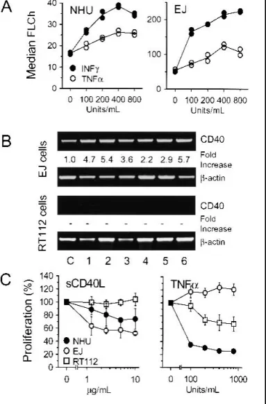
Using flow cytometry, we found that, in agreement with pre-vious reports (28,73), CD40 expression on all NHU cell lines and CD40-positive transitional cell carcinoma cell lines was increased when the cells were treated with IFN␥ or TNF-␣. IFN ␥ was more efficient than TNF-␣ at mediating an increase in CD40 expression in all the CD40-positive cell lines (Fig. 1, A). These results were confirmed by RT–PCR; CD40 mRNA was readily detectable in the CD40-positive EJ cells and showed further increases in expression after the cells were pretreated with IFN␥, TNF-␣, or a combination of both agents (Fig. 1, B). By contrast, treatment with IL-4 had no effect on CD40 expres-sion in either normal urothelial cells or in RT4, RT112, and EJ carcinoma cell lines (data not shown).
Treatment with TNF-␣, IFN ␥, or IL-4 did not induce CD40 protein expression in any of the CD40-negative cell lines. Fur-thermore, CD40 mRNA transcripts were undetectable by RT– PCR in the CD40-negative RT112 cell line. No de novo
induc-tion of CD40 mRNA expression was detected in any cell line after pretreatment with TNF-␣, and only minimal levels of CD40 mRNA were apparent following exposure to IFN ␥ (Fig. 1, B).
Effects of CD40L on Urothelial Cell Proliferation and Survival
Presentation of CD40L as soluble ligand. sCD40L had a pronounced growth inhibitory effect on both NHU cells and CD40-positive EJ cells (Fig. 1, C, left), in agreement with pre-vious reports (23,30–32). However, sCD40L alone did not sta-tistically significantly induce apoptosis in any cell line tested (see Figs. 4, A, and 5, B). sCD40L had no effect on proliferation (Fig. 1, C) or apoptosis (see Figs. 4, A, and 5, B) in the CD40-negative RT112 cell line.
Presentation of CD40L by coculture with CD40L-transfected fibroblasts. By contrast to the results obtained when CD40L was presented as a soluble ligand, when EJ cells were presented with CD40L on the cell surface (e.g., by cocul-turing with 3T3CD40L cells), they underwent extensive apop-tosis compared with EJ cells cocultured with control (3T3neo) cells (Figs. 2 and 3). In all experiments, the percentage of cells undergoing apoptosis at 48 hours was always at least 70%–80%, as assessed by flow cytometry. Because the presence of cocul-tured 3T3 cells was a potential confounding factor in the flow cytometric analysis, we confirmed these results in the same two types of cocultures by using the JAM test, which allowed us to estimate DNA fragmentation specifically in the prelabeled target cell population (Fig. 3, A). We further confirmed these results by using another independent CD40L-transfected murine fibroblast cell line (CD40L-L cells; Fig. 3, A) which, as assessed by flow cytometric analyses, expresses CD40L at densities comparable to that of the 3T3CD40L cell line.
All of the CD40-positive transitional cell carcinoma cell lines similarly underwent extensive apoptosis when they were cocul-tured with 3T3CD40L cells. The degree of apoptosis was not statistically significantly increased by pretreating the cells for up to 72 hours with IFN␥ or TNF-␣ (data not shown), despite the ability of these cytokines to increase CD40 expression, implying that even a low surface density of CD40 was adequate to trigger a maximal apoptotic response. Pretreatment of the CD40-negative RT112 cells with IFN␥ or TNF-␣ did not render them susceptible to CD40-mediated apoptosis (data not shown).
None of the NHU cell lines tested showed susceptibility to apoptosis when they were cocultured with 3T3CD40L cells (Fig. 2), even though all expressed CD40 on their surfaces at densities comparable to those in the CD40-positive tumor cell lines. Pre-treatment of NHU cells with IFN␥ or TNF-␣ also did not result in any CD40L-mediated apoptosis (data not shown). CD40-negative RT112 cells were unaffected in terms of survival by coculture with 3T3CD40L cells (Fig. 2, B).
CD40 cross-linking experiments. Because sCD40L and sur-face-presented CD40L appeared to induce different responses in tumor cells, we tested whether these differences were due to cross-linking of CD40. The FLAG-tagged sCD40L construct, with and without the cross-linking enhancer, did not induce any statistically significant apoptosis compared with untreated cells (data not shown). Similarly, the G28–5 agonistic anti-CD40 an-tibody did not induce any statistically significant apoptosis ei-ther used alone or in conjunction with a goat anti-mouse IgG cross-linking secondary antibody (Fig. 3, B). Nevertheless, the anti-CD40 antibody was functionally capable of inducing apop-tosis in EJ cells when it was presented on the surface of L cell fibroblasts transfected with the Fc␥ receptor CD32 (Fig. 3, B). These findings demonstrate that soluble CD40 agonists, even if
Fig. 1. CD40 expression in urothelial cells and effects of CD40 ligation and tumor necrosis factor-␣ (TNF-␣) on proliferation. A) Normal human urothelial (NHU) cells and transitional cell carcinoma (TCC)-derived EJ cells (both CD40-positive) were seeded in replicate cultures at 105cells/well and cultured for 72
hours with TNF-␣ or interferon gamma (IFN ␥) at 0–800 U/mL in 24-well plates. Cell-surface expression of CD40 was measured by flow cytometry in duplicate runs, and results are expressed as median fluorescence channel (FLCh). B) CD40-positive EJ cells and CD40-negative RT112 cells were cultured for 48 hours with 200 or 800 U/mL of IFN␥ (lanes 1 and 2, respectively), 200 or 800 U/mL of TNF-␣ (lanes 3 and 4, respectively), or with both cytokines together at 100 or 400 U/mL each (lanes 5 and 6, respectively). Control cells were cultured without cytokines (lane C). Messenger RNA was isolated from each culture and used to prepare complementary DNAs, which were used as templates for poly-merase chain reaction (PCR) amplification of the CD40 and-actin coding sequences that used gene-specific oligonucleotide primers. PCR products were resolved and visualized on agarose gels. Gel bands were quantified by densi-tometry, and results are expressed as fold induction relative to the intensity of the bands obtained for-actin. “–” indicates no increase in CD40 mRNA compared with-actin mRNA. C) NHU, EJ, and RT112 cells were seeded into 96-well plates at 104cells/well and exposed to soluble trimeric CD40 ligand (sCD40L;
range⳱ 0–10 g/mL) or TNF-␣ (range ⳱ 0–800 U/mL) in sextuplicate wells for 72 hours. [3H]Thymidine (0.5Ci) was added to each well for the final
18-hour incubation period. The cells were then harvested onto glass fiber filters, and [3H]thymidine incorporation into DNA was quantitated by scintillation
spec-trometry. The results are expressed as means of percentages of the “no ligand” controls for each cell line. Error bars correspond to 95% confidence intervals.
extensively cross-linked, cannot transmit apoptotic signals to tumor-derived urothelial cells, because cell death occurred only when the ligating receptor agonist was presented on the surface of cells. Western blots of EJ cells induced to undergo apoptosis by coculture with 3T3CD40L cells showed that levels of the anti-apoptotic protein Bcl-2 decreased markedly. By contrast, in these same cocultures, levels of the pro-apoptotic Bax protein increased, as did those of Bak, although to a lesser extent (Fig. 3, C).
Effects of TNF-␣ and Fas Ligation on Urothelial Cell Proliferation and Survival
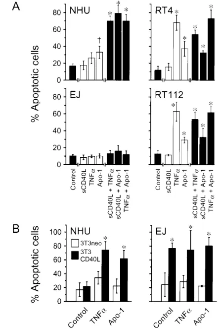
To determine the effects of ligation of other members of the TNFR superfamily, we used TNF-␣ and an agonistic anti-Fas antibody. TNF-␣ inhibited the growth of NHU cells (Fig. 1, C). However, TNF-␣ had only minor pro-apoptotic effects on nor-mal cells: In nine of 11 independent NHU cell lines tested, the percentage of apoptotic and/or dead cells was increased slightly in TNF-␣-treated cells compared with untreated cells (Fig. 4, A). The other two NHU cell lines did not undergo TNF-␣-mediated apoptosis. TNF-␣ had a small but statistically significant stimu-latory effect on growth in EJ cells (Fig. 1, C) but did not promote apoptosis in these cells (Fig. 4, A). By contrast, RT4 and RT112 cells were highly sensitive to TNF-␣-mediated apoptosis, with TNF-␣ exposure typically resulting in 60%–70% dead cells after 48 hours (Fig. 4, A).
NHU cells showed modest sensitivity to Fas-mediated apop-tosis following surface ligation of Fas with anti-Fas antibody cross-linked with a goat anti-mouse IgG secondary antibody (Fig. 4, A). We observed, by using acridine orange staining, that NHU cell cultures contained a few apoptotic bodies at 24 hours after incubation with antibody. By 48 hours, the percentage of apoptotic and/or dead cells had approximately doubled in all nine independent NHU cell lines tested.
After 48 hours of incubation with cross-linked Fas anti-body, RT112 and RT4 cells displayed a 30%–40% increase in apoptosis compared with control (i.e., untreated) cells (Fig. 4, A). By contrast, EJ cells were completely resistant to Fas-mediated apoptosis and showed no increase in the percentage of apoptotic cells following incubation with anti-Fas antibody, in-dependent of cross-linking with secondary antibody (Fig. 3). In control experiments, the efficacy of the cross-linked anti-Fas antibody was demonstrated by using the Fas-sensitive Jurkat T lymphoblastoid cell line, in which up to 96% of cells underwent apoptosis by 48 hours of incubation with the antibody (data not shown).
Effects of Multiple Receptor Ligation on Urothelial Cell Survival
Although NHU cells did not undergo apoptosis when they were cocultured with 3T3CD40L cells alone (Fig. 2), addition of either TNF-␣ or anti-CD95 antibody to such cocultures resulted in extensive apoptosis (Fig. 4, B). However, cocultures of NHU cells with 3T3neo cells did not confer any additional apoptotic sensitivity over that induced by exposing NHU cells to TNF-␣ (Fig. 4, B), anti-Fas antibody (Fig. 4, B), or sCD40L (data not shown). By contrast, addition of soluble ligands to EJ cells co-cultured with 3T3CD40L or 3T3neo cells did not increase either the very high apoptotic rate seen with ligand-transfected fibro-blasts or the low spontaneous rates, respectively, seen in control cultures (Fig. 4, B).
To examine whether apoptotic responses in NHU cells could be elicited by other ligands of the TNF family, we tested the effects of combinations of sCD40L with TNF-␣, sCD40L with anti-Fas antibody, or TNF-␣ with anti-Fas antibody. All three combinations showed synergistic enhancement of apoptosis in NHU cells over the amounts of apoptosis induced by the single effectors alone, typically inducing 60%–80% apoptosis. By con-trast, tumor-derived RT4 and RT112 cells showed no synergistic
Fig. 2. Induction of apoptosis in malignant urothelial cells by coculture with CD40 ligand (CD40L)-transfected 3T3 fibroblasts. Mitomycin C-treated 3T3 fibroblasts expressing either the neomycin resistance gene alone (3T3neo, left column) or the CD40L gene (3T3CD40L, right column) were seeded at 105
cells/well in 24-well plates. After the fibroblasts had attached to the substrate, normal human urothelial (NHU) cells or transitional cell carcinoma-derived EJ or RT112 bladder tumor cells were seeded on top of them at 9 × 10⁴cells/well.
A) Phase–contrast micrographs of CD40-positive NHU and EJ cells taken after 72 hours of coculture. Scale bar⳱ 100 m. B) Nonadherent and adherent cells harvested after 72 hours of coculture of transfected fibroblasts with NHU, EJ, or RT112 cells were incubated in suspension with fluorescein isothiocyanate-conjugated Annexin V and propidium iodide (PI) and then analyzed by flow cytometry. The results are plotted as three-dimensional density plots of mean cell number on the z-axis versus Annexin V fluorescence on the x-axis and PI fluorescence on the y-axis. Cells that did not stain for either dye were considered “live,” Annexin V-positive cells that stained medium-low with PI were consid-ered apoptotic (i.e., in the process of undergoing apoptosis), and Annexin V-positive cells that stained brightly with PI were considered as overtly dead cells (i.e., cells with disrupted membrane integrity), as indicated by the arrows.
or even additive responses when exposed to multiple soluble ligands. Thus, EJ cells remained refractory to all combinations of soluble ligands, whereas in RT4 and RT112 cells, the apop-totic response to combinations of soluble ligands did not exceed that of the most potent single soluble ligand (TNF-␣) (Fig. 4, A). These results demonstrate that receptor cooperation is neces-sary for the efficient induction of apoptosis in normal bladder epithelial cells, whereas responses of their carcinoma-derived counterparts were dependent solely on the individual receptors. This concept was further supported by two additional observa-tions (data not shown): first, that expression of Fas was not statistically significantly increased, as determined by flow cy-tometry, on EJ cells undergoing apoptosis induced by coculture with 3T3CD40L cells; and second, that addition of the Fas-blocking NOK-1 antibody had no effect on 3T3CD40L cell-induced apoptosis.
Effect of Protein Synthesis Inhibition on Apoptosis
In the majority of studies of the effects of CD40 ligation on carcinoma cells (17,35,36), apoptosis is evident only if protein synthesis is blocked by treating the cells with cycloheximide. We therefore sought to determine whether cycloheximide would affect apoptotic responses in our urothelial cell model. Cyclo-heximide had no effect on apoptosis in NHU cells or in EJ cells cocultured with 3T3CD40L cells beyond that caused by toxicity of the cycloheximide alone (Fig. 5, A). Similarly, pretreatment of NHU cells with cycloheximide did not affect the levels of apoptosis induced by sCD40L, TNF-␣, or CD95 ligation (Fig. 5, B). By contrast, pretreatment of EJ cells with cycloheximide rendered them sensitive to all three soluble effectors. In RT4 and RT112 cells, apoptotic responses were augmented following cy-cloheximide treatment, exceeding 95% killing in TNF-␣-treated
Fig. 3. Analysis of apoptosis induced in malignant bladder epithelial cells by CD40 ligation. A) EJ cells were cocultured with 3T3 cells or L fibroblast cells that were transfected with the coding sequence for CD40 ligand (CD40L) (3T3CD40L cells and CD40L-L cells, respectively) and with control-transfected 3T3neo cells or untransfected (NT-L) fibroblasts. The fibroblasts were treated with mitomycin C, plated at 104 cells/well in 96-well plates, and allowed to
attach overnight. The EJ cells were prelabeled while they were in the exponential growth phase with [3H]thymidine overnight and then harvested, washed, and
plated onto the fibroblasts at 9 × 10³cells/well. After 48 hours of coculture, cell
contents were harvested onto glass fiber filters, and the amount of3H-labeled
nuclear DNA retained was estimated by liquid scintillation spectrometry. The percentage of nuclear DNA fragmentation was calculated according to the for-mula (S – E × 100)/S, where S⳱ cpm recovered from control cultures and E ⳱ cpm recovered from test cultures, after background subtraction. Results are means of six replicates; error bars correspond to 95% confidence intervals. * indicates a statistically significant difference (P<.001; two-tailed Student’s t test) in the percentage of DNA fragmentation between EJ cell–3T3neo cell cocultures and EJ cell–3T3CD40L cell cocultures and between EJ cell–NT-L cell cocul-tures and EJ cell–CD40L-L cell coculcocul-tures. Bars in (A) are colored for clarity and contrast. B) EJ cells prelabeled with [3H]thymidine were plated out as in A
and incubated with native G28-5 agonistic anti-CD40 antibody (G28-5), G28-5 that was cross-linked with affinity-isolated human serum protein-adsorbed goat anti-mouse immunoglobulin (G28-5 + GaMIg), or G28-5 that was presented on the surface of L cells transfected with CD32 (G28-5 + CD32-L cells). CD32-L cells were used as a control. To present the antibody on fibroblasts, CD32-L cells were pretreated with mitomycin C, washed and plated out as in A above, and then incubated with G28-5 antibody or isotype-matched control before addition of epithelial cells as in A. Results are means of six replicates; error bars correspond to 95% confidence intervals. * indicates a statistically significant difference (P<.001; two-tailed Student’s t test) in the percentage of DNA frag-mentation between EJ cells that were exposed to G28-5 and CD32-L cells and EJ cells that were exposed to any of the other experimental conditions. C) Western blotting was performed on lysates of 24-hour cocultures of EJ cells and 3T3CD40L or 3T3neo fibroblasts. Cell lysates were separated by 12% sodium dodecyl sulfate–polyacrylamide gel electrophoresis, transferred to nitrocellulose membranes, and probed with human-specific antibodies against cytokeratins 8 and 18 (CK8 and CK18), and against Bcl-2, Bax, and Bak. Lysates from fibro-blasts cultured alone were included as controls. Track loading was adjusted for equal amounts of epithelial cell protein on the basis of immunoreactivity with antibodies to CK8 and CK18 in western blots. Antibody binding was detected by enhanced chemiluminescence. Lane 1, EJ cells only (control); lane 2, EJ cells cocultured with 3T3CD40L cells; lane 3, EJ cells cocultured with 3T3neo cells; lane 4, 3T3CD40L cells only; lane 5, 3T3neo cells only.
RT4 cells. Cycloheximide did not induce sensitivity to sCD40L in CD40-negative RT112 cells (Fig. 5, B).
These findings suggest that tumor cells rely on the synthesis of apoptosis-inhibitory proteins to escape cell death induced by soluble TNF superfamily ligands, whereas normal cells do not. This dependence on synthesis of apoptosis inhibitory proteins appears to be particularly the case for EJ cells. The inability of these cells to resist 3T3CD40L-mediated apoptosis, even when protein synthesis is allowed, further suggests that the two modes of receptor ligation resulted in different downstream conse-quences.
Expression of Genes Associated With Apoptotic and TNF Signaling Pathways
Multi-probe ribonuclease protection assays were used to identify differences in gene expression between normal and ma-lignant bladder epithelial cells that might account for the differ-ential responses of tumor-derived versus normal urothelial cells to ligation of CD40. NHU cells and transitional cell carcinoma-derived cell lines expressed mRNA transcripts for TNF family receptors (data not shown) and intracellular interacting proteins
involved in the transduction of apoptotic signals. The abundance of mRNA species that encode proteins involved in death recep-tor signaling, such as Fas-associated death domain-containing protein (FADD), Fas-associated protein factor 1 (FAF1), and receptor interacting protein (RIP), was lower in NHU cells and the Fas-resistant EJ cells than in Fas-sensitive RT4 or RT112 cells. TNF-␣-resistant EJ cells exhibited relatively low levels of TNFR1-associated protein (TRADD) mRNA compared with RT4 and RT112 cells, which were susceptible to TNF- ␣-mediated apoptosis (Table 1). Expression of transcripts for Bcl-2 family members was similar between cell types (data not shown).
We also examined the abundance of mRNA transcripts for proteins reported to inhibit apoptosis mediated by TNF family members. All cell lines studied expressed variable amounts of mRNA for members of the TNF receptor-associated factor (TRAF) family (TRAF1, TRAF2, and TRAF3) and inhibitor of apoptosis protein (IAP) family (IAP1 and X-linked IAP [XIAP]). IAP2 mRNA was expressed at high abundance in EJ cells compared with the other cell types and was undetectable in NHU cells and RT112 cells (Table 1).
Fig. 4. Apoptotic responses to ligation with other receptors, both singly and in combination. A) Normal human urothelial (NHU) cells and the transitional cell carcinoma-derived cell lines RT4 (well-differentiated, CD40-positive), RT112 (moderately-differ-entiated, CD40-negative), and EJ (anaplastic, CD40-positive) were plated in replicates at 105cells/well in 24-well plates and incubated
for 72 hours with soluble receptor agonists alone or in combination. Soluble trimeric CD40 ligand (sCD40L) was used at 10g/mL, tumor necrosis factor-␣ (TNF-␣) was used at 300 U/mL, and ago-nistic anti-Fas antibody (Apo-1) was used at 0.5g/mL with a cross-linking goat anti-mouse immunoglobulin (Ig) at 10g/mL. The contents of each well were harvested, labeled in suspension with Annexin V fluoroconjugate and propidium iodide, and ana-lyzed by flow cytometry. Gates were set to detect viable and apop-totic cells (compare Fig. 2), and the percentage of apopapop-totic cells in each culture was plotted. Bars represent mean values for 11 inde-pendent NHU cell lines and for at least six indeinde-pendent determi-nations for each transitional cell carcinoma-derived cell line; error bars represent 95% confidence intervals. For NHU cells, * indicates a statistically significant difference (P<.001) in the per-centage of apoptotic cells between cultures treated with combina-tions of agonists and control cultures or cultures treated with single agonists, and † indicates a statistically significant difference (P<.01) in the percentage of apoptotic cells between cells treated with Apo-1 antibody and control cultures. For RT4, EJ, and RT112 cell cultures, * indicates a statistically significant difference (P<.001) in the percentage of apoptotic cells between cells treated with TNF-␣, Apo-1, or any combination of agonists and control cultures. Bars in (A) are colored for clarity and contrast. B) Mito-mycin-C pretreated control- or CD40L-transfected fibroblasts (3T3neo and 3T3CD40L cells, respectively) were plated at 105
cells/well and cocultured with 9 × 10⁴NHU or EJ cells, in the
presence or absence of TNF-␣ or Apo-1 antibody for 72 hours. The cells were harvested and labeled with Annexin V and PI, and the percentage of apoptotic cells in each culture was assessed by flow cytometry. Bars represent mean values for six replicates; error bars represent 95% confidence intervals. * indicates a statistically significant difference in the percentage of apoptotic cells between NHU or EJ cells that were cocultured with 3T3neo cells and NHU or EJ cells that were cocultured with 3T3CD40L cells for each treatment (P<.001). All statistical significance was determined by analysis of variance using the Tukey–Kramer multiple compari-sons test.
Involvement of NFB in Responses to CD40, Fas, and TNFR Ligation
Because NFB has been implicated in influencing cell-survival outcomes mediated by members of the TNFR super-family (74,75), we studied its involvement in signaling mediated by CD40, TNFR, and Fas in our urothelial cell system. All normal and cancer-derived urothelial cell lines showed compa-rable constitutive expression of NFB by immunoblotting (data not shown). Indirect immunofluorescence microscopy revealed that NFB was localized to the cytoplasm in both normal and malignant urothelial cells; incubation of those cells with sCD40L, TNF-␣, or anti-CD95 antibody did not induce the nuclear translocation of NFB. By contrast, in human foreskin-derived fibroblasts, which were used as a positive control, NFB translocated to the nucleus within 10 minutes of exposing the cells to 400 U/mL TNF-␣, where it remained for up to 120 minutes (data not shown).
Ligation of the CD40 receptor in EJ cells following coculture with 3T3CD40L cells resulted in NFB translocation to the nucleus, whereas, in EJ cells cocultured with 3T3neo cells, it
remained in the cytoplasm (Fig. 6). By contrast, coculturing CD40-positive NHU and RT4 cells and CD40-negative RT112 cells with 3T3CD40L cells did not trigger NFB translocation. Moreover, the NFB inhibitors BAY 11–7082 and SN50 did not inhibit apoptosis of EJ cells cocultured with 3T3CD40L cells (data not shown). These data suggest that, despite the ability of NFB to translocate to the nucleus in EJ cells, NFB activation is not involved in the execution phase of apoptosis induced by 3T3CD40L fibroblasts in CD40-expressing tumor cells. Responses of p53-Disabled Urothelial Cells to TNF Family Signaling
Because loss of p53 function is a key genetic event for ma-lignant progression in bladder cancer (48,49), we investigated whether deletion of p53 function in otherwise normal urothelial cells would alter their responses to CD40, Fas, and TNFR liga-tion. We found that NHU cells with disabled p53 function (HU-E6 cells) expressed TNFR and CD95 at levels comparable to those in parental NHU cells; CD40 expression was slightly higher in HU-E6 cells than in the parental NHU cells (data not shown).
Fig. 5. Effects of blocking protein synthesis on apoptotic responses. The effects of blocking protein synthesis was assessed by pretreat-ing normal human urothelial (NHU) cells with 0.01g/mL cyclo-heximide (CHX), and the transitional cell carcinoma-derived RT4 (well-differentiated, positive), EJ (anaplastic, CD40-positive), and RT112 (moderately-differentiated, CD40-negative) cell lines with 1g/mL cycloheximide for 30 minutes, after which agonists were added to the cells, which were cultured in the con-tinued presence of cycloheximide. Control cultures were performed in the absence of cycloheximide. A) Mitomycin C pretreated con-trol- or CD40 ligand (CD40L)-transfected fibroblasts (3T3neo and 3T3CD40L cells, respectively) were plated at 105 cells/well and
cocultured with 9 × 10⁴NHU or the transitional cell carcinoma EJ
cells for 72 hours. The cells were harvested and labeled with An-nexin V and propidium iodide (PI), and the percentage of apoptotic cells in each culture was assessed by flow cytometry. Bars repre-sent mean values for four replicates; error bars reprerepre-sent 95% confidence intervals. * indicates a statistically significant difference (P<.001) in the percentage of apoptotic cells between EJ cells that were cocultured with 3T3neo cells and EJ cells that were cocultured with 3T3CD40L cells, regardless of CHX treatment. B) NHU cells and the transitional cell carcinoma-derived cell lines RT4, RT112, and EJ were plated in replicates at 105cells/well in 24-well plates
and incubated for 72 hours with soluble receptor agonists. Soluble trimeric CD40 ligand (sCD40L) was used at 10 g/mL, tumor necrosis factor-␣ (TNF-␣) was used at 300 U/mL, and Apo-1 ago-nistic anti-Fas antibody was used at 0.5g/mL with a cross-linking goat anti-mouse immunoglobulin (Ig) at 10g/mL. The contents of each well were harvested, labeled in suspension with Annexin V fluoroconjugate and PI, and analyzed by flow cytometry. Gates were set to detect viable and apoptotic cells (compare Fig. 2), and the percentage of apoptotic cells in each culture was plotted. Bars represent mean values of 4–17 replicates; error bars represent 95% confidence intervals. † indicates a statistically significant difference (P<.01) in the percentage of apoptotic cells between control cells and cells treated with Apo-1 in the absence of CHX; § indicates a statistically significant difference (P<.001) in the percentage of apoptotic cells between control cells and cells treated with TNF-␣ or Apo-1 in the absence of CHX; ‡ indicates a statistically signifi-cant difference in the percentage of apoptotic cells (P<.001) be-tween control cells and treated cells in cultures that were pretreated with CHX. The statistical significance of all differences was deter-mined by analysis of variance using the Tukey–Kramer multiple comparisons test.
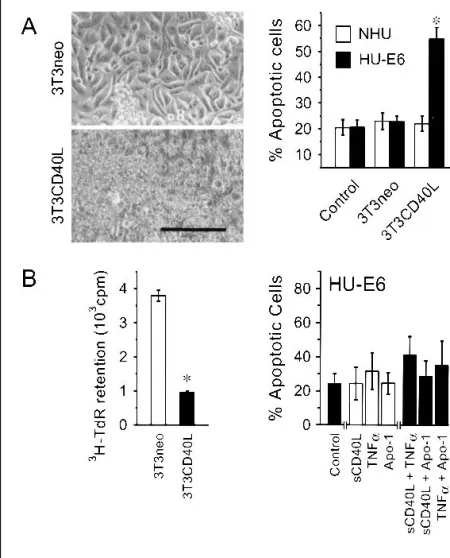
HU-E6 cells, despite showing no karyologic abnormalities, showed the same overall pattern of sensitivity to TNF family ligands as fully malignant (i.e., tumor-derived) cells rather than that shown by the untransfected NHU parental cells. HU-E6 cells were highly sensitive to apoptosis induced by coculture with 3T3CD40L cells (Fig. 7, A) but were not sensitive to ap-optosis induced by exposure to any soluble ligand (e.g., sCD40L, TNF-␣, or anti-Fas), either alone or in combination (Fig. 7, B). No nuclear translocation of NFB was seen
follow-ing coculture of HU-E6 cells with either 3T3CD40L cells or 3T3neo cells (data not shown), and no major differences were detected in the expression of apoptosis-related mRNA species between HU-E6 cells and NHU cells (data not shown). These results suggest that even the early genetic events of malignant transformation, typified by loss of p53 function (48,49), pro-foundly alter the apoptotic susceptibility and response patterns of human urothelial cells to signaling by members of the TNF family.
D
ISCUSSIONResults from our study reveal three novel aspects of CD40 biology in urothelial cells: 1) that the effects of CD40 ligation, either alone or in conjunction with ligation of other members of the TNFR family, are context-specific, in that outcome depends on additional factors; 2) that the mode of CD40 ligation is criti-cal to the outcome; and 3) that genetic changes that lead to malignant transformation alter the responses of urothelial cells to TNF family signaling.
Our data were obtained by using established cell lines and therefore may not be entirely applicable to the situation in vivo. Nevertheless, the established transitional cell carcinoma-derived cell lines that we used have been shown, both in vitro (76) and in vivo (52), to faithfully recapitulate the grade and stage of the originating tumor. The ability of NHU cell lines to re-form stratified transitional epithelia (47) also supports the biologic relevance of our model system. The findings of antitumor effects of CD40L in SCID mice inoculated with established breast car-cinoma cell lines (33,34) has recently been extended to xeno-grafts of surgical specimens of ovarian adenocarcinoma (39), thereby supporting the use of cell lines as indicators of CD40 sensitivity. These and other studies have also shown that carci-noma cells from a number of epithelial origins are susceptible to the antitumor effects of CD40 ligation.
Fig. 6. Nuclear translocation of nuclear factor-B (NFB) in re-sponse to CD40 signaling. Mitomycin C-treated 3T3neo or 3T3CD40L fibroblasts were seeded at 1.5 × 10⁴cells/spot on
12-spot glass slides and incubated overnight before addition of 104
normal human urothelial (NHU) cells or transitional cell carci-noma-derived EJ cells per spot. The cells were cocultured for 2 hours, then washed, fixed in a 1 : 1 mixture of methanol/acetone, air-dried, and processed for two-color indirect immunofluores-cence. NFB (red) was detected with a rabbit anti-NFB p65 an-tibody and visualized with goat anti-rabbit immunoglobulin (Ig)– Texas Red conjugate. Monoclonal antibodies specific for cytokeratin 8 (CK8) or for CD40 ligand (CD40L) were used to detect epithelial cells and CD40L-transfected 3T3 fibroblasts, re-spectively. Monoclonal antibodies were visualized by using a fluo-rescein isothiocyanate-conjugated goat anti-mouse Ig (green). Yel-low indicates colocalization of red- and green-tagged antibodies. Arrows indicate cells that displayed nuclear translocation of NFB. Scale bar⳱ 50 m.
Table 1. Expression of mRNA for apoptosis-associated proteins in urothelial cells* Protein Cell lines NHU HU-E6 RT4 RT112 EJ Adaptors FADD 9.90 23.73 82.54 29.49 25.43 FAF-1 10.21 5.63 86.88 32.97 30.40 FAP-1 52.29 7.33 82.30 60.00 18.65 RIP 5.60 6.58 35.55 42.73 7.56 TRAF1 5.70 3.72 7.55 9.83 5.20 TRAF2 16.90 11.86 6.75 19.48 10.80 TRAF3 19.39 7.50 11.28 5.00 13.10 TRADD 32.28 18.4 31.61 29.95 7.05 Apoptosis inhibitors IAP1 (IAP-C) 23.46 12.89 8.85 15.5 7.05 IAP2 (IAP-B) — 10.19 4.05 — 53.00 XIAP 26.69 13.50 26.91 23.84 20.13
*Messenger RNA (mRNA) was quantitated by ribonuclease protection assays; values are expressed as a percentage of the mRNA for the glyceraldehyde-3-phosphate dehydrogenase in each cell line. NHU⳱ normal human urothelial; FADD ⳱ associated death domain-containing protein; FAF ⳱ Fas-associated protein factor; FAP⳱ Fas-associated phosphatase 1; RIP ⳱ receptor interacting protein; TRAF⳱ tumor necrosis factor receptor-associated factor; TRADD⳱ tumor necrosis factor receptor 1-associated protein; IAP ⳱ inhibitor of apoptosis protein; XIAP⳱ X-linked inhibitor of apoptosis protein; — ⳱ a value too low to quantitate.
Context-specific responses to CD40 signaling have been well established in B cells and their malignant counterparts, where, depending on endogenous and external conditions, CD40 liga-tion may either induce apoptosis or promote survival (2,8,10– 12,77,78). Our study is the first to demonstrate that a similar situation may apply in urothelial cells. Importantly, our data demonstrate that urothelial cell survival is highly dependent on the mode of CD40 ligation. Thus, sCD40L inhibited the growth of both normal and malignant urothelial cells but did not induce apoptosis, whereas surface-presented CD40L, while having no substantial inhibitory effects on growth (data not shown), spe-cifically induced apoptosis in transformed cells but not in nor-mal cells. Our findings with soluble ligands are consistent with the findings of others who have shown that growth inhibition is a consequence of CD40 ligation in normal keratinocytes (23,29,30) and in carcinoma cells in vitro (29–32) and in vivo (33,34,39).
It is well established that in B cells, responses to CD40 de-pend on the mode of ligation. Factors such as epitope specificity and degree of cross-linking of the ligating CD40 antibody (53,54), the use of agonistic antibody versus cell-surface pre-sentation of CD40L (55), and even the density of CD40L ex-pression on transfected fibroblasts in a coculture system (56) can critically affect functional outcome [reviewed in (9)]. Thus, it would appear that in B cells and epithelial cells, the precise nature of the response to CD40 ligation, be it survival or death, depends on the nature of the ligating signal, probably as a
func-tion of the quality of receptor cross-linking. A close analogy is provided by the Fas/FasL system, where killing or protection from killing depends on whether FasL is presented in soluble or membrane-bound form (79,80).
Our studies using CD40L-transfected fibroblasts also reveal that CD40 ligation can induce a dramatic apoptotic response in malignant, and even in “premalignant” (i.e., normal cells with disabled p53 function) urothelial cells, while sparing their nor-mal counterparts. These novel observations are supported by previous reports of CD40-dependent apoptotic responses in other tumor cell systems, such as CD40-transfected HeLa cells after cycloheximide treatment (17,35) and cycloheximide-treated hepatocellular and ovarian carcinoma cells (17,36).
Although a wealth of information has been gathered about the downstream effectors involved in CD40 signaling, particularly those in B cells, the mechanisms by which these effectors co-operate with those mechanisms elicited by other members of the TNFR family to transmit apoptotic versus survival signals is far from clear. Although CD40 has no intrinsic death domain of its own, its ability to transmit cell death signals by recruiting mem-bers of the TRAF family of adaptor molecules may allow it to interact with the apoptotic pathways known to be associated with other TNFR superfamily molecules in addition to directly activating NFB and/or stress kinases (8,75,78,81,82). For ex-ample, CD40 ligation has been reported to promote (16,29,31) and to inhibit (40–44) Fas-mediated apoptosis. Recently, CD40 has been shown to induce autotropic cell death via other
mem-Fig. 7. Responses of p53-disabled Human Urothelial (HU-E6) cells to CD40, tumor necrosis factor receptor (TNFR), and Fas ligation. A) Mitomycin C-treated 3T3 fibroblasts expressing the neomycin resistance gene alone (3T3neo cells) or with the gene for CD40 ligand (CD40L) (3T3CD40L cells) were seeded at 105cells/well in
24-well plates. Following fibroblast attachment, untransfected nor-mal human urothelial (NHU) cells or urothelial cells with disabled p53 function (HU-E6 cells) were seeded on top of the fibroblasts at 9 × 10⁴cells/well. Left panel shows a phase–contrast micrograph
of HU-E6 cells after 72 hours of coculture with 3T3neo cells (top) or with 3T3CD40L cells (bottom). Scale bar⳱ 100 m. Cells harvested from the 72-hour cocultures were labeled in suspension with Annexin V fluoroconjugate and propidium iodide and ana-lyzed by flow cytometry to determine percentages of viable and apoptotic cells (right panel). Results are expressed as mean value of 4–9 replicate determinations; error bars represent 95% confi-dence intervals. * indicates a statistically significant difference in the percentage of apoptotic cells between HU-E6:3T3CD40L co-cultures and HU-E6:3T3neo coco-cultures (P<.001; two-tailed Stu-dent’s t test). B) Mitomycin C-treated 3T3neo or 3T3CD40L fibro-blasts were seeded at 104cells/well in 96-well plates and cocultured
for 72 hours with 9 × 10³HU-E6 cells that were previously labeled
with [3H]thymidine (3H-TdR). The cells were then harvested onto
glass fiber filters, and the amount of DNA retained was estimated by measuring3H-TdR using liquid scintillation spectrometry (left
panel). Results are expressed as the mean value of sextuplicate determinations; error bars represent 95% confidence intervals. * indicates a statistically significant difference in the amount of
3H-TdR retained by 3T3CD40L cocultures and the amount retained
by 3T3neo cocultures (P<.001; two-tailed Student’s t test). HU-E6 cells were also plated alone (right panel) at 9 × 10⁴cells/well and
cultured in the presence of soluble trimeric CD40 ligand (sCD40L) at 10g/mL, tumor necrosis factor-␣ (TNF-␣) at 300 U/mL, and agonistic anti-Fas antibody (Apo-1) at 0.5 g/mL with cross-linking goat anti-mouse immunoglobulin at 10g/mL, alone or in combination. Adherent and nonadherent cells were harvested at 72 hours, labeled with Annexin V and propidium iodide, and analyzed by flow cytometry. Bars in (B) are colored for clarity and contrast.