Visual Neuroscience (1996), 13, 997-1009. Printed in the USA.
Copyright © 1996 Cambridge University Press 0952-5238/96 $11.00 + .10
Calcium-binding proteins immunoreactivity in the
human subcortical and cortical visual structures
G. LEUBA AND K. SAINI
University Psychogeriatrics Hospital, CH-1008 Lausanne-Prilly, Switzerland
(RECEIVED July 5, 1995; ACCEPTED March 1, 1996)
Abstract
The distribution of neurons and fibers immunoreactive (ir) to the three calcium-binding proteins
parvalbumin (PV), calbindin D-28k (CB), and calretinin (CR) was studied in the human lateral geniculate nucleus (LGN), lateral inferior pulvinar, and optic radiation, and related to that in the visual cortex. In the LGN, PV, CR, and CB immunoreactivity was present in all laminae, slightly stronger in the magnocellular than in the parvocellular laminae for CB and CR. PV-ir puncta, representing transversally cut axons, and CR-ir fibers were revealed within the laminae and interlaminar zones, and just beyond the outer border of lamina 6 in the geniculate capsule. In the optic radiation both PV- and CR-immunoreactive neurons, puncta, and fibers were present. CB immunoreactivity was revealed in neurons of all laminae of the lateral geniculate nucleus, including S lamina and interlaminar zones. There were hardly any CB-ir puncta or fibers in the laminae, interlaminar zones, geniculate capsule, or optic radiation. In the lateral inferior pulvinar, immunoreactive neurons for the three calcium-binding proteins were present in smaller number than in the LGN, as well as PV-ir puncta and CR-ir fibers within the nucleus and in the pulvinar capsule. In the white matter underlying area 17, fibers intermingled with a few scattered neurons were stained for both PV and CR, but very rarely for CB. These fibers stopped at the limit between areas 17 and 18. Area 17 showed a dense plexus of PV-ir puncta and neurons in the thalamo-receptive layer IV and CR-ir puncta and neurons both in the superficial layers I—II, IIIC, and in layer VA. Cajal-Retzius CR-ir neurons were present in layer I. CB-ir puncta were almost confined to layer I—III and CB-ir neurons to layer II. Finally the superior colliculus exhibited mostly populations of PV and CR pyramidal-like immunoreactive neurons, mainly in the intermediate tier. These data suggest that in the visual thalamus most calcium-binding protein immunoreactive neurons project to the visual cortex, while in the superior colliculus a smaller immunoreactive population represent projection neurons.
Keywords: Calcium-binding proteins, Lateral geniculate nucleus, Visual cortex, Superior colliculus, Human
Introduction
Although the role of calcium-binding proteins is not yet com-pletely understood, the cellular distribution of parvalbumin (PV) and calbindin D-28k (CB) has been extensively studied during the last few years in the brain of rodents and monkeys but com-paratively less in the human brain. In the cerebral cortex of monkeys, PV- and CB-positive neurons have been described as two nonoverlapping populations of GABAergic interneurons (Blumcke et al., 1990; Bliimcke & Celio, 1992; Hendry & Carder, 1993; Jones, 1993). In most areas, GABAergic cells immunoreactive (ir) for parvalbumin are chandelier and basket interneurons concentrated in layers III and IV, while those immunoreactive for calbindin are mainly double bouquet inter-neurons distributed in layers II and III, less in layers V-VI (Jones, 1993). In addition, PV immunoreactivity reveals a zone of thalamic afferents in lamina IV of the primary visual cortex
Reprint requests to: G. Leuba, University Psychogeriatrics Hospi-tal, CH-1008 Lausanne-Prilly, Switzerland.
(Blumcke etal., 1991; DeFelipe & Jones, 1991). In the human visual cortex, the distribution of both PV-ir and CB-ir inter-neurons is comparable to that in the monkey (Blumcke et al., 1990; Hendry & Carder, 1993), although PV-ir interneurons are about 11 % of the neuronal population in the monkey and only about 7% in human (Blumcke et al., 1990). The distribution of calretinin (CR)-positive interneurons has been less extensively studied than that of PV-ir and CB-ir interneurons in the cere-bral cortex. However, a population of CR-ir cells has been described in the primary visual cortex of dolphins and humans; in the latter, these cells, mainly bipolar or fusiform in shape, are distributed largely in superficial layers II to IVA, while large multipolar cells are observed in layers I and VI (Glezer et al., 1992).
In the thalamus of Macaque monkeys, and more specifically in the lateral geniculate nucleus (LGN) and pulvinar, there are both PV-ir and CB-ir neuronal populations, with the CB-ir neu-rons localized exclusively in the interlaminar and S laminae (Jones & Hendry, 1989). The presence of a large number of PV-positive asymmetric synaptic contacts in the thalamo-recip-997
ient layer IV of the primary visual cortex suggests that these afferents derive from the PV-ir neurons located in the LGN (Bliimckeetal., 1991; DeFelipe & Jones, 1991). Intheprosim-ian Galago, a complementary pattern of PV-ir and CB-ir neu-rons has also been described in the LGN, differentiating parallel visual pathways to the visual cortex (Diamond et al., 1993; Casagrande, 1994; Johnson & Casagrande, 1995). The presence of CB-ir neurons in the koniocellular layers, receiving fibers both from the retina and from the superior colliculus, suggests that calbindin is a marker for the LGN cells that belong to the third visual pathway, the K pathway, resembling the W pathway in cat (Casagrande, 1994). Furthermore, combined retrograde- and double-labeling experiments have shown that both PV-ir and CB-ir LGN neurons are projecting to different parts of the visual cortex, none being GABAergic (Johnson & Casagrande, 1995). Finally in the lateral inferior pulvinar (LIP), a chemoarchitec-tonic subdivision based on PV and CB immunocytochemistry was demonstrated, suggesting that the PV-positive neurons were the fast firing cells projecting to the medial temporal area MT in the monkey, while the CB-positive cells were scarce (Cusick et al., 1993). PV-ir and CB-ir neurons seem thus to be projec-tion neurons in the thalamus of monkeys, while they are mostly GABAergic interneurons in the cerebral cortex.
CR-ir neurons have not, to our knowledge, been demon-strated in the thalamus of primates, but in the retina and LGN of different species (Pasteels et al., 1990) and in the optic tract and LGN of rodents (Jacobowitz & Winsky, 1991; Araiet al., 1992), suggesting that there may be a CR-positive projection from the retina to the LGN. In the present paper, we study the distribution of PV, CB, and CR in the LGN, LIP, optic radia-tion (OR), and superior colliculus (SC) of humans, and com-pare it to that in the primary visual cortex and underlying white matter.
Materials and methods
Tissue blocks were taken from the brains of ten normal human adults, both males and females, without neurological disease (Table 1). The brains were fixed by submersion in 10% formal-dehyde or, for some of them, frozen directly at autopsy (-70°C). The postmortem delay was between 10 and 20 h and the time
Table 1. Description of the 10 cases used in this study" Cases 1 2 3 4 5 6 7 8 9 10 Age 30 48 59 65 71 76 78 82 84 96 Sex M M M F M M M F M M Cause of death myocardial infarct myocardial infarct esophageal carcinoma accident pancreatic carcinoma bronchial carcinoma thrombosis hypothermia glioblastoma pneumonia
"Age is in years; sex: (M) male, (F) female; cause of death is not neuro-logical, except for case 9, whose glioblastoma was not in the visual region. Postmortem delay between 10 and 20 h (see Methods).
of fixation generally did not exceed 1 year. However, two cases with a slightly longer fixation gave excellent results. Blocks were dissected out from the middle of the calcarine sulcus (area 17), from the thalamic region containing the LGN and LIP and from the mesencephalon containing the SC. Frozen 50 /jm sections were cut in a frontal plane and kept at 4°C in cold Tris-buffered saline (TBS 0.1 M, pH 7.3) until immunohistochemical processing. Immunohistochemical staining was performed using three dif-ferent primary antibodies: Anti-Parvalbumin 235 mouse mono-clonal IgG 1, Anti-Calbindin D-28K mouse monomono-clonal 300 IgG 1, and Anti-Calretinin 7696 rabbit polyclonal donated by M.R. Celio. Free-floating sections were incubated for 3 days at 4°C either with PV, CR (dilution 1:1000), or CB (dilution 1:500) in TBS buffer containing 10% calf serum and 0.1 % Triton X100. After incubation with the primary antibody, the sections were rinsed in TBS ( 6 x 5 min) and incubated for 2 h at room tem-perature in biotinylated antimouse IgG (for PV and CB) or anti-rabbit IgG (for CR) from Vector Laboratories, Burlingame, CA (dilution 1:200 in TBS containing 10% calf serum). Sections were rinsed again in TBS ( 6 x 5 min) and incubated for 3 h at room temperature in avidin-biotin-peroxidase complex (4.5 pi/ml in TBS containing 10% calf serum). The bound peroxidase was revealed by incubating the sections for 10-20 min at room tem-perature in a mixture of 0.05% 3,3-diaminobenzidine (Sigma Chemical Co., St. Louis, MO) and 0.01 % H2O2 in 0.05 M TBS.
Immunoreactive cells and neuropil were visualized under bright-field illumination using a Zeiss Axioplan microscope.
Results
Lateral geniculate nucleus, pregeniculate nucleus, and optic radiation (Figs. 1-3; Table 2)
In the LGN, we found PV-ir neurons in all of the laminae and the intensity of staining did not depend on the age of the sub-jects (Figs. 1A-1D). Neuronal perikarya were diffusely stained without clear dendritic arborization in both magnocellular (M) and parvocellular (P) laminae (Figs. 1A-1D, 3A, and 3B). Inter-laminar zones were mostly free of PV-ir neurons, although a few positive neurons were scattered close to the laminar bor-ders. Numerous PV-ir puncta, which represent axonal and den-dritic terminals (see Discussion), were distributed across the laminae and the interlaminar zones, as well as in the geniculate capsule at the outer border of lamina 6 (Figs. 3A-3C). CR-ir neurons were also observed in all M and P laminae, diffusely marked with a stronger staining in the M than in the P laminae (Figs. 2A, 2C, and 3D). In the interlaminar zones, only neurons close to the borders showed CR immunoreactivity. CR-ir puncta were present in the interlaminar zones (Fig. 3D), while CR-ir fibers were observed mainly in the geniculate capsule (Fig. 3E). CB-ir neurons were present in all laminae with well-marked cell bodies but poorly marked dendrites. Magnocellular neurons ap-peared to be slightly more intense than the parvocellular ones but this difference was not as clear as for CR (compare Figs. 2A, 2C with Figs. 2B, 2D). Scattered well-marked CB-ir neurons but no fibers or puncta were observed in the interlaminar zones or in the geniculate capsule (Figs. 2B, 2D, 3F, and 3G). When an S lamina was present in our sections, it showed a similar CB immunostaining as the magnocellular laminae 1 and 2 (Fig. 2D).
In the pregeniculate nucleus, which lies in close vicinity to the LGN dorsally and caudally, we found rare CB-ir neurons,
Calcium-binding proteins immunoreactivity in the human visual system 999
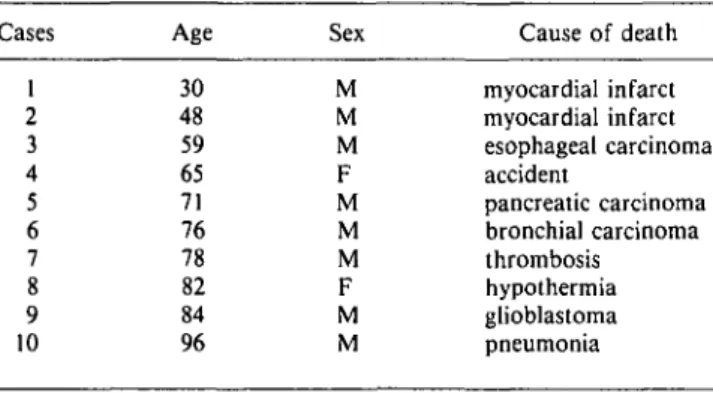
Fig. 1. Light micrographs demonstrating PV immu-noreactivity in the human LGN from different cases. (A) case 6, age 76 years (see Table 1); (B) case 9, age 84 years; (C) case 5, age 71 years; and (D) case 1, age 30 years. PV immunoreactivity in M laminae 1 and 2 appears almost similar to that in P laminae 3-6 and the intensity of immunostaining is not different between young and old cases. At the outer border of lamina 6, the geniculate capsule (GC) shows PV-ir puncta. Puncta are also observed within the interlam-inar zones (*) and their density varies slightly from one case to the other. Scale bar in A-D = 500 jim.
but a few large strongly PV-ir and CR-ir neurons (Fig. 3E), mostly multipolar and bipolar in shape.
Finally in the OR, we found many scattered and well-marked PV-ir and CR-ir interstitial neurons as well as well-defined PV-ir and CR-ir fiber bundles. CB-ir neurons and fibers were only few in the optic radiation.
Lateral inferior pulvinar (Fig. 4; Table 2)
In the lateral inferior pulvinar (LIP), the immunostaining of calcium-binding proteins was similar to that in the LGN but the proportion of stained neurons was less. PV-ir neurons as well as puncta were well marked (Figs. 4A and 4B). CR-ir neurons
GO
B
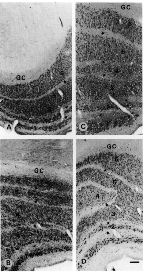
Fig. 2. Light micrographs demonstrating CR-ir (A,C) and CB-ir (B,D) in the human LGN. (A) case 1 (see Table 1); (B) case 1; (C) case 6; and (D) case 3. Note that in (A) and (C) the CR-ir is higher in the M (1 and 2) than in the P (3-6) laminae, whereas, in (B) and (D) the difference between the CB-ir M and P laminae is not as striking. GC: geniculate capsule, and (*) interlami-nar zones. S represents a part of the ventral-most lamina which is also CB-ir. Scale bar in A-D = 500 jim.
and fibers running roughly parallel to each other were also present (Figs. 4C and 4D). CB-ir neurons were less strongly marked. As in the LGN, there were neither fibers nor puncta which were CB-ir. In the pulvinar capsule, we observed both PV-ir puncta and CR-ir fibers (Fig. 4), but CB-ir fibers or puncta were rare.
Primary visual cortex and underlying white matter (Figs. 5-7; Table 2)
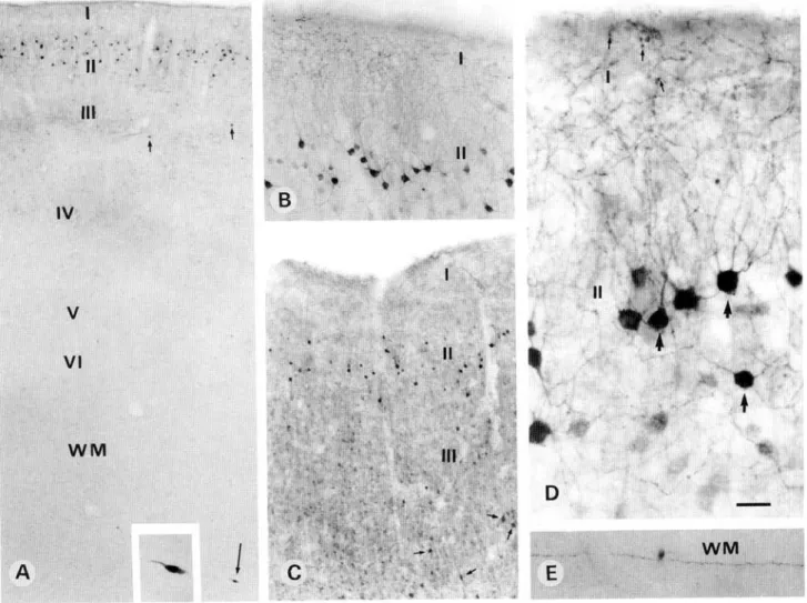
Primary area 17 showed a stronger PV immunoreactivity in the recipient layer IV than in layers II—III and V-VI (Figs. 5A and 5C). This was mainly due to a high density of puncta, repre-senting transversally cut axons and dendrites (see Discussion), as well as to numerous PV-ir neurons (Figs. 5C and 5D), most of which resembled small granule cells with multipolar dendritic
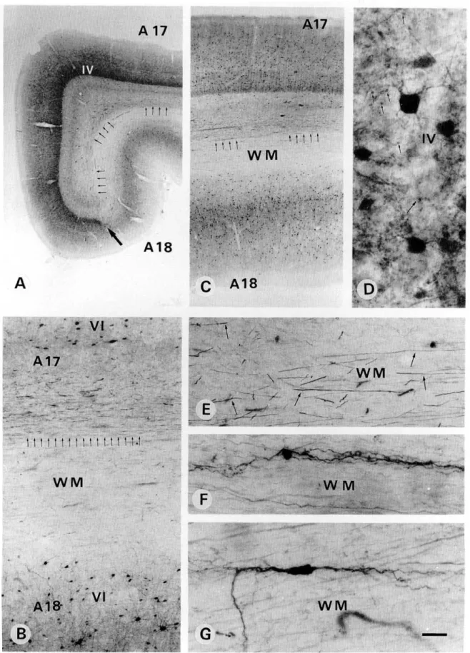
arborization. In other layers PV-ir neurons resembled either chandelier or basket types. In the white matter, PV-ir fibers ran parallel to the cortical layers and were confined mostly to the region underlying layer VI of area 17 (Figs. 5A-5C and 5E). In the white matter underlying layer VI of area 18, such dense fibers were not observed (Figs. 5A-5C). A few PV-ir intersti-tial neurons, mostly of bipolar shape with long dendrites ori-ented parallel to the fibers, were encountered in the white matter (Figs. 5F and 5G). The pattern of CR immunoreactivity was completely different. A high density of puncta, together with numerous CR-ir neurons, was observed in the superficial lay-ers I and II (Figs. 6A and 6B). On the surface of layer I, a few CR-ir Cajal-Retzius neurons, showing a cell body and dendritic arborization oriented parallel to the pial surface, were encoun-tered (Figs. 6B and 6C). In layers II-IV, most CR-ir neurons showed a bipolar or bitufted dendritic arbor oriented
Calcium-binding proteins immunoreactivity in the human visual system 1001
GC
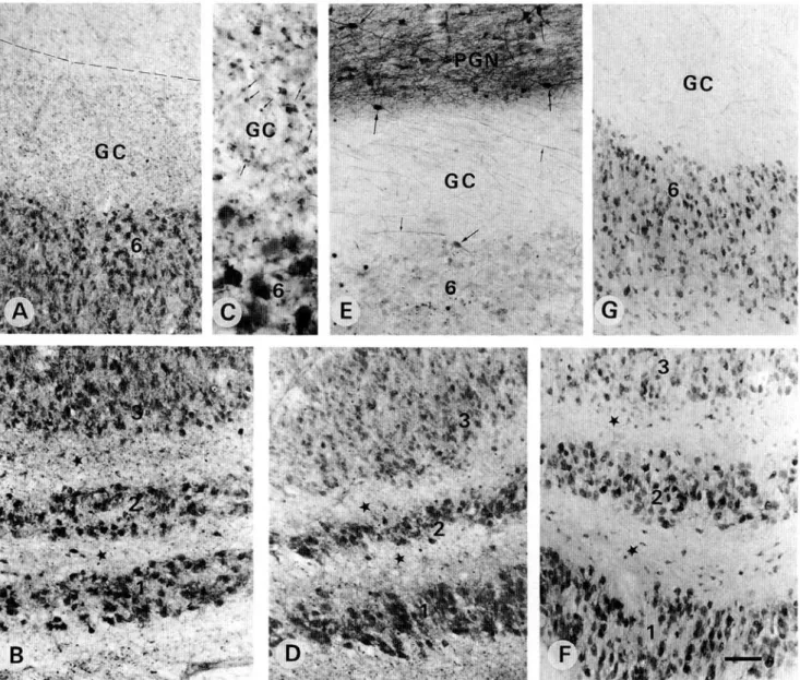
Fig. 3. (A-C) Photomicrographs showing the presence of numerous PV-ir puncta in the geniculate capsule (GC) and the laminar zones (*). Such puncta (arrows) are better seen at a higher magnification in (C). (D) shows CR-ir puncta in the inter-laminar zones (•). (E) shows numerous CR-ir fibers (small arrows) in the geniculate capsule (GC). Note that the CR-ir neurons (big arrows) are better marked in the pregeniculate nucleus (PGN) than those in lamina 6 of the LGN. (F) and (G) show hardly any CB-ir fibers and/or puncta in the geniculate capsule (GC) and interlaminar zones (•). In (F), a few scattered CB-ir neurons are also present within the interlaminar zones (*). Scale bar in A, B, D, E, F, and G = 100 ^m; C = 50 ^m.
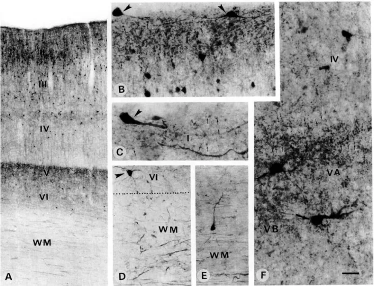
dicular to the cortical layers, while others showed a small multi-polar arborization. In layer V, the density of CR-ir puncta was as strong as in layers I—II and a few CR-ir neurons of multi-polar and sometimes pyramidal shape were also encountered (Fig. 5F). In the deepest layer VI, a few CR-ir multipolar neu-rons were also observed (Fig. 6D). In the white matter, numer-ous CR-ir fibers running parallel to each other were present below layer VI of area 17 but not 18. Widely scattered CR-ir interstitial neurons of bipolar and multipolar types including the pyramidal shape (Fig. 6E) were observed with their dendritic arborization oriented either parallel or perpendicular to the fibers (Fig. 6E). The pattern of CB immunoreactivity was also very clear (Fig. 7). CB-ir neurons were confined mainly to layer II, less to layer III, and very few to the deeper layers. They showed a clear and intensely immunoreactive dendritic arborization
mostly of double bouquet or multipolar shape (Figs. 7A-7D). The ascending dendrites contributed to the density of puncta which was highest in the superficial layer I (Figs. 7A-7D). CB-ir fibers and neurons in the white matter were rare (Figs. 7A and 7E).
Superior colliculus (Figs. 8-9; Table 2)
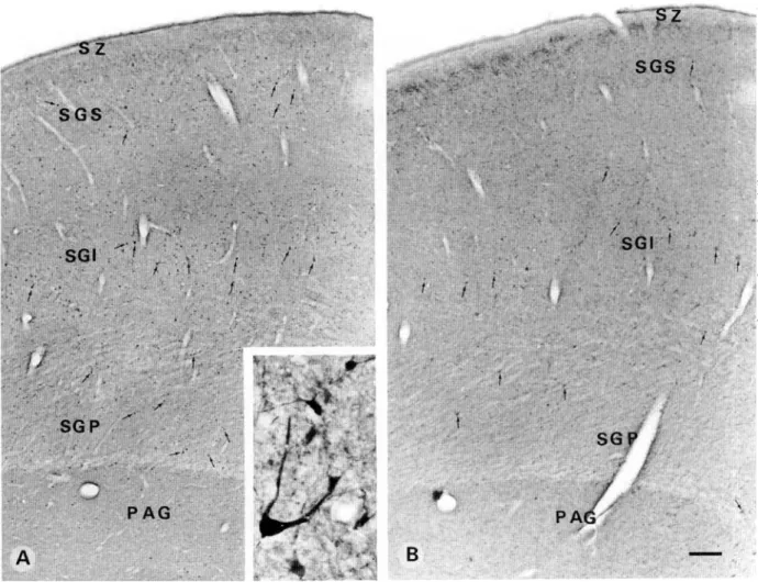
In the SC, PV-ir neurons were widely scattered throughout its depth (Fig. 8A). They included small- to large-size neurons rang-ing between 8-25 /tm in their maximum diameter, mostly multi-polar, pyramidal-like and a few bipolar in shape. Stratum griseum superficiale (SGS) showed lightly labelled PV-ir puncta mainly in its upper part, below which small-size neurons mostly of multipolar shape were encountered. Stratum griseum
inter-Table 2. Distribution of immunoreactivity for parvalbumin (PV), catretinin (CR), and calbindin (CB) in different structures of the human visual system"
Neurons Fibers and/or puncta
PV CR CB PV CR CB LGN P LGN M LGN ILZ LGN capsule PC nucleus LIP OR SCSGS SCSGI SCSGP A17 GM A17 WM a
LGN: lateral geniculate nucleus; P: parvocellular laminae; M: mag-nocellular laminae; ILZ: interlaminar zones; PG: pregeniculate nucleus; LIP: lateral inferior pulvinar; OR: optic radiation; SC: superior col-liculus; SGS: stratum griseum superficiale; SGI: stratum griseum inter-medium; SGP: stratum griseum profundum; A17: area 17; GM: grey matter; and WM: white matter. The intensity of the immunoreactivity was appreciated as strong ++ medium + or absent - ; intermediate sit-uations were not uncommon (+), (-).
medium (SGI) showed a relatively higher density of neurons most of which were of medium to large size. A few small bipo-lar cells oriented perpendicubipo-lar to the pial surface were also encountered. Stratum griseum profundum (SGP) demonstrated a mixed population of small- to medium-size neurons. A few large immunoreactive neurons (Fig. 8A, inset) in the upper and lower part of the SGP were also observed. CB-ir neurons were weakly stained and more widely scattered (Fig. 8B) as compared to the PV-ir ones. However, the puncta were more intensely stained and showed a highest density (Fig. 8B) just below the stratum zonale (SZ). SGS showed only a few widely scattered neurons of small size. In SGI, many large neurons together with a few small- to medium-size cell bodies were observed. SGP showed as widely scattered a population of neurons as the SGS but the neurons were of medium to large size. CR-ir neurons appeared to be more numerous than PV-ir or CB-ir neurons throughout the SC (Fig. 9). Even in the stratum zonale a few small immunoreactive neurons were observed (Fig. 9A, inset). Stratum griseum superficiale showed immunoreactive neurons of small- to medium-size mainly in its lower half. The upper half of SGS revealed a high density of puncta. SGI revealed a broad band of immunoreactive, small- to large-size neurons. Several showed a pyramidal-shaped cell body with a thick prox-imal dendrite oriented towards the pial surface (Fig. 9B and inset). Small- to medium-size neurons were multipolar and bipo-lar in shape. SGP showed a mixed population of a few bipo-large and many small- to medium-size neurons of mostly multipolar and some pyramidal-like in shape.
Discussion
In primates, visual information is processed along two main allel visual pathways, referred to as magnocellular (M) and par-vocellular (P) pathways, carrying specific clues relative to the
perception of motion (M) or color and form (P) (Livingstone & Hubel, 1988). However, a third visual pathway which seems to play a role in the integration of visual information has been described as anatomically, neurophysiologically, and neuro-chemically distinct —the koniocellular (K) pathway (Casagrande, 1994; Johnson & Casagrande, 1995). Indeed, in prosimian mon-keys such as Galago crassicaudatus, the LGN has six laminae, two of which — 4 and 5, koniocellular —have smaller relay cells which stain specifically for calbindin, while there is a comple-mentary pattern of staining for parvalbumin in the other M and P laminae (Diamond et al., 1993; Casagrande, 1994; Johnson & Casagrande, 1995). In simian monkeys, such as Macaqua mulatto or fascicularis, the koniocellular laminae correspond mostly to the interlaminar zones (ILZ) and S laminae, while the remaining laminae 1 and 2 are magnocellular and laminae 3-6 are parvocellular. The projection from the SC to the LGN, which reaches the K laminae in prosimians (Lachica & Casa-grande, 1993), seems to reach mainly the ILZ in simians (Casa-grande & Norton, 1991). In simians, the complementary pattern of PV and CB staining is found respectively in M and P lami-nae on the one hand, and in the ILZ and S lamilami-nae on the other hand, although there is some PV staining in ILZ and S lami-nae as well (Jones & Hendry, 1989). CR staining has not been described in primate LGN. Our data in human bring some new information about common and different features between pri-mate species.
PV immunoreactivity is found in all two M and four P lam-inae of the human LGN, with a high proportion of immuno-reactive neurons. Since most neurons are marked, including the larger ones with a clear multipolar shape, this indicates that they likely represent projection neurons rather than interneurons which are only 8% in the human LGN (Braak & Bachmann, 1985). As LGN neurons project almost exclusively to the pri-mary visual area in primates (Casagrande & Norton, 1991), the dense plexus of PV-ir puncta in the geniculate capsule and optic radiation probably represents transversally cut axons. Indeed, immunoreactive puncta have been described as corresponding to axonal and dendritic terminals by several authors (Stichel et al., 1988; Bliimcke et al., 1991; Fonseca et al., 1993). In the white matter underlying area 17, PV-ir fibers stop at the limit with area 18, turning often perpendicularly toward the cortical layers, where dense PV-ir puncta are observed in the thalamo-recipient layer IV. In Macaque monkey where similar PV-ir puncta have been observed, the presence of a high number of PV-ir asymmetric contacts in layer IV of area 17 suggested that they were derived from the axons of the LGN PV-ir neurons (Bliimcke et al., 1991; DeFelipe& Jones, 1991). A recent study in Galago, combining cell-size analysis, retrograde and double labelling, demonstrated that PV-ir LGN neurons project to layer IV and III of area 17 (Johnson & Casagrande, 1995). Our data provide only indirect evidence of PV-ir projections from M and P LGN laminae to primary area 17 in human but com-parison with other simians makes it reasonable. Moreover, PV-ir neurons in interlaminar zones were almost absent in humans, as in Macaque monkeys (Jones & Hendry, 1989).
However, we did not find a complementary pattern of CB immunoreactivity in the LGN interlaminar zones as described in other simians (Jones & Hendry, 1989), but on the contrary, a very well-marked population of CB-ir neurons in all laminae of the LGN, with a slightly denser staining in the M laminae and scattered neurons in the ILZ. Questions about the antibody specificity in human may be addressed, but the specificity of
Calcium-binding proteins immunoreactivity in the human visual system
1003
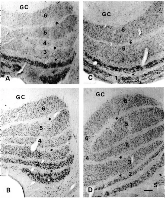
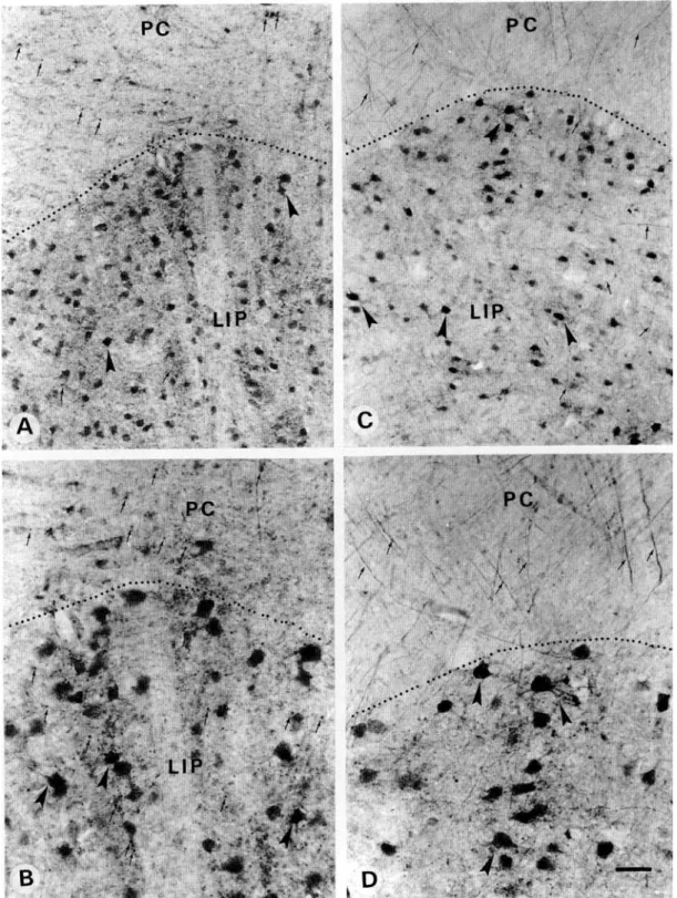
Fig. 4. Photomicrographs demonstrating PV (A,B) and CR (C,D) immunolabeling in the lateral inferior pulvinar (LIP). (A,B) Note the presence of PV-ir neurons (arrowheads) in LIP. PV-ir puncta (arrows) are seen in LIP as well as in the pulvinar cap-sule (PC). (C,D) Note the presence of CR-ir neurons (arrowheads) and fibers (arrows) in LIP as well as in the capcap-sule (PC). Scale bar in A, C = 75 ^m; B, D = 40 urn.
our Celio monoclonal antibody against calbindin-D28K is well
known and in area 17 of the same cases, it works remarkably
well, marking only specific populations of interneurons with a
defined morphology. Furthermore, it has been used in other
human cortical regions with the same success (Tunon et al.,
1992). Puncta in layers I—III of area 17 are CB-ir, but neither
puncta nor fibers are positive along the geniculo-cortical
path-way. In Macaque monkey, CB immunoreactivity has been
described as very light and PV immunoreactivity as particularly
strong in the optic tract (Jones & Hendry, 1989). It may
hap-pen that only the thalamic perikarya and their cortical
termi-nals are stained while the intervening axons remain negative;
Fig. 5. Photomicrographs demonstrating PV-ir neurons and fibers in the primary visual area 17. (A,C) Note that the thalamo-cortical recipient layer IV is strongly immunoreactive. PV-ir neurons are more numerous in layers III and IV than in layers II, V, and VI. Layer I is totally devoid of PV immunoreactivity. The white matter (WM) underlying layer VI shows a bundle of PV-ir fibers (small arrows) which extends to the limit between area 17 and 18. (B) A higher magnification of the PV-ir fibers underlying layer VI of area 17 but not of area 18. (D) shows PV-ir puncta (arrows) and multipolar neurons in layer IV at a high magnification. (E-G) show at a high magnification the PV-ir fibers and neurons oriented parallel to the fibers in the white matter (WM) underlying area 17. Scale bar in A =1 mm; B = 100 /im; C = 750 ^m; D = 25 jtm; E-G = 40 urn.
Calcium-binding proteins immunoreactivity in the human visual system
1005
WM
D
Fig. 6. Light micrographs demonstrating CR-ir neurons and puncta in the primary visual area 17. (A) CR-ir neurons are seen mainly in the superficial layers (II-III), whereas, in the deep layers (IV-VI) they are sparse and widely scattered. CR immuno-reactivity in the neuropil of layer V is as strong as in layers I and II. (B,C) At a higher magnification, layer I shows densely stained CR-ir puncta (arrows) as well as Cajal-Retzius cells (arrowheads) oriented parallel to the pial surface. (D) Several CR-ir fibers (arrows) of different size, as well as scattered neurons (E) are present in the white matter (WM) underlying the deep layer VI. (F) A higher magnification of layer V reveals the density of immunoreactive puncta (big arrows) concentrated mainly in its upper part, layer VA and a few large multipolar immunoreactive neurons. Scale bar in A = 250 ^m; B, D = 40 fim; C, E = 25 pm.
this intracellular segregation of immunoreactivity has been
observed in various instances (M.R. Celio, personal
communi-cation). Thus, a nonspecific projection from the CB-ir LGN
neurons to superficial layers of primary area 17 is quite
feasi-ble in human. It would however originate in all LGN laminae,
contrary to the other primate's pattern where only the smallest
laminar and interlaminar cells, as well as neurons of the S
lam-inae, appear to send projections to the superficial visual layers
(Casagrande & Norton, 1991; Johnson & Casagrande, 1995).
In only a few cases, we could see one of the S laminae, and when
present it was CB positive. Such CB-ir projections from the
koniocellular laminae of the LGN to the superficial layers of
the visual cortex were demonstrated recently in the prosimian
Galago (Johnson & Casagrande, 1995) as well as in Macaque
monkey, from the medial geniculate nucleus to layer I of the
cerebral cortex (Hashikawa et al., 1991). Because
calcium-bind-ing immunostaincalcium-bind-ing combined with either retrograde-labelcalcium-bind-ing
(Johnson & Casagrande, 1995) or fluorescent tracer
(Hashi-kawa et al., 1991) experiments is not possible in human,
addi-tional information should be obtained by double labeling and
quantitative distribution of PV-ir and CB-ir neurons (study in
progress).
CR-ir neurons have not been described in the primate LGN,
but our data demonstrate immunoreactive neurons and fibers
in all laminae, with a more intense staining in the M laminae
and only rare neurons in the ILZ. The presence of fibers in the
geniculate capsule, optic radiation, and underlying white
mat-ter of area 17 suggests that there are CR-positive visual thalamic
projections in human. Within area 17, there are two dense zones
of CR-ir puncta, one at the level of superficial layers I and II
and the other at the level of layer V. Although puncta
repre-sent largely the terminals of cortical interneurons, they may
rep-resent partly the terminals of subcortical afferents, as it was
shown for PV in monkeys (Bliimcke et al., 1991; DeFelipe &
• U'
Ill
IV
.H
" %
V
VI
WM
WM,
Fig. 7. Light photomicrographs demonstrating CB-ir neurons and puncta in the primary visual area 17. (A, C) CB-ir neurons are confined mainly to the superficial layers 11 and III, whereas, the deep layers 1V-V1 are relatively free of immunoreactive neurons. Arrow in the white matter (WM) indicates a very rare CB-ir neuron, enlarged in the inset. (B, D) show layer II CB-ir neurons whose ascending dendrites contribute to the puncta seen in layer I. (E) A rare CB-ir white matter neuron. Scale bar in A = 250 ^m; B = 75 fim; C = 150 urn; D = 25 jim; E = 50 ^m.
Jones, 1991; Johnson & Casagrande, 1995). As there are no
comparative CR data in primates, we can only suggest that
CR-positive LGN projections could reach specific layers of area 17.
However, the distribution of CR-ir cortical interneurons matches
completely with that already described (Glezer et al., 1992) and
the presence of Cajal-Retzius CR-ir neurons in layer I of area 17
is in accordance with recent observations in humans (Belichenko
et al., 1995).
In the optic radiation and in the white matter underlying area
17, PV-ir and more particularly CR-ir neurons are also present.
Their morphology is clearly neuronal, with long dendrites
par-allel or perpendicular to the fibers, and our preliminary
ob-servations, combining calcium-binding proteins with GFAP
immunostaining, have shown that they were not glial cells.The
role of these white matter neurons is still unclear but seems to
be essential in development for the final target selection of
tha-lamic axons to the cortical recipient layers (Ghosh & Shatz,
1992). In close vicinity to the LGN, the pregeniculate nucleus
receives also direct retinal afferents and seems to be the
homo-logue of the rodent intergeniculate leaflet playing a role in the
organization of the primate circadian system (Moore, 1993). It
projects to different subcortical structures, including the LGN
itself, the suprachiasmatic nucleus (Moore, 1993), the nucleus
of the optic tract (Mustari et al., 1994), and seems reciprocally
connected with the visual association cortex and temporal area
MT (Maioli et al., 1984; Leichnetz, 1990). The few large PV-ir
and CR-ir neurons probably represent projection neurons
al-ready described in the monkey pregeniculate nucleus (Jones &
Hendry, 1989).
In the pulvinar capsule, PV-ir puncta and CR-ir fibers seem
to be axons from the PV-ir and CR-ir neurons, which will partly
join the optic radiation and contribute to the immunoreactive
fiber bundles of the white matter underlying area 17. However,
primate cortical projections originating in the pulvinar not only
reach the primary visual cortex, but also several temporal,
pari-etal, and orbito-frontal visually related areas (Morecraft et al.,
1992; Baizer et al., 1993). From the pattern of connections, it
was inferred that the PV-ir perikarya, particularly rich in the
medial division of the pulvinar, were mainly the fast-firing
neu-rons projecting to area MT (Cusick et al., 1993).
Finally, in the three grey laminae of the SC, the PV-ir and
CR-ir neurons are mostly of medium to large size, which may
Calcium-binding proteins immunoreactivity in the human visual system
1007
I *'•?; \fr™}&2&m
Lv.fe^Oi
Fig. 8. Light photomicrographs demonstrating PV (A) and CB (B) immunostaining in the superior colliculus. (A) Note that PV-ir neurons (arrows) are widely scattered in all the layers. They show a slightly greater concentration in the stratum griseum intermedium (SGI) and include small- to large-size neurons of multipolar and pyramidal-like shape (Inset). Lightly stained PV-ir puncta show a greater density in the upper part of the stratum griseum superficial (SGS). (B) CB-ir neurons (arrows) are more widely scattered and not as strongly marked as the PV-ir ones. They include also small- to large-size neurons of multi-polar and pyramidal-like shape. Intensely stained CB-ir puncta concentrate in the upper part of the SGS. Stratum zonale (SZ) is free of immunoreactive neurons. Scale bar in A,B = 350 ^m; inset = 30 /xm.
project outside the nucleus towards the pontine nuclei and the
medulla or towards the LGN (Stein & Meredith, 1993). Smaller
neurons are less immunoreactive and may include both
projec-tion neurons and interneurons. However, neurons making a
local axonal arborization are rare in the primate SC (Ma et al.,
1990). In the cat SC, about 84% of the projection neurons
iden-tified by horseradish peroxidase injections in several visually
related structures such as the LGN, the lateral posterior nucleus,
the opposite SC, or descending tectal pathways are PV-positive,
while the CB-ir neurons are smaller and identified as
interneu-rons (Mize et al., 1992a). This characteristic complementary
dis-tribution of PV-ir and CB-ir neurons was not observed in
human, where CB-ir neurons are widely scattered throughout
the nucleus, but until now there are no comparative data in
primates.
The reason why calcium-binding proteins are expressed
mainly by interneurons in the cerebral cortex and projection
neu-rons in subcortical structures is unknown and their distribution
is also largely species dependent (Baimbridge et al., 1992). Our
data bring some more information on these species differences
in the visual system. In primates, these differences appear to
be more prevalent in subcortical than in cortical structures. The
primary effect of these proteins involves mainly the promotion
and maintenance of neuronal activity by controlling the entrance
of calcium into the cell, and thus a series of intracellular
sig-nals; but it could include as well the regulation of cytoskeletal
organization, cell motility and differentiation, fast axonal
trans-port, and synthesis and release of some neurotransmitters
(Baimbridge et al., 1992; Heizmann, 1992, 1993). The
colocali-zation of PV with Cat-301 (Rausell & Jones, 1991) suggested
a higher calcium-buffering requirement for the fast-firing
neu-rons of the magnocellular pathway (Mize & Hockfield, 1989;
Hockfield & Sur, 1990; Hockfield et al., 1990). It has also been
suggested that interneurons containing calcium-binding proteins
would be more resistant to degeneration (Heizmann, 1992,1993;
Eckert et al., 1994), but data in this domain are conflicting, at
least in the human (Heizmann & Braun, 1992). In monkeys,
experiments of visual deprivation fail to reduce CB
immuno-Fig. 9. Light photomicrographs demonstrating CR immunostaining in the superior colliculus. (A) CR-ir neurons (arrows) are widely scattered but more numerous in the stratum griseum intermedium (SGI). They include many large neurons of multi-polar and pyramidal-shaped cell bodies among which smaller neurons of different size are also marked. (A, inset) Arrow indi-cates a small CR-ir neuron in the stratum zonale (SZ) and the region below it shows a high density of puncta in the most upper part of the SGS. (B) shows a higher magnification of CR-ir neurons in SGI. Inset shows a large neuron (long arrowhead) and a few smaller neurons (small arrowhead) Scale bar in A,B = 350 («n; inset = 30 urn.
reactivity in the SC (Mize & Luo, 1992) or PV immunoreactiv-ity in the LGN (Tigges & Tigges, 1993), but deafferentation produces a discrete reduction of CB immunoreactivity in the LGN (Mize et al., 1992ft) as well as of PV immunoreactivity in the LGN and visual cortex (Bliimcke et al., 1994). However, when neurons still express calcium-binding proteins in these experimental conditions, a shift in the expression of PV and CB may occur, for instance coexpression of both PV and CB in re-sponse to neuronal injury (Gutierrez & Cusick, 1994).
Although it is not yet possible to make a precise link between transmitter and neuronal type on the one hand and a PV, CR, or CB content on the other hand, the calcium-binding protein profile in the human visual system seems to present both simi-larities and differences with other primates. The functional dis-tinctiveness of these different neuronal populations remains to be explored in greater detail.
Acknowledgments
This study was supported by the Swiss National Science Foundation (No. 3100-034491.92 and 3100-043573.95). We thank Professor J.
Wert-heimer for his great encouragement and support; Professor L.J. Garey and Professor M.R. Celio for critical review of the manuscript; Professor M.R. Celio for donating the antibodies; Professor R.C. Janzer, Dr. J. Miklossy (Lausanne), and Professor J. Ulrich (Basel) for providing autopsy material; and C. Dumas for technical assistance.
References
ARAI, M., ARAI, R., KANI, K. & JACOBOWITZ, D.M. (1992).
Immuno-histochemical localization of calretinin in the rat lateral geniculate nucleus and its retino-geniculate projection. Brain Research 596, 215-222.
BAIMBRIDGE, K.G., CELIO, M.R. & ROGERS, J.H. (1992).
Calcium-binding proteins in the nervous system. Trends in Neurosciences 15, 303-308.
BAIZER, J.S., DESIMONE, R. & UNGERLEIDER, L.G. (1993).
Compari-son of subcortical connections of inferior temporal and posterior parietal cortex in monkeys. Visual Neuroscience 10, 59-72.
BELICHENKO, P.V., VOGT WEISENHORN, D.M., MYKLOSSY, J. & CELIO,
M.R. (1995). Calretinin-positive Cajal-Retzius cells persist in the adult human neocortex. Neuroreport 6, 1869-1874.
BLUMCKE, I., H O F , P.R., MORRISON, J.H. & CELIO, M.R. (1990).
Dis-tribution of parvalbumin immunoreactivity in the visual cortex of
Calcium-binding proteins immunoreactivity in the human visual system
1009
old world monkeys and humans. Journal of Comparative Neurol-ogy 301, 417-432.
BLOMCKE, 1., HOF, P.R., MORRISON, J.H. & CELIO, M.R. (1991).
Parv-albumin in the monkey striate cortex: A quantitative immunoelectron-microscopy study. Brain Research 554, 237-243.
BLOMCKE, I., WERUAGA, E., KASAS, S., HENDRICKSON, A.E. & CELIO,
M.R. (1994). Discrete reduction patterns of parvalbumin and cal-bindin D-28k immunoreactivity in the dorsal lateral geniculate nucleus and the striate cortex of adult macaque monkeys after mon-ocular enucleation. Visual Neuroscience 11, 1-11.
BLOMCKE, I. & CELIO, M.R. (1992). Parvalbumin and calbindin D-28k immunoreactivities coexist within cytochrome oxidase-rich compart-ments of squirrel monkey area 18. Experimental Brain Research 92, 39-45.
BRAAK, H. & BACHMANN, A. (1985). The percentage of projection neu-rons and interneuneu-rons in the human lateral geniculate nucleus. Human Neurobiology 4, 91-95.
CASAORANDE, V.A. (1994). A third parallel visual pathway to primate area VI. Trends in Neurosciences 17, 305-310.
CASAGRANDE, V.A. & NORTON, T.T. (1991). Lateral geniculate nucleus: A review of its physiology and function. In Vision and Visual Dys-function, Vol. 4, The Neural Basis of Visual Function, ed. LEVEN-THAL, E. & CRONLEY-DILLON, J.R., pp. 41-84. London: Macmillan Press.
CUSICK, C.G., SCRIPTER, J.L., DARENSBOURG, J.G. & WEBER, J.T.
(1993). Chemoarchitectonic subdivisions of the visual pulvinar in monkeys and their connectional relations with the middle temporal and rostral dorsolateral visual areas, MT and DLr. Journal of Com-parative Neurology 336, 1-30.
DEFELIPE, J. & JONES, E.G. (1991). Parvalbumin immunoreactivity reveals layer IV of monkey cerebral cortex as a mosaic of micro-zones of thalamic afferent terminations. Brain Research 362, 39-47.
DIAMOND, I.T., FITZPATRICK, D. & SCHMECHEL, D. (1993).
Calcium-binding proteins distinguish large and small cells of the ventral pos-terior and lateral geniculate nuclei of the prosimian galago and the tree shrew (Tupaia belanger). Proceedings of the National Acad-emy of Sciences of the U.S.A. 90, 1425-1429.
ECKERT, A., HARTMANN, H., FORSTL, H. & MULLER, W.E. (1994).
Alter-ations of intracellular calcium regulation during aging and Alzhei-mer's disease in nonneuronal cells. Life Sciences 55, 2019-2029.
FONSECA, M., SORIANO, E., FERRER, I., MARTINEZ, A. & TUNON, T.
(1993). Chandelier cell axons identified by parvalbumin-immunore-activity in the normal human temporal cortex and in Alzheimer's disease. Neuroscience 55, 1107-1116.
GHOSH, A. & SHATZ, C.J. (1992). Pathfinding and target selection by developing geniculocortical axons. Journal of Neuroscience 12,39-55.
GLEZER, 1.1., HOF, P.R. & MORGANE, P.J. (1992).
Calretinin-immunore-active neurons in the primary visual cortex of dolphin and human brains. Brain Research 595, 181-188.
GUTIERREZ, C. & CUSICK, C.G. (1994). Effects of chronic monocular enucleation on calcium binding proteins calbindin-D28k a nd
parv-albumin in the lateral geniculate nucleus of adult rhesus monkeys. Brain Research 651, 300-310.
HASHIKAWA, T., RAUSELL, E., MOLINARI, M. & JONES, E.G. (1991).
Parv-albumin- and calbindin-containing neurons in the monkey medial geniculate complex: Differential distributions and cortical layer spe-cific projections. Brain Research 544, 335-341.
HEIZMANN, C.W. (1992). Calcium-binding proteins: Basic concepts and clinical implications. General Physiology and Biophysics 11,411 -425. HEIZMANN, C.W. (1993). Calcium signaling in the brain. Ada
Neuro-biologiae Experimental 53, 15-23.
HEIZMANN, C.W. & BRAUN, K. (1992). Changes in Ca2+-binding pro-teins in human neurodegenerative disorders. Trends in Neurosciences 15, 259-264.
HENDRY, S.H.C. & CARDER, R.K. (1993). Neurochemical compart-mentation of monkey and human visual cortex: Similarities and variations in calbindin immunoreactivity across species. Visual Neuro-science 10, 1109-1120.
HOCKFIELD, S., TOOTELL, R.B. & ZAREMBA, S. (1990). Molecular
dif-ferences among neurons reveal an organization of human visual cor-tex. Proceedings of the National Academy of Sciences of the U.S.A. 87, 3027-3031.
HOCKFIELD, S. & SUR, M. (1990). Monoclonal antibody Cat-301 iden-tifies Y-cells in the dorsal lateral geniculate nucleus of the cat. Jour-nal of Comparative Neurology 300, 320-330.
JACOBOWITZ, D.M. & WINSKY, L. (1991). Immunocytochemical local-ization of calretinin in the forebrain of the rat. Journal of Com-parative Neurology 304, 198-218.
JOHNSON, J.K. & CASAGRANDE, V.A. (1995). Distribution of calcium-binding proteins within the parallel visual pathways of a primate
(Galago crassicaudatus). Journal of Comparative Neurology 356, 238-260.
JONES, E.G. (1993). GABAergic neurons and their role in cortical plas-ticity in primates. Cerebral Cortex 3, 361-372.
JONES, E.G. & HENDRY, S.H.C. (1989). Differential calcium binding protein immunoreactivity distinguishes classes of relay neurons in monkey thalamic nuclei. European Journal of Neuroscience 1, 222-246.
LACHICA, E.A. & CASAGRANDE, V.A. (1993). The morphology of
col-licular and retinal axons ending on small relay (W-like) cells of the primate lateral geniculate nucleus. Visual Neuroscience 10, 403-418. LEICHNETZ, G.R. (1990). Preoccipital cortex receives a differential input from the frontal eye field and projects to the pretectal olivary nucleus and other visuomotor-related structures in the rhesus monkey. Visual Neuroscience 5, 123-133.
LIVINGSTONE, M. & HUBEL, D. (1988). Segregation of form, color, move-ment, and depth: Anatomy, physiology, and perception. Science 240, 740-749.
MA, T.P., CHENG, H.-W., CZECH, J.A. & RAFOLS, J.A. (1990).
Inter-mediate and deep layers of the macaque superior colliculus: A Golgi study. Journal of Comparative Neurology 295, 92-110.
MAIOLI, M., GALLETTI, C , SQUATRITO, S., BATTAGLINI, P.P. & RIVA
SAN-SEVERINO, E. (1984). Projections from the cortex of the superior tem-poral sulcus to the dorsal lateral geniculate and pregeniculate nuclei in the macaque monkey. Archives italiennes de Biologie 122, 301-309.
MIZE, R.R., L u o . Q . , BUTLER, G., JEON, C.-J. &NABORS, B. ( 1 9 9 2 O ) .
The calcium binding proteins parvalbumin and calbindin-D 28K form complementary patterns in the cat superior colliculus. Journal of Comparative Neurology 320, 243-256.
MIZE, R.R., Luo, Q. & TIGGES, M. (19926). Monocular enucleation reduces immunoreactivity to the calcium-binding protein calbindin 28kD in the Rhesus monkey lateral geniculate nucleus. Visual Neuro-science 9, 471-482.
MIZE, R.R. & HOCKFIELD, S. (1989). Cat-301 antibody selectively labels neurons in the Y-innervated laminae of the cat superior colliculus.
Visual Neuroscience 3, 433-443.
MIZE, R.R. & Luo, Q. (1992). Visual deprivation fails to reduce cal-bindin 28kD or GABA immunoreactivity in the Rhesus monkey superior colliculus. Visual Neuroscience 9, 157-168.
MOORE, R.Y. (1993). Organization of the primate circadian system. Review. Journal of Biological Rhythms 8 (Suppl.) S3-S9.
MORECRAFT, R.J., GEULA, C. & MESULAM, M.M. (1992).
Cytoarchi-tecture and neural afferents of orbitofrontal cortex in the brain of the monkey. Journal of Comparative Neurology 323, 341-358.
MUSTARI, M.J., FUCHS, A.F., KANEKO, C.R. & ROBINSON, F.R. (1994).
Anatomical connections of the primate pretectal nucleus of the optic tract. Journal of Comparative Neurology 349, 111-128.
PASTEELS, B., ROGERS, J., BLACKER, F. & POCHET, R. (1990). Calbindin
and calretinin localization in retina from different species. Visual Neuroscience 5, 1-16.
RAUSELL, E. & JONES, E.G. (1991). Histochemical and immunocyto-chemical compartments of the thalamic VPM nucleus in monkeys and their relationship to the representation map. Journal of Neuro-science 11, 210-225.
STEIN, B.E. & MEREDITH, M.E. (1993). Anatomical organization of the superior colliculus. In The Merging of the Senses, ed. GAZZANIGA, M.S., pp. 41-49. Cambridge, London: A Bradford Book, The MIT Press.
STICHEL, C.C., SINGER, W. & HEIZMANN, C.W. (1988). Light and
elec-tron immunocytochemical localization of parvalbumin in the dor-sal geniculate nucleus of the cat: Evidence for coexistence with GABA. Journal of Comparative Neurology 268, 29-37. TIGGES, M. & TIGGES, J. (1993). Parvalbumin immunoreactivity in the
lateral geniculate nucleus of rhesus monkeys raised under monocu-lar and binocumonocu-lar deprivation conditions. Visual Neuroscience 10, 1043-1053.
TUNON, T., INSAUSTI, R., FERRER, I., SOBREVIELA, T. & SORIANO, E.
(1992). Parvalbumin and calbindin D-28K in the human entorhinal cortex. An immunohistochemical study. Brain Research 589, 24-31.