N-Glycan structures and N-glycosylation sites of mouse soluble intercellular adhesion
molecule-1 revealed by MALDI-TOF and FTICR mass spectrometry
Vivianne I. Otto1,2, Eugen Damoc4,5, Leah N. Cueni2, Thomas Schürpf2, Renate Frei2, Sarah Ali2,
Nico Callewaert3, Adrian Moise4, Julie A. Leary5, Gerd Folkers2, and Michael Przybylski4
2Institute of Pharmaceutical Sciences, Department of Chemistry and
Applied Biosciences; 3GlycoINIT and Institute of Microbiology, Department of Biology, ETH Zurich, Switzerland; 4Department of Chem-istry, Laboratory of Analytical ChemChem-istry, University of Konstanz, 78457 Konstanz, Germany; and 5Genome Center, UC Davis, Davis, CA 95616
Received on January 2, 2006; revised on July 26, 2006; accepted on July 26, 2006
Intercellular adhesion molecule-1 (ICAM-1) is a heavily N-glycosylated transmembrane protein comprising five extra-cellular Ig-like domains. The soluble isoform of ICAM-1 (sICAM-1), consisting of its extracellular part, is elevated in the cerebrospinal fluid of patients with severe brain trauma. In mouse astrocytes, recombinant mouse sICAM-1 induces the production of the CXC chemokine macrophage inflam-matory protein-2 (MIP-2). MIP-2 induction is glycosylation dependent, as it is strongly enhanced when sICAM-1 carries sialylated, complex-type N-glycans as synthesized by wild-type Chinese hamster ovary (CHO) cells. The present study was aimed at elucidating the N-glycosylation of mouse sICAM-1 expressed in wild-type CHO cells with regard to sialylation, N-glycan profile, and N-glycosylation sites. Ion-exchange chromatography and matrix-assisted laser desorption ionization time-of-flight (MALDI-TOF) mass spectrometry (MS) of the released N-glycans showed that sICAM-1 mostly carried di- and trisialylated complex-type N-glycans with or without one fucose. In some sialylated glycans, one N-acetylneuraminic acid was replaced by N-glycolylneuraminic acid, and ~4% carried a higher number of sialic acid residues than of antennae. The N-glycosylation sites of mouse sICAM-1 were analyzed by MALDI-Fourier transform ion cyclotron resonance (FTICR)-MS and nanoLC-ESI-FTICR-MS of tryptic digests of mouse sICAM-1 expressed in the Lec1 mutant of CHO cells. All nine consensus sequences for N-glycosylation were found to be glycosylated. These results show that the N-glycans that enhance the MIP-2-inducing activity of mouse sICAM-1 are mostly di- and tri-sialylated complex-type N-glycans including a small fraction carrying more sialic acid residues than antennae and that the nine N-glycosylation sites of mouse sICAM-1 are all glycosylated.
Key words: Fourier transform ion cyclotron resonance mass spectrometry/glycosylation–function relationships/ N-glycosylation/oligosialic acid/mouse soluble intercellular adhesion molecule-1
Introduction
Intercellular adhesion molecule-1 (ICAM-1) is a heavily N-glycosylated type I transmembrane protein belonging to the Ig superfamily of adhesion molecules. It consists of five extracellular Ig-like domains, a single transmembrane seg-ment, and a short intracellular domain. ICAM-1 also occurs as a soluble glycoprotein consisting of the extracellu-lar part of membrane-bound ICAM-1. Soluble ICAM-1 (sICAM-1) may derive from either alternative RNA splic-ing or proteolytic cleavage of membrane-bound ICAM-1 (King et al., 1995; Budnik et al., 1996; Lyons and Benveniste, 1998). ICAM-1 is normally expressed at low levels on vascular endothelium, lymphocytes, and mac-rophages. In response to inflammatory stimuli and injury, ICAM-1 expression is enhanced and can also be induced on many other cell types (Dustin et al., 1986; Sobel et al., 1990). After severe brain trauma, the number of ICAM-1-positive brain vessels and the concentration of sICAM-1 in cerebrospinal fluid are strongly increased (Otto et al., 2000; Rancan et al., 2001).
The biological role of membrane-bound ICAM-1 in leu-kocyte extravasation has been studied extensively. ICAM-1 on the surface of vascular endothelial cells mediates the firm adhesion and transendothelial migration of activated leukocytes by binding to the β2 integrins leukocyte function-associated antigen-1 (LFA-1) and macrophage antigen-1 (Mac-1) (Marlin and Springer, 1987; Diamond et al., 1990). The binding sites for LFA-1 and Mac-1 are located on the first and third N-terminal Ig domain of ICAM-1, respectively (Diamond et al., 1991). Beyond its ability to provide the adhesion sites for leukocytes, mem-brane-bound ICAM-1 behaves as a signal transducer, allowing brain endothelial cells and astrocytes to respond actively to leukocyte adhesion (Greenwood et al., 2002; Millan and Ridley, 2005). The functions of sICAM-1 are less well understood. Some studies suggest that sICAM-1 may have anti-inflammatory properties, as human sICAM-1 expressed in baby hamster kidney (BHK) and Chinese ham-ster ovary (CHO) cells and mouse sICAM-1 expressed in Sf9 insect cells acted as competitive inhibitors of leukocyte adhesion to vascular endothelium (Meyer et al., 1995; Rieckmann et al., 1995; Kusterer et al., 1998). Other studies suggest that sICAM-1 has pro-inflammatory signaling functions. In this regard, recombinant human sICAM-1
1To whom correspondence should be addressed; e-mail:
expressed in CHO cells was shown to enhance lymphocyte proliferation and cytokine production during the mixed lymphocyte response (McCabe et al., 1993). Furthermore, sICAM-1 seems to be involved in neuroinflammation. sICAM-1 levels are elevated in the cerebrospinal fluid of patients with severe brain trauma, and recombinant mouse sICAM-1 expressed in mouse fibroblasts induces the pro-duction of the CXC chemokine macrophage inflammatory protein-2 (MIP-2) in cultured mouse astrocytes (Otto et al., 2000, 2002). Although the signaling function of human sICAM-1 in lymphocytes is mediated through its binding to LFA-1, MIP-2 induction in mouse astrocytes is mediated by an as yet undefined receptor.
The various functions of ICAM-1 and sICAM-1 appear to be differentially regulated by glycosylation. An early study reported that binding of human ICAM-1 present on transfected L-cells to immobilized LFA-1 was not influ-enced by glycosylation (Diamond et al., 1991). The binding of human ICAM-1 to Mac-1 was however found to be hin-dered if sialylated or desialylated complex-type N-glycans were present on the third Ig domain of ICAM-1 (Figure 1; Diamond et al., 1991). In contrast, one study dealing with mouse sICAM-1 showed that sialylated and galactosylated complex-type N-glycans strongly enhanced the ability of recombinant mouse sICAM-1 expressed in wild-type CHO cells to induce MIP-2 production in astrocytes (Otto et al., 2004). It seems therefore that binding to different binding partners or receptors of ICAM-1/sICAM-1 is differentially regulated by glycosylation.
The mechanisms by which the glycosylation of a protein may influence its overall three-dimensional structure, favor or hinder its interactions with binding partners and/or its ability to induce cell signaling are poorly understood. To get a handle on these questions, the elucidation of the gly-can structures on proteins with glycosylation-dependent functions and of the sites to which they are attached is of
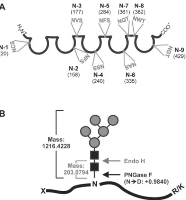
primary importance. The glycosylation and N-glycosylation sites of human ICAM-1 have been analyzed in a few previ-ous studies. In human sICAM-1 expressed in CHO cells, all eight potential N-glycosylation sites were found to be glyco-sylated (Bloom et al., 1996). They carried mostly sialylated, complex-type N-glycans with or without a fucose, but some hybrid-type N-glycans were present as well. In contrast, a study based on the crystal structure of Ig domains 3–5 of human ICAM-1 expressed in the Lec3.2.8.1 mutant of CHO cells suggested that one potential N-glycosylation site (N-379) was not glycosylated (Yang et al., 2004). To date, the glycosy-lation of mouse ICAM-1 has not been analyzed in detail. Mouse ICAM-1 displays 55% amino acid sequence homology with human ICAM-1. Five consensus sequences for N-glycosylation are conserved in human and mouse ICAM-1 (N-156/158, N-175/177, N-240, N-358/361, N-379/382). All potential N-glycosylation sites of human ICAM-1 are located between Ig domain 2 and the interdomain interface following Ig domain 4 (Figure 1). In contrast, the mouse ICAM-1 sequence additionally comprises potential N-glycosylation sites in the first and fifth Ig domains (Figure 3A).
Owing to its functional importance in the regulation of astrocytic MIP-2 production, we aimed at elucidating the N-linked glycosylation of mouse sICAM-1 expressed in wild-type CHO cells with regard to the number of sialic acids, structures, and relative abundances of the sialylated N-glycans as well as the sites on the protein to which N-glycans are attached. Previously, we had analyzed the sialylation of sICAM-1 from wild-type CHO cells by isoelectric focusing (IEF) before and after treatment with neuraminidase (Otto et al., 2004). Neuraminidase-treated sICAM-1 had an apparent isoelectric point (pI) of 5.9. However, untreated sICAM-1 displayed a pI of <3.6 and was therefore not visi-ble on the IEF gels. To overcome the limitations of IEF, we chose an approach based on the enzymatic release of N-glycans, fluorescent labeling, and high-performance ion-exchange chromatography (HP-IEC) followed by matrix-assisted laser desorption ionization-time of flight (MALDI-TOF) mass spectrometry (MS). The sites on mouse sICAM-1 car-rying N-glycans were determined by MALDI- and electro-spray ionization (ESI)-Fourier transform ion cyclotron resonance mass spectrometry (FTICR-MS) of tryptic pep-tides obtained from mouse sICAM-1 expressed in the Lec1 mutant of CHO cells with or without prior endoglycosidase treatment. sICAM-1 expressed in Lec1 cells was used for this analyses, because its homogeneous glycosylation allows for unambiguous interpretation of MS results. Additionally, the knowledge of the N-glycosylation pathway allows to predict that the occupancy of N-glycosylation sites is the same in Lec1 and wild-type CHO cells.
Results
N-Glycans present on mouse sICAM-1 from wild-type CHO cells
To characterize the function-enhancing, sialylated N-glycans of mouse sICAM-1 expressed in wild-type CHO cells, we used an approach based on HP-IEC followed by MALDI-TOF-MS (Townsend et al., 1996; Viseux et al., 2001). N-Glycans were released from sICAM-1 by peptide N-glycosidase F Fig. 1. Biological functions of human ICAM-1 and how they are
influ-enced by glycosylation. The five open circles symbolize the five Ig domains of human ICAM-1 and the eight predicted N-glycosylation sites are pointed out by gray triangles. Binding of the first N-terminal Ig domain of human ICAM-1 on endothelial cells (ECs) to LFA-1 (heterodimer of the αL and β2 integrin subunits) on leukocytes (LCs) is not altered by glyco-sylation, whereas the presence of large complex-type N-glycans (symbolic representation of monosaccharides as in Figure 2B) on the third Ig domain hinders binding to Mac-1 (heterodimer of the αM and β2 integrin subunits) (Diamond et al., 1991).
(PNGase F) treatment, labeled with fluorescent 2-aminoben-zamide (2-AB) by reductive amination, and separated by HP-IEC. The peak fractions were collected and analyzed by MALDI-TOF-MS. To determine the composition and struc-tures of the N-glycans, we compared the masses obtained by MALDI-TOF-MS with molecular masses calculated from N-linked oligosaccharide structures commonly found in CHO cells, and the identity and number of terminal nega-tively charged substituents of the more abundant compo-nents were confirmed by MALDI-TOF/TOF tandem MS.
HP-IEC separated the N-glycans released from sICAM-1 into 11 major peak fractions (Figure 2A), and
MALDI-TOF-MS allowed for the identification of 61 dif-ferent oligosaccharides (Table SI), of which the most abun-dant ones are shown in Figure 2B. Of the 61 glycans, 56 were of the complex type. Thirty-four carried one fucose residue (Fuc) which most probably was a core α6 Fuc as usually seen in wild-type CHO cell-derived glycoproteins. A small amount (fraction 1) of neutral N-glycans was eluted shortly before the remaining free 2-AB label. The major glycan among the neutral N-glycans was of the high man-nose type and contained five manman-noses (Figure 2B). The other glycans in fraction 1 were nonsialylated complex-type N-glycans with or without one fucose. The negative charges on the glycans in the later eluting fractions were mostly due to sialic acid as evidenced by MALDI-TOF/TOF tandem MS analysis. The only exception was a phosphorylated spe-cies identified in peak 4 (PhosHex6HexNAc2; Table SI), probably corresponding to PhosMan6GlcNAc2 (Man-6-P). Peaks 3 and 4 contained 55% of all glycans, and the most abundant glycans in these peak fractions according to MALDI-TOF peak heights carried two sialic acid residues. Peaks 5–8 represented 22% of the N-glycans and the most prominent peaks in the corresponding MALDI-TOF spec-tra could be assigned to N-glycans carrying three sialic acid residues. The glycans contained in peak 2 (15%) and peaks 9–11 (9%) were of similar abundance and contained mono-sialylated and mostly tetramono-sialylated N-glycans, respec-tively. The largest glycans found were the fully sialylated, fucosylated tetraantennary structures with one or two N-acetyllactosamine repeats contained in fractions 8 and 9. Interestingly, the most abundant glycan contained in frac-tion 8 carried three N-acetylneuraminic acid (NeuAc) resi-dues on only two antennae, and the predominant glycan in fraction 11 carried four NeuAc residues on maximally three antennae (Figure 2B). In some mono-, bi-, tri-, and tetrasia-lylated glycans, one NeuAc was replaced by N-glycolyl-neuraminic acid (NeuGc; Table SI). The relative amounts (as judged from the peak heights in the MALDI-TOF-MS) of the glycans with one NeuGc compared with the corre-sponding glycans carrying only NeuAc were estimated to ∼16% in the monosialylated glycans, 24–30% in the disialy-lated, 44% in the trisialydisialy-lated, and 51% in the tetrasialy-lated glycans (data not shown). Interestingly, the relative amount of the NeuGc-carrying variant of the tetrasialy-lated glycan with maximally three antennae from fraction 11 amounted to only ∼32%.
N-Glycosylation sites of mouse sICAM-1 expressed in the Lec1 mutant of CHO cells
To understand how N-glycosylation influences the biologi-cal function of a protein on the molecular level, it is indis-pensable not only to elucidate the composition and structures of the N-glycans but also to identify the sites on the protein that are glycosylated. Mouse sICAM-1 contains nine potential N-glycosylation sites (Figure 3A). For the identification of the ones that are glycosylated, we used two complementary high-resolution MS approaches: (1) nanoLC-FTICR-MS of sICAM-1 trypsinized in solution and (2) direct MALDI-FTICR-MS of sICAM-1 treated with the endoglycosidases endoglycosidase H (Endo H) or PNGase F and trypsinized in silver-stained SDS–PAGE Fig. 2. N-Glycans present on mouse sICAM-1 from CHO cells. (A) The
N-glycans of mouse soluble ICAM-1 from CHO cells were released by
PNGase F treatment, purified over Carbograph, fluorescently labeled with 2-AB, and injected onto the GlycoSep C HP-IEC column, which had been equilibrated with 80% ACN. The ACN content was linearly decreased to 20% over 13 min and then maintained at this level. Between 13 and 48 min, a linear gradient of ammonium acetate pH 4.5 from 0 to 160 mM was run. The separation of fluorescent compounds was moni-tored by fluorescence detection. (B) The relative amounts (rel. amt.) of the total glycans contained in each peak of the chromatogram shown in A were calculated from the peak areas (mean from two independent analy-ses; the sum of the areas of the peaks 2–11 was set to 100%). As the neutral
N-glycans in peak 1 coeluted with free 2-AB, their relative abundance
could not be assessed (NA). The single peak fractions were collected and analyzed by MALDI-TOF-MS. One possible structure of the major gly-can species in each peak fraction is represented schematically. Its com-pounds are represented by the symbols proposed by the nomenclature committee of the Consortium for Functional Glycomics (black rectangle, GlcNAc; gray circle, mannose; gray triangle, fucose; white circle, galac-tose; black diamond, sialic acid; http://glycomics.scripps.edu/CFGnomen-clature.pdf). The peak fractions marked with # (A) contained impurities unrelated to N-glycans. Asterisks designate glycans whose number of ter-minal negatively charged substituents (sialic acid residues) was confirmed by MALDI-TOF/TOF tandem MS.
gels. All experiments were carried out with affinity-purified sICAM-1 expressed in the Lec1 mutant of CHO cells. The Lec1 mutant of CHO cells has a single insertion mutation that generates an inactive, truncated N-acetylglucosamine (GlcNAc) transferase I. Lec1 cells hence stop processing N-glycans at the Man5GlcNAc2-Asn stage (Stanley and Chaney, 1985; Chen and Stanley, 2003). sICAM-1 expressed in Lec1 cells has the following analytical advan-tages: (1) its glycosylation is more homogeneous than the glycosylation of sICAM-1 expressed in wild-type CHO cells (Otto et al., 2004) and (2) being of the high mannose-type, its glycans are sensitive to Endo H which cleaves the glyco-sidic linkage between the two GlcNAcs of the chitobiose core leaving one GlcNAc residue on the protein (Figure 3B). For analysis by nanoLC-FTICR-MS, sICAM-1 from Lec1 cells was reduced and alkylated in solution and then digested with trypsin and the resulting (glyco-) peptide mix-ture was directly injected into a nanoLC C18 column cou-pled to a Finnigan LTQ-FTICR mass spectrometer. The FTICR-MS data allowed to identify the glycosylated pep-tides comprising N-1, N-2, N-3, N-6, N-7, N-8, and N-9 (Figure 4, Table I), and the presence of Hex5HexNAc2 gly-cans was confirmed by MS/MS analysis (data not shown).
As sICAM-1 from Lec1 cells could theoretically carry gly-cans with five to nine mannoses depending on whether the action of mannosidase I went to completion or not, we elu-cidated the masses and composition of the N-glycans of sICAM-1 expressed in Lec1 cells. N-Glycans were released by PNGase F treatment and then directly analyzed by MALDI-FTICR-MS (Figure 5A). The largest m/z mea-sured was 1257.43161, which is in good agreement with the calculated m/z of the [M + Na]+ ion of Hex5HexNAc2 (Δm = 6.8 ppm). Hence, sICAM-1 expressed in Lec1 cells only carries glycans of the composition Man5GlcNAc2.
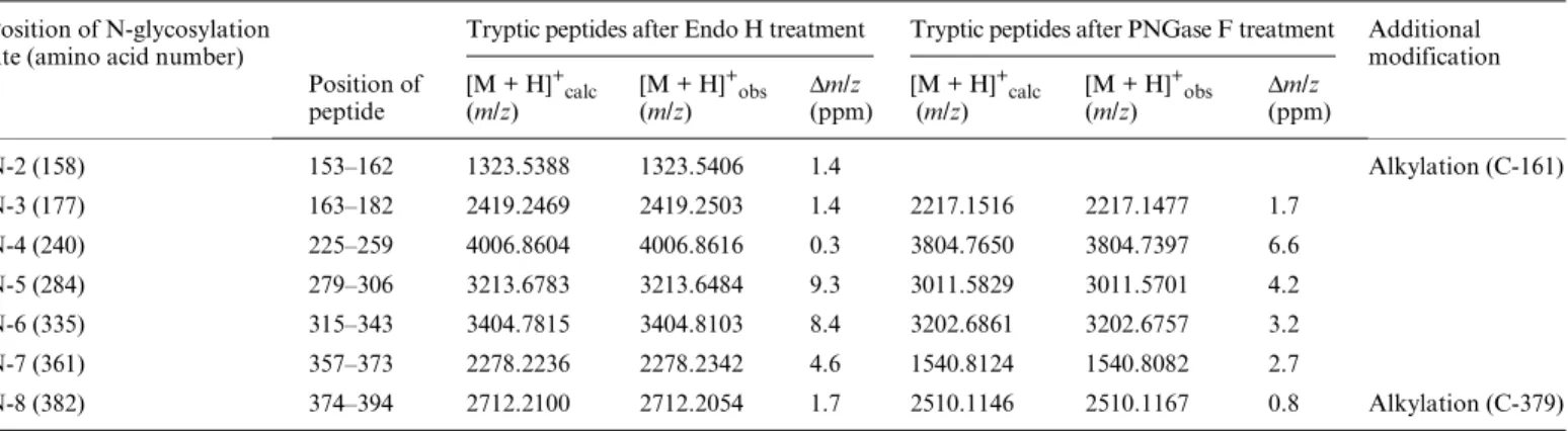
Next, the reduced and alkylated band of sICAM-1 iso-lated by SDS–PAGE was subjected to in-gel deglycosyla-tion with either PNGase F or Endo H, then to proteolysis by trypsin, and the resulting (glyco-) peptide mixtures were directly analyzed by MALDI-FTICR-MS. Figure 5B shows a MALDI-FTICR-MS of the peptide mixture obtained after deglycosylation with Endo H, which provided the identification of the peptides comprising the glycosylation sites N-2, N-3, N-4, N-5, N-6, N-7, and N-8 each carrying one remaining GlcNAc (Table II). Six of the seven glyco-peptides were also found with the expected deamidated N-residues after PNGase F treatment (Table II). Taken together, the results from the two complementary high-resolution MS approaches show that all nine consensus sequences for N-glycosylation of mouse sICAM-1 are glyc-osylated.
Discussion
In a previous study, we showed that sialylated and galacto-sylated complex-type N-glycans as synthesized by wild-type CHO cells strongly enhance the ability of mouse sICAM-1 to induce MIP-2 production in astrocytes (Otto et al., 2004). The present study was aimed at defining the sialyla-tion and structures of the funcsialyla-tion-enhancing glycans of mouse sICAM-1 expressed in wild-type CHO cells as well as the sites on mouse sICAM-1 which are N-glycosylated.
For defining the sialylation and structures of highly com-plex mixtures of glycans, combinations of chromatography with high-sensitivity MS methods have proven to be partic-ularly powerful (Dell and Morris, 2001). The method used here for profiling of the sialylated N-glycans of mouse sICAM-1 was based on enzymatic N-glycan release, fluo-rescent labeling of the reducing end, separation, and analy-sis by fluorometric HP-IEC and MALDI-TOF-MS. This analysis showed that the majority of N-glycans of mouse sICAM-1 from wild-type CHO cells were of the sialylated complex type, confirming earlier data based on IEF after neuraminidase treatment, SDS–PAGE after Endo H treat-ment, and monosaccharide compositional analysis (Otto et al., 2004). HP-IEC separated these glycans according to charge (number of sialic acid residues) and according to the size/ number of N-acetyllactosamine units (Figure 2B). All charged groups were due to sialic acid with the only excep-tion of one phosphorylated glycan (PhosHex6HexNAc2, probably corresponding to PhosMan6GlcNAc2/Man-6-P)
in fraction 4 (Table SI). The phosphorylated glycotope Man-6-P is the key structural determinant in targeting gly-coproteins to the lysosomal compartment but is also found Fig. 3. Schematic representations of mouse sICAM-1, its nine potential
N-glycosylation sites, and the strategies used for identification of the N-glycosylated sites. (A) Schematic representation of mouse sICAM-1. The five open circles symbolize the five Ig domains and the connecting straight lines the interdomain interfaces. The positions (amino acid number) and amino acid sequences of the nine potential N-glycosylation sites (N-1 to N-9) are shown. (B) To identify the N-glycosylation sites of mouse sICAM-1, we used sICAM-1 expressed in Lec1 cells, which carries only high mannose-type N-glycans, as a sample. The scheme shows the cleavage sites of Endo H and PNGase F and the corresponding mass increments to be expected for glycosylated tryptic peptides. Symbolic representation of monosaccharides is as in Figure 2B.
on recombinant glycoproteins with high expression levels (Varki et al., 1999; Lee et al., 2003). Besides this phosphory-lated high mannose-type N-glycan, the only N-glycans not being of the complex type were a small amount of neutral high mannose-type N-glycans found in fraction 1. These
high mannose-type N-glycans probably derive from a small amount of sICAM-1 carrying only high mannose-type N-glycans detected in our previous study and probably rep-resent incompletely processed N-glycans (Otto et al., 2004). The complex-type N-glycans of sICAM-1 ranged from Fig. 4. Full-scan ESI-FTICR-MS of the tryptic (glyco-) peptide mixture of mouse sICAM-1 expressed in Lec1 cells. Purified sICAM-1 from Lec1 cells was
reduced, alkylated, and digested with trypsin in solution. The resulting (glyco-) peptide mixture was injected into a nanoLC column coupled to a hybrid LTQ-FTICR mass spectrometer. Exact monoisotopic masses of the ions detected are shown above the peaks. For assignment of peptides carrying a Hex5HexNAc2 glycan (highlighted by asterisks), see Table I. The insets show the isotopic distributions of the [M + 4H]4+ ion of the glycopeptide (426–448) and of the [M + 3H]3+ ion of the glycopeptide (8–26) containing the N-9 and N-1 glycosylation sites, respectively.
Table I. NanoLC-FTICR-MS identification of tryptic peptides containing N-glycosylation sites carrying Hex5HexNAc2 glycans (m/z 1216.4228)
Position of N-glycosylation site (amino acid number)
Tryptic glycopeptides Additional modification
Position of peptide
m/zcalc m/zobs Charge state (z) Δm/z (ppm)
N-1 (20) 8–26 1091.1220 1091.1270 3+ 4.58 Alkylation (C-21, C-25) N-2 (158) 143–162 864.3797 864.3844 4+ 5.44 Alkylation (C-161) N-2 (158) 143–162 1152.1704 1152.1766 3+ 5.38 Alkylation (C-161) N-3 (177) 163–182 1144.8687 1144.8692 3+ 0.43 N-6 (335) 315–342 1066.2618 1066.2615 4+ 0.28 N-7 (361) 361–373 919.4223 919.4238 3+ 1.63 N-8 (382) 374–394 1242.5230 1242.5313 3+ 6.68 Alkylation (C-379) N-9 (429) 426–448 970.8918 970.8950 4+ 3.30 Alkylation (C-434) N-9 (429) 426–448 1294.1864 1294.952 3+ 6.80 Alkylation (C-434)
Fig. 5. MALDI-FTICR-MS of the N-glycans of mouse sICAM-1 from Lec1 cells and of the tryptic (glyco-) peptide mixture obtained after Endo H
treat-ment. (A) N-Glycans from mouse sICAM-1 expressed in Lec1 cells were released by PNGase F treatment, purified over Carbograph, and analyzed by MALDI-FTICR-MS. The inset shows the monoisotopic peak of the largest m/z detected, that is, 1257.43161 corresponding to the depicted Hex5HexNAc2 with a Na+ adduct (calculated m/z of the [M + Na]+ ion: 1257.4231, Δm = 6.8 ppm). The m/z 1036.33234 can be assigned to the same glycan having lost one HexNAc at its reducing end. Symbolic representation of monosaccharides is as in Figure 2B. (B) Purified sICAM-1 from Lec1 cells was isolated by SDS–PAGE, reduced, alky-lated, and digested in gel with Endo H followed by trypsin and analyzed by MALDI-FTICR-MS. Exact monoisotopic m/z values of [M + H]+ ions are shown above the peaks. For assignment of peptides carrying one GlcNAc residue after Endo H treatment (highlighted by asterisks), see Table II.
small biantennary to large tetraantennary structures with two N-acetyllactosamine repeats. Of the 61 N-glycan struc-tures identified, 34 contained one fucose and 28 two or three sialic acid residues. Interestingly, a significant fraction (∼4% according to HP-IEC peak area and MALDI-TOF peak height) was found to carry a higher number of sialic acid residues than of antennae. The additional sialic acid residues can theoretically be attached either to subterminal GlcNAc residues in α2,6 linkage as seen on mouse transfer-rin (Coddeville et al., 2000) or to a terminal sialic acid in α2,8 linkage, forming di/oligosialic acid. We favor the latter interpretation because CHO cells are known to express the polysialyltransferase ST8SiaIV responsible for the synthesis of di/oligosialic acid (Muhlenhoff et al., 2001) but have never been reported to attach α2,6-linked sialic acid to sub-terminal GlcNAc residues. A disialic acid motif often termi-nates the oligosaccharide chains of gangliosides but was also found on glycoproteins derived from mammalian brain (Sato et al., 2000). The function of di- and oligosialic acids on glycoproteins is presently unclear. One study demon-strated that disialic acid on O-glycans of CD166 may be involved in neurite outgrow (Sato et al., 2002). Another study demonstrated that the integrin α5 subunit carries oli-gosialic acid and that olioli-gosialic acid is required for the adhesion of the integrin α5 subunit to fibronectin (Nadanaka et al., 2001). Lectins that specifically bind disialic acid were recently described. Human siglec-11 occurring on macroph-ages in various tissues and on microglia was found to specifically recognize α2,8-linked sialic acid (Angata et al., 2002) and mouse siglec-E expressed on neutrophils bound preferentially to sialic acid in α2,8 linkage (Zhang et al., 2004).
In some of the more abundant, mostly fucosylated mono-, di-, tri-, and tetrasialylated glycans of sICAM-1, one NeuAc residue was replaced by a NeuGc residue. NeuGc is found on glycoproteins of virtually all verte-brates, except for humans and chicken (Chenu et al., 2003). In animals, NeuGc is formed as the sugar nucleotide cyti-dine monophosphate-N-glycolylneuraminic acid (CMP-NeuGc) by the action of CMP-NeuAc hydroxylase, an enzyme which is inactivated through the loss of an exon in humans (Chenu et al., 2003). The relative amounts of the
sICAM-1-derived N-glycans carrying one NeuGc as com-pared with the corresponding glycan carrying only NeuAc suggest that ∼13% of the total CMP-sialic acid available for sialylation in wild-type CHO cells is NeuGc. Interestingly, the relative amount of the NeuGc-carrying isoform of the tetrasialylated triantennary glycan probably containing the di/oligosialic acid motif was only ∼32% suggesting some selectivity for CMP-NeuAc in di/oligosialic acid synthesis by wild-type CHO cells.
The glycosylation of mouse sICAM-1 from wild-type CHO cells presented here is similar to the glycosylation of human sICAM-1 from wild-type CHO cells (Bloom et al., 1996) in that both glycoproteins carry mostly heavily sialy-lated, fucosylated complex-type N-glycans. In contrast to human sICAM-1, mouse sICAM-1 seems not to carry any hybrid-type glycans. Furthermore, the presence of N-glycans with more sialic acid residues than antennae (prob-ably carrying di/oligosialic acid) seems to be a characteristic feature of mouse sICAM-1, which is not found on human sICAM-1. The enhanced ability of sialylated mouse sICAM-1 from wild-type CHO cells to induce MIP-2 pro-duction may hence be due to a high extent of sialylation, the presence of the di/oligosialic acid motif, and/or the vari-ety of large complex-type N-glycans with terminal nega-tively charged sialic acid residues (Figure 6). A high extent of sialylation could protect sICAM-1 from uptake by the mannose receptor into astrocytes, thereby prolonging the interaction time of sICAM-1 with its astrocytic receptor. A high number of negatively charged sialic acid residues could also alter the conformation of sICAM-1 and/or its recogni-tion by the receptor in a funcrecogni-tion-enhancing manner. If the fraction of glycans probably containing di/oligosialic acid were to contribute to the increase in MIP-2-inducing activ-ity, specific recognition by the astrocytic receptor would be a probable mechanism. Another possibility is that the vari-ety and complexity of the sialylated N-glycans are recog-nized by astrocytic cell surface proteins in a manner that is comparable to pattern recognition by innate immunity receptors and that this pattern recognition is translated into enhanced MIP-2 induction.
To understand how glycosylation may influence the bio-logical function of a protein, we need to know not only the Table II. MALDI-FTICR-MS identification of tryptic peptides carrying one GlcNAc residue (m/z 203.0794) at the N-glycosylation site after Endo H
treatment or having deamidated N-residues (+m/z 0.9840) after deglycosylation with PNGase F
Position of N-glycosylation site (amino acid number)
Tryptic peptides after Endo H treatment Tryptic peptides after PNGase F treatment Additional modification Position of peptide [M + H]+calc (m/z) [M + H]+obs (m/z) Δm/z (ppm) [M + H]+calc (m/z) [M + H]+obs (m/z) Δm/z(ppm) N-2 (158) 153–162 1323.5388 1323.5406 1.4 Alkylation (C-161) N-3 (177) 163–182 2419.2469 2419.2503 1.4 2217.1516 2217.1477 1.7 N-4 (240) 225–259 4006.8604 4006.8616 0.3 3804.7650 3804.7397 6.6 N-5 (284) 279–306 3213.6783 3213.6484 9.3 3011.5829 3011.5701 4.2 N-6 (335) 315–343 3404.7815 3404.8103 8.4 3202.6861 3202.6757 3.2 N-7 (361) 357–373 2278.2236 2278.2342 4.6 1540.8124 1540.8082 2.7 N-8 (382) 374–394 2712.2100 2712.2054 1.7 2510.1146 2510.1167 0.8 Alkylation (C-379)
structures and relative amounts of the glycans but also the sites on the protein that are glycosylated. Therefore, we analyzed which of the nine consensus sequences for N-glycosylation of mouse sICAM-1 expressed in the Lec1 mutant of CHO cells carry N-glycans. For this purpose, we used two complementary high-resolution MS approaches: nanoLC-ESI-FTICR-MS of sICAM-1 trypsinized in solu-tion and direct MALDI-FTICR-MS of sICAM-1 which had been treated with Endo H or PNGase F and trypsinized in silver-stained SDS–PAGE gels. The combi-nation of these approaches led to the discovery that all nine potential N-glycosylation sites of mouse sICAM-1 from Lec1 cells were actually glycosylated with Man5GlcNAc2.
All previous studies on the glycosylation of ICAM-1 were performed using human sICAM-1. Five consensus sequences for N-glycosylation are conserved between human and mouse ICAM-1 (N-156/158, N-175/177, N-240, N-358/361, N-379/382). One previous study based on reversed-phase-HPLC and ESI-MS of tryptic peptides showed that all eight N-glycosylation sites of human ICAM-1 were glycosylated (Bloom et al., 1996). Another study based on crystallization and electron density maps of Endo H-treated domains 3–5 of human ICAM-1 expressed in Lec3.2.8.1 cells reported that N-379 was not glycosylated and that this was probably due to the unfavorable presence of a tryptophan in the NWT N-glycosylation sequon (Yang
et al., 2004). Here, the glycosylation site corresponding to human N-379, N-8 (382) was glycosylated although the NWT N-glycosylation sequon is conserved in the mouse protein.
The glycosylation sites on domain 3 of human ICAM-1 were previously reported to be important for binding to Mac-1. Large complex-type glycans at N-240 and N-269 inhibited the binding of human ICAM-1 to Mac-1 (Figure 1; Diamond et al., 1991). Interestingly, N-240 is conserved in mouse sICAM-1, whereas the N-glycosylation site that had an even larger impact on binding of human ICAM-1 to Mac-1, N-269 is not.
The potential N-glycosylation sites of human ICAM-1 are all located in the middle part of the molecule (between domain 2 and the interdomain interface following domain 4), whereas mouse sICAM-1 additionally has potential N-glycosylation sites in the first (N-1) and the fifth Ig-like domain (N-9). N-1 is of particular interest, as it is located close to the binding site of LFA-1. Even though absent in human ICAM-1, it is present in all other members of the human ICAM family (ICAM-2 to ICAM-5; Jimenez et al., 2005). The crystal structure of human ICAM-2 showed that the first GlcNAc at N-1 interacted with the tryptophan res-idue W51. Both N-1 and the interacting tryptophan resres-idue (glycan1-W motif) are highly conserved in the ICAM sub-family. The only ICAM subfamily members not having a glycan1-W motif are ICAM-1 from humans and chimpan-zees. Interestingly, when human ICAM-1 was mutated to contain the glycan1-W motif (T20N, R49W), its binding to LFA-1 was not altered (Jimenez et al., 2005). In contrast, the binding of ICAM-2 and ICAM-3 to LFA-1 was abol-ished when the glycan1-W motif was eliminated by site-directed mutagenesis (Klickstein et al., 1996; Jimenez et al., 2005). It may therefore be that the presence of a glycan con-taining at least one GlcNAc at N-1 and of W-48 is required for a proper folding of domain 1 of mouse sICAM-1 allow-ing for bindallow-ing to LFA-1.
The induction of MIP-2 production by mouse sICAM-1 in astrocytes occurs by binding to an as yet undefined receptor and is enhanced by sialylated complex-type N-glycans. Because neither the astrocytic receptor nor its binding site on sICAM-1 are known, further research should be aimed at defining whether enhanced MIP-2 induction is due to the sialylated glycans attached to a specific N-glycosylation site or to a specific protein conformation stabilized by the glycosylation of the nine N-glycosylation sites with the sialylated N-glycans described here.
The glycosylation of the nine N-glycosylation sites of mouse sICAM-1 from Lec1 cells can be expected to occur also on mouse sICAM-1 from natural sources, as the occu-pancy of N-glycosylation sites is determined by their pro-tein environment (Petrescu et al., 2004). The observation of changes in sialylation and galactosylation of N-glycans altering the ability of recombinant mouse sICAM-1 from wild-type CHO cells to induce MIP-2 production leads to the hypothesis that the MIP-2-inducing activity of sICAM-1 may also be regulated by glycosylation in vivo. Glycosyla-tion by wild-type CHO cells is similar to glycosylaGlycosyla-tion in mice in various aspects (e.g., the synthesis of NeuGc, α2,8-linked disialic acid; Yasukawa et al., 2006) and differs in others (e.g., the absence of α2,6-linked sialic acid and of Fig. 6. The ability of mouse sICAM-1 to induce MIP-2 production in
astrocytes and how it is influenced by glycosylation. The N-glycans present on mouse sICAM-1 expressed in wild-type CHO cells and in the Lec1 mutant of CHO cells as determined in the present study are juxta-posed to the MIP-2-inducing activities of the two sICAM-1 glycoforms as determined in our previous study (Otto et al., 2004). To determine the MIP-2-inducing activity, we treated primary mouse astrocytes with increasing concentrations of purified sICAM-1 expressed in either wild-type CHO or Lec1 cells. After 24 h, the culture supernatants were harvested, and secreted MIP-2 was measured by enzyme-linked immun-osorbent assay. The data are presented as average ± SD of four assays derived from two independent experiments, each performed in duplicate.
antenna fucosylation). It will be interesting to extend the analysis of the N-glycosylation of mouse sICAM-1 expressed in wild-type CHO cells performed here to mouse sICAM-1 present in mouse brain and serum under normal and under inflammatory conditions. Inflammatory stimuli have been shown to up-regulate the expression of glyco-syltransferases that are responsible for the biosynthesis of sialylated N-glycans, and an increased expression of sialylglycotopes on serum glycoproteins was found in response to turpentine oil-induced inflammation in mice (Delmotte et al., 2002; Garcia-Vallejo et al., 2005; Yasukawa et al., 2005). If an increase in (α2,3 and/or α2,8-linked) sialylation of sICAM-1 in serum and/or brain of mice with experimental brain trauma was observed, it may in fact be that MIP-2 induction is regulated by the sia-lylation of mouse sICAM-1 after brain trauma.
Materials and methods
Materials
PNGase F (catalog number 1 365 193) and Endo H (cata-log number 1 088 726) were purchased from Roche Diagnostics GmbH (Penzberg, Germany). The Signal 2-AB labeling kit (GKK-404), the GlycoSep C column (I-4721), and GlycoClean S cartridges (GKI-4726) were obtained from Prozyme-Glyko (San Leandro, CA). Carbograph SPE columns were purchased from Alltech Associates (Deerfield, IL). NuTip graphitized carbon 10-μL pipette tips were from Glygen (Columbia, MD). Trifluoroacetic acid (TFA) and 5-methoxysalicylic acid were purchased from Sigma-Aldrich (St. Louis, MO). 2,5-Dihydroxybenzoic acid (DHB) puriss. p.a. was from Fluka (Buchs, Switzerland). Sequencing grade trypsin was purchased from Promega (Mannheim, Germany).
Preparation of sICAM-1 expressed in wild-type CHO and Lec1 cells
The extracellular portion of mouse ICAM-1 with the 12-amino acid HPC4 epitope tag attached to its C-terminus after G441 was expressed in either dihydrofolate reductase-deficient CHO cells (designated as wild-type CHO) or the Lec1 mutant of CHO cells and affinity-purified from cul-ture supernatants as previously described (Otto et al., 2004). Eluted sICAM-1 was dialyzed against three changes of low-salt TBS (5 mM Tris–HCl, pH 7.5, 15 mM NaCl) and concentrated 10–20-fold with a Centricon 30 concen-trator (Millipore, Billerica, MA). The concentrations of the two sICAM-1 glycoforms were assessed by western blot analysis and comparison to an sICAM-1 standard of known concentration (Otto et al., 2004).
Release and purification of N-glycans from sICAM-1 expressed in wild-type CHO and Lec1 cells
An aliquot containing 1 nmol of sICAM-1 expressed in either wild-type CHO or Lec1 cells was lyophilized and dis-solved in 8.2 μL of ultrapure water. Then, sICAM-1 was denatured by boiling for 10 min in 0.5% sodium dodecyl sulfate (SDS) and 1% β-mercaptoethanol. Nonidet P-40 (NP-40) was added in 7.5× excess of SDS. The N-glycans
were released from the protein by PNGase F treatment, using 525 U/mL of enzyme for 2 nmol/mL (corresponding to ∼190 μg/mL) of sICAM-1 from wild-type CHO cells and 315 U/mL of enzyme for 2 nmol/mL (corresponding to ∼125 μg/mL) of sICAM-1 from Lec1 cells in phosphate-buffered saline pH 7.4, and incubating for 21 h at 37 °C. The completeness of N-glycan release was confirmed by SDS–PAGE and western blot analysis (Otto et al., 2004). The released N-glycans were separated from the proteins and other contaminants (salts, detergents, and reagents) using nonporous graphitized carbon black Carbograph SPE columns (150 mg, corresponding to a bed volume of ∼0.5 mL) as previously described (Packer et al., 1998). Briefly, columns were conditioned with three bed volumes of 80% (v/v) acetonitrile (ACN) containing 0.1% (v/v) TFA followed by three bed volumes of ultrapure water. Samples were loaded onto the columns, and glycans were left to adsorb to the carbon for 1–2 min. Contaminants were washed off with three bed volumes of ultrapure water, and glycans were eluted with three bed volumes of 25% (v/v) ACN containing 0.05% (v/v) TFA. Flow was generated by gravity and was between 0.5 and 1 mL/min. The eluates were lyophilized and stored at –20°C.
Preparation of fluorescently labeled N-glycans from sICAM-1 expressed in wild-type CHO cells
The glycan pool of sICAM-1 from wild-type CHO cells was reductively aminated with 2-AB (molecular weight 136.15) using the Signal 2-AB labeling kit, according to the manu-facturer’s instructions. Briefly, the lyophilized sample was dissolved in 5 μL of a solution of 2-AB (0.35 M) and sodium cyanoborohydride (1 M) in 30% (v/v) acetic acid in dimethylsulfoxide (DMSO). After incubation at 65°C for 3 h, the excess reagents were separated from the labeled gly-cans using a GlycoClean S cartridge, following the manu-facturer’s instructions. The oligosaccharide solution was then filtered through an IC Millex-LG Filter Unit (Millipore, Billerica, MA; pore size 0.2 μm), lyophilized, and stored protected from light at –20°C.
HP-IEC of fluorescently labeled N-glycans
HP-IEC analyses were performed on a Merck Hitachi LaChrom D-7000 system (Merck KGaA, Darmstadt, Germany/Hitachi High Technologies America, San Jose, CA) equipped with a fluorescence detector L-7480. The dried 2-AB-labeled oligosaccharides from sICAM-1 expressed in wild-type CHO cells were dissolved in 250 μL of water. A 50-μL aliquot of this solution, undiluted or 4-fold diluted, was injected onto the GlycoSep C HP-IEC column, and the 2-AB-labeled glycans were separated at room temperature using a flow rate of 0.4 mL/min and an ACN/water/ammo-nium acetate gradient as modified from the work of Viseux and others (2001). In a first step, the ACN content was decreased from 80 to 20% within 13 min, whereas the water proportion was increased accordingly. In a second step, in which ACN was kept constant at 20%, the concentration of ammonium acetate (pH 4.5) was increased from 0 to 160 mM in 35 min. The eluate was monitored by fluorescence detec-tion (λex 330 nm, λem 420 nm). A Merck-Hitachi Model D-7000 Chromatography Data Station software version 4.0
was used for recording the spectra and for calculating the peak areas. Peak fractions were collected, lyophilized, and stored at –20°C.
MALDI-TOF-MS of chromatographic fractions
The peak fractions collected from HP-IEC were desalted prior to MS analysis using NuTip graphitized carbon 10-μL pipette tips (Glygen, Columbia, MD). Tips were conditioned by pipetting five times 10 μL of 60% (v/v) ACN containing 0.05% (v/v) TFA followed by three times 10 μL of water. Dried HP-IEC fractions were dis-solved in 5 μL of water. Samples were bound to the car-bon by pipetting up and down 50 times 2 μL volumes (20 times oversampling). Contaminants were washed off by pipetting 10 times 10 μL of water, and glycans were eluted with five times 1.5 μL of 60% (v/v) ACN contain-ing 0.05% (v/v) TFA. Each 1.5 μL aliquot of eluent was pipetted up and down 10 times before collection of the eluate. Samples were then vacuum dried.
MALDI-TOF-MS was carried out using a 4700 Pro-teomics Analyzer (Applied Biosystems, Foster City, CA) equipped with a nitrogen UV laser (λmax 337 nm). Mass spectra were recorded at matrix-optimized laser fluency, with an accelerating voltage of 20 kV and in negative polar-ity linear mode. The spectra were obtained by summation of 50 subspectra, each averaged over 50 laser shots. Dried, desalted HP-IEC fractions were dissolved in 2 μL of 70% (v/v) methanol and mixed with 2 μL of superDHB matrix solution (9:1 mixture of 20 mg/mL DHB and 20 mg/mL 5-methoxysalicylic acid in 70% methanol, containing 10 mM ammonium citrate). After the transfer of 1.4 μL of this mix-ture to the target, the solution was allowed to dry at ambi-ent temperature and pressure. External on-plate mass calibration in the m/z range 750–4250 was performed using a reference peptide mixture (Applied Biosystems). The MALDI-TOF procedure was optimized for sensitivity, res-olution, and minimal sialic acid loss using a 2-AB-labeled trisialylated triantennary reference N-glycan (A3; Dextra, Reading, UK). Of the more abundant components, the number of terminal negatively charged substituents was confirmed by MALDI-TOF/TOF tandem MS at 1 keV col-lisional energy in the negative-ion mode, using air at 5.10E–7 Torr as the collision gas. Control analyses as above were conducted with 1 nmol of human α-1 acid glycoprotein (Sigma).
Tryptic digestion of sICAM expressed in Lec1 cells in solution
A 100-μg sample of sICAM expressed in Lec1 cells was reduced for 45 min at 56°C in 270 μL of 50 mM ammonium bicarbonate containing 5.5 mM dithiothreitol (DTT) and then S-alkylated for 1 h at 20°C in the dark by adding 30 μL of 100 mM iodoacetamide (IAA) to a final concentration of 10 mM IAA. Trypsin was added in a final enzyme:substrate ratio of 1:50, and the digestion was carried out for 2 h at 37 °C. The reaction was stopped by freezing the sample with liquid nitrogen. The tryptic digest was then lyophilized and redissolved in 2% ACN in 0.1% TFA just prior to nanoLC-MS/MS analysis.
NanoLC-MS/MS
NanoLC-MS/MS experiments were performed on a Finnigan LTQ-FT hybrid linear ion trap/7T FTICR mass spectrometer (Thermo Electron, San Jose, CA), equipped with a Finnigan Nanospray ion source (Thermo Electron), a Finnigan Surveyor MS pump (Thermo Electron), and a Finnigan micro-autosampler (Thermo Electron). The sICAM-1 tryp-tic peptide mixture was separated on a 50-μm ID PicoFrit column packed in-house with Magic C18AQ material (Michrom BioResources, Auburn, CA). The column was packed to a length of 12 cm with a 100% MeOH slurry of C18 reversed-phase material (100 Å pore size, 3 μm particle size) using a high-pressure cell pressurized with helium. The column was equilibrated before sample injection for 10 min at 2% solvent B (0.1% [v/v] formic acid in ACN) and 98% solvent A (0.1% [v/v] formic acid in water) at a flow rate of 140 nL/min. Separation was achieved by using a linear gradi-ent from 2 to 50% solvgradi-ent B in 24 min at a flow rate of 320 nL/min. The LTQ-FT mass spectrometer was operated in the data-dependent acquisition mode using the TOP10 method: a full-scan MS acquired in the FTICR mass spectrometer was followed by 10 MS/MS experiments performed with the LTQ on the 10 most abundant ions detected in the full-scan MS. Analysis of the N-glycans from sICAM-1 from Lec1 cells by MALDI-FTICR-MS
The N-glycan pool of sICAM-1 from Lec1 cells eluted from the Carbograph SPE column was directly analyzed by MALDI-FTICR-MS as described below.
In-gel deglycosylation and proteolytic digestion of sICAM-1 from Lec1 cells
The silver-stained band of sICAM-1 from Lec1 cells was excised from a 7.5% SDS–PAGE gel, washed, reduced with DTT, and S-alkylated with IAA as previously described (Shevchenko et al., 1996). After reduction and alkylation, gel pieces were washed with 50–100 μL of 50 mM ammo-nium bicarbonate for 10 min, dehydrated by the addition of ACN, swelled by rehydration in 50 mM ammonium bicar-bonate, and shrunk again by the addition of the same vol-ume of ACN. The liquid phase was removed, and the gel pieces were dried in a vacuum centrifuge.
sICAM-1 was then treated in gel with Endo H or PNGase F. Although PNGase F treatment allows for easy identification of the deglycosylated peptides, Endo H treat-ment allows for a more secure identification of glycosylated peptides. The presence of a remaining GlcNAc is a more reliable sign of previous N-glycosylation than the transfor-mation of asparagine into aspartic acid by PNGase F (Fig-ure 3B), because such a transformation may also be due to spontaneous asparagine oxidation. For in-gel deglycosyla-tion, the gel pieces were swollen in 50 μL of deglycosylation buffer containing either Endo H (500 mU/mL in 25 mM ammonium bicarbonate adjusted to pH 5.5 with HCl) or PNGase F (100 U/mL in 50 mM ammonium bicarbonate). If all the liquid was taken up by the gel pieces, further deglycosylation buffer (but without Endo H or PNGase F) was added to keep the samples wet during overnight incubation at 37°C (Kuster and Mann, 1999). To avoid interference from PNGase F- or Endo H-related peptides
in the MS spectra, the deglycosylation enzymes were removed prior to proteolysis as previously described (Kuster and Mann, 1999), but using ammonium bicarbon-ate at 50 mM (instead of 100 mM), and then the gel pieces were dried in a vacuum centrifuge.
The dried gel pieces were rehydrated in the protease solu-tion (12.5 ng/μL of trypsin in 50 mM ammonium bicarbon-ate) at 4°C for 45 min. The supernatant solution was removed and replaced by 10–20 μL of 50 mM ammonium bicarbonate without protease to keep the gel pieces wet during overnight incubation (37°C) as previously described (Shevchenko et al., 1996). The supernatant was removed, lyophilized, and analyzed by MALDI-FTICR-MS. Protein fragments were additionally eluted from the gel pieces by shaking them twice with a mixture of one part 20 mM ammonium bicarbonate and two parts of ACN for 2 h at 20°C. The eluates were combined, lyophilized, and dis-solved just before MALDI-FTICR-MS analysis in 5 μL of ACN/0.1% TFA in water (2:1).
MALDI-FTICR-MS
The tryptic digests of mouse sICAM-1 were directly analyzed by MALDI-FTICR-MS using a Bruker APEX II FTICR instrument equipped with an actively shielded 7T supercon-ducting magnet (Magnex, Oxford, UK), a cylindrical infin-ity ICR analyzer cell, and an external Scout 100 fully automated X–Y target stage MALDI source with pulsed collision gas. A 100 mg/mL solution of DHB in ACN/0.1% TFA in water (2:1) was used as the matrix. A 0.5-μL aliquot of matrix solution and 0.5 μL of sample solution were mixed on the stainless-steel MALDI target and allowed to dry. The pulsed nitrogen laser was operated at 337 nm, and the ions generated by 20–30 laser shots were accumulated in the hexapole for 0.5–1 s. Mass spectra were obtained by the acquisition of typically 32–64 scans. External calibration was carried out using the monoisotopic masses of singly protonated ion signals of bovine insulin (5730.609), oxi-dized bovine insulin B-chain (3494.651), bee venom melittin (2845.762), human adrenocorticotropic hormone (ACTH) fragment (2465.199), human neurotensin (1672.917), human angiotensin I (1296.685), human bradykinin (1060.569), and human angiotensin II (1046.542). The acquisition and processing of spectra were performed with the XMASS software (Bruker Daltonics, Bremen, Germany).
Supplementary data
Supplementary data are available at Glycobiology online (http://glycob.oxfordjournals.org/). Table SI: supplementary mass spectrometric data to Figure 2.
Acknowledgments
We thank Matthias Meyer from the Institute of Pharma-ceutical Sciences, ETH Zurich for excellent technical assis-tance with protein purification. We thank Dr. Catalina Damoc and Dr. Brett Phinney from UC Davis Proteomic Core Facility for expert help with Finnigan LTQ-FT mass spectrometer.
Conflict of interest statement
None declared.
Abbreviations
2-AB, 2-aminobenzamide; ACN, acetonitrile; CHO, Chinese hamster ovary; CMP, cytidine monophosphate; DHB, 2,5-dihydroxybenzoic acid; DTT, dithiothreitol; Endo H, endoglycosidase H; ESI, electrospray ionization; FTICR, Fourier transform ion cyclotron resonance; Fuc, fucose; Gal, galactose; GlcNAc, N-acetylglucosamine; HP-IEC, high-performance ion-exchange chromatography; IAA, iodoacetamide; ICAM-1, intercellular adhesion molecule-1; IEF, isoelectric focusing; LFA-1, leukocyte function-associated antigen-1; Mac-1, macrophage antigen-1; MALDI-TOF, matrix-assisted laser desorption ionization-time of flight; Man, mannose; MIP-2, macrophage inflammatory protein-2; MS, mass spectrometry; NeuAc, N-acetylneuraminic acid; NeuGc, N-glycolylneuraminic acid; PNGase F, peptide N-glycosidase F; sICAM-1, solu-ble intercellular adhesion molecule-1; TFA, trifluoroacetic acid.
References
Angata, T., Kerr, S.C., Greaves, D.R., Varki, N.M., Crocker, P.R., and Varki, A. (2002) Cloning and characterization of human Siglec-11. A recently evolved signaling that can interact with SHP-1 and SHP-2 and is expressed by tissue macrophages, including brain microglia. J. Biol.
Chem., 277, 24466–24474.
Bloom, J.W., Madanat, M.S., and Ray, M.K. (1996) Cell line and site spe-cific comparative analysis of the N-linked oligosaccharides on human ICAM-1des454–532 by electrospray ionization mass spectrometry.
Biochemistry, 35, 1856–1864.
Budnik, A., Grewe, M., Gyufko, K., and Krutmann, J. (1996) Analysis of the production of soluble ICAM-1 molecules by human cells. Exp.
Hematol., 24, 352–359.
Chen, W. and Stanley, P. (2003) Five Lec1 CHO cell mutants have distinct Mgat1 gene mutations that encode truncated N-acetylglucosaminyl-transferase I. Glycobiology, 13, 43–50.
Chenu, S., Gregoire, A., Malykh, Y., Visvikis, A., Monaco, L., Shaw, L., Schauer, R., Marc, A., and Goergen, J.L. (2003) Reduction of CMP-N-acetylneuraminic acid hydroxylase activity in engineered Chinese hamster ovary cells using an antisense-RNA strategy. Biochim. Biophys.
Acta, 1622, 133–144.
Coddeville, B., Regoeczi, E., Strecker, G., Plancke, Y., and Spik, G. (2000) Structural analysis of trisialylated biantennary glycans isolated from mouse serum transferrin. Characterization of the sequence Neu5Gc(alpha 2–3)Gal(beta 1–3)[Neu5Gc(alpha 2–6)]GlcNAc(beta 1–2)Man. Biochim. Biophys. Acta, 1475, 321–328.
Dell, A. and Morris, H.R. (2001) Glycoprotein structure determination by mass spectrometry. Science, 291, 2351–2356.
Delmotte, P., Degroote, S., Lafitte, J.J., Lamblin, G., Perini, J.M., and Roussel, P. (2002) Tumor necrosis factor alpha increases the expres-sion of glycosyltransferases and sulfotransferases responsible for the biosynthesis of sialylated and/or sulfated Lewis x epitopes in the human bronchial mucosa. J. Biol. Chem., 277, 424–431.
Diamond, M.S., Staunton, D.E., de Fougerolles, A.R., Stacker, S.A., Garcia-Aguilar, J., Hibbs, M.L., and Springer, T.A. (1990) ICAM-1 (CD54): a counter-receptor for Mac-1 (CD11b/CD18). J. Cell Biol.,
111, 3129–3139.
Diamond, M.S., Staunton, D.E., Marlin, S.D., and Springer, T.A. (1991) Binding of the integrin Mac-1 (CD11b/CD18) to the third immunoglobulin-like domain of ICAM-1 (CD54) and its regulation by glycosylation.
Dustin, M.L., Rothlein, R., Bhan, A.K., Dinarello, C.A., and Springer, T.A. (1986) Induction by IL, 1 and interferon-gamma: tissue distribu-tion, biochemistry, and function of a natural adherence molecule (ICAM-1). J. Immunol., 137, 245–254.
Garcia-Vallejo, J.J., van Dijk, W., van Die, I., and Gringhuis, S.I. (2005) Tumor necrosis factor-alpha up-regulates the expression of beta1,4-galactosyltransferase I in primary human endothelial cells by mRNA stabilization. J. Biol. Chem., 280, 12676–12682.
Greenwood, J., Etienne-Manneville, S., Adamson, P., and Couraud, P.O. (2002) Lymphocyte migration into the central nervous system: Impli-cation of ICAM-1 signalling at the blood–brain barrier. Vasc.
Pharma-col., 38, 315–322.
Jimenez, D., Roda-Navarro, P., Springer, T.A., and Casasnovas, J.M. (2005) Contribution of N-linked glycans to the conformation and func-tion of intercellular adhesion molecules (ICAMs). J. Biol. Chem., 280, 5854–5861.
King, P.D., Sandberg, E.T., Selvakumar, A., Fang, P., Beaudet, A.L., and Dupont, B. (1995) Novel isoforms of murine intercellular adhesion molecule-1 generated by alternative RNA splicing. J. Immunol., 154, 6080–6093.
Klickstein, L.B., York, M.R., Fougerolles, A.R., and Springer, T.A. (1996) Localization of the binding site on intercellular adhesion mole-cule-3 (ICAM-3) for lymphocyte function-associated antigen, 1 (LFA-1).
J. Biol. Chem., 271, 23920–23927.
Kuster, B. and Mann, M. (1999), 18O-labeling of N-glycosylation sites to improve the identification of gel-separated glycoproteins using peptide mass mapping and database searching. Anal. Chem., 71, 1431–1440. Kusterer, K., Bojunga, J., Enghofer, M., Heidenthal, E., Usadel, K.H.,
Kolb, H., and Martin, S. (1998) Soluble ICAM-1 reduces leukocyte adhesion to vascular endothelium in ischemia-reperfusion injury in mice. Am. J. Physiol., 275, G377–G380.
Lee, K., Jin, X., Zhang, K., Copertino, L., Andrews, L., Baker-Malcolm, J., Geagan, L., Qiu, H., Seiger, K., Barngrover, D., McPherson, J.M., and Edmunds, T. (2003) A biochemical and pharmacological comparison of enzyme replacement therapies for the glycolipid storage disorder Fabry disease. Glycobiology, 13, 305–313.
Lyons, P.D. and Benveniste, E.N. (1998) Cleavage of membrane-associated ICAM-1 from astrocytes: involvement of a met alloprotease. Glia, 22, 103–112.
Marlin, S.D. and Springer, T.A. (1987) Purified intercellular adhesion molecule-1 (ICAM-1) is a ligand for lymphocyte function-associated antigen, 1 (LFA-1). Cell, 51, 813–819.
McCabe, S.M., Riddle, L., Nakamura, G.R., Prashad, H., Mehta, A., Berman, P.W., and Jardieu, P. (1993) sICAM-1 enhances cytokine production stimulated by alloantigen. Cell. Immunol., 150, 364–375.
Meyer, D.M., Dustin, M.L., and Carron, C.P. (1995) Characterization of intercellular adhesion molecule-1 ectodomain (sICAM-1) as an inhibi-tor of lymphocyte function-associated molecule–1 interaction with ICAM-1. J. Immunol., 155, 3578–3584.
Millan, J. and Ridley, A.J. (2005) Rho GTPases and leucocyte-induced endothelial remodelling. Biochem. J., 385, 329–337.
Muhlenhoff, M., Manegold, A., Windfuhr, M., Gotza, B., and Gerardy-Schahn, R. (2001) The impact of N-glycosylation on the functions of polysialyltransferases. J. Biol. Chem., 276, 34066–34073.
Nadanaka, S., Sato, C., Kitajima, K., Katagiri, K., Irie, S., and Yamagata, T. (2001) Occurrence of oligosialic acids on integrin alpha, 5 subunit and their involvement in cell adhesion to fibronectin. J. Biol. Chem., 276, 33657–33664.
Otto, V.I., Gloor, S.M., Frentzel, S., Gilli, U., Ammann, E., Hein, A.E., Folkers, G., Trentz, O., Kossmann, T., and Morganti-Kossmann, M.C. (2002) The production of macrophage inflammatory protein-2 induced by soluble intercellular adhesion molecule-1 in mouse astro-cytes is mediated by src tyrosine kinases and p42/44 mitogen-activated protein kinase. J. Neurochem., 80, 824–834.
Otto, V.I., Heinzel-Pleines, U.E., Gloor, S.M., Trentz, O., Kossmann, T., and Morganti-Kossmann, M.C. (2000) sICAM-1 and TNF-alpha
induce MIP-2 with distinct kinetics in astrocytes and brain microvascu-lar endothelial cells. J. Neurosci. Res., 60, 733–742.
Otto, V.I., Schurpf, T., Folkers, G., and Cummings, R.D. (2004) Sialy-lated complex-type N-glycans enhance the signaling activity of soluble intercellular adhesion molecule-1 in mouse astrocytes. J. Biol. Chem.,
279, 35201–35209.
Packer, N.H., Lawson, M.A., Jardine, D.R., and Redmond, J.W. (1998) A general approach to desalting oligosaccharides released from glycopro-teins. Glycoconj. J., 15, 737–747.
Petrescu, A.J., Milac, A.L., Petrescu, S.M., Dwek, R.A., and Wormald, M.R. (2004) Statistical analysis of the protein environment of N-glyco-sylation sites: implications for occupancy, structure, and folding.
Glycobiology, 14, 103–114.
Rancan, M., Otto, V.I., Hans, V.H., Gerlach, I., Jork, R., Trentz, O., Kossmann, T., and Morganti-Kossmann, M.C. (2001) Upregulation of ICAM-1 and MCP-1 but not of MIP-2 and sensorimotor deficit in response to traumatic axonal injury in rats. J. Neurosci. Res., 63, 438–446.
Rieckmann, P., Michel, U., Albrecht, M., Bruck, W., Wockel, L., and Felgenhauer, K. (1995) Soluble forms of intercellular adhesion molecule-1 (ICAM-1) block lymphocyte attachment to cerebral endot-helial cells. J. Neuroimmunol., 60, 9–15.
Sato, C., Fukuoka, H., Ohta, K., Matsuda, T., Koshino, R., Kobayashi, K., Troy, F.A., 2nd, and Kitajima, K. (2000) Frequent occurrence of pre-existing alpha 2→8-linked disialic and oligosialic acids with chain lengths up to 7 Sia residues in mammalian brain glycoproteins. Preva-lence revealed by highly sensitive chemical methods and anti-di-, oligo-, and poly-Sia antibodies specific for defined chain lengths. J. Biol.
Chem., 275, 15422–15431.
Sato, C., Matsuda, T., and Kitajima, K. (2002) Neuronal differentiation-dependent expression of the disialic acid epitope on CD166 and its involvement in neurite formation in Neuro2A cells. J. Biol. Chem., 277, 45299–45305.
Shevchenko, A., Wilm, M., Vorm, O., and Mann, M. (1996) Mass spectro-metric sequencing of proteins silver-stained polyacrylamide gels. Anal.
Chem., 68, 850–858.
Sobel, R.A., Mitchell, M.E., and Fondren, G. (1990) Intercellular adhe-sion molecule-1 (ICAM-1) in cellular immune reactions in the human central nervous system. Am. J. Pathol., 136, 1309–1316.
Stanley, P. and Chaney, W. (1985) Control of carbohydrate processing: the lec1A CHO mutation results in partial loss of N-acetylglucosami-nyltransferase I activity. Mol. Cell. Biol., 5, 1204–1211.
Townsend, R.R., Lipniunas, P.H., Bigge, C., Ventom, A., and Parekh, R. (1996) Multimode high-performance liquid chromatography of fluo-rescently labeled oligosaccharides from glycoproteins. Anal. Biochem.,
239, 200–207.
Varki, A., Cummings, R.D., Esko, J., Freeze, H., Hart, G., and Marth, J. (1999) Essentials of Glycobiology. Cold Spring Harbor Laboratory Press, Cold Spring Harbor, New York.
Viseux, N., Hronowski, X., Delaney, J., and Domon, B. (2001) Qualitative and quantitative analysis of the glycosylation pattern of recombinant proteins. Anal. Chem., 73, 4755–4762.
Yang, Y., Jun, C.D., Liu, J.H., Zhang, R., Joachimiak, A., Springer, T.A., and Wang, J.H. (2004) Structural basis for dimerization of ICAM-1 on the cell surface. Mol. Cell, 14, 269–276.
Yasukawa, Z., Sato, C., and Kitajima, K. (2005) Inflammation-dependent changes in alpha2,3-, alpha2,6-, and alpha2,8-sialic acid glycotopes on serum glycoproteins in mice. Glycobiology, 15, 827–837.
Yasukawa, Z., Sato, C., Sano, K., Ogawa, H., and Kitajima, K. (2006) Identification of disialic acid-containing glycoproteins in mouse serum: a novel modification of immunoglobulin light chains, vitronectin, and plasminogen. Glycobiology, 16, 651–665.
Zhang, J.Q., Biedermann, B., Nitschke, L., and Crocker, P.R. (2004) The murine inhibitory receptor mSiglec-E is expressed broadly on cells of the innate immune system whereas mSiglec-F is restricted to eosino-phils. Eur. J. Immunol., 34, 1175–1184.