HAL Id: inserm-02321840
https://www.hal.inserm.fr/inserm-02321840
Submitted on 21 Oct 2019
HAL is a multi-disciplinary open access
archive for the deposit and dissemination of
sci-entific research documents, whether they are
pub-lished or not. The documents may come from
teaching and research institutions in France or
abroad, or from public or private research centers.
L’archive ouverte pluridisciplinaire HAL, est
destinée au dépôt et à la diffusion de documents
scientifiques de niveau recherche, publiés ou non,
émanant des établissements d’enseignement et de
recherche français ou étrangers, des laboratoires
publics ou privés.
To cite this version:
Alfonso Represa. Why Malformations of Cortical Development Cause Epilepsy. Frontiers in
Neuro-science, Frontiers, 2019, 13, pp.250. �10.3389/fnins.2019.00250�. �inserm-02321840�
MINI REVIEW published: 29 March 2019 doi: 10.3389/fnins.2019.00250
Edited by: Kazunori Nakajima, Keio University, Japan Reviewed by: Carlos Cepeda, Independent Researcher, Los Angeles, CA, United States Mitsuhiro Kato, Showa University, Japan Stéphanie Baulac, INSERM U1127 Institut du Cerveau et de la Moelle épinière, France *Correspondence: Alfonso Represa [email protected] Specialty section: This article was submitted to Neurogenesis, a section of the journal Frontiers in Neuroscience Received: 21 November 2018 Accepted: 04 March 2019 Published: 29 March 2019 Citation: Represa A (2019) Why Malformations of Cortical Development Cause Epilepsy. Front. Neurosci. 13:250. doi: 10.3389/fnins.2019.00250
Why Malformations of Cortical
Development Cause Epilepsy
Alfonso Represa*
INSERM, Institut de Neurobiologie de la Méditerranée, Aix-Marseille University, Marseille, France
Malformations of cortical development (MCDs), a complex family of rare disorders,
result from alterations of one or combined developmental steps, including progenitors
proliferation, neuronal migration and differentiation. They are an important cause of
childhood epilepsy and frequently associate cognitive deficits and behavioral alterations.
Though the physiopathological mechanisms of epilepsy in MCD patients remain poorly
elucidated, research during the past decade highlighted the contribution of some factors
that will be reviewed in this paper and that include: (i) the genes that caused the
malformation, that can be responsible for a significant reduction of inhibitory cells
(e.g., ARX gene) or be inducing cell-autonomous epileptogenic changes in affected
neurons (e.g., mutations on the mTOR pathway); (ii) the alteration of cortical networks
development induced by the malformation that will also involve adjacent or distal cortical
areas apparently sane so that the epileptogenic focus might be more extended that
the malformation or even localized at distance from it; (iii) the normal developmental
processes that would influence and determine the onset of epilepsy in MCD patients,
particularly precocious in most of the cases.
Keywords: cortical malformation, epileptogenesis, developmental disorder, ARX, mTOR, focal cortical dysplasia, gray matter heterotopia
INTRODUCTION
Malformations of cortical development (MCD) are a complex family of rare disorders that result
from alterations of one or combined developmental steps, including proliferation of neural
progenitors, migration of neuroblasts to the cortical plate, layer organization and neuronal
maturation (
Barkovich et al., 2012
). Thus, in general, alterations of neuronal and glial proliferation
associating neuronal dysgenesis are a cause of focal cortical dysplasia type II (FCD-type
II); alterations of neuronal migration leading to ectopic localization of neurons are a cause
of periventricular nodular heterotopia (neurons accumulate along the ventricular walls) and
subcortical band heterotopia (SBH; neurons accumulate in the white matter between the cortex and
the ventricular wall); alterations on processes subsequent to neuronal migration are at the origin of
polymicrogyria, characterized by a cortex organized in multiple small gyri (
Barkovich et al., 2012
).
Malformations of cortical development are an important cause of childhood epilepsy. Though
the precise incidence of MCDs is not known, it is estimated that they account for up to 40% of cases
of intractable or medication-resistant childhood epilepsies (
Barkovich et al., 2012
;
Guerrini and
Dobyns, 2014
) and that at least 75% of the patients with MCDs will have epilepsy (
Leventer et al.,
1999
). Associated to seizures, the patients display different comorbidities, particularly cognitive
deficits, that are more frequent and severe when epilepsy begins early in life (
Berg et al., 2017
).
MCDs consequently represent a severe burden for patients, families and society.
Though the physiopathological mechanisms of epilepsy in
MCD patients remain unclear, clinical and experimental data
suggest that epileptogenesis results from diverse developmental
processes that can be cell autonomous or not, directly imputable
to the genetic cause of the malformation or linked to an
abnormal development of neuronal networks. In this review,
we will discuss the pathophysiological mechanisms involved
in MCD epileptogenesis through the analysis of three distinct
matters: the genetic causes of MCDs, the localization of the
epileptogenic focus and the age at the epilepsy onset in patients
and animal models.
CAUSATIVE GENES AND
EPILEPTOGENESIS
The classification of cortical malformations according to
the developmental step involved (proliferation, migration or
differentiation) or the type of malformation generated (e.g., gray
matter heterotopia, lissencephaly, focal cortical dysplasia) does
not provide clear indications about the epileptogenic process.
However, the analysis of causative genes and their respective
cellular roles, provided in some cases interesting data that would
help the understanding of the epileptogenic process.
The pathogenesis of MCDs is multifactorial involving different
genes and environmental factors. There is a large and increasing
number of genes identified during the past decade as causative of
MCD (
Guerrini and Dobyns, 2014
). This is an exciting research
field that is also contributing to our knowledge of genetic factors
controlling brain development. However, the objective of this
report is not to provide a complete overview of the genetic
causes of MCDs, but to discuss some examples that help our
understanding of epileptogenesis.
One of the more fascinating genes causing brain malformation
is ARX (aristaless related homeobox gene).
In vitro and in vivo
studies have shown that the Arx gene is both a positive
and a negative regulator of gene transcription important for
brain development (
Collombat et al., 2003
;
Seufert et al.,
2005
;
McKenzie et al., 2007
;
Fullenkamp and El-Hodiri, 2008
).
Among the roles of this gene are the regionalization of the
brain, the proliferation of cortical progenitors, the migration
of interneurons and early commitment of cholinergic neurons
(
Colombo et al., 2004
;
Marsh et al., 2009, 2016
;
Friocourt and
Parnavelas, 2010
). Numerous mutations of the ARX gene have
been reported in more than a dozen different early neurological
disorders, where intellectual disability is associated or not
with epileptic seizures (
Bienvenu et al., 2002
;
Kitamura et al.,
2002
;
Stromme et al., 2002
). These conditions do or do not
associate brain malformations during embryonic development
(
Shoubridge et al., 2010
). Phenotypic heterogeneity may, in part,
be explained by the nature and location of ARX mutations (
Kato
et al., 2004
;
Olivetti and Noebels, 2012
). Indeed, phenotypes
without malformation are mainly caused by mutations that
are in the polyalanine domains and outside the homeodomain.
Conversely, the more severe phenotypes with brain malformation
are mostly associated with mutations leading to protein
truncation or located in the homeodomain; this is the case for
the XLAG syndrome characterized by a severe lissencephaly,
agenesis of the corpus callosum and abnormal genitalia. Animal
models so far generated have shown that Arx deficient mice or
Knockin mice displaying Arx mutations associate a more or less
pronounced reduction of cortical GABAergic and cholinergic
neurons (reviewed in
Olivetti and Noebels, 2012
) and the analysis
of post-mortem brain tissue reported a three-layered cortex
containing exclusively pyramidal neurons in XLAG patients from
three different families (
Bonneau et al., 2002
). In the context of
developmental malformations, ARX related syndromes can be
thus considered as “interneuronopathies” and the epilepsy and
cognitive deficits reported in patients and animal models, would
be directly related to the reduction of inhibition. Glutamatergic
neurons do not express ARX and are not directly affected by
the mutation of ARX but support the consequences and are thus
responsible for the expression of epileptic seizures.
Mutations affecting the activation of the mammalian target
of rapamycin (mTOR)-signaling pathway have been identified
in focal malformations of cortical development associating
alterations of progenitor cell proliferation, defective neuronal
migration and lamination and the presence of cytomegalic
neurons and balloon cells as a result of a defective differentiation
program of cortical cells. These malformations include FCD
type II and Hemimegalencephaly (
Harvey et al., 2008
;
Blümcke
et al., 2011
;
D’Gama et al., 2017
). Tuberous sclerosis, a rare
multisystem genetic disease condition that in the brain generates
cortical tubers (focal distortions in cellular organization and
morphology which extend into the subcortical white matter) is
also caused by a hyperactivation of mTORC1, due to mutations in
either TSC1 or TSC2 genes (
European Chromosome 16 Tuberous
Sclerosis Consortium, 1993
). This disorder presents intractable
epilepsy, cognitive disability, and autism spectrum disorders.
Interestingly, tuberal lesion display cellular features similar
to FCD type II (i.e., cytomegalic neurons and balloon cells).
Therefore, collectively, these disorders might be referred to as
“mTORopathies” (Reviewed by
Crino, 2015
;
Marsan and Baulac,
2018
). During the past decade somatic activation mutations in
mTOR itself have been identified in these syndromes (
Lim et al.,
2015
;
Mirzaa et al., 2016
;
Møller et al., 2016
;
D’Gama et al.,
2017
;
Ribierre et al., 2018
). In addition, positive (e.g., gain of
function mutations in Akt1 or AKT3;
Lee et al., 2012
;
Poduri
et al., 2012
;
Jansen et al., 2015
) or negative (e.g., TSC2, or
DEPDC5;
Baulac et al., 2015
;
D’Gama et al., 2017
;
Lim et al.,
2017
) regulators of mTOR have been implicated in FCD type II
and Hemimegalencephaly.
Because some patients with FCD are surgically treated, there
have been opportunities for investigating on resected tissue
neuronal properties (reviewed by
Abdijadid et al., 2015
). These
investigations described for example an abnormal expression
of glutamate and GABA receptor in dysplastic and heterotopic
neurons (
Crino et al., 2001
;
Lozovaya et al., 2014
), a reduction
of GABAA
-receptor-mediated inhibition (
Calcagnotto et al.,
2005
), an altered pattern of expression and distribution of
synaptic protein SV2 (
Toering et al., 2009
). Carlos Cepeda and
coworkers nicely evaluated the electrophysiological properties
of cytomegalic neurons and balloon cells and identified in
dysplastic areas the presence of neurons with immature cellular
Represa Epileptogenic Cortical Malformations
and synaptic properties (
Cepeda et al., 2007
); these observations
gave origin to the hypothesis that local interactions of dysmature
cells, that would be directly affected by the mutations, with
normal postnatal neurons produce seizures (
Cepeda et al.,
2007
). Though these investigations did not elucidate the exact
mechanisms of epileptogenesis in FCD, they indicate clearly that
different cell types, molecular changes and cellular interactions
are contributing factors.
Many neuronal changes reported on neurons from FCD type
II or tuber resections would be considered as cell autonomous
as they indicate that the hyperactivation of the mTOR pathway
leads to neuronal and synaptic dysfunctions, that contribute to
epilepsy, independently from the cytoarchitectonic alteration.
This notion has been confirmed by
Hsieh et al. (2016)
when
investigating a murine model of type II FCDs by increasing
mTOR activity in layer 2/3 neurons of the medial prefrontal
cortex. When the hyperactivation of mTOR was induced
in cortical progenitors by in utero electroporation, animals
displayed dyslamination and cytomegalic neurons; associated
to these changes, animals developed spontaneous tonic-clonic
seizures. However, when mTOR hyperactivation was induced
after corticogenesis, thanks to the use of inducible vectors,
animals still displayed an epileptic condition without neuronal
misplacement and dysmorphogenesis. In agreement with this,
my lab has shown that heterozygous mice from a Tsc1-KO line,
develop spontaneous seizures during the first month of life in the
absence of any apparent cortical dysplasia (
Lozovaya et al., 2014
).
In this study we demonstrated that Tsc1+/−neurons, particularly
L4 spiny stellate cells, display an anomalous expression of
NR2C receptors resulting in a change on the kinetics of
NMDA currents and that this change was sufficient to induce
the epileptic condition. Indeed, the treatment of pups with
antagonists specific for NR2C transiently abolished seizures
(
Lozovaya et al., 2014
). We also demonstrated in this report
that treatment of newborn mice with rapamycin was sufficient
to reverse the phenotype confirming the link between mTORC1
hyperactivation and NMDA receptor changes. Interestingly,
cortical resections from patients with tuberous sclerosis and FCD
type II also demonstrated higher expression levels of NR2C
as compared with control fetal or adult samples (
Lozovaya
et al., 2014
); in addition to this, patch-clamp recordings on
these cortical resections demonstrated the contribution of NR2C
to NMDA currents confirming the potential role of NR2C to
epileptogenesis in mTORpathies.
It is also important to remind, however, that human samples
investigated were obtained from severely affected,
pharmaco-resistant patients with resection of the epileptogenic zone being
the only therapeutic option. The epileptic process by itself
can be cause of many subsequent alterations, including an
excitatory/inhibitory imbalance. Thus, based on investigations
of a Tsc1-KO mice line,
Bateup et al. (2013)
reported that
many biochemical, transcriptional and functional changes in Tsc1
neurons arise secondarily, due to increased network activity.
To study the epileptogenic process itself, we must develop
appropriate experimental paradigms in order to evaluate the
changes that increase the excitability of neurons and networks
and that take place before epilepsy onset.
In conclusion, the two paradigmatic examples discussed
before, suggest that the initial genetic alteration that yields
cytoarchitectonic disruptions of cortical development, might
also be responsible for the clinical manifestations. However,
these investigations also highlighted to a certain extent the
importance of cellular interactions and the possibility that
developmental changes, that might involve non-mutated neurons
during a particularly vulnerable developmental period, could also
contribute to the emergence of epilepsy and/or cognitive deficits.
THE EPILEPTOGENIC NETWORK
Investigations combining EEG and functional imaging have
demonstrated that patients with focal cortical malformations
display interictal or ictal events not only in the affected area (the
malformation itself) but also in more or less distal cortical areas.
Thus, reports of patients with SBH or periventricular nodular
heterotopia indicate that the epileptogenic network is restricted
to the heterotopia, or involves both the heterotopia and the
surrounding cortex or localizes out of the heterotopia (
Mai et al.,
2003
;
Tassi et al., 2005
;
Kobayashi et al., 2006
;
Tyvaert et al.,
2008
;
Valton et al., 2008
;
Christodoulou et al., 2012
;
Shafi et al.,
2015
;
Pizzo et al., 2017
). In patients with FCD, it has also been
reported that epileptogenicity extends beyond the limit of the
malformation in many patients (
Aubert et al., 2009
). Another
pathological conditions in which the adjacent or even more distal
cortex can be the primary origin of epileptiform activity is the
tuberous sclerosis. Although tubers are thought to be the initial
heart of the epileptogenic zone, electrocorticographic recordings
of some patients revealed epileptiform activities and ictal onsets
in the perituberal cortex (
Madhavan et al., 2007
;
Major et al.,
2009
;
Ma et al., 2012
).
Though conclusions are hampered by the diversities of clinical
courses, that includes variabilities on epilepsy onset, type of
seizures, efficiency of AEDs, cortical area affected, etc., these
data tend to support the notion that epileptic network frequently
involves supposedly “healthy” cortical areas that are affected by
the presence of a cortical malformation. We investigated this
notion in a rat model of SBH induced by in utero (by embryonic
day 15) knockdown (KD) of Dcx, the main causative gene of this
condition (
des Portes et al., 1998
;
Gleeson et al., 1998
;
Pilz et al.,
1998
). In Dcx-KD rats, affected neurons fail to migrate to the
cortical plate and form a band of ectopic neurons in the white
matter of the electroporated hemisphere (
Bai et al., 2003
).
Dcx-KD rats display altered neocortical excitability already present at
the second postnatal week, resulting in an increased propensity
for convulsant-induced seizures and spontaneous absence-like
seizures in adulthood (
Ackman et al., 2009
;
Manent et al., 2009
;
Lapray et al., 2010
).
We first investigated in Dcx-KD juvenile rats the phenotype
of ectopic neurons and found that they displayed a reduced
dendritic tree as compared with control (mismatch) neurons
and a reduced density of dendritic spines (
Ackman et al.,
2009
;
Martineau et al., 2018
). We also performed genetic
labeling of scaffolding proteins PSD-95 and gephyrin for
quantifying, respectively, glutamatergic and GABAergic synapses
in ectopic neurons and observed that they were severely reduced
(
Martineau et al., 2018
). These structural changes were associated
with a decrease of the frequency of glutamatergic and GABAergic
synaptic currents in patch-clamp recordings (
Ackman et al.,
2009
;
Martineau et al., 2018
). Though DCX also plays a role on
neuron maturation (
Martineau et al., 2018
), the ectopic position
of neurons is mainly responsible for the impaired development
and synaptogenesis.
Reports on other animal models of cortical migration defects
revealed similar features: neurons displayed a simplification of
their dendritic arbors in ectopic gray masses induced by either
fetal irradiation (
Ferrer et al., 1984
) or treatment of pregnant
rats with methylazoxymethanol (MAM) (
Singh, 1980
;
Chevassus
au Louis et al., 1998
;
Rafiki et al., 1998
;
Sancini et al., 1998
);
a significant diminution in spine numbers was also reported in
MAM treated offspring (
Colciaghi et al., 2014
). All together these
observations confirm that the positioning of neocortical neurons
significantly impacts the subsequent development of dendrites
and synapses impairing their integration in functional networks.
In contrast to these dendritic and synaptic defects, ectopic
neurons develop axonal projections toward targets that are
considered to be “normal” for cortical neurons. In DCX-KD rats,
ectopic neurons form cortico-cortical projections like Layer II/III
neurons: they reach layers V-VI in the ipsilateral cortex and send
axonal projections to the contralateral cortex (
Ackman et al.,
2009
); they also form cortico-striatal connections like Layer V
neurons. Similarly, ectopic neurons in irradiated rats and in Tish
rats, respectively, lesional and genetic models of SBH, project
as normal Layer V neurons to the spinal cord and thalamus
(
D’Amato and Hicks, 1980
;
Jensen and Killackey, 1984
;
Lee et al.,
1997
). Therefore, ectopic neurons, though poorly innervated as
compared with their normotopic counterparts, are in the position
of spreading signals to typical cortical targets and participating in
the propagation of epileptic activities.
We then analyzed the synaptic properties of normotopic
neurons from DCX-KD rats to evaluate if the SBH altered the
development of normal positioned neurons. Our studies revealed
that normotopic neurons displayed an increased frequency
of spontaneous glutamatergic post-synaptic currents while
GABAergic currents were unaffected (
Ackman et al., 2009
). As
a consequence, the excitatory/inhibitory ratio of synaptic inputs
was increased in normotopic neurons (
Ackman et al., 2009
).
Calcium imaging analysis also revealed that more neurons were
active in the cortex overlaying SBH than in the cortex of control
rats and that they displayed higher frequency of spontaneous
events. In addition, more neurons exhibited synchronized events
(
Ackman et al., 2009
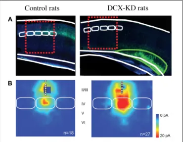
). More recently, in a rat model of bilateral
double cortex we also observed that the strength of excitatory
L4 to L2/3 synapses (Figure 1), the intrinsic properties of
L4 glutamatergic cells and the excitation/inhibition ratio in
L2/3 converge into making the early stage of cortical sensory
integration abnormally strong in somatosensory cortex adjacent
to heterotopia, thus demonstrating developmental alterations of
cortical functional circuits that likely play a major role in the
cortical dysfunction of the malformed brain (
Plantier et al., 2018
).
Investigations of animal models of periventricular heterotopia
induced by irradiation (
Zhu and Roper, 2000
) or treatment
FIGURE 1 | Collateral Alterations of Functional Cortical Circuits in a Rat Model of Subcortical Band Heterotopia. (A) Representative examples of
across-barrel brain slices from control (Mismatch) and Dcx-KD rats. Fluorescence microphotographs of electroporated cells are superimposed on schematized views of barrels (in white) and SBH (in green). Red dashed line rectangles illustrate the cortical columns evaluated in the study. (B) Excitatory synaptic input maps for L2/3 neurons in mismatch and Dcx-KD rats. Colors indicate the mean amplitude of excitatory synaptic responses in a 100-ms time window. White circles show soma positions of recorded L2/3 neurons. Solid white lines delineate barrels. Note that in DCX-KD rats the strength of excitatory inputs to L2/3 neurons are dramatically increased (for more details seePlantier et al., 2018).
with carmustine (
Benardete and Kriegstein, 2002
) also found an
excitatory/inhibitory misbalance in normotopic cortex.
Taken together these data support the notion that normotopic
cortex becomes hyper excitable during postnatal development
and that it would be responsible for epileptogenesis in
DCX-KD rats. Interestingly, the suppression of excitability of
ectopic neurons by their transfection with potassium channels
Kir2.1, did not alter the high propensity of DCX-KD rats
to experience seizures, while the transfection of ectopic and
normotopic neurons significantly reduces it (
Petit et al., 2014
).
These observations prompted us to evaluate the origin of
interictal events on acute slices from DCX-KD rats using
60-channels microelectrode arrays (
Petit et al., 2014
). Our data
demonstrated that most of interictal discharges initiated in
normotopic cortex and propagated secondarily to the SBH.
In vivo recording with deep electrodes in Tish rats (
Chen et al.,
2000
) also indicated that normotopic neurons were more prone
to exhibit epileptiform activities than heterotopic neurons and
that blocking the connectivity between the two fields by a local
TTX injection inhibited the firing of ectopic but not that of
normotopic neurons.
Another pathological condition in which the dysplastic or
malformed brain area is not the primary origin of epileptiform
activities is provided by the rat model of microgyria induced
in rats by freeze-lesioning of deep layer neurons at neonatal
stage. Though injured animals did not display spontaneous
Represa Epileptogenic Cortical Malformations
epileptic events, it has been proven that there is a focal region
of hyperexcitability around the lesion (
Jacobs et al., 1996
); In
the cortex lateral to the microgyria, an increased excitation
of L5 neurons and an increased inhibition of L5 and L2/3
neurons were observed (
Jacobs and Prince, 2005
;
Brill and
Huguenard, 2010
;
Jin et al., 2014
). In addition to this, a rather
widespread cortical reduction of GABA
Areceptor subunits
α1,
α2, α3, α5, and γ2 expression has been reported (
Redecker
et al., 2000
). These alterations together would account for the
increased excitability of an apparently normal cortex lateral
to the microgyria.
Collectively, these clinical and experimental observations
support the notion that anatomically unaltered cortical
regions surrounding the malformation are included in a
large epileptogenic network. We propose that developmental
changes in these areas play a major role in the generation of more
or less large epileptogenic networks.
THE TIME OF ONSET OF EPILEPSY
The age at epilepsy onset in patients with MCD is largely variable
ranging from newborn to adulthood. This is observed even within
the same type of cortical malformations. For example, in a series
of 132 patients with polymicrogyria (
Leventer et al., 2010
), the
mean age of epilepsy onset was 4.9 ± 6.7 years but ranging
from 1 day to 34 years of age. However, 43% of the patients had
the first seizures during the first year of life, coinciding with an
important period of synaptogenesis. In a similar way, the analysis
of 86 female patients with mutations of DCX (
Bahi-Buisson et al.,
2013
) indicate an early onset, during the first year of life, in 55%
of cases, but the age at onset varied between the first month of
life to 17 years.
These data indicate that the majority of patients with MCD
develop the first clinical manifestations of epilepsy during the
first year of life, a period of brain development characterized
by an intense neuronal maturation and synaptogenesis. It is
thus plausible that epilepsy onset is facilitated in these patients
by the increasing weigh of maturing synaptic inputs and/or
the maturation of the axonal initial segment responsible for
the genesis of action potentials and/or other molecular and
synaptic changes linked to neuronal maturation. Interestingly,
in Dravet syndrome (
Dravet, 1978
), a severe encephalopathy
due to
de novo loss-of-function mutations in the SCN1A gene,
leading to haploinsufficiency of NaV1.1 channel (reviewed by
Brunklaus and Zuberi, 2014
), epilepsy typically presents around
6 months of age. Expression analysis on human temporal cortex
and hippocampus demonstrated that Na(v)1.1 immunoreactivity
increased significantly during the late fetal and postnatal periods,
reaching peaks 7–9 months after birth (
Wang et al., 2011
).
Hence, epilepsy onset in Dravet patients compares with the
developmental course of the affected channel. It is not unlikely
that such developmental pattern plays a role on the onset of
epileptic manifestations in Dravet and would be also participating
in other epileptic syndromes.
It is well established that GABAergic synapses play a major
pathophysiological role in epilepsy and thus GABAergic
transmission is targeted by many antiepileptic drugs. Synaptic
currents induced by the activation of GABA
Areceptors
are carried by chloride and consequently the intracellular
concentrations of this anion determine the type of response
evoked by the transmitter. In adult neurons, the
potassium-chloride
cotransporter
KCC2
usually
extrudes
chloride
promoting hyperpolarizing, inhibitory, responses. In immature
neurons the Na
+-K
+-2Cl
−cotransporter NKCC1 loads them
with chloride and favors depolarizing responses to GABA so
that the transmitter might have in developmental brain an
excitatory effect (
Ben-Ari et al., 1989
). However, though the
topic remains controversial, the action of GABA on developing
cortical neurons
in vivo seems to be inhibitory (
Tyzio et al.,
2008
;
Kirmse et al., 2015
;
Valeeva et al., 2016
), maybe because of
its shunting effect (
Staley and Mody, 1992
) that is independent
from the polarity of GABAergic signals. Interestingly, in
epileptic tissue from patients with temporal lobe epilepsy it
has been shown that changes in chloride homeostasis switch
GABAergic signaling from hyperpolarizing to depolarizing
(
Cohen et al., 2002
;
Khalilov et al., 2003
;
Huberfeld et al.,
2007
). It is therefore likely that GABA may play an important
role in childhood epilepsy and that its depolarizing actions in
immature neurons would contribute to epilepsy onset. However,
in cortical resections from pediatric (6 to 14 months old)
Sturge-Weber patients, a severe epileptogenic neurocutaneous
syndrome, we found that GABA played mainly an inhibitory
and anticonvulsive role (
Tyzio et al., 2009
). On the other hand,
investigations on resections from pediatric patients with FCD,
Hemimegalencephaly and tuberous sclerosis (
Aronica et al.,
2007
;
Talos et al., 2012
) suggest a possible dysregulation on the
expression of cation-chloride co-transporters. These changes
may be activity-dependent and secondary to the epileptic
process itself (
Puskarjov et al., 2012
); they would thus participate
more in the expression and evolution of the epileptic disease
than at its onset.
The occurrence of a large time window between the initial
insult and the onset of clinical manifestations is a common
feature for many neurological disorders, including Parkinson
and Alzheimer diseases. In these neurodegenerative diseases the
latency window can be related to the evolution of the disease,
for example the progressive degeneration of dopaminergic
neurons in Parkinson disease. Following this reasoning, the
late onset of epilepsy reported in some patients might be
due to developmental apoptotic processes and/or synaptic
(
Bourgeois and Rakic, 1993
) and dendritic (
Zehr et al., 2006
)
pruning. It is interesting to note that synaptic pruning is
particularly important around puberty. For example, electron
microscopy studies in primates (
Bourgeois and Rakic, 1993
)
showed a significant reduction in the density of synapses
in cortex and hippocampus, during this period of time and
imaging analysis of human cortex depicted a reduction in
cortical thickness during equivalent periods (
Giedd et al.,
2006
;
Raznahan et al., 2011
). Would these changes contribute
to the peak of incidence of epilepsy that was observed at
this period of life (
Doherty et al., 2003
)? this including
juvenile myoclonic epilepsy, emerging mainly in
mid-to-late childhood (
Martínez-Juárez et al., 2006
;
Kasteleijn-Nolst
Trenité et al., 2013
;
Ochoa-Gómez et al., 2017
)? We do not
have an obvious answer on this, but it is clear that we
are facing a particular vulnerable period of brain life and
that a deregulation of axonal pruning processes, for example
affecting more inhibitory synapses or stabilizing recurrent
axonal loops, could reveal at last a neuronal excitability status
concealed until then.
Alternatively, the intervention of additional precipitating
factors during specific time windows, can be at play. Among
the factors that would increase neuronal excitability one
can mention hormones like estrogens (
Zehr et al., 2006
) or
progesterone (
Smith et al., 2002
) and inflammation (
Bartolini
et al., 2018
). There is an increasing interest for the role of
inflammation and glia in neurodevelopmental disorders like
autism spectrum disorders (ASD), schizophrenia, cerebral palsy,
cognitive impairment, epilepsy and depression (
Galic et al.,
2012
;
Devinsky et al., 2013
;
Vezzani, 2013
;
Knuesel et al.,
2014
;
Marchi et al., 2014
;
Rosenblat et al., 2014
;
Jiang et al.,
2018
). Indeed, microglia could have a significant physiological
role, contributing to the regulation of cell death/survival,
synapse pruning and neurogenesis (
Stolp et al., 2011
;
Vukovic
et al., 2012
;
Kettenmann et al., 2013
;
Wake et al., 2013
).
Microglia thus contribute to the regulation of maturation
and plasticity of developing neuronal circuits. Furthermore,
it has been suggested that microglia would act as a versatile
modulator of neurogenesis depending on its activation state:
pro-inflammatory microglia would reduce neurogenesis while
anti-inflammatory microglia could increase neurogenesis through
release of trophic factors (e.g.,
Kyritsis et al., 2012
) and it is
suspected that similar dual action of microglia applies to synaptic
functioning, plasticity and stability (
Marin and Kipnis, 2013
;
Nisticò et al., 2017
).
The contribution of inflammation and glial cells in epilepsy
has been particularly investigated (reviewed by
Devinsky et al.,
2013
;
Vezzani, 2013
) and it has been proposed that inflammation
plays an important role in the onset of pediatric seizures
(
Bartolini et al., 2018
). While some infections (e.g., bacterial
meningitis, herpes virus, toxoplasmosis) are known to cause
acute seizures (
Lowenstein et al., 2014
;
Vezzani et al., 2016
)
an actual epileptogenic process would take place in some
patients after an initial infection. The precise mechanisms
remain to be elucidated but they seem to depend on “the
pathogen itself, the developmental stage, the degree of
cytokine-mediated inflammatory response and the genotype-phenotype of
the person concerned” (
Vezzani, 2013
). It can be thus proposed
that in a patient with a susceptible brain condition like MCDs,
inflammation-induced responses would act as a second-hit, a
trigger or an aggravating factor. Interestingly, signs of activation
of both innate and adaptative immunities have been found
in dysplastic tissue from FCD type II patients (
Iyer et al.,
2010
), suggesting that at least in this type of malformation
an inflammatory process is engaged. To note, however, that
some of these changes can be related to the action of mTOR
on glial cells or be a consequence of the epileptic activity per
se. Future research is required for better understanding this
important question.
CONCLUSION
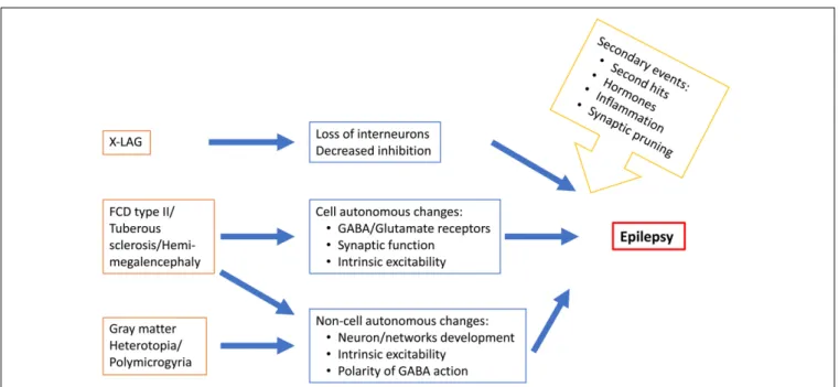
In conclusion, epileptogenesis in MCDs occur during a period
of brain development characterized by many molecular, cellular
and structural changes (Figure 2) that determine the features
of brain operation and functioning and impact epileptogenic
processes and epilepsy expression. Epileptogenesis in MCDs
involves complex multifactorial causes that would relate to
Represa Epileptogenic Cortical Malformations
the type of gene or insult involved in the malformation,
normal developmental processes and developmental adaptative
or reactive changes in cortical circuitry. The emergence
of new animal models reproducing focal mosaic lesions
associating manifestation reminiscent of human clinical
symptoms, open promising vistas for better understanding
the physiopathology of these disorders and testing new
therapeutic options.
AUTHOR CONTRIBUTIONS
The author confirms being the sole contributor of this work and
has approved it for publication.
FUNDING
The financial support from European Community 7th
Framework programs [Development and Epilepsy – Strategies
for Innovative Research to improve diagnosis, prevention and
treatment in children with difficult to treat Epilepsy (DESIRE)],
EraNet Neuron [Deciphering hyperexcitable networks associated
with neurodevelopmental lesions (DeCipher),
#ANR-15-NEUR-0001-03], INSERM and CNRS are acknowledged.
ACKNOWLEDGMENTS
The author thanks Drs. P. P. Lenck-Santini, V. Crepel and M.
Milh for critically reading the manuscript.
REFERENCES
Abdijadid, S., Mathern, G. W., Levine, M. S., and Cepeda, C. (2015). Basic mechanisms of epileptogenesis in pediatric cortical dysplasia.CNS Neurosci. Ther. 21, 92–103. doi: 10.1111/cns.12345
Ackman, J. B., Aniksztejn, L., Crepel, V., Becq, H., Pellegrino, C., Cardoso, C., et al. (2009). Abnormal network activity in a targeted genetic model of human double cortex.J. Neurosci. 29, 313–327. doi: 10.1523/JNEUROSCI.4093-08.2009 Aronica, E., Boer, K., Redeker, S., Spliet, W. G., van Rijen, P. C., Troost, D.,
et al. (2007). Differential expression patterns of chloride transporters, nNa+-K+-2Cl–cotransporter and K+-Cl–cotransporter, in epilepsy-associated malformations of cortical development.Neuroscience 145, 185–196. doi: 10. 1016/j.neuroscience.2006.11.041
Aubert, S., Wendling, F., Regis, J., McGonigal, A., Figarella-Branger, D., Peragut, J. C., et al. (2009). Local and remote epileptogenicity in focal cortical dysplasias and neurodevelopmental tumours.Brain 132, 3072–3086. doi: 10.1093/brain/ awp242
Bahi-Buisson, N., Souville, I., Fourniol, F. J., Toussaint, A., Moores, C. A., Houdusse, A., et al. (2013). New insights into genotype-phenotype correlations for the doublecortin-related lissencephaly spectrum. Brain 136, 223–244. doi: 10.1093/brain/aws323
Bai, J., Ramos, R. L., Ackman, J. B., Thomas, A. M., Lee, R. V., and LoTurco, J. J. (2003). RNAi reveals doublecortin is required for radial migration in rat neocortex.Nat. Neurosci. 6, 1277–1283. doi: 10.1038/nn1153
Barkovich, A. J., Guerrini, R., Kuzniecky, R. I., Jackson, G. D., and Dobyns, W. B. (2012). A developmental and genetic classification for malformations of cortical development: update 2012.Brain 135, 1348–1369. doi: 10.1093/brain/aws019 Bartolini, L., Libbey, J. E., Ravizza, T., Fujinami, R. S., Jacobson, S., and Gaillard,
W. D. (2018). Viral triggers and inflammatory mechanisms in pediatric epilepsy.Mol. Neurobiol. 56, 1897–1907. doi: 10.1007/s12035-018-1215-5 Bateup, H. S., Johnson, C. A., Denefrio, C. L., Saulnier, J. L., Kornacker, K.,
and Sabatini, B. L. (2013). Excitatory/inhibitory synaptic imbalance leads to hippocampal hyperexcitability in mouse models of tuberous sclerosis.Neuron 78, 510–522. doi: 10.1016/j.neuron.2013.03.017
Baulac, S., Ishida, S., Marsan, E., Miquel, C., Biraben, A., Nguyen, D. K., et al. (2015). Familial focal epilepsy with focal cortical dysplasia due to DEPDC5 mutations.Ann. Neurol. 77, 675–683. doi: 10.1002/ana.24368
Benardete, E. A., and Kriegstein, A. R. (2002). Increased excitability and decreased sensitivity to GABA in an animal model of dysplastic cortex.Epilepsia 43, 970–982. doi: 10.1046/j.1528-1157.2002.40901.x
Ben-Ari, Y., Cherubini, E., Corradetti, R., and Gaiarsa, J. L. (1989). Giant synaptic potentials in immature rat CA3 hippocampal neurones.J. Physiol. 416, 303–325. doi: 10.1113/jphysiol.1989.sp017762
Berg, A. T., Tarquinio, D., and Koh, S. (2017). Early life epilepsies are a comorbidity of developmental brain disorders. Semin. Pediatr. Neurol. 24, 251–263. doi: 10.1016/j.spen.2017
Bienvenu, T., Poirier, K., Friocourt, G., Bahi, N., Beaumont, D., Fauchereau, F., et al. (2002). ARX, a novel Prd-class-homeobox gene highly expressed in the
telencephalon, is mutated in X-linked mental retardation.Hum. Mol. Genet. 11, 981–991. doi: 10.1093/hmg/11.8.981
Blümcke, I., Thom, M., Aronica, E., Armstrong, D. D., Vinters, H. V., Palmini, A., et al. (2011). The clinicopathologic spectrum of focal cortical dysplasias: a consensus classification proposed by an ad hoc task force of the ILAE diagnostic methods commission.Epilepsia 52, 158–174. doi: 10.1111/j.1528-1167.2010. 02777.x
Bonneau, D., Toutain, A., Laquerriere, A., Marret, S., Saugier-Veber, P., Barthez, M., et al. (2002). X-linked lissencephaly with absent corpus callosum and ambiguous genitalia (XLAG): clinical, magnetic resonance imaging, and neuropathological findings.Ann. Neurol. 51, 340–349. doi: 10.1002/ana. 10119
Bourgeois, J. P., and Rakic, P. (1993). Changes of synaptic density in the primary visual cortex of the macaque monkey from fetal to adult stage.J. Neurosci. 13, 2801–2820. doi: 10.1523/JNEUROSCI.13-07-02801.1993
Brill, J., and Huguenard, J. R. (2010). Enhanced infragranular and supragranular synaptic input onto layer 5 pyramidal neurons in a rat model of cortical dysplasia.Cereb. Cortex 20, 2926–2938. doi: 10.1093/cercor/bhq040
Brunklaus, A., and Zuberi, S. M. (2014). Dravet syndrome–from epileptic encephalopathy to channelopathy. Epilepsia 55, 979–984. doi: 10.1111/epi. 12652
Calcagnotto, M. E., Paredes, M. F., Tihan, T., Barbaro, N. M., and Baraban, S. C. (2005). Dysfunction of synaptic inhibition in epilepsy associated with focal cortical dysplasia.J. Neurosci. 25, 9649–9657. doi: 10.1523/JNEUROSCI.2687-05.2005
Cepeda, C., André, V. M., Wu, N., Yamazaki, I., Uzgil, B., Vinters, H. V., et al. (2007). Immature neurons and GABA networks may contribute to epileptogenesis in pediatric cortical dysplasia.Epilepsia 48(Suppl. 5), 79–85. doi: 10.1111/j.1528-1167.2007.01293.x
Chen, Z. F., Schottler, F., Bertram, E., Gall, C. M., Anzivino, M. J., and Lee, K. S. (2000). Distribution and initiation of seizure activity in a rat brain with subcortical band heterotopia.Epilepsia 41, 493–501. doi: 10.1111/j.1528-1157. 2000.tb00201.x
Chevassus au Louis, N., Rafiki, A., Jorquera, I., Ben-Ari, Y., and Represa, A. (1998). Neocortex in the hippocampus: morphofunctional analysis of CA1 heterotopiae in the hippocampus of MAM treated rats.J. Comp. Neurol. 394, 520–536.
Christodoulou, J. A., Walker, L. M., Del Tufo, S. N., Katzir, T., Gabrieli, J. D., Whitfield-Gabrieli, S., et al. (2012). Abnormal structural and functional brain connectivity in gray matter heterotopia.Epilepsia 53, 1024–1032. doi: 10.1111/ j.1528-1167.2012.03466.x
Cohen, I., Navarro, V., Clemenceau, S., Baulac, M., and Miles, R. (2002). On the origin of interictal activity in human temporal lobe epilepsy in vitro.Science 298, 1418–1421. doi: 10.1126/science.1076510
Colciaghi, F., Finardi, A., Nobili, P., Locatelli, D., Spigolon, G., and Battaglia, G. S. (2014). Progressive brain damage, synaptic reorganization and NMDA activation in a model of epileptogenic cortical dysplasia.PLoS One 9:e89898. doi: 10.1371/journal.pone.0089898
Collombat, P., Mansouri, A., Hecksher-Sorensen, J., Serup, P., Krull, J., Gradwohl, G., et al. (2003). Opposing actions of Arx and Pax4 in endocrine pancreas development.Genes Dev. 17, 2591–2603. doi: 10.1101/gad.269003 Colombo, E., Galli, R., Cossu, G., Gecz, J., and Broccoli, V. (2004). Mouse
orthologue of ARX, a gene mutated in several X-linked forms of mental retardation and epilepsy, is a marker of adult neural stem cells and forebrain GABAergic neurons.Dev. Dyn. 231, 631–639. doi: 10.1002/dvdy.20164 Crino, P. B. (2015). mTOR signaling in epilepsy: insights from malformations of
cortical development.Cold Spring Harb. Perspect. Med. 5:a022442. doi: 10.1101/ cshperspect.a022442
Crino, P. B., Duhaime, A. C., Baltuch, G., and White, R. (2001). Differential expression of glutamate and GABA-A receptor subunit mRNA in cortical dysplasia.Neurology 56, 906–913. doi: 10.1212/WNL.56.7.906
D’Amato, C. J., and Hicks, S. P. (1980). Development of the motor system: effects of radiation on developing corticospinal neurons and locomotor function.Exp. Neurol. 70, 1–23. doi: 10.1016/0014-4886(80)90002-3
des Portes, V., Francis, F., Pinard, J. M., Desguerre, I., Moutard, M. L., Snoeck, I., et al. (1998). Doublecortin is the major gene causing X-linked subcortical laminar heterotopia (SCLH).Hum. Mol. Genet. 7, 1063–1070. doi: 10.1093/ hmg/7.7.1063
Devinsky, O., Vezzani, A., Najjar, S., De Lanerolle, N. C., and Rogawski, M. A. (2013). Glia and epilepsy: excitability and inflammation.Trends Neurosci. 36, 174–184. doi: 10.1016/j.tins.2012.11.008
D’Gama, A. M., Woodworth, M. B., Hossain, A. A., Bizzotto, S., Hatem, N. E., LaCoursiere, C. M., et al. (2017). Somatic mutations activating the mTOR pathway in dorsal telencephalic progenitors cause a continuum of cortical dysplasias.Cell Rep. 21, 3754–3766. doi: 10.1016/j.celrep.2017.11.106 Doherty, M. J., Simon, E., De Menezes, M. S., Kuratani, J. D., Saneto, R. P.,
Homles, M. D., et al. (2003). When might hemispheric favouring of epileptiform discharges begin?Seizure 12, 595–598.
Dravet, C. (1978). Severe epilepsies in infancy and childhood [in French].Vie Méd. 8, 548–1548.
European Chromosome 16 Tuberous Sclerosis Consortium (1993). Identification and characterization of the tuberous sclerosis gene on chromosome 16.Cell 75, 1305–1315.
Ferrer, I., Xumetra, A., and Santamaría, J. (1984). Cerebral malformation induced by prenatal X-irradiation, an autoradiographic and Golgi study.J. Anat. 138, 81–93.
Friocourt, G., and Parnavelas, J. G. (2010). Mutations in ARX result in several defects involving GABAergic neurons.Front. Cell. Neurosci. 4:4. doi: 10.3389/ fncel.2010.00004
Fullenkamp, A. N., and El-Hodiri, H. M. (2008). The function of the Aristaless-related homeobox (Arx) gene product as a transcriptional repressor is diminished by mutations associated with X-linked mental retardation (XLMR). Biochem. Biophys. Res. Commun. 377, 73–78. doi: 10.1016/j.bbrc.2008.09.116 Galic, M. A., Riazi, K., and Pittman, Q. J. (2012). Cytokines and brain excitability.
Front. Neuroendocrinol. 33, 116–125. doi: 10.1016/j.yfrne.2011.12.002 Giedd, J. N., Clasen, L. S., Lenroot, R., Greenstein, D., Wallace, G. L., Ordaz, S., et al.
(2006). Puberty-related influences on brain development.Mol. Cell. Endocrinol. 254–255, 154–162. doi: 10.1016/j.mce.2006.04.016
Gleeson, J. G., Allen, K. M., Fox, J. W., Lamperti, E. D., Berkovic, S., Scheffer, I., et al. (1998). Doublecortin, a brain-specific gene mutated in human X-linked lissencephaly and double cortex syndrome, encodes a putative signaling protein. Cell 92, 63–72. doi: 10.1016/S0092-8674(00)80899-5
Guerrini, R., and Dobyns, W. B. (2014). Malformations of cortical development: clinical features and genetic causes.Lancet Neurol. 13, 710–726. doi: 10.1016/ S1474-4422(14)70040-7
Harvey, A. S., Cross, J. H., Shinnar, S., Mathern, G. W., and ILAE Pediatric Epilepsy Surgery Survey Taskforce (2008). Defining the spectrum of international practice in pediatric epilepsy surgery patients.Epilepsia 49, 146–155. doi: 10. 1111/j.1528-1167.2007.01421.x
Hsieh, L. S., Wen, J. H., Claycomb, K., Huang, Y., Harrsch, F. A., Naegele, J. R., et al. (2016). Convulsive seizures from experimental focal cortical dysplasia occur independently of cell misplacement.Nat. Commun. 7:11753. doi: 10.1038/ ncomms11753
Huberfeld, G., Wittner, L., Clemenceau, S., Baulac, M., Kaila, K., Miles, R., et al. (2007). Perturbed chloride homeostasis and GABAergic signaling in human
temporal lobe epilepsy.J. Neurosci. 27, 9866–9873. doi: 10.1523/JNEUROSCI. 2761-07.2007
Iyer, A., Zurolo, E., Spliet, W. G., van Rijen, P. C., Baayen, J. C., Gorter, J. A., et al. (2010). Evaluation of the innate and adaptive immunity in type I and type II focal cortical dysplasias.Epilepsia 51, 1763–1773. doi: 10.1111/j.1528-1167. 2010.02547.x
Jacobs, K. M., Gutnick, M. J., and Prince, D. A. (1996). Hyperexcitability in a model of cortical maldevelopment.Cereb. Cortex 6, 514–523. doi: 10.1093/cercor/6.3. 514
Jacobs, K. M., and Prince, D. A. (2005). Excitatory and inhibitory postsynaptic currents in a rat model of epileptogenic microgyria.J. Neurophysiol. 93, 687– 696. doi: 10.1152/jn.00288.2004
Jansen, L. A., Mirzaa, G. M., Ishak, G. E., O’Roak, B. J., Hiatt, J. B., Roden, W. H., et al. (2015). PI3K/AKT pathway mutations cause a spectrum of brain malformations from megalencephaly to focal cortical dysplasia. Brain 138, 1613–1628. doi: 10.1093/brain/awv045
Jensen, K. F., and Killackey, H. P. (1984). Subcortical projections from ectopic neocortical neurons.Proc. Natl. Acad. Sci. U.S.A. 81, 964–968. doi: 10.1073/ pnas.81.3.964
Jiang, N. M., Cowan, M., Moonah, S. N., and Petri, W. A. Jr. (2018). The impact of systemic inflammation on neurodevelopment.Trends Mol. Med. 24, 794–804. doi: 10.1016/j.molmed.2018.06.008
Jin, X., Jiang, K., and Prince, D. A. (2014). Excitatory and inhibitory synaptic connectivity to layer V fast-spiking interneurons in the freeze lesion model of cortical microgyria.J. Neurophysiol. 112, 1703–1713. doi: 10.1152/jn.00854. 2013
Kasteleijn-Nolst Trenité, D. G., Schmitz, B., Janz, D., Delgado-Escueta, A. V., Thomas, P., Hirsch, E., et al. (2013). Consensus on diagnosis and management of JME: from founder’s observations to current trends.Epilepsy Behav. 28(Suppl. 1), S87–S90. doi: 10.1016/j.yebeh.2012.11.051
Kato, M., Das, S., Petras, K., Kitamura, K., Morohashi, K., Abuelo, D. N., et al. (2004). Mutations of ARX are associated with striking pleiotropy and consistent genotype-phenotype correlation.Hum. Mutat. 23, 147–159. doi: 10.1002/humu. 10310
Kettenmann, H., Kirchhoff, F., and Verkhratsky, A. (2013). Microglia: new roles for the synaptic stripper.Neuron 77, 10–18. doi: 10.1016/j.neuron.2012.12.023 Khalilov, I., Holmes, G. L., and Ben-Ari, Y. (2003). In vitro formation of a
secondary epileptogenic mirror focus by interhippocampal propagation of seizures.Nat. Neurosci. 6, 1079–1085. doi: 10.1038/nn1125
Kirmse, K., Kummer, M., Kovalchuk, Y., Witte, O. W., Garaschuk, O., and Holthoff, K. (2015). GABA depolarizes immature neurons and inhibits network activity in the neonatal neocortexin vivo. Nat. Commun. 6:7750. doi: 10.1038/ ncomms8750
Kitamura, K., Yanazawa, M., Sugiyama, N., Miura, H., Iizuka-Kogo, A., Kusaka, M., et al. (2002). Mutation of ARX causes abnormal development of forebrain and testes in mice and X-linked lissencephaly with abnormal genitalia in humans. Nat. Genet. 32, 359–369. doi: 10.1038/ng1009
Knuesel, I., Chicha, L., Britschgi, M., Schobel, S. A., Bodmer, M., Hellings, J. A., et al. (2014). Maternal immune activation and abnormal brain development across CNS disorders.Nat. Rev. Neurol. 10, 643–660. doi: 10.1038/nrneurol. 2014.187
Kobayashi, E., Hawco, C. S., Grova, C., Dubeau, F., and Gotman, J. (2006). Widespread and intense BOLD changes during brief focal electrographic seizures.Neurology 66, 1049–1055. doi: 10.1212/01.wnl.0000204232.37720.a4 Kyritsis, N., Kizil, C., Zocher, S., Kroehne, V., Kaslin, J., Freudenreich, D., et al.
(2012). Acute inflammation initiates the regenerative response in the adult zebrafish brain.Science 338, 1353–1356. doi: 10.1126/science.1228773 Lapray, D., Popova, I. Y., Kindler, J., Jorquera, I., Becq, H., Manent, J. B., et al.
(2010). Spontaneous epileptic manifestations in a DCX knockdown model of human double cortex.Cereb. Cortex 20, 2694–2701. doi: 10.1093/cercor/bh q014
Lee, J. H., Huynh, M., Silhavy, J. L., Kim, S., Dixon-Salazar, T., Heiberg, A., et al. (2012). De novo somatic mutations in components of the PI3K-AKT3-mTOR pathway cause hemimegalencephaly.Nat. Genet. 44, 941–945. doi: 10.1038/ng. 2329
Lee, K. S., Schottler, F., Collins, J. L., Lanzino, G., Couture, D., Rao, A., et al. (1997). A genetic animal model of human neocortical heterotopia associated
Represa Epileptogenic Cortical Malformations
with seizures.J. Neurosci. 17, 6236–6242. doi: 10.1523/JNEUROSCI.17-16-06236.1997
Leventer, R. J., Jansen, A., Pilz, D. T., Stoodley, N., Marini, C., Dubeau, F., et al. (2010). Clinical and imaging heterogeneity of polymicrogyria: a study of 328 patients.Brain 133, 1415–1427. doi: 10.1093/brain/awq078
Leventer, R. J., Phelan, E. M., Coleman, L. T., Kean, M. J., Jackson, G. D., and Harvey, A. S. (1999). Clinical and imaging features of cortical malformations in childhood.Neurology 53, 715–722. doi: 10.1212/WNL.53.4.715
Lim, J. S., Gopalappa, R., Kim, S. H., Ramakrishna, S., Lee, M., Kim, W. I., et al. (2017). Somatic mutations in TSC1 and TSC2 cause focal cortical dysplasia.Am. J. Hum. Genet. 100, 454–472. doi: 10.1016/j.ajhg.2017.01.030
Lim, J. S., Kim, W. I., Kang, H. C., Kim, S. H., Park, A. H., Park, E. K., et al. (2015). Brain somatic mutations in MTOR cause focal cortical dysplasia type II leading to intractable epilepsy.Nat. Med. 21, 395–400. doi: 10.1038/nm.3824 Lowenstein, D. H., Walker, M., and Waterhouse, E. (2014). Status epilepticus in the
setting of acute encephalitis.Epilepsy Curr. 14, 43–49. doi: 10.5698/1535-7511-14.s2.43
Lozovaya, N., Gataullina, S., Tsintsadze, T., Tsintsadze, V., Pallesi-Pocachard, E., Minlebaev, M., et al. (2014). Selective suppression of excessive GluN2C expression rescues early epilepsy in a tuberous sclerosis murine model.Nat. Commun. 5:4563. doi: 10.1038/ncomms5563
Ma, T. S., Elliott, R. E., Ruppe, V., Devinsky, O., Kuzniecky, R., Weiner, H. L., et al. (2012). Electrocorticographic evidence of perituberal cortex epileptogenicity in tuberous sclerosis complex.J. Neurosurg. Pediatr. 10, 376–382. doi: 10.3171/ 2012.8.PEDS1285
Madhavan, D., Weiner, H. L., Carlson, C., Devinsky, O., and Kuzniecky, R. (2007). Local epileptogenic networks in tuberous sclerosis complex: a case review. Epilepsy Behav. 11, 140–146. doi: 10.1016/j.yebeh.2007.03.017
Mai, R., Tassi, L., Cossu, M., Francione, S., Lo Russo, G., Garbelli, R., et al. (2003). A neuropathological, stereo-EEG, and MRI study of subcortical band heterotopia.Neurology 60, 1834–1838. doi: 10.1212/01.WNL.0000065884. 61237.24
Major, P., Rakowski, S., Simon, M. V., Cheng, M. L., Eskandar, E., Baron, J., et al. (2009). Are cortical tubers epileptogenic? Evidence from electrocorticography. Epilepsia 50, 147–154. doi: 10.1111/j.1528-1167.2008.01814.x
Manent, J. B., Wang, Y., Chang, Y., Paramasivam, M., and LoTurco, J. J. (2009). Dcx reexpression reduces subcortical band heterotopia and seizure threshold in an animal model of neuronal migration disorder. Nat. Med. 15, 84–90. doi: 10.1038/nm.1897
Marchi, N., Granata, T., and Janigro, D. (2014). Inflammatory pathways of seizure disorders.Trends Neurosci. 37, 55–65. doi: 10.1016/j.tins.2013.11.002 Marin, I., and Kipnis, J. (2013). Learning and memory... and the immune system.
Learn. Mem. 20, 601–606. doi: 10.1101/lm.028357.112
Marsan, E., and Baulac, S. (2018). Mechanistic target of rapamycin (mTOR) pathway, focal cortical dysplasia and epilepsy.Neuropathol. Appl. Neurobiol. 44, 6–17. doi: 10.1111/nan.12463
Marsh, E., Fulp, C., Gomez, E., Nasrallah, I., Minarcik, J., Sudi, J., et al. (2009). Targeted loss of Arx results in a developmental epilepsy mouse model and recapitulates the human phenotype in heterozygous females.Brain 132, 1563– 1576. doi: 10.1093/brain/awp107
Marsh, E. D., Nasrallah, M. P., Walsh, C., Murray, K. A., Nicole Sunnen, C., McCoy, A., et al. (2016). Developmental interneuron subtype deficits after targeted loss of Arx.BMC Neurosci. 17:35. doi: 10.1186/s12868-016-0265-8 Martineau, F. S., Sahu, S., Plantier, V., Buhler, E., Schaller, F., Fournier, L.,
et al. (2018). Correct laminar positioning in the neocortex influences proper dendritic and synaptic development.Cereb. Cortex 28, 2976–2990. doi: 10.1093/ cercor/bhy113
Martínez-Juárez, I. E., Alonso, M. E., Medina, M. T., Durón, R. M., Bailey, J. N., López-Ruiz, M., et al. (2006). Juvenile myoclonic epilepsy subsyndromes: family studies and long-term follow-up.Brain 129, 1269–1280. doi: 10.1093/brain/ awl048
McKenzie, O., Ponte, I., Mangelsdorf, M., Finnis, M., Colasante, G., Shoubridge, C., et al. (2007). Aristaless-related homeobox gene, the gene responsible for West syndrome and related disorders, is a Groucho/transducin-like enhancer of split dependent transcriptional repressor.Neuroscience 146, 236–247. doi: 10.1016/j. neuroscience.2007.01.038
Mirzaa, G. M., Campbell, C. D., Solovieff, N., Goold, C., Jansen, L. A., Menon, S., et al. (2016). Association of MTOR mutations with developmental brain
disorders, including megalencephaly, focal cortical dysplasia, and pigmentary mosaicism.JAMA Neurol. 73, 836–845. doi: 10.1001/jamaneurol.2016.0363 Møller, R. S., Weckhuysen, S., Chipaux, M., Marsan, E., Taly, V., Bebin, E. M., et al.
(2016). Germline and somatic mutations in the MTOR gene in focal cortical dysplasia and epilepsy.Neurol. Genet. 2:e118.
Nisticò, R., Salter, E., Nicolas, C., Feligioni, M., Mango, D., Bortolotto, Z. A., et al. (2017). Synaptoimmunology - roles in health and disease.Mol. Brain 10:26. doi: 10.1186/s13041-017-0308-9
Ochoa-Gómez, L., López-Pisón, J., Lapresta Moros, C., Fuertes Rodrigo, C., Fernando Martínez, R., Samper-Villagrasa, P., et al. (2017). A study of epilepsy according to the age at onset and monitored for 3 years in a regional reference paediatric neurology unit.Ann. Pediatr. 86, 11–19. doi: 10.1016/j.anpedi.2016. 05.002
Olivetti, P. R., and Noebels, J. L. (2012). Interneuron, interrupted: molecular pathogenesis of ARX mutations and X-linked infantile spasms.Curr. Opin. Neurobiol. 22, 859–865. doi: 10.1016/j.conb.2012.04.006
Petit, L. F., Jalabert, M., Buhler, E., Malvache, A., Peret, A., Chauvin, Y., et al. (2014). Normotopic cortex is the major contributor to epilepsy in experimental double cortex.Ann. Neurol. 76, 428–442. doi: 10.1002/ana.24237
Pilz, D. T., Matsumoto, N., Minnerath, S., Mills, P., Gleeson, J. G., Allen, K. M., et al. (1998). LIS1 and XLIS (DCX) mutations cause most classical lissencephaly, but different patterns of malformation. Hum. Mol. Genet. 7, 2029–2037. doi: 10.1093/hmg/7.13.2029
Pizzo, F., Roehri, N., Catenoix, H., Medina, S., McGonigal, A., Giusiano, B., et al. (2017). Epileptogenic networks in nodular heterotopia: a stereoelectroencephalography study. Epilepsia 58, 2112–2123. doi: 10.1111/epi.13919
Plantier, V., Watrin, F., Buhler, E., Martineau, F. S., Sahu, S., Manent, J. B., et al. (2018). Direct and collateral alterations of functional cortical circuits in a rat model of subcortical band heterotopia.Cereb. Cortex doi: 10.1093/cercor/ bhy307 [Epub ahead of print].
Poduri, A., Evrony, G. D., Cai, X., Elhosary, P. C., Beroukhim, R., Lehtinen, M. K., et al. (2012). Somatic activation of AKT3 causes hemispheric developmental brain malformations.Neuron 74, 41–48. doi: 10.1016/j.neuron.2012.03.010 Puskarjov, M., Ahmad, F., Kaila, K., and Blaesse, P. (2012). Activity-dependent
cleavage of the K-Cl cotransporter KCC2 mediated by calcium-activated protease calpain.J. Neurosci. 32, 11356–11364. doi: 10.1523/JNEUROSCI.6265-11.2012
Rafiki, A., Chevassus-au-Louis, N., Ben-Ari, Y., Khrestchatisky, M., and Represa, A. (1998). Glutamate receptors in dysplasic cortex: an in situ hybridization and immunohistochemistry study in the brain of rats with prenatal treatment with methylazoxymethanol.Brain Res. 782, 142–152. doi: 10.1016/S0006-8993(97) 01273-0
Raznahan, A., Lerch, J. P., Lee, N., Greenstein, D., Wallace, G. L., Stockman, M., et al. (2011). Patterns of coordinated anatomical change in human cortical development: a longitudinal neuroimaging study of maturational coupling. Neuron 72, 873–884. doi: 10.1016/j.neuron.2011.09.028
Redecker, C., Luhmann, H. J., Hagemann, G., Fritschy, J. M., and Witte, O. W. (2000). Differential downregulation of GABAA receptor subunits in widespread brain regions in the freeze-lesion model of focal cortical malformations. J. Neurosci. 20, 5045–5053. doi: 10.1523/JNEUROSCI.20-13-05045.2000 Ribierre, T., Deleuze, C., Bacq, A., Baldassari, S., Marsan, E., Chipaux, M., et al.
(2018). Second-hit mosaic mutation in mTORC1 repressor DEPDC5 causes focal cortical dysplasia-associated epilepsy.J. Clin. Invest. 128, 2452–2458. doi: 10.1172/JCI99384
Rosenblat, J. D., Cha, D. S., and Mansur, R. B. (2014). McIntyre RS. Inflamed moods: a review of the interactions between inflammation and mood disorders. Prog. Neuropsychopharmacol.Biol. Psychiatry. 53, 23–34. doi: 10.1016/j.pnpbp. 2014.01.013
Sancini, G., Franceschetti, S., Battaglia, G., Colacitti, C., Di Luca, M., Spreafico, R., et al. (1998). Dysplastic neocortex and subcortical heterotopias in methylazoxymethanol-treated rats: an intracellular study of identified pyramidal neurones.Neurosci. Lett. 246, 181–185. doi: 10.1016/S0304-3940(98) 00258-4
Seufert, D. W., Prescott, N. L., and El-Hodiri, H. M. (2005). Xenopus aristaless-related homeobox (xARX) gene product functions as both a transcriptional activator and repressor in forebrain development.Dev. Dyn. 232, 313–324. doi: 10.1002/dvdy.20234
Shafi, M. M., Vernet, M., Klooster, D., Chu, C. J., Boric, K., Barnard, M. E., et al. (2015). Physiological consequences of abnormal connectivity in a developmental epilepsy.Ann. Neurol. 77, 487–503. doi: 10.1002/ana.24343 Shoubridge, C., Fullston, T., and Gecz, J. (2010). ARX spectrum disorders: making
inroads into the molecular pathology.Hum. Mutat. 31, 889–900. doi: 10.1002/ humu.21288
Singh, S. C. (1980). Deformed dendrites and reduced spine numbers on ectopic neurones in the hippocampus of rats exposed to methylazoxymethanol-acetate. A Golgi-Cox study.Acta Neuropathol. 49, 193–198. doi: 10.1007/BF007 07106
Smith, M. J., Adams, L. F., Schmidt, P. J., Rubinow, D. R., and Wassermann, E. M. (2002). Effects of ovarian hormones on human cortical excitability.Ann. Neurol. 51, 599–603. doi: 10.1002/ana.10180
Staley, K. J., and Mody, I. (1992). Shunting of excitatory input to dentate gyrus granule cells by a depolarizing GABAA receptor-mediated post- synaptic conductance.J. Neurophysiol. 68, 197–212. doi: 10.1152/jn.1992.68.1.197 Stolp, H. B., Turnquist, C., Dziegielewska, K. M., Saunders, N. R., Anthony,
D. C., and Molnár, Z. (2011). Reduced ventricular proliferation in the foetal cortex following maternal inflammation in the mouse.Brain 134, 3236–3248. doi: 10.1093/brain/awr237
Stromme, P., Mangelsdorf, M. E., Shaw, M. A., Lower, K. M., Lewis, S. M. E., Bruyere, H., et al. (2002). Mutations in the human ortholog of Aristaless cause X-linked mental retardation and epilepsy.Nat. Genet. 30, 441–445. doi: 10.1038/ ng862
Talos, D. M., Sun, H., Kosaras, B., Joseph, A., Folkerth, R. D., Poduri, A., et al. (2012). Altered inhibition in tuberous sclerosis and type IIb cortical dysplasia. Ann. Neurol. 71, 539–551. doi: 10.1002/ana.22696
Tassi, L., Colombo, N., Cossu, M., Mai, R., Francione, S., Lo Russo, G., et al. (2005). Electroclinical, MRI and neuropathological study of 10 patients with nodular heterotopia, with surgical outcomes.Brain 128, 321–337. doi: 10.1093/brain/ awh357
Toering, S. T., Boer, K., de Groot, M., Troost, D., Heimans, J. J., Spliet, W. G., et al. (2009). Expression patterns of synaptic vesicle protein 2A in focal cortical dysplasia and TSC-cortical tubers.Epilepsia 50, 1409–1418. doi: 10.1111/j.1528-1167.2008.01955.x
Tyvaert, L., Hawco, C., Kobayashi, E., LeVan, P., Dubeau, F., and Gotman, J. (2008). Different structures involved during ictal and interictal epileptic activity in malformations of cortical development: an EEG-fMRI study. Brain 131, 2042–2060. doi: 10.1093/brain/awn145
Tyzio, R., Khalilov, I., Represa, A., Crepel, V., Zilberter, Y., Rheims, S., et al. (2009). Inhibitory actions of the gamma-aminobutyric acid in pediatric Sturge-Weber syndrome.Ann. Neurol. 66, 209–218. doi: 10.1002/ana.21711
Tyzio, R., Minlebaev, M., Rheims, S., Ivanov, A., Jorquera, I., Holmes, G. L., et al. (2008). Postnatal changes in somatic gamma-aminobutyric acid signalling in
the rat hippocampus.Eur. J. Neurosci. 27, 2515–2528. doi: 10.1111/j.1460-9568. 2008.06234.x
Valeeva, G., Tressard, T., Mukhtarov, M., Baude, A., and Khazipov, R. (2016). An optogenetic approach for investigation of excitatory and inhibitory network GABA actions in mice expressing channelrhodopsin-2 in GABAergic neurons. J. Neurosci. 36, 5961–5673. doi: 10.1523/JNEUROSCI.3482-15.2016
Valton, L., Guye, M., McGonigal, A., Marquis, P., Wendling, F., Regis, J., et al. (2008). Functional interactions in brain networks underlying epileptic seizures in bilateral diffuse periventricular heterotopia.Clin. Neurophysiol. 119, 212–223. doi: 10.1016/j.clinph.2007.09.118
Vezzani, A. (2013). Fetal brain inflammation may prime hyperexcitability and behavioral dysfunction later in life.Ann. Neurol. 74, 1–3. doi: 10.1002/ana. 23930
Vezzani, A., Fujinami, R. S., White, H. S., Preux, P. M., Blümcke, I., Sander, J. W., et al. (2016). Infections, inflammation and epilepsy.Acta Neuropathol. 131, 211–234. doi: 10.1007/s00401-015-1481-5
Vukovic, J., Colditz, M. J., Blackmore, D. G., Ruitenberg, M. J., and Bartlett, P. F. (2012). Microglia modulate hippocampal neural precursor activity in response to exercise and aging.J. Neurosci. 32, 6435–6443. doi: 10.1523/JNEUROSCI. 5925-11.2012
Wake, H., Moorhouse, A. J., Miyamoto, A., and Nabekura, J. (2013). Microglia: actively surveying and shaping neuronal circuit structure and function.Trends Neurosci. 36, 209–217. doi: 10.1016/j.tins.2012.11.007
Wang, W., Takashima, S., Segawa, Y., Itoh, M., Shi, X., Hwang, S. K., et al. (2011). Nabeshima K, Takeshita M, Hirose S. The developmental changes of Na(v)1.1 and Na(v)1.2 expression in the human hippocampus and temporal lobe.Brain Res. 1389, 61–70. doi: 10.1016/j.brainres.2011.02.083
Zehr, J. L., Todd, B. J., Schulz, K. M., McCarthy, M. M., and Sisk, C. L. (2006). Dendritic pruning of the medial amygdala during pubertal development of the male Syrian hamster.J. Neurobiol. 66, 578–590. doi: 10.1002/neu.20251 Zhu, W. J., and Roper, S. N. (2000). Reduced inhibition in an animal model of
cortical dysplasia.J. Neurosci. 20, 8925–8931. doi: 10.1523/JNEUROSCI.20-23-08925.2000
Conflict of Interest Statement: The author declares that the research was conducted in the absence of any commercial or financial relationships that could be construed as a potential conflict of interest.
Copyright © 2019 Represa. This is an open-access article distributed under the terms of the Creative Commons Attribution License (CC BY). The use, distribution or reproduction in other forums is permitted, provided the original author(s) and the copyright owner(s) are credited and that the original publication in this journal is cited, in accordance with accepted academic practice. No use, distribution or reproduction is permitted which does not comply with these terms.