Block Copolymer-Templated Iron Oxide Nanoparticles for Bimodal Growth of Multi-walled Carbon Nanotubes
By
KYLE E YAZZIE
Submitted to the Department of Materials Science and Engineering in Partial Fulfillment
of the Requirements for the Degree of
Bachelor of Science
at theMassachusetts Institute of Technology June 2008
C 2008 Kyle E Yazzie All rights reserved
~AS6ACHUSEnrs
INSTMIME~
OFTEOHNOLOGY
JUN 18
2008
LIBRARIES
The author hereby grants to MIT permission to reproduce and to distribute publicly paper and electronic copies of this thesis document in whole or in part in any medium now
. know, or hereafter created.
Signature of Author...
Department of Materials Scie and Engineering
V
May 16, 2008
Certified by... ... ...-. ... Robert E. Cohen, Ph.D.
St. Laurent Professor of Chemical Engineering, MIT Thesis Advisor
Accepted by... ... Caroline A. Ross
Block Copolymer-Templated Iron Oxide Nanoparticles for Bimodal Growth of Multi-walled Carbon Nanotubes
by
KYLE E YAZZIE
Submitted to the Department of Materials Science and Engineering in Partial Fulfillment of the Requirements for the Degree of
Bachelor of Science at the
Massachusetts Institute of Technology
Abstract
Since their discovery carbon nanotubes (CNTs) have sparked great interest due to their exceptional mechanical, electrical, and thermal properties.' These properties make carbon nanotubes desirable for numerous applications including: nanoelectronics, high-strength composites, energy storage, superhydrophobic surfaces, sensors, and biomaterial interfaces.2,3 Bulk synthesis of carbon nanotubes with controlled physical features, i.e.
length, diameter, multiwalled vs. single walled, carbon nanotube chirality, etc. is necessary to make full use of carbon nanotubes' exceptional properties in commercial aspects.
Typical carbon nanotube synthesis processes use chemical vapor deposition (CVD), arc-discharge, and laser ablation.' Synthesizing carbon nanotubes via CVD typically involves depositing a thin metal film on a silicon substrate, and heating the substrate so that the thin metal film dewets and forms metallic nanoparticles. A hydrocarbon gas is then flowed over the nanoparticles to initiate carbon nanotube growth.4 Though these thin metal film catalysts are easy to prepare, they offer poor control over nanoparticle diameters and areal density.4 It has been shown that physical
properties of carbon nanotubes, such as diameter and uniformity of growth, are directly related to the diameter of the catalyst nanoparticle, and that chirality of the carbon nanotube is inversely related to the catalyst nanoparticle diameter.2'3'4 Therefore, fully exploiting the unique properties of carbon nanotubes requires an understanding of how to control catalyst nanoparticle diameters, and thereby carbon nanotube physical characteristics. Bennett et al demonstrated that controllability of nanoparticle diameters is possible using a simple poly(styrene-b-acrylic acid) (PS-b-PAA) amphiphilic block copolymer.4 The amphiphilic PS-b-PAA block copolymer forms micelles, when
dissolved in toluene, with anionic carboxylic acid groups available from the PAA. The anionic PAA carboxylic acid groups can be used to sequester metal cations, so that metal is effectively loaded into the micelles. The size of nanoparticles can be controlled by the size of the PAA portion of the block copolymer.5 When spin cast onto a substrate, the metal-loaded PS-b-PAA micelles form a quasi-ordered block copolymer thin film. Maximizing the amount of metal-loaded micelles in solution can maximize the resulting areal density of nanoparticles, thereby forming a monodisperse, quasi-hexagonal nanoparticle array.5 The deposited micellular thin film and substrate can then be etched with oxygen plasma, removing the organic polymer so that only the nanoparticle array is left, and the substrate is ready for carbon nanotube growth.
Acknowledgements
I would like to extend my most sincere gratitude to Eric Verploegen and the many colleagues that he has introduced me to. His breadth and depth of scientific knowledge and unmitigated enthusiasm has made this thesis project truly educational and fulfilling. I feel incredibly fortunate to have worked with him on this project. Work performed by A. J. Hart and his team of graduate students from the University of Michigan, including Sang Han and Eric Meshot, has contributed substantial matter to this thesis. I would also like to thank Andy Miller for plasma etching several samples, and Teijia Zang for characterizing the nanoparticle arrays with AFM. My thesis advisor, Professor Robert E. Cohen, has my deepest respect and gratitude. His convivial nature and astute intellect has made the process of constructing this thesis rewarding and educational.
Finally, I would like to thank my family, who have always supported me, and without whom none of my accomplishments would be possible.
Table of Contents LIST OF FIGURES ... ... 6 LIST OF TABLES ... ... 11 IIntroduction ... 12 2 Experimental Details ... .... 17 2.1 Materials ... ... 17 2.2 Sample Preparation... 18
2.3 Transmission Electron Microscopy ... 20
2.4 Small-Angle X-ray Scattering and Grazing Incidence Small-Angle X-ray Scattering ... ... 21
2.5 Growth of Carbon Nanotubes from Nanoparticle Arrays ... 23
2.6 Statistical Analysis of Nanoparticle Diameter Distributions ... ... 24
3 Results and Discussion ... 26
3.1 Significantly Different Mean Particle Diameters ... 26
3.2 Single PS-b-PAA Block Copolymer and Metal Loading Systems ... 29
3.2.1 Small ... 29
3.2.2 Medium ... 30
3.2.3 Large ... 32
3.3 Summary of nanoparticle diameters available using single systems of one PS-b-PAA block copolymer and one metal loading ... ... 34
3.4 Combinations of Two Block Copolymer and Metal Loading Systems...34
3.4.1 Small + Medium ... 36
3.4.2 Small + Large ... 39
3.4.3 Small + XLarge ... 42
3.5 Additive Relationship between single and combined systems...45
3.5.1 Small, Medium, and Small + Medium ... 45
3.5.2 Small, Large, and Small + Large ... 47
3.5.3 Small, and Small + XLarge ... 48
3.6 Summary of nanoparticle diameters available using additive system of nanoparticle arrays... ... 50
3.7 Factors affecting the nanoparticle diameter distributions ... 51
3.8 Grazing Incidence Small-angle X-ray Scattering (GISAXS) of Nanoparticle A rrays ... 53
3.9 Small-Angle X-ray Scattering of Carbon Nanotubes Grown From Nanoparticle Arrays ... ... 56
4 Summary and Conclusions ... 60
5 References... ... 63
Appendix A ... 67
A ppendix B ... .. 68
List of Figures
Figure 1 The arrangement of repeat units in a block copolymer, where A and B are chemically dissimilar repeat units... 12 Figure 2 Chemical formula for poly(styrene-block-acrylic acid). n and m are the number
of repeat units constituting each block. The COOH group of the acrylic acid portion of the block copolymer ionizes in solution, and constitutes the anionic core of the m icelles... ... 12 Figure 3 Microstructure of a micelle formed with block copolymers. Solutions of such micelles, with anionic cores were loaded with Fe3+ via a reduction of FeCl3 salt in
solution. This mechanism was used to synthesize the Fe20 3 nanoparticles studied
herein ... ... . . 12 Figure 4 Strategies for synthesizing bimodal nanoparticles, using PS-b-PAA block copolymer micelles. In Strategy A a single block copolymer with two different metal loadings was used. In Strategy B in two block copolymers with dissimilar molecular weights, containing one metal loading was used. In Strategy C two block copolymers with dissimilar molecular weights, containing two different metal loadings was utilized. Strategy C potentially yields the largest size discrepancy between nanoparticles ... 16 Figure 5 Typical setup for SAXS of carbon nanotube tubes. A motorized stage allows for progressive vertical scans to be taken in the y-direction, at a height h from the substrate surface. (used with permission, Verploegen 2008) ... 22 Figure 6 A schematic of a typical GISAXS experiment for a thin film. A collimated X-ray beam is grazed off the sample at an angle, a. Scattering in the qy direction results from features in the plane of the sample surface. Scattering in the qx direction results from features parallel to the sample surface. (used with permission, Verploegen 200 8)...2 3
Figure 7 The left portion of the figure shows the distribution of nanoparticle diameters, and box-and-whisker plots indicating their mean values and range. The particle counts are; 49 for Small, 187 for Medium, and 151 for Large. The right portion of the figure shows the Tukey-Kramer Honestly Significant Difference (HSD) test of the mean nanoparticle diameters. The diameters of the circles represent the nanoparticle diameters that fall within the 95% confidence levels. Circles that intersect at less than or equal to 900 are considered to represent diameters with significantly different means. The Tukey-Kramer HSD shows that the Small, Medium, and Large nanoparticle have significantly different mean diameters. However, the Small and Medium nanoparticles have only borderline significantly different mean diameters. This is not surprising considering that the Small and Medium nanoparticles are synthesized using the same block copolymer, PS11000-b-PAA1 200, only with different metal loadings...26
Figure 8 Distribution of particle diameters for Small (PSl1ooo-b-PAA 1200 Metal Loading =
0.5). The total particle count is 49. The normal-quantile plot above the nanoparticle diameter distribution indicates that the diameters are normally distributed. A normal distribution curve was fitted to the Small nanoparticle distribution to give a mean nanoparticle diameter of 5.8 nm with a standard deviation of 1.2 nm. The inset is a representative TEM image of the Small nanoparticle array. The dark blotches, such as the one circled with the dashed line, appear in several TEM images. They did not significantly affect the morphology of the nanoparticles or the particle diameter analysis. The blotches were determined to be due to debris from undissolved polymer in solution or debris ejected onto the backside of the TEM window during the spin casting process... ... 30 Figure 9 Distribution of particle diameters for Medium (PS11000-b-PAA1200 Metal Loading
= 5). The total particle count is 187 particles. The normal-quantile plot above the
nanoparticle diameter distribution indicates that the diameters are normally distributed. A normal distribution curve was fitted to the Medium nanoparticle distribution to give a mean nanoparticle diameter of 6.8 nm with a standard deviation of 2.2 nm. The inset is a representative TEM image of the Medium nanoparticle array... 31
Figure 10 Distribution of particle diameters for Large (PS16 500-b-PAA4500 Metal Loading
= 5). The total particle count is 150. The normal-quantile plot above the nanoparticle
diameter distribution indicates that the diameters are normally distributed. A normal distribution curve was fitted to the Large nanoparticle distributions to give a mean nanoparticle diameter of 12.07 nm with a standard deviation of 2.70 nm. The inset is a representative TEM image of the Large nanoparticle array. The hollow appearance of the nanoparticles was determined to be caused by focusing effects in the TEM, and do not reflect a true morphological characteristic of the nanoparticles. Similar conclusions about nanoparticles that appear hollow were reached by Bennett 2007. The spread of the nanoparticle diameter distribution can be attributed to the irregularly shaped Large nanoparticles, as seen in the inset TEM image...33 Figure 11 Distribution of particle diameters for a combined solution of Small and Medium solutions (PSooo1000-b-PAA1200 Metal Loading = 0.5 + PSooo1000-b-PAA1 200 Metal
Loading = 5). The normal-quantile plot above the nanoparticle diameter distribution indicates that the diameters are not normally distributed. The inset is a representative TEM image of the Small + Medium nanoparticle array... 37 Figure 12 The unimodal nanoparticle diameter distributions that constitute the non-normal Small + Medium nanoparticle diameter distribution are shown extracted to the right. The unimodal nanoparticle diameter distributions are normally distributed with means and standard deviations of 5.19 nm ± 0.89 nm for the top distribution and 9.8 nm ±
1.06 nm for the bottom distribution. The unimodal nanoparticle diameter distributions represent the Small and Medium constituents of the Small + Medium nanoparticle diameter distribution because the standard deviations for the Small distributions overlap and the standard deviations for the Medium distributions overlap ... 38
Figure 13 Distribution of particle diameters for a combined solution of Small and Large solutions (PSllooo-b-PAA12o0 Metal Loading = 0.5 + PS16500-b-PAA4500 Metal Loading =
5). The total particle count is 97 particles. The normal-quantile plot above the nanoparticle diameter distribution indicates that the diameters are not normally distributed. The inset is a representative TEM image of the Small + Large nanoparticle array ... ... ... 4 0 Figure 14 The unimodal nanoparticle diameter distributions that constitute the non-normal Small + Large nanoparticle diameter distribution are shown extracted to the right. The unimodal nanoparticle diameter distributions are normally distributed with means and standard deviations of 5.55 nm + 1.40 nm for the top distribution and 11.70 nm ± 1.80 nm for the bottom distribution. The unimodal nanoparticle diameter distributions represent the Small and Large constituents of the Small + Large nanoparticle diameter distribution because the standard deviations for the Small distributions overlap and the standard deviations for the Large distributions overlap... .... 41 Figure 15 Distribution of particle diameters for a combined solution of Small and XLarge solutions (PSo1 000-b-PAA1200 Metal Loading = 0.5 + PS22 00-b-PAAl1 500 Metal Loading =
5). The total particle count is 137 particles. The normal-quantile plot above the nanoparticle diameter distribution indicates that the diameters are not normally distributed. The inset is a representative TEM image of the Small + XLarge nanoparticle array ... ... 4 3 Figure 16 The unimodal nanoparticle diameter distributions that constitute the non-normal Small + XLarge nanoparticle diameter distribution are shown, extracted to the right. The unimodal nanoparticle diameter distributions are normally distributed with means and standard deviations of 5.55 nm ± 1.40 nm for the top distribution and 12.47 nm ± 1.46 nm for the bottom distribution. The unimodal nanoparticle diameter distributions represent the Small and XLarge constituents of the Small + XLarge nanoparticle diameter distribution...44 Figure 17 A superposition of the single Small and Medium diameter distributions, and the Small + Medium diameter distribution is shown. The diameter distribution for the Small nanoparticles is indicated in gray, and has a mean diameter of 5.8 nm with a standard deviation of 1.2 nm. The diameter distribution for the Medium nanoparticles is indicated in black, and has a mean diameter of 6.8 nm with a standard deviation of 2.2 nm. The diameter distribution for the combined Small + Medium nanoparticles is indicated in diagonal black lines. The decomposed unimodal distributions of Small + Medium have a low mean of 5.19 nm with a standard deviation of 0.89 nm, and a high mean of 9.8 nm with a standard deviation of 1.06 nm. The low and high unimodal means correspond to the means of the single Small and Medium diameter distributions, with standard deviations that overlap... ... 46 Figure 18 A superposition of the single Small and Large diameter distributions, and the
nanoparticles is indicated in gray, and has a mean diameter of 5.80 nm with a standard deviation of 1.20 nm. The diameter distribution for the Large nanoparticles is indicated in black, and has a mean diameter of 12.09 nm with a standard deviation of 2.70 nm. The diameter distribution for the combined Small + Large nanoparticles is indicated in diagonal black lines. The decomposed unimodal distributions of Small + Large have a low mean of 5.50 nm with a standard deviation of 1.40 nm, and a high mean of 11.70 nm with a standard deviation of 1.80 nm. The low and high unimodal means correspond to the means of the single Small and Large diameter distributions, with standard deviations that overlap... ... 48 Figure 19 An overlay of the single solution Small, and the combined solution Small +
XLarge histograms is shown. The diameter distribution for the Small nanoparticles is indicated in gray and has a mean diameter of 5.80 nm with a standard deviation of 1.20 nm. The diameter distribution for the combined Small + XLarge nanoparticles is indicated in diagonal black lines. The decomposed unimodal distributions of Small +
XLarge have a low mean of 5.51 nm with a standard deviation of 1.50 nm, and a high mean of 12.47 nm with a standard deviation of 1.46 nm. The low and high unimodal means correspond to the means of the single Small and XLarge diameter distributions, with standard deviations that overlap for the Small diameter nanoparticles...50 Figure 20 A representative grazing incidence small-angle X-ray scattering (GISAXS) image taken of nanoparticle array. Structure factor scattering related to spacing between the nanoparticles is indicated with a black arrow...54 Figure 21 GISAXS intensities for a Large nanoparticle array that is heated from room temperature (30 oC) to the typical carbon nanotube growth temperature (820 'C) are shown. This process is termed annealing. Annealing is performed routinely on metallic nanoparticle arrays used for catalyzing carbon nanotube growth. The structure factor peak is indicated with a black arrow. It can be seen that the intensity of the structure factor peak disappears as the Large nanoparticle array is annealed. This indicates a slight rearrangem ent of the nanoparticles... ... 55 Figure 22 A small-angle X-ray scattering (SAXS) image of a carbon nanotube forest grown from a Large diameter nanoparticle array. The SAXS image indicates vertical alignment for the carbon nanotube forest... 56 Figure 23 SAXS intensities for a carbon nanotube forest grown from an array of Large diameter nanoparticles. For this analysis c = 0.5, the average diameter was 6.9 nm, the standard deviation was 0.82 nm, and the Hermans orientation factor was 0.52...57 Figure 24 TEM image of carbon nanotubes grown from a Large diameter nanoparticle array. SAXS analysis indicated that the carbon nanotubes grown from the Large diameter nanoparticle arrays had an average diameter of 6.9 nm with a standard deviation of 0.82 nm. The Hermans orientation factor was 0.52. The carbon nanotubes are supported on a holey carbon film ... 58
Figure 25 SAXS intensities for a carbon nanotube forest grown from an array of XLarge diameter nanoparticles. For this analysis c = 0.7, the average diameter was 6.4 nm, the standard deviation was 1.2 nm, and the Hermans orientation factor was close to
0 ... 5 8 Figure 26 A defect in the walls of a multi-walled carbon nanotube... 67 Figure 27 GISAXS intensity plot for the Medium 0.5 wt% (black curve) and Medium 0.2 wt% (gray curve) nanoparticle arrays. The Medium 0.5 wt% solution used to fabricate the nanoparticle array had equilibrated for 6 months prior to the sample being spin cast. The Medium 0.2 wt% solution used to fabricate the nanoparticle array had equilibrated for 7 months prior to the sample being spin cast. The inter-particle for the Medium 0.5 wt% 6 month and Medium 0.2 wt% 7 month nanoparticle arrays are 20 nm and 17 nm, respectively... ... 69 Figure 28 GISAXS intensity plot for the Large 0.5 wt% (black curve) and Large 0.2 wt% (gray curve) nanoparticle arrays. The Large 0.5 wt% solution used to fabricate the nanoparticle array had equilibrated for 6 months prior to the sample being spin cast. The Large 0.2 wt% solution used to fabricate the nanoparticle array had equilibrated for 7 months prior to the sample being spin cast. The inter-particle for the Large 0.5 wt% 6 month and Large 0.2 wt% 7 month nanoparticle arrays are 28 nm and 27 nm, respectively ... 70 Figure 29 GISAXS intensity plot for the Small + Large 0.5 wt% (black curve) and Small
+ Large 0.2 wt% (gray curve) nanoparticle arrays. The Small + Large 0.5 wt% solution used to fabricate the nanoparticle array had equilibrated for 6 months prior to the sample being spin cast. The Small + Large 0.2 wt% solution used to fabricate the nanoparticle array had equilibrated for 7 months prior to the sample being spin cast. The inter-particle for the Small + Large 0.5 wt% 6 month and Small + Large 0.2 wt% 7 month nanoparticle arrays are 28 nm and 26 nm, respectively... 71
List of Tables
Table 1 The poly(styrene-block-acrylic acid) copolymers used and their respective block lengths are listed below. The molecular weights of each block are denoted in subscript, in units of g/mol. The metal loadings that were used with each block copolymer in solution are also listed. Metal loading is the metal ion equivalents per carboxylic acid group. Relative size refers to the expected size of each micelle, relative to one another...18 Table 2 A summary of the mean nanoparticle diameters available, and the standard
deviations for the Small, Medium, and Large PS-b-PAA block copolymer and metal loading systems is shown below. No data was available for XLarge at the time that this report was written. The XLarge category is included for completeness...34 Table 3 The peak nanoparticle diameters available for a combined set of block copolymers and metal loadings are indicated in the table... .... 51 Table 4 Listed below are the nanoparticle arrays for which there were adequate data to plot intensities. The plotted intensities yielded intensity peaks, due to the structure factor of the nanoparticles, that made it possible to calculate the inter-particle spacing. The solution concentration, equilibrium time, and inter-particle spacing are listed...72 Table 5 The diameters of carbon nanotubes grown from nanoparticle arrays that were spin cast from 0.5 wt% solutions are shown. Only the XLarge and XLarge + Small nanoparticle arrays that grew carbon nanotubes are listed. These carbon nanotube forests produced enough SAXS intensity to be analyzed... ... ... 74
1. Introduction
Copolymers are polymers that have more than one chemically dissimilar repeat unit in their molecular chain.6 The arrangement of the dissimilar repeat units may be
block, random, alternating, or graft. The block, random, and alternating arrangement of copolymer repeat units produce linear polymer chains. In block copolymers the copolymer repeat units are arranged in continuous sections of one type of repeat unit, as illustrated in Figure 1.
-(A-A-A-A-A-A)-(B-B-B-B-B-B)-Figure 1 The arrangement of repeat units in a block copolymer, where A and B are chemically dissimilar repeat units.
In this study, the diblock copolymer used was poly(styrene-block-acrylic acid) PS-b-PAA.
-(-CI-
)n-(-CH2-CH-)m-C=O
Figure 2 Chemical formula for poly(styrene-block-acrylic acid). n and m are the number of repeat units constituting each block. The COOH group of the acrylic acid portion of the block copolymer ionizes in solution, and constitutes the anionic core
The dissimilar block sections of a copolymer produce a range of morphologically distinct phases if the blocks are immiscible with the surrounding medium. Increasing the asymmetry of the diblock copolymer sections can result in lamellar, rod, and spherical morphologies.7Spherical morphologies of diblock copolymers are termed micelles.
Figure 3 Microstructure of a micelle formed with block copolymers. Solutions of such micelles, with anionic cores were loaded with Fe3+ via a reduction of FeCl3 salt in solution. This mechanism was used to synthesize the Fe20 3 nanoparticles studied
herein.
The arrangements of diblock copolymer micelles in solution are such that block sections, that are soluble in the solution, make up the surface, or corona, of the micelle. The interior of the micelle is composed of the diblock section that is insoluble in the solution.7'8 For this report PS-b-PAA was dissolved in toluene. The PS polymer unit is
soluble in toluene and forms the corona of the PS-b-PAA micelle. The self-assembly of diblock copolymers into micelles makes them ideal structures for sequestering inorganic or organic species and keeping those species separate from the surrounding medium. The carboxylic acid groups in PS-b-PAA become anionic in solution, losing H÷ ions. When
ionized in solution, the acrylic acid groups make excellent reducers for metallic species. When PS-b-PAA is in the micelle phase, the micelles serve as nanoreactors.9 The internal
environment of micelles is often purer than bulk processing environments.
-(A-A-A-A-A-A)-(B-B-B-B-B-B)-PS-b-PAA may be used to synthesize inorganic nanoparticles from precursor metallic salts.9'10 In several cases this mechanism of synthesizing inorganic nanoparticles is used to create metal-polymer nanocomposites. In those studies no particular attention was paid to controlling the diameters of the metallic nanoparticles that were synthesized. Recent work done by Bennett et al. 2006 has elaborated on the metal-loaded micelle technique for creating inorganic nanoparticles, by introducing block copolymers with varying block lengths of PAA. Varying the block length of the PAA, and varying the amount of metal species that was put into solution with the micelles was shown to provide an elegant means of controlling the diameters of metallic nanoparticles synthesized via that technique.4'5
Inorganic nanoparticles can be used as catalysts for synthesizing carbon nanotubes.4'5'11 A hydrocarbon gas, such as ethylene, is flowed over inorganic
nanoparticles that are supported on a heated substrate. The hydrocarbon gas decomposes on the surfaces of the inorganic nanoparticles and begins to catalyze carbon nanotube growth. The specific mechanism by which carbon nanotubes grow on the surface of the inorganic nanoparticles is not fully understood at present. It has been proposed that the size of the inorganic nanoparticle catalyst can control the number of walls that the carbon nanotube has, and its chirality.2,3,4 Controlling the morphology and chirality of carbon nanotubes becomes necessary when specific applications are addressed, e.g. composite reinforcement, electronic components, biomaterials, etc.
The technique developed by Bennett et al. 2006 has been shown to yield inorganic nanoparticles with diameters that are tunable using PS-b-PAA block copolymers with varying molecular weights and metal loadings. Bennett et al. showed that this technique
could be used to synthesize a normally distributed, narrow range of carbon nanotube diameters from a monodisperse array of inorganic nanoparticles. This technique is also useful for varying the areal density of nanoparticle arrays. Varying the areal density directly affects the morphology of the carbon nanotube forests grown from the inorganic nanoparticle arrays.
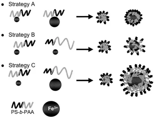
In this study, the concept of using tunable micelles to synthesize inorganic nanoparticle arrays for catalyzing carbon nanotube growth is extended to the synthesis of bimodal nanoparticle arrays. Several types of PS-b-PAA block copolymer with varying block lengths were put into solution with toluene to create micelles with different domain sizes. The relative amounts of metal species that were loaded into the micelles were also varied. Varying both the size of the PAA core of the micelles and their metal loading would provide several options for creating nanoparticles of dissimilar sizes. The combinations of PS-b-PAA block copolymer and metal loadings resulted in three strategies to obtain a bimodal distribution of nanoparticle diameters. In the first strategy, referred to as Strategy A in Figure 4, a single type of block copolymer with two different metal loadings was used. In the second strategy, referred to as Strategy B in Figure 4, two types of block copolymers with dissimilar molecular weights, containing one metal loading, was used. In the third strategy, referred to as Strategy C in Figure 4, two types of block copolymers with dissimilar molecular weights, containing two different metal loadings was utilized. Strategy A potentially yields the smallest size discrepancy between nanoparticles. Strategy C potentially yields the largest size discrepancy. Strategy B was not analyzed in this study, because Strategies A and C were thought to provide
greater insight into the maximum and minimum discrepancy possible using the metal loaded PS-b-PAA block copolymer micelle systems.
* Strategy A
* Strategy
B
* Strategy
C
0
PS-b-PAA
.4AýW
0
0
Figure 4 Strategies for synthesizing bimodal nanoparticles, using PS-b-PAA block copolymer micelles. In Strategy A a single block copolymer with two different metal loadings was used. In Strategy B in two block copolymers with dissimilar molecular weights, containing one metal loading was used. In Strategy C two block copolymers with dissimilar molecular weights, containing two different metal loadings was utilized. Strategy C potentially yields the largest size discrepancy between nanoparticles.
Nanoparticle arrays fabricated in this study were characterized using transmission electron microscopy (TEM) and small-angle X-ray scattering (SAXS) techniques. Techniques such as angle X-ray scattering (SAXS), and grazing incidence small-angle X-ray scattering (GISAXS) are powerful tools used to gain insight into the morphology of a large number of discrete features; as is the case with carbon nanotube
--*3LC
forests and nanoparticle arrays. In SAXS a high-energy (- 10's - 100's of eV) collimated
X-ray beam is aimed at a sample. Elastic bombardment of the X-rays gives information about the sample. The small-angle scattering (1-100 typically) provides nanometer scale structural information about the sample.12 In GISAXS, a high-energy beam of X-rays is
aimed at a sample, at a grazing angle a. Aiming the GISAXS beam in this fashion provides structural information about primarily two-dimensional samples on a surface, e.g. thin forests, nanoparticle arrays, spin cast polymers, etc.12 In this study SAXS was
used to determine the morphology of carbon nanotube forests synthesized from the bimodal nanoparticle arrays. GISAXS was used to determine the morphology and spacing of nanoparticles in arrays.
2. Experimental Details 2.1. Materials
Table 1 shows the PS-b-PAA block copolymers that were used to create micelles in solution with toluene, and the molecular weights of each block. The PS-b-PAA block copolymers were used as received from Polymer Source, Inc.
Table 1 The poly(styrene-block-acrylic acid) copolymers used and their respective block lengths are listed below. The molecular weights of each block are denoted in subscript, in units of g/mol. The metal loadings that were used with each block copolymer in solution are also listed. Metal loading is the metal ion equivalents per carboxylic acid group. Relative size refers to the expected size of each micelle, relative to one another.
PS-b-PAA Block Copolymers and Metal Loadings
Relative Sizes Block Copolymers Metal Loading FeC13required (g/mL)
Small PS11ooo-b-PAA1200 0.5 0.00055
Metal loadings are metal ion equivalents per carboxylic acid group. The metal salt used for metal loading of the poly(styrene-block-acrylic acid) micelles was anhydrous iron(III) chloride, FeC13. The anhydrous iron(III) chloride was used as received from Sigma-Aldrich Co. Toluene was obtained from an Innovative Technology Pure-Solv 400 Solvent Purification System.
2.2. Sample Preparation
Poly(styrene-block-acrylic acid) was measured and put into solution with toluene at a
concentration of 0.005 g/mL. Early samples indicated that 0.005 g/mL may have been too concentrated to provide a monolayer of micelles when spin cast. Consequently, a second set of poly(styrene-block-acrylic acid) and toluene solutions* were created with a
Previous work had indicated that the poly(styrene-block-acrylic acid) solution had to be heated and cooled in order to kinetically lock the block copolymers into the micelle phase.4
concentration of 0.002 g/mL. The poly(styrene-block-acrylic acid) copolymers with the lower PAA block lengths dissolved easily in the toluene solution. However the
poly(styrene-block-acrylic acid) copolymer with the largest PAA block length, PS2200
-b-PAA11 500, did not dissolve easily. In this case, the poly(styrene-block-acrylic acid) and
toluene solution was sonicated on low power for up to 1 and V2 hours.t
The amount of FeCl3 that is required for a particular concentration of
poly(styrene-block-acrylic acid) in toluene, and for a particular metal loading is calculated in the
following manner; given block copolymer PSn-b-PAAn, where n and m are the respective molar masses of each block length, the metal loading is calculated as:
n + m = total molar mass (g/mol)
m (2/mol) = % PAA
total molar mass (g/mol)
"Effective acrylic acid molar mass" = acrylic acid molar mass (72 g/mol) % PAA
Acrylic acid groups available = Solution concentration (0.5 wt%) "Effective acrylic acid molar mass"
Grams FeCI3 = Acrylic acid groups required x molar mass FeCI3 (162 g/mol)
per mL solution
necessary to obtain micelles. Further more it was apparent that the heat treatment may have created block copolymer films and debris that contaminated spin cast samples.
t Miniscule pieces of undissolved PS2200-b-PAA11500 was still visible, even after
sonication. Debris in the solutions was allowed to settle before using the solutions for spin casting. Clear solution above the debris layer that settled on the bottom was also transferred to new vials.
Grams FeCl3 = metal loading (0.5 or 5) x Grams FeCl3 per mL solution
per mL solution required for metal loading
Once the proper metal loading is added to the poly(styrene-block-acrylic acid) and toluene solution, the solution is gently shaken and allowed to equilibrate for 24 hours. Spin casting was performed at 8000 rpm for 1 minute. For single solution spin casts, the substrate surface is completely covered with metal-loaded poly(styrene-block-acrylic acid) micelle solution and the spin speed rapidly increased to 8000 RPM. Combinations of solutions were prepared by combining equal amounts of each solution in a vial and mixing for no longer than 5 seconds, before covering the substrate surface with the combined solutions and spin casting. Allowing the metal loaded PS-b-PAA block copolymer micelle solutions to mix longer than 5 seconds was found to result in homogenization of metal loadings within the micelles. This phenomena is discussed further in 3.7. Table 1 lists the combinations of poly(styrene-block-acrylic acid) solution and metal loadings that were spin cast to obtain bimodal nanoparticle arrays. Spin cast samples were oxygen plasma etched at 8-12 MHz for 10-15 minutes. Oxygen plasma etching removed the block copolymer thin film and oxidized the Fe3+ nanoparticles so
that an array of Fe203 nanoparticles remained on the sample surface.
2.3. Transmission Electron Microscopy
Nanoparticle arrays prepared for transmission electron microscopy (TEM) were spin cast on electron-transparent silicon nitride (Si3N4) TEM windows. The window
region of the silicon nitride was 100 nm thick. Silicon nitride TEM windows were purchased from SPI Supplies. Carbon nanotube samples were prepared by removing a piece of the carbon nanotube forest from the bulk sample, immersing in isopropyl alcohol, and sonicating on low power for less than 10 seconds to break up the forest. The dispersed carbon nanotubes in isopropyl alcohol was then dropped, with a pipette, onto holey carbon film coated copper grids. Holey carbon film coated copper grids were purchased from SPI Supplies. Transmission electron microscopy was performed on a JEOL 2011 at 200 kV.
2.4. Small-Angle X-ray Scattering and Grazing Incidence Small-Angle X-ray Scattering
Small-angle X-ray scattering (SAXS) and grazing-incidence small-angle X-ray scattering (GISAXS) experiments were performed at the G1 beamline at the Cornell High Energy Synchrotron Source (CHESS). The wavelength of the X-rays was 1.239A and silver behenate was used to calibrate the sample to detector distance with a first order scattering vector of q of 1.076nm' (with q = 4x sin0/X, where 20 is the
scattering angle and X is the wavelength). A slow-scan CCD-based X-ray detector, home built by Drs. M.W. Tate and S.M. Gruner of the Cornell University Physics Department, was used for data collection. Additional SAXS studies were performed at the X27 beamline at the National Synchrotron Light Source (NSLS) at Brookhaven National Laboratory (BNL), where the wavelength was 0.1371nm. Data was collected with a MarCCD X-ray detector.
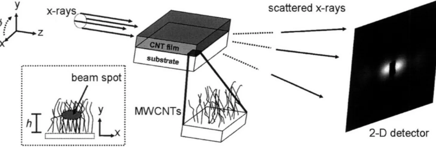
SAXS was used to characterize the morphology of carbon nanotube forests grown using the PS-b-PAA block copolymer templated nanoparticle arrays. SAXS gave
information about the alignment of the carbon nanotubes and about the modality of their diameter distribution. Figure 5 below shows a schematic of a typical SAXS experiment setup for carbon nanotube forests.
y
ei
c %.n+,nrnr, v-r ic
3\JLALL~I ~iI. iA-I LA,.
x-rays
Z
beam spotiY
h
j
L-LJ UMLCULUIFigure 5 Typical setup for SAXS of carbon nanotube tubes. A motorized stage allows for progressive vertical scans to be taken in the y-direction, at a height h from the substrate surface. (used with permission, Verploegen 2008)
GISAXS was used to characterize the morphology of the Fe20 3 nanoparticle
arrays templated using block copolymer micelles. GISAXS gave information about the inter-particle spacing and about the modality of the nanoparticle diameter distributions. Figure 6 below is a schematic of a typical GISAXS experiment setup for thin films. Thin films were not analyzed in this report, but the extension of GISAXS from thin film analysis to nanoparticle array analysis is trivial.
.. ... , ......
q
7-Thin
film
Jbstrate
Figure 6 A schematic of a typical GISAXS experiment for a thin film. A collimated X-ray beam is grazed off the sample at an angle, a. Scattering in the qy direction results from features in the plane of the sample surface. Scattering in the qx
direction results from features parallel to the sample surface. (used with permission, Verploegen 2008)
2.5. Growth of Carbon Nanotubes from Nanoparticle Arrays
Stacking and agglomeration of nanoparticles was found to be a problem for PS-b-PAA solutions of 0.5 wt%. Nanoparticle agglomerates are not desirable for carbon nanotube synthesis. The spin cast nanoparticle arrays used to synthesize carbon nanotube growth were from PS-b-PAA solutions that were diluted from 0.5 wt% to 0.2 wt%. After the PS-b-PAA solutions were diluted from 0.5 wt% to 0.2 wt% no
agglomeration was noticed in TEM. While dilution prevented nanoparticle agglomerates from forming the diluted solutions did not provide nanoparticle arrays with enough areal density to catalyze carbon nanotube growth.t These results are discussed further in 3.9. The samples were placed in a tube furnace that housed a slightly conductive p-doped Si substrate. Current was passed through the p-doped Si substrate so that the surface temperature of the sample placed on top of the substrate could be controlled using resistive heating. The resistive heater and sample are enclosed in a small gas flow chamber. The procedure used to synthesize carbon nanotubes using the nanoparticle arrays was the following:
* He gas was flowed at 400 sccm. The sample is heated from room temperature
to 775 oC for 10 minutes.
* He gas flow of 400 sccm is held for 9 minutes at 775 OC.
* He/H2 gases were flowed at 100/400 sccm, respectively, and held at 775 TC
for 1 minute.
* C2H4/He/H2 gases were flowed at 100/100/400 sccm, respectively, and held at
775 OC for 15 minutes.
2.6. Statistical Analysis of Nanoparticle Diameter Distributions
Statistical analysis of nanoparticle diameter distributions is utilized as a quantitative measurement of the overall distribution of nanoparticle sizes present in a nanoparticle array. Though the concept of using TEM images of particles to obtain a
: Recent work using 0.5 wt% metal loaded PS-b-PAA block copolymer micelle solutions has yielded nanoparticle arrays with a high enough areal density to catalyze significant carbon nanotube growth.
statistical measurement of the particles is not new, Woehrle et al. 2006 described how this process should be implemented using a public domain image processing software, ImageJ. ImageJ was used to perform particle counts on selected regions of TEM images of the PS-b-PAA templated Fe203 nanoparticles. Great care was taken
to ensure that the selected regions analyzed were representative of the whole sample. Large areas of nanoparticle arrays, on several samples, were surveyed to ensure that the selected image area was representative of that sample set.
To perform the particle count, the TEM image is opened in ImageJ. The TEM image scale bar is used to calibrate the pixel-per-length scale provided by ImageJ. A threshold is taken of a selected region, so that only the nanoparticles visible in the TEM image are highlighted for counting. Thresholding effectively sets a cutoff intensity for the features of the TEM image that will be counted. Particle counting is calibrated using three variables in ImageJ; particle circularity, 'exclude on edges', and 'include holes'. Circularity refers to how circular a particle is. Circularity is quantified by the value, circularity = 4nt(area/perimeter2), where area and perimeter are for the measured particle. Circularity ranges from 0 to 1, where 0 is an increasingly elongated particle, and 1 is a perfect circle. Intensity inhomogenieties over the surface of the TEM image, and the over the surface of the nanoparticle often causes thresholding to yield particles that have circularity > 0.25. Intensity inhomogenieties can also produce holes in the center of the particles when the threshold is taken. Therefore circularity of 0.25-1 was used. This circularity range was found to reliably exclude erroneous particle counts due to background noise. The 'include holes' and 'exclude on edges' options were chosen to optimize the particle
count. Once a particle count is tallied for the image, the particle diameters are calculated from the particle areas and plotted in a histogram with bin sizes equal to 1 nm. JMP 7 Statistical Discovery software was used to fit normal distribution curves to the single solution nanoparticle arrays, and for the combined solution nanoparticle arrays. Normal-quantile plots were obtained for each normal curve fit.
3. Results and Discussion
3.1. Significantly Different Mean Particle Diameters
The statistically significant difference between the mean particle diameters for the single systems of one block copolymer and one metal loading was determined using a Tukey-Kramer Honestly Significant Difference (HSD) test, with a p-value of > 0.05. Tukey-Kramer HSD test for statistically significant difference was chosen because it is a test typically used for data sets of different sizes. Figure 7 below shows the outcome of the Tukey-Kramer HSD test for the nanoparticle arrays synthesized from one block copolymer and one metal loading: Small, Medium, and Large. No data was available for XLarge nanoparticle arrays at the time this report was written.
19 18 17 16 15 14-13 12 E '$11 5 8 7 6 4-3 2 1.
Large ) Medium
0
Small oTukey-Kramer HSD
Figure 7 The left portion of the figure shows the distribution of nanoparticle diameters, and box-and-whisker plots indicating their mean values and range. The particle counts are; 49 for Small, 187 for Medium, and 151 for Large. The right portion of the figure shows the Tukey-Kramer Honestly Significant Difference (HSD) test of the mean nanoparticle diameters. The diameters of the circles represent the nanoparticle diameters that fall within the 95% confidence levels. Circles that intersect at less than or equal to 900 are considered to represent diameters with significantly different means. The Tukey-Kramer HSD shows that the Small, Medium, and Large nanoparticle have significantly different mean diameters. However, the Small and Medium nanoparticles have only borderline significantly different mean diameters. This is not surprising considering that the Small and Medium nanoparticles are synthesized using the same block copolymer, PS11ooo-b-PAA200oo, only with different metal loadings.
The Tukey-Kramer HSD test in Figure 7 represents the distribution of nanoparticle diameters for Large, Medium, and Small arrays, using circles. The particle counts are; 49 for Small, 187 for Medium, and 151 for Large. The
-i
O
Large
(E)
Medium
Small
~I.
j
diameters of the circles represent the distribution of nanoparticle diameters that falls within the 95 % confidence level range. Significantly different distributions are indicated by circles for which the outside angle of intersection is less than or
equal to 900. Circles that intersect at a 900 angle are considered to be borderline
significantly different. Using the Tukey-Kramer HSD test it is shown that the Large nanoparticles have a mean diameter that is significantly different from the Small and Medium mean nanoparticle diameters. The Small and Medium nanoparticles have mean diameters which are borderline significantly different. It is not surprising that the Small and Medium nanoparticles have only borderline significantly different mean diameters, because the Small and Medium nanoparticles were synthesized from the same block copolymer, PS 10
ooo-b-PAA1200, only with different metal loadings.
That fact that the nanoparticles synthesized from the single block copolymer and single metal loadings systems have mean diameters that are significantly different infers that the mean diameter values calculated for the nanoparticle arrays can be considered distinct from one another. Furthermore, knowing that the single block copolymer and metal loading systems yield nanoparticles with significantly different mean diameters leads to the hypothesis that these single block copolymer and metal loading systems may be combined to produce nanoparticle arrays with diameter distributions that reflect the significantly different mean diameters of their constituents; in other words bimodal nanoparticle arrays.
3.2. Single PS-b-PAA Block Copolymer and Metal Loading Systems 3.2.1. Small
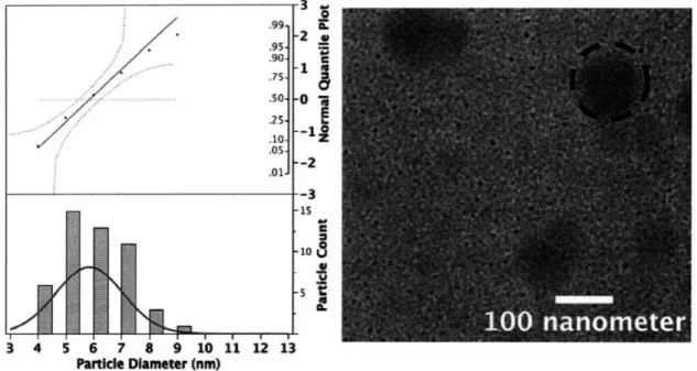
Figure 8 below shows the diameter distribution calculated for the Small (PSulooo-b-PAA1200 Metal Loading = 0.5) nanoparticles. The total number of particles analyzed was 49. The micelle solution was 0.5 wt%, and had equilibrated for 1 week before this sample was spin cast. A red normal curve was fitted to the distribution of nanoparticle diameters. A normal-quantile plot was produced with the distribution of nanoparticle diameters. As seen in Figure 8, the distribution of nanoparticle diameters falls within the 95% confidence bounds, in red. The distribution of nanoparticle diameters in the normal-quantile plot also fits reasonably well to the straight, red normal-quantile line. These characteristics of the distribution of Small nanoparticle diameters indicate that it is normally distributed. Therefore, a mean nanoparticle diameter can be calculated for the Small nanoparticles. The mean Small nanoparticle diameter is 5.8 nm with a standard deviation of 1.2 nm.
Small
10. . . * aer
3 4 5 6 7 8 9 10 11 12 13
Particle Diameter (nm)
Figure 8 Distribution of particle diameters for Small (PS1 100 0o-b-PAA1200 Metal Loading = 0.5). The total particle count is 49. The normal-quantile plot above the nanoparticle diameter distribution indicates that the diameters are normally distributed. A normal distribution curve was fitted to the Small nanoparticle distribution to give a mean nanoparticle diameter of 5.8 nm with a standard deviation of 1.2 nm. The inset is a representative TEM image of the Small nanoparticle array. The dark blotches, such as the one circled with the dashed line, appear in several TEM images. They did not significantly affect the morphology of the nanoparticles or the particle diameter analysis. The blotches were determined to be due to debris from undissolved polymer in solution or debris ejected onto the
backside of the TEM window during the spin casting process.
3.2.2. Medium
Figure 9 below shows the diameter distribution calculated for the Medium (PS1 10oo-b-PAA1 200 Metal Loading = 5) nanoparticles. The total number of
particles analyzed was 187. The block copolymer solution concentration was 0.5
wt%, and had equilibrated for 1 week before this sample was spin cast. A red normal curve was fitted to the distribution of nanoparticle diameters. A
normal-quantile plot was produced with the distribution of nanoparticle diameters. As seen in Figure 9, the distribution of nanoparticle diameters falls within the 95%
confidence bounds, in red. The distribution of nanoparticle diameters in the normal-quantile plot also fits reasonably well to the straight, red normal-quantile
line. These characteristics of the distribution of Medium nanoparticle diameters indicate that it is normally distributed. Therefore, a mean nanoparticle diameter can be calculated for the Medium nanoparticles. The mean Medium nanoparticle diameter is 6.80 nm with a standard deviation of 2.20 nm.
Medium
3 4 5 6 7 8 9 10 11 12 13
Partidcle Diameter (nm)
Figure 9 Distribution of particle diameters for Medium (PS1 00oo-b-PAA1 200 Metal
Loading = 5). The total particle count is 187 particles. The normal-quantile plot above the nanoparticle diameter distribution indicates that the diameters are normally distributed. A normal distribution curve was fitted to the Medium nanoparticle distribution to give a mean nanoparticle diameter of 6.8 nm with a standard deviation of 2.2 nm. The inset is a representative TEM image of the Medium nanoparticle array.
3.2.3. Large
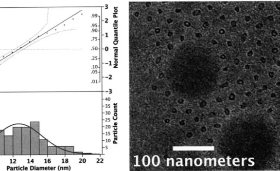
Figure 10 below shows the distribution of particle diameters calculated for the Large (PS16500-b-PAA4500 Metal Loading = 5) nanoparticle array. The particle
count was 150. A red normal curve was fitted to the distribution of Large nanoparticle diameters. A normal-quantile plot was produced with the distribution of nanoparticle diameters. As seen in Figure 10.
Figure 10 the distribution of nanoparticle diameters falls within the 95% confidence bounds, in red. The distribution of nanoparticle diameters in the normal-quantile plot also fits reasonably well to the straight, red normal-quantile
line. These characteristics of the distribution of Large nanoparticle diameters indicate that it is normally distributed. Therefore, a mean nanoparticle diameter can be calculated for the Large nanoparticles. The mean Large nanoparticle diameter is 12.09 nm with a standard deviation of 2.7 nm. The inset TEM in
Figure 10 suggests that the wide range is due to irregularly shaped nanoparticles. In most cases that were observed via TEM the Large solution of nanoparticles was susceptible to stacking through multilayering. The stacked nanoparticles would form irregularly shaped agglomerates after plasma etching. Though that is not considered to be the cause of the irregularly shaped nanoparticles in this particular analysis, because the nanoparticles appear to be regularly spaced and discrete. Attempts were made to decrease the stacking effect by decreasing the concentration of the PS-b-PAA block copolymer solution from 0.5 wt% to 0.2 wt%. The Large micelle solution yielded the most hexagonally close-packed array of nanoparticles.
Large
.95- .90- .75- .50- .25- .10- 05;- .01-8 -2 . C -1 E z -0 *i6 --12 --3 -40 -35 C -30= -25 -20 -10~ -15 -l 10 12 14 16 18 20 22 Particle Diameter (nm)Figure 10 Distribution of particle diameters for Large (PS1600oo-b-PAA 4500 Metal
Loading = 5). The total particle count is 150. The normal-quantile plot above the nanoparticle diameter distribution indicates that the diameters are normally distributed. A normal distribution curve was fitted to the Large nanoparticle distributions to give a mean nanoparticle diameter of 12.07 nm with a standard deviation of 2.70 nm. The inset is a representative TEM image of the Large nanoparticle array. The hollow appearance of the nanoparticles was determined to be caused by focusing effects in the TEM, and do not reflect a true morphological
characteristic of the nanoparticles. Similar conclusions about nanoparticles that appear hollow were reached by Bennett 2007. The spread of the nanoparticle diameter distribution can be attributed to the irregularly shaped Large nanoparticles, as seen in the inset TEM image. In this image it can be seen that the dark blotch due to undissolved polymer debris has caused the nanoparticle array to form around it. While this debris does affect the nanoparticle array, it does not affect the particle diameter analysis or the general modality of the nanoparticle diameters. r .,o CI I
.01-I 8 pl-! I
3.3. Summary of nanoparticle diameters available using single systems of one PS-b-PAA block copolymer and one metal loading
Mean nanoparticle diameters and standard deviations were calculated for the nanoparticles synthesized from single PS-b-PAA block copolymers and metal loadings: Small, Medium, and Large. These values are shown below in Table 2.
Table 2 A summary of the mean nanoparticle diameters available, and the standard deviations for the Small, Medium, and Large PS-b-PAA block copolymer and metal loading systems is shown below. No data was available for XLarge at the time that this report was written. The XLarge category is included for completeness.
Single Block Copolymer
-
Metal Loading Systems
Mean Mean
Block Metal
Relative Size Diameter Diameter Std Particle Count
Copolymer Loading nm) T•v ("nmn
Medium PSu1ooo-b- 5.00 6.80 2.20 187
XLarge PS22o-b- 5.00 unavailable unavailable unavailable
PAA1150o
3.4. Combinations of Two Block Copolymer and Metal Loading Systems
A priori, it is expected that the number of 'big' nanoparticles, in the array produced from combined solutions, would be much less than the number of 'small' nanoparticles, because the PS-b-PAA micelle and metal loading solutions were prepared with respect to a weight percentage concentration of PS-b-PAA block copolymer in toluene. Statistically this weight-averaged concentration does not reflect the number of micelles in solution. Therefore, combining equal volumes of a 'big'
micelle solution and a 'small' micelle solution results in a solution with a number average of micelle sizes that is skewed to a large number of 'small' micelles. This 'small' micelle rich solution produces nanoparticle arrays that reflect that skew. The particle diameter histograms presented in this section exhibit the expected skew to 'small' micelles .
As discussed previously in 3.1, because the Small, Medium, and Large nanoparticle arrays are shown to be normally-distributed and their respective means are significantly different, it can be expected that a combination of the Small and Medium, or Small and Large, and by extension the Small and XLarge block copolymer and metal loading systems will yield nanoparticle diameter distributions that are bimodal. Taken alone, a non-normal nanoparticle distribution is not concrete evidence of bimodality of the physical nanoparticles. However, the nanoparticle distributions represent the physical sizes of the nanoparticles as imaged with TEM. Consulting TEM images of the nanoparticle arrays in conjunction with the nanoparticle diameter distributions lends significant confidence to the conclusions that may be reached using both analyses.
To aid in identifying the single modes of nanoparticle diameter distributions that constitute the nanoparticle diameter distributions, each distribution for Small + Medium, Small + Large, and Small + XLarge have been decomposed. The non-normal nanoparticle distributions have been decomposed into data sets that are normally distributed. The decomposed normally distributed nanoparticle diameter
§ Attempts were made to produce nanoparticle arrays that had a greater number of 'big' micelles with respect to the 'small' micelles. However these results have not yet shown significant improvement over the particle diameter distributions shown herein.
distributions have mean diameters and standard deviations that represent the unimodal diameter distributions of the constituent single Small, Medium, Large, or XLarge nanoparticles.
3.4.1. Small + Medium
Figure 11 below shows the distribution of particle diameters calculated for the combined Small (PS110oo-b-PAA1 2oo Metal Loading = 0.5) and Medium
(PS1100o-b-PAA1200 Metal Loading = 5) nanoparticle systems. The total particle
count is 95. The solutions were 0.5 wt% and had equilibrated for 1 week before spin casting. A normal-quantile plot was produced with the distribution of Small
+ Medium nanoparticle diameters. As seen in Figure 11 the distribution of nanoparticle diameters does fall within the 95% confidence bounds, in red. However, the distribution of nanoparticle diameters in the normal-quantile plot does not fit well to the straight, red normal-quantile line .These characteristics of the distribution of Small + Medium nanoparticle diameters indicate that it is not normally distributed. Therefore, a mean nanoparticle diameter cannot be
Small + Medium
3 4 5 6 7 8 9 10 12 14 16 18 Particle Diameter (nm)
Figure 11 Distribution of particle diameters for a combined solution of Small and Medium solutions (PSllooo-b-PAA1200 Metal Loading = 0.5 + PSlo0 00-b-PAA1200 Metal
Loading = 5). The normal-quantile plot above the nanoparticle diameter distribution
indicates that the diameters are not normally distributed. The inset is a representative TEM image of the Small + Medium nanoparticle array.
The nanoparticle diameter distribution for the Small + Medium nanoparticles has been decomposed into unimodal constituents that represent the single Small and Medium nanoparticles. The decomposed diameter distributions are shown below in Figure 12. The decomposed unimodal diameter distributions represent true constituent distributions of the combined bimodal Small + Medium distribution. The normal-quantile plots for each decomposed unimodal diameter distribution shows that the distribution is normally distributed. The mean and standard deviations for the decomposed Small and Medium distributions are 5.19 nm + 0.89 nm and 9.8 nm + 1.06 nm, respectively. The standard deviations of the decomposed Small and Medium distributions overlap with the standard
deviations of the single Small and Medium nanoparticle diameter distributions calculated in 3.2. The fact that the standard deviations for the decomposed Small distributions overlap, and the standard deviations for the decomposed Medium distributions overlap indicates that the decomposed Small and Medium distributions reflect the unimodal Small and Medium constituents.
Small + Medium
Particle Diameter (nm)
3 4 5 6
Particle Diameter (nm)
Particle Diameter (nm)
Figure 12 The unimodal nanoparticle diameter distributions that constitute the non-normal Small + Medium nanoparticle diameter distribution are shown extracted to
the right. The unimodal nanoparticle diameter distributions are normally
distributed with means and standard deviations of 5.19 nm h 0.89 nm for the top distribution and 9.8 nm * 1.06 nm for the bottom distribution. The unimodal
nanoparticle diameter distributions represent the Small and Medium constituents of the Small + Medium nanoparticle diameter distribution because the standard deviations for the Small distributions overlap and the standard deviations for the Medium distributions overlap.
.99 .95 .. JS i" .011 1 -r
3.4.2. Small + Large
Figure 13 below shows the distribution of particle diameters calculated for the combined Small (PS110ooo-b-PAA1200 Metal Loading = 0.5) and Large (PS16500-b-PAA4500 Metal Loading = 5) nanoparticle systems. The total
particle count is 97. The solutions were 0.5 wt% and had equilibrated for 4 months before spin casting. A normal-quantile plot was produced with the distribution of nanoparticle diameters. As seen in Figure 13 the distribution of nanoparticle diameters does not fall completely within the 95%
confidence bounds, in red. The distribution of nanoparticle diameters in the normal-quantile plot does not fit well to the straight, red normal-quantile line. These characteristics of the distribution of Small + Large nanoparticle diameters indicate that it is not normally distributed. Therefore, a mean nanoparticle diameter cannot be calculated for the Small + Large nanoparticles.
Small + Large
I l I I I I I I I I I I
3 4 5 6 7 8 9 10 12 14 16
Particle Diameter (nm)
Figure 13 Distribution of particle diameters for a combined solution of Small and Large solutions (PS11ooo-b-PAA 1200 Metal Loading = 0.5 + PS165oo-b-PAA4500 Metal
Loading = 5). The total particle count is 97 particles. The normal-quantile plot
above the nanoparticle diameter distribution indicates that the diameters are not normally distributed. The inset is a representative TEM image of the Small + Large nanoparticle array.
The nanoparticle diameter distribution for the Small + Large nanoparticles has been decomposed into unimodal constituents that represent the single Small and Large nanoparticles. The decomposed diameter distributions are shown below in Figure 14. The decomposed unimodal diameter distributions represent the constituent distributions of the combined bimodal Small + Large distribution. The normal-quantile plots for each decomposed unimodal diameter distribution shows that the distribution is normally distributed. The mean and standard deviations for the decomposed Small and Large distributions are 5.50 nm -+ 1.40 nm and 11.70 nm _ 1.80 nm, respectively. The standard


