HAL Id: hal-03156785
https://hal.archives-ouvertes.fr/hal-03156785
Preprint submitted on 2 Mar 2021
HAL is a multi-disciplinary open access
archive for the deposit and dissemination of sci-entific research documents, whether they are pub-lished or not. The documents may come from teaching and research institutions in France or abroad, or from public or private research centers.
L’archive ouverte pluridisciplinaire HAL, est destinée au dépôt et à la diffusion de documents scientifiques de niveau recherche, publiés ou non, émanant des établissements d’enseignement et de recherche français ou étrangers, des laboratoires publics ou privés.
interactions in mice.
Emmanuel Bourinet, Miquel Martin, Damien Huzard, Freddy Jeanneteau,
Pierre-François Méry, Amaury François
To cite this version:
Emmanuel Bourinet, Miquel Martin, Damien Huzard, Freddy Jeanneteau, Pierre-François Méry, et al.. The impact of C-Tactile Low threshold mechanoreceptors on affective touch and social interactions in mice.. 2021. �hal-03156785�
1
The impact of C-Tactile Low threshold mechanoreceptors on affective touch and social
1
interactions in mice.
2 3
Authors: Emmanuel Bourinet1, Miquel Martin1,2, Damien Huzard1, Freddy Jeanneteau1,
4
Pierre-Francois Mery1, Amaury François1,3,
5
1
IGF, Université de Montpellier, CNRS, INSERM, Montpellier, France
6
Institut de Génomique Fonctionnelle, 141 rue de la Cardonille 34090 Cedex 5 Montpellier,
7France,
82
present address: Laboratory of Neuropharmacology-Neurophar, Universitat Pompeu Fabra
9
(UPF), Barcelona, Spain.
103
Lead contact. Correspondence:
amaury.francois@igf.cnrs.fr
11 12
Abstract
13
Affective touch is necessary for proper neurodevelopment and sociability. However, it
14
is still unclear how the neurons innervating the skin detect affective and social
15
behaviours. To clarify this matter, we targeted a specific population of somatosensory
16
neurons in mice, named C-low threshold mechanoreceptors (C-LTMRs), that appears
17
particularly well suited physiologically and anatomically to perceive affective and social
18
touch but whose contribution to these processes has not yet been resolved. Our
19
observations revealed that C-LTMRs functional deficiency from birth induced social
20
isolation and reduced tactile interactions in adults. Conversely, transient increase in
C-21
LTMRs excitability in adults using chemogenetics was rewarding, temporally promoted
22
touch seeking behaviours and thus had pro-social effects on group dynamics. This
23
work provides the first empirical evidence that specific peripheral inputs alone can drive
24
complex social behaviour, demonstrating the existence of a specialised neuronal circuit
25
originating from the skin wired to promote interaction with other individuals.
26 27
Introduction
28
The rewarding value that emerges from touch is essential for decision making and motivation, 29
especially in social animals, and dysregulation of this process leads to debilitating psychiatric 30
or neurologic conditions, including autism, anxiety or depression 1–3. Nonetheless, the neural 31
mechanisms underlying emotional tactile sensory processing relationship with social behaviour 32
are at the early stage of their understanding. 33
2 The skin is innervated by an array of functionally distinct populations of receptors contributing 34
to touch, which can be distinguished by their response properties, activation threshold, 35
conduction velocity, and the type of end organ that they innervate 4.
36
One class of these receptors, the C-Tactile fibers (CT), are particularly well responsive to tactile 37
stimulation categorized as “pleasant” and “affective”, by human subjects 5,6. These neurons
38
are unmyelinated low-threshold mechanoreceptors (LTMRs) and respond to non-noxious 39
touch with a predilection for slow-moving and low-force, stroking stimuli, such as gentle 40
brushing 6–8. Activation of CTs in humans provides poor conscious spatial and qualitative 41
information to the subjects, who nonetheless still carry a positive feeling related to the gentle 42
brushing of the skin 9. Furthermore, the positively valent tactile information conveyed by CTs
43
make them particularly well suited to link tactile information to social bonding 10,11.
44
Evidence for pleasant touch contribution to sociability and related disorders was also found in 45
laboratory animal models. For example, playful and pleasant touch is rewarding and 46
contributes greatly to social development in adolescent rats 12. In mice, two recent studies
47
linked the disruption of LTMR functions with alterations of social behaviour typical of autism 48
spectrum disorder (ASD) 13,14. Specifically, these studies used genetically modified mice
49
recapitulating mutations found in human patients within the Mecp2, Shank3B, and Fmr1 genes. 50
Peripherally restricted conditional Knock-Out (KO) of these genes greatly altered Low-51
threshold-mechanoreceptors (LTMRs) function which induced multiple phenotypes associated 52
with ASD, especially social deficits that are usually observed in constitutive KOs of these 53
genes. However, LTMR sensory neurons are known to be a highly heterogeneous population, 54
leaving open the question regarding the contribution of CT and pleasant touch to these social 55
deficits. 56
We and others genetically identified a population of primary sensory neurons in mice, named 57
C-Low-threshold-mechanoreceptors (C-LTMRs), with similar functional properties than CTs. 58
These studies characterized genes specific to C-LTMRs in rodents such as TAFA4, but also a 59
set of genes allowing to differentiate C-LTMRs from other sensory neurons, such as tyrosine 60
hydroxylase (TH), VGlut3, TAFA4 or Cav3.2, that may be used to gain genetic access to these
61
neurons 15–20. These studies unveiled the contribution of C-LTMRs, and these genes, to pain 62
chronification in the context of neuropathic or inflammatory pathological pain. However, none 63
of these studies considered the role of C-LTMRs in touch sensation, within naturalistic 64
conditions and especially their role in social behaviours. 65
In the present study, we explored the role of C-LTMRs in social interactions. Using two mouse 66
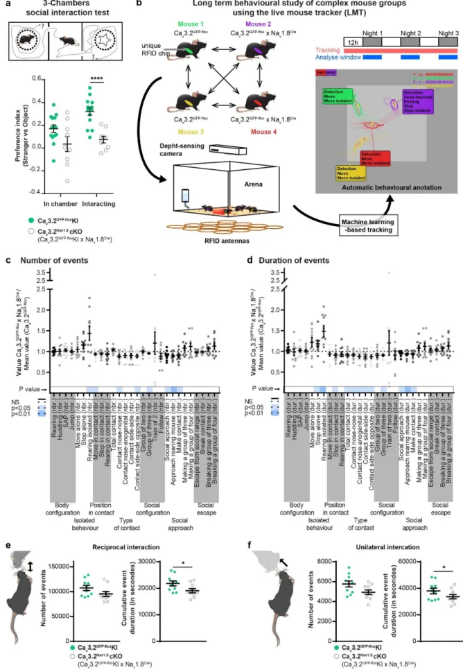
transgenic models to decrease or facilitate C-LTMRs excitability, and a new tracking system 67
3 to automatically annotate social behaviour in groups of 4 animals, we clarified the specific 68
function of C-LTMRs in rodent inter-individual relations. 69
Results
70
Social behaviours are impaired in Cav3.2Nav1.8cKO
71
First, we aimed at investigating the consequence of C-LTMRs hypofunction on social 72
behaviour. For that purpose, we used a genetic mouse model where the expression of the low 73
threshold calcium channel Cav3.2 is conditionally knocked out in C-LTMRs, by crossing Cav3.2
74
GFP-floxKI with Na
v1.8cre mice as we previously described 16. In this mouse model
75
(Cav3.2Nav1.8cKO), C-LTMR impaired function starts just before birth. Indeed, removing Ca v3.2
76
expression from C-LTMRs increases the firing threshold of action potentials, reduces the firing 77
frequency and lowers the conduction velocity of these fibres transforming their mechanical 78
sensitivity into High Threshold Mechanoreceptors (HTMR) phenotype 16.
79
To evaluate the consequences of C-LTMRs deficiency on social preference behaviour, we 80
used the three-chamber paradigm (ie. Crawley test). Interestingly, when compared to control 81
Cav3.2GFP-floxKI littermates, male Cav3.2Nav1.8cKO mice spent less time interacting with an
82
unfamiliar mouse than with an unanimated object, which is summarized by the reduction in the 83
preference index (Figure 1a, Supplementary Fig. 1a). To further investigate the precise 84
quality of the social and tactile interactions that appears to be impaired in this model we used 85
a novel paradigm where mice could interact freely with each other. The live mouse tracker 86
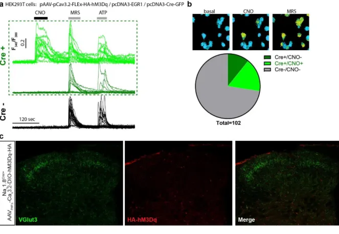
(LMT), based on a machine learning analysis framework, was designed by de Chaumont and 87
colleagues for that specific purpose 21. This system allows the tracking and automatic
88
annotation of mice behaviours and social interactions in their environment for multiple days 21.
89
Using this system, we analysed the behaviour of 5 groups of 4 male mice (each group 90
composed of 2 controls Cav3.2GFP-floxKI, and 2 C-LTMRs-impaired Cav3.2Nav1.8cKO, all
91
littermates) for three consecutive nights (Figure 1b). For every time frame, the LMT detects 92
head, tail, ears, eyes and nose position, producing a geometrical mask for each mouse. These 93
data permit the computation of different behavioural events based on mice geometries 94
movements and localization compared to other mice. Overall, we detected 28 events that were 95
separated into seven categories: body configuration, isolated behaviour, position in contact, 96
type of contact, social configuration, social approach and social escape. These events were 97
analysed separately for both their number (Figure 1c) and their duration (Figure 1d) for each 98
individual. To compare C-LTMR-impaired mice and their control cagemates with the same 99
baseline and to reduce inter-experiment variability, the value of each behavioural trait for one 100
Cav3.2Nav1.8cKO mouse was compared with the mean level of this trait from the two control
101
cagemates (called LMT index thereafter). An LMT index value above one indicates that the 102
4 Cav3.2Nav1.8cKO mice perform more occurrences of a specific trait compared to controls
103
whereas a value below one means that the mutant mice do less of that trait than controls. 104
The LMT-index indicates that Cav3.2Nav1.8cKO mice spent more time isolated than controls
105
(time stop alone: +18.2%±7), Figure 1d), without any noticeable differences in locomotor 106
activity (Supplementary Fig. 1b) or exploratory behaviour (stretch attending posture: (SAP) 107
Figure 1e&d). In social events, C-LTMR impaired mice showed a small, but statistically
108
significant, decrease in time spent engaged in all type of contacts (-15.7%±3.2 in average per 109
interaction bout, Supplementary Fig. 1c). Moreover, the duration of reciprocal (here 110
associated with nose-to-nose contacts and side-by-side contacts) and unilateral social 111
interactions (associated in these experiments with giving ano-genital contacts) were reduced, 112
even if the number of events is not statistically different (Figure 1e&1f). However passive 113
social interaction (associated in our experiment with receiving ano-genital contacts) were not 114
altered (Supplementary Fig. 1d). Cav3.2Nav1.8cKO mice also displayed shorter social
115
approaches leading to a contact (social approach: -10.9%±1.9 and make contact: -7.3%±2.8, 116
Figure 1d) and spent less time in groups of three mice (-9.27%±1.9) (Figure 1d). However,
117
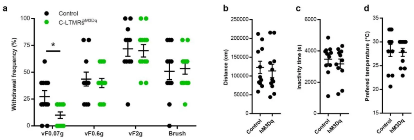
no difference was observed in the social escape behaviours category. Taking together, the 118
results from the 3-chamber test and the LMT revealed a deficit in sociability of Cav3.2Nav1.8cKO
119
mice compared to their control littermates. 120
A new viral strategy to specifically target C-LTMRs in mice
121
Next, we deepened our investigations on C-LTMR role in sociability by designing a 122
chemogenetic strategy to selectively excite C-LTMRs remotely in adult mice independently of 123
Cav3.2 protein function and, importantly, without any postnatal functional perturbation of
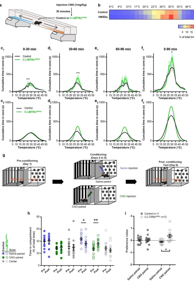
C-124
LTMR. Our strategy to target C-LTMRs in adult mice, illustrated in Figure 2a and b, consists 125
on expressing a gene of interest under the control of the mini-Cav3.2 promoter in a
Cre-126
dependent manner. Indeed, C-LTMRs can be defined by the expression of both the sodium 127
channel Nav1.8 and the calcium channel Cav3.2 16,22. We previously engineered adeno
128
associated viruses (AAV) with a mini-Cav3.2 sequence and validated its faithful expression in 129
Cav3.2 positive neurons within the dorsal horn of the spinal cord 23. Here we used
AAV-PHP-130
S serotype that has a high tropism for peripheral sensory neurons 24, which we delivered into
131
Nav1.8Cre heterozygote mice. This intersectional strategy combining the expression pattern of
132
Cav3.2 and Nav1.8 aims at restricting the expression of the viral payload into C-LTMRs. To 133
achieve C-LTMRs chemogenetic stimulation with this strategy, we inserted within the pAAV 134
vector the HA tagged-hM3Dq excitatory DREADD cassette. 135
HEK293T cells were transfected with the pAAV vector (pAAV-pCav3.2-FLEx-HA-hM3Dq) to 136
validate its Cre-dependency and functionality. As the Cav3.2 promoter activity is highly 137
5 enhanced by the EGR1 transcription factor 25, we co-expressed the murine EGR1 cDNA and
138
added or not the Cre recombinase (Cre-GFP fusion). We analysed the DREADD functionality 139
using Calcium fluorimetry in Fura2 loaded Cre-GFP positive cells. In a representative field, out 140
of 102 cells recorded, 28 were GFP positive and of those 17 responded to the hM3Dq 141
pharmacological actuator Clozapine-N-oxyde (CNO) (Supplementary Fig. 2a & b). No 142
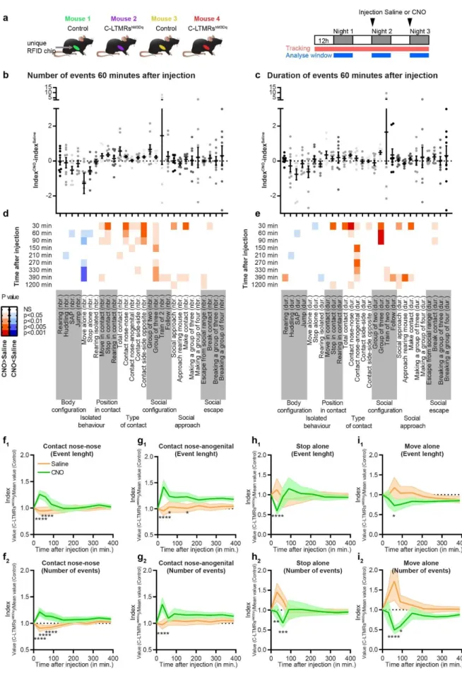
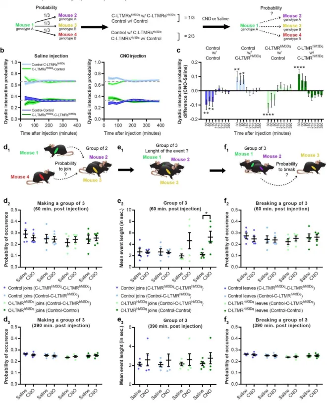
responses to CNO were observed in non-GFP cells. 143
We then packaged viral particles with this validated construct using the AAV-PHP-S capsid. 144
Although AAV PHP-S can be delivered systematically to access sensory neurons, we locally 145
injected the virus intrathecally with minimal invasiveness and a sole access to DRGs and no 146
other sensory neurons such as those of the vagal ganglia (Figure 2b)24. Consistent with an
147
efficient C-LTMR targeting strategy, intrathecal injection of this viral construct leads to the 148
expression of the excitatory DREADD receptor HA-hM3Dq in C-LTMRs labelled by the tyrosine 149
hydroxylase (TH), 8 weeks post injection (Figure 2c). Overall, 71.2%±2.4 of HA-hM3Dq cells 150
were also positive to TH (N=5 mice). Inversely, HA-hM3Dq is present in 25.9%±2.6 of TH 151
positive dorsal root ganglia (DRG, T7 to L6) neurons (Figure 2c). In addition, no observation 152
was made of any specific HA immunostaining in the dorsal horn of the spinal cord 153
(Supplementary Fig. 2c) 154
To further confirm that hM3Dq was selectively expressed in C-LTMRs accordingly to the 155
defined strategy and kept its pro-excitatory nature, we evaluated the effect of CNO on cultured 156
DRG neurons from animal injected intrathecally with the rAAVPHPs-pCav3.2-FLEx-HA-hM3Dq
157
cells were loaded with the Fura2 radiometric calcium indicator, and labelled with red-dye 158
conjugated IB4 to access the pharmacological and functional properties of large population of 159
neurons at the same time. Application of CNO (30µM) induced intracellular calcium increase 160
in neurons also responding to the TRPA1 agonist, allyl isothiocyanate (AITC, 200µM), and to 161
the P2Y1R agonist, MRS2365 (200nM) (Figure2d and 2e). Of all CNO responsive neurons, 162
the large majority responded to AITC and MRS (15), 3 also responded to Capsaicin on top of 163
AITC and MRS, and only one was not responding to anything else. Among the MRS 164
responders, 56.2% were also responding to CNO while among AITC responders only 26.4% 165
were CNO responders. In mouse DRGs, TRPA1 is weakly expressed in C-LTMRs and P2YR1 166
receptor is only expressed in C-LTMRs and TrkB positive A-LTMRs 18,20. As TRPA1 is not
167
express in TrkB neurons, responses to both MRS and AITC can only be observed in C-LTMRs. 168
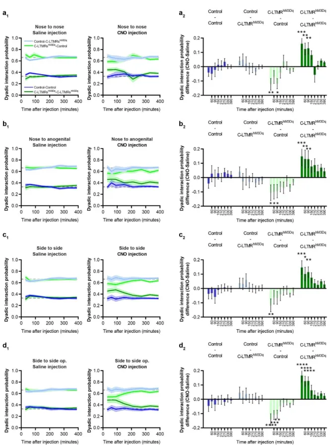
Moreover, none of the CNO responsive neurons were labelled by IB4 (Figure 2f), in agreement 169
with the lack of reactivity of the lectin in mouse TH positive C-LTMR neurons 26. Taken together,
170
our morphological and functional data provide supporting evidence toward a specific 171
expression of HA-hM3Dq in a large population of C-LTMRs (10.7% of all DRG neurons 172
6 recorded, N= 3 mice). Accordingly, we used this experimental approach in vivo to investigate 173
the impact of C-LTMRs stimulation in social behaviours. 174
Effect of exogenous C-LTMR activation on somatosensory perception
175
First, we assessed the consequences of C-LTMRs exogenous activation on somatosensory 176
perception. Eight weeks after intrathecal injections of rAAVPHPs-pCav3.2-FLEx-HA-hM3Dq
177
(named further: C-LMTRshM3Dq) or rAAV
PHPs-CAG-mCherry (Control) in Nav1.8cre mice, CNO
178
was administrated intraperitoneally (1mg/kg, IP) to all animals. 30 to 45 minutes after, a 179
decrease in reflex paw withdrawal frequency to low force von Frey stimulation (0.07g) was 180
observed, but not for higher forces (0.6g and 2g) or brushing in C-LMTRshM3Dq mice when
181
compared to control mice (Supplementary Fig. 3a). To note, CNO injection did not trigger any 182
alteration of motor activity or spontaneous nocifensive behaviours, such as paw shaking, 183
guarding, grooming, licking or jumping compared to the control group. 184
As C-LTMRs have been implicated into temperature perception 15,16,27, we probed thermal
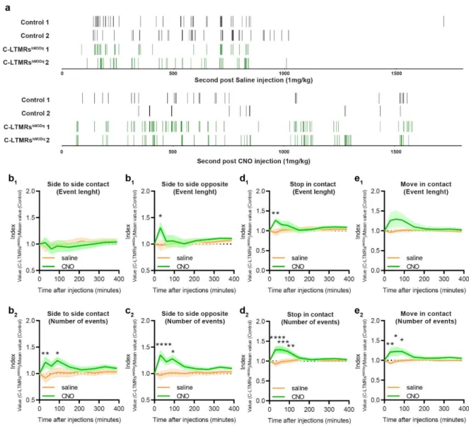
185
sensitivity of control and C-LMTRshM3Dq mice in the thermal gradient test. 30 minutes after CNO
186
injection, mice were placed into a 1.5m corridor with the floor at one extremity cooled down to 187
5°C and the other heated up to 50°C, creating a gradient of temperature between the two 188
extremities. Once placed in the corridor, mice were allowed to explore the thermal gradient to 189
reveal their thermotaxis behaviour. The exploration was tracked during 90 minutes and the 190
animal’s position was annotated according to the temperature zones they visited (Figure 3a). 191
When compared to control animals, CNO treated C-LMTRshM3Dq mice settled more quickly at
192
the comfort temperature of 30°C (Figure 3c and 3d) and spend more time overall at this 193
temperature (Figure 3b and 3f), without affecting locomotor (Supplementary Fig. 3b and 3c) 194
nor the preferred temperature that was similar between the two groups (Supplementary Fig. 195
3d). This interesting result suggests that exogenous activation of C-LTMRs can reinforce
196
motivational behaviour towards a pleasurable somatosensory stimulation. 197
Positive valent information is associated with C-LTMRs stimulation
198
As activation of LTMRs may results in positive feeling, next we investigated whether C-199
LTMRs activation could be rewarding on its own by using the conditioned place preference 200
paradigm (CPP). Following one day of habituation to the CPP arena, control and C-LMTRshM3Dq
201
mice were conditioned to receive saline injection (CNO vehicle) in the compartment they 202
preferred during habituation and CNO (1mg/kg in saline) in the other compartment, for 3 203
consecutive days (Figure 3g). On the last day, mice were free to explore the entire arena and 204
their position was video tracked. While control animals did not develop a preference for the 205
side in which they either received saline or CNO injections, C-LMTRshM3Dq mice showed a
206
marked preference for the compartment associated with CNO injections (32%±1.4 increase, 207
7
Figure 3h and 3i). Overall, these two experiments suggest that activation of C-LTMRs is
208
rewarding and can increase the rewarding value of other sensory modalities. 209
C-LTMR stimulation induces touch seeking and pro-social behaviours
210
Taking into consideration the intrinsic emotional value conveyed by C-LTMRs activation, we 211
finally investigated whether C-LTMR activation can affect social behaviours and social group 212
organization. The LMT system was again used to analyse the behaviour of 5 groups of 4 mice 213
independently during 3 nights. Each group was composed of 2 Nav1.8cre mice injected with
214
rAAVPHPs-CAG-mCherry (Control) and 2 mice injected with rAAVPHPs-pCav
3.2-FLEx-HA-215
hM3Dq (C-LTMRshM3Dq), all male littermates. The first 24 hours were used as a habituation
216
phase, and just before the second dark cycle, we injected either a saline (CNO vehicle) or 217
CNO solution (IP, 1mg/kg in saline) to all animals (Control and C-LTMRshM3Dq). At the
218
beginning of the third dark cycle, groups that received a saline solution for the second dark 219
cycle received CNO and vice versa (Figure 4a). As for Cav3.2Nav1.8cKO mice, we calculated
220
the LMT-index by taking the value of each trait for one C-LMTRshM3Dq mouse and compared it
221
with the mean level of that trait for the two control cagemates (Figure 4 f to i). The LMT-index 222
was calculated from cumulated value for both saline and CNO injection during the first half of 223
the dark cycle, at 30 minutes, 60 minutes, 90 minutes, 150 minutes (2.5 hours), 210 minutes 224
(3.5 hours), 270 minutes (4.5 hours), 330 minutes (5.5 hours), and 390 minutes (6.5 hours) 225
post injection. In addition, the LMT-index was also calculated at 1200 minutes during the 226
following light cycle (20 hours) post injection. To highlight the behaviours that were 227
exacerbated or inhibited by activation of the C-LTMRs following CNO injection, we then 228
computed another ratio for each behaviour by subtracting the LMT-indexCNO to LMT-indexsaline.
229
The p-value of these ratios was then calculated for each different time points and represented 230
as a two-colour gradient heatmap (Figure 4b, c d and e). 231
The index ratios indicate that, overall, CNO but not saline injection, significantly reduced the 232
number of isolated events while it increased inter-individual events in C-LMTRshM3Dq mice and
233
not in controls 1 hour post-injection (Move alone: -122%±2.1, 60 minutes post CNO injection; 234
stop alone: -57%±1.2, 60 minutes post CNO injection; move in contact: +24%±0.7, 60 minutes
235
post CNO injection; stop in contact: +30%±0.7, 60 minutes post CNO injection; Figure 4b, c, 236
d, e, h and I; Supplementary Fig. 4 d and e). It is noteworthy that a single injection of CNO
237
was sufficient to significantly decrease the number of events corresponding to isolated 238
movement up to 6.5 hours following the injection (Figure 4b and i). 239
C-LMTRshM3Dq mice also appeared to be engaging more and, for longer periods, in all the
240
different types of contact post CNO injection. We also observed that some behavioural traits 241
were transiently altered, lasting for up to 30 to 90 minutes, while other were more durable (up 242
8 to 6.5 hours post injection, a time point largely exciding the CNO clearance 28). Specifically,
243
behavioural traits associated with the time spent on social exploration were significantly 244
increased immediately after CNO injection and lasted up to 6 hours (duration of contact nose-245
anogenital: +20.4%±0.7 at 60 minutes post CNO, number of group of three: +22% at 60
246
minutes post CNO; Figure 4d, e and g), whereas other behaviours were only significantly 247
increased for the first 60 to 90 minutes post CNO injection (duration and number of contact 248
nose-nose: +25%±0.4 and +20%±0.3 respectively 60 minutes post CNO injection, contact
249
side-by-side: +22.2%±0.8 90 minutes post CNO injection , and contact side-by-side opposite:
250
+26%±0.9 90 minutes post CNO injection; Figure 4d, e and f; Supplementary Fig. 4a, b and 251
c).
252
In conclusion activation of C-LTMRs transiently increases all kind of social interaction between 253
animals to the expense of isolated behaviour for up to 90 minutes post CNO injections, 254
including behaviour related to skin to skin contacts and social exploration. The impact of such 255
behavioural alteration appears to impact group dynamics for longer periods, especially groups 256
of 3 mice. Remarkably, some behavioural traits were still significantly increased up to 20 hours 257
after CNO injection, such as the duration of nose-to-nose interaction, social approach and 258
stops in contact (Figure 4e) as well as the number of social approach and social escape 259
(Figure 4d). In contrast, all behavioural traits in C-LMTRshM3Dq mice came back to control mice
260
level after 20 hours post injection. 261
Next, the relationship between each mouse and the group dynamics was analysed. First, we 262
focused on group of two mice and, in our condition, one mouse of a given genotype had a 263
probability of 1/3 to interact with a mouse from the same genotype, and 2/3 to interact with a 264
mouse from the other genotype (Figure 5a). Remarkably, after CNO but not saline injection, 265
C-LMTRshM3Dq mice interacted more with each other than expected by chance level
266
(+11.7%±0.2 compared to saline), while Control mice interacted less with each other (-267
9.8%±0.2 compared to saline) (Figure 5b and c). In addition, Control mice had a higher 268
probability of contacting C-LMTRshM3Dq mice than expected (+9.7%±0.3 compared to saline).
269
This effect was visible for 30- and 90-minutes post CNO injection, depending on the couple 270
formed. These observations are only valid when all types of contacts are analysed as a whole 271
but not for individual behaviours related to social exploration where only C-LTMRshm3Dq mice
272
hada higher probability of interacting with each other (Supplementary Fig. 5). The mean 273
duration of the time spent in a group of 2 was however similar. Next, we focused our 274
investigation of the dynamic of groups of three mice. While looking at the combination of mouse 275
making or breaking groups-of-three, we did not observe any differences after CNO injection 276
compared to saline (Figure 5d and f), and this held true at all time point. However, it appears 277
that the groups of three mice formed by a C-LMTRshM3Dq mouse lasted longer than those
9 created by a Control mouse, especially if the C-LMTRshM3Dq mouse joined two other animals
279
to create a mix group (one C-LMTRshM3Dq mouse and two Controls) (Figure 5e
2). This effect
280
was particularly striking 1 hour after CNO injection, but decreased after 90 minutes and was 281
completely abolished after 390 minutes (Figure 5e3).
282
Discussion.
283
Understanding how touch shapes social interactions, while keeping a level of ethological 284
validity is particularly challenging. In this study we overcame this challenge by combining a 285
unique genetic strategy and new tracking technologies to characterize the contribution of C-286
LTMRs to affective and social touch in mice. By reducing, just before birth, the activity of this 287
specific population of primary sensory neurons defined genetically and physiologically, we 288
observed in adults a reduction of contacts with other mice, leading to an increase in isolated 289
behaviours. Conversely, exacerbation of C-LTMR excitability with a chemogenetic approach 290
in adults, led to an increase in social interaction, reduced isolated behaviour, and changed 291
groups social dynamics. We present evidence that C-LTMRs may be one of the main 292
contributors to social development and a potential target of treatment for neurodevelopmental 293
disorders. 294
C-LTMRs, Cav3.2 and sociability 295
Even if the C-LTMR hypofunction and the C-LTMRs remote activation share a similar genetic 296
strategy, the behavioural phenotypes observed in adults may results from totally different 297
mechanism. C-LTMRs inhibition using Cav3.2 cKONav1.8 mice is a conditional knock-out where
298
pro-excitatory calcium channel Cav3.2 expression is removed in C-LTMRs as soon as Nav1.8
299
promoter and the Cre recombinase start to be active, around E16-E17 29,30. Thus, this “late”
300
DRG Cav3.2 conditional KO spare any in utero development presumably dependent of Cav3.2, 301
while it starts to abolish Cav3.2 function in neuronal excitability at perinatal stage. We and 302
other documented the impact of Cav3.2 in helping LTMR neurons to fire in burst by lowering
303
action potential threshold and by generating an after depolarisation potential 16,31–33. 304
Accordingly, C-LTMR remained mechanosensitive after Cav3.2 conditional knock out, but
305
responded to higher threshold stimuli as the result of dampened excitability 16. Touch
306
perception defects from birth may have dramatic consequences on nurturing touch which can 307
lead to impaired somatosensory development, increased stereotyped behaviours and deficits 308
in social behaviour and cognitive abilities 34–36. Because of the affective nature of the 309
information carried by CTs, and potentially their rodent equivalent, C-LTMRs, it has been 310
hypothesized that these neurons play a critical role in this process. Interestingly, 311
Cav3.2Nav1.8cKO mice share some phenotypes with two mouse models of ASD which have
312
been phenotyped with the LMT (Shank 2 and Shank 3 KOs)21. Specifically, Shank 2 KO and
10 Cav3.2Nav1.8cKO mice share common phenotypes with regards to isolated behaviours and the
314
contact reduction. On the other hand, C-LTMRs Cav3.2 cKO mice do not show any deficit in
315
exploratory behaviours Figure1 c and d and Supplementary Fig. 1)21. The fact that no
316
difference was observed in either social escape behaviours or in passive social interaction in 317
these mice while unilateral and reciprocal interactions were decreased also suggests that 318
Cav3.2Nav1.8cKO mice do not seek to avoid inter-individual tactile stimuli, but rather do not
319
actively seek it. Nonetheless, if these mice do not show signs of tactile avoidance, based on 320
the parameters automatically extracted by the LMT and on the Crawley test results where only 321
the time of interaction is affected, this could be due to the experimental paradigm. Indeed, it 322
may be difficult for a mouse to avoid 3 others in the LMT arena (50*50cm). 323
It is also interesting to note that Cav3.2Nav1.8cKO mice have an excessive rearing behaviour
324
when isolated (Figure1 c and d), which can be considered a manifestation of anxiety 37.
325
Rearing behaviour’s relationship with anxiety level is a matter of debate, this phenotype, 326
associated with decreased sociability in the LMT and in the Crawley test is strikingly similar to 327
those observed in the Mecp2 and Shank3 peripherally restricted KO 14. These observations
328
strongly suggest that C-LTMRs are a key component of social interactions and may play a 329
critical role in neurodevelopmental disorder such as ASD, as suggested from human 330
observation. Indeed, individuals with ASD have altered tactile sensitivities and autism-331
associated behavioural deficits and neural responses to C-LTMR-triggered affective touch 332
stimuli are inversely correlated 38. Such observations suggest that people with greater numbers
333
of autism-relevant traits have impaired processing of affective touch. Because most young 334
children with ASD are averse to touch, caregivers often provide less nurturing touch, and this 335
lack of tactile input may have a profound impact on subsequent behaviour and development 336
39. The asocial phenotypes observed in this study in adult C-LTMRs Ca
v3.2Nav1.8cKO could then
337
be the reflect of long-term consequences of altered bottom up effect of touch on shaping the 338
developing social brain during early life. Finally, the asocial traits revealed here may have 339
translational relevance in clinic. Indeed, they nicely parallel the presence of congenital 340
missense mutations in distinct ASD patients within the Cacna1H gene leading to Cav3.2 341
functional defects 40. While in this case, mutations have body wide consequences, we can
342
speculate a substantial contribution of tactile sensory deficits to explain the genotype-343
phenotype relations. Consequently, a perspective of developing peripherally restricted Cav3.2 344
selective T-type calcium channel activators could represent a therapeutic opportunity to correct 345
early ASD defects. Conversely, caution should be warranted with the use of T-type calcium 346
channels inhibitors specifically with the risk of hitting the critical period of social brain 347
development in child’s early life. 348
C-LTMRs and pleasant touch.
11 Using a model of chemogenetic activation of C-LTMRs we demonstrated that this population 350
of neurons is sufficient to create a pleasant experience. Not only does C-LTMR stimulation 351
induce a rewarding experience in a CCP paradigm with no specific context, but it also 352
reinforces thermotaxis centred around 30°C, consistent with the idea that the functioning of 353
these neurons is tuned to the temperature of a skin-stroking caress (~30°C) as in humans 41.
354
Consistently, we previously documented that Cav3.2Nav1.8cKO mice has the exact opposite
355
phenotype with a weakened thermotaxis 16. Furthermore, the temperature at which the LMT
356
experiments are performed is 24°C, which is cool for mice. Therefore, the CNO reinforcement 357
of thermotaxia evidenced in the gradient paradigm, is likely to contribute to the animal seeking 358
for group formation within the LMT where the warm body temperature of each other mouse in 359
duo, trio, or even tetrad further amplify the C-LTMR activation during skin to skin contacts. This 360
data reinforces our confidence that the population we defined as C-LTMRs by using different 361
gene expression such as TAFA4, TH and the duo Cav3.2-Nav1.8 is the correlate of the sensory
362
fibres supporting affective touch in humans. 363
It is interesting to note that the mini Cav3.2 promoter used in our viral constructs may not be
364
as efficient as those usually used in similar strategy such as CAG, hSyn, or Ef1A. By contrast, 365
our approach likely results in hM3Dq expression levels just sufficient to potentiate C-LTMRs 366
excitability rather than over-excite them, thus explaining the subtle behaviour changes 367
observed. In vitro calcium imaging in cultured DRG from DREADD expressing mice confirms 368
that most of the neurons functionally responding to DREADD agonist CNO had a unique C-369
LTMR chemo response pattern consistent with their transcriptional profile regarding the 370
expression of channels and receptors coupled to cytosolic calcium variations. This includes 371
responses to agonists of TRPA1 agonist as reported before 15,16, as well as to the purinergic
372
metabotropic receptor P2RY1 that is consistently reported to be highly expressed in C-LTMRs 373
across species from rodents to non-human primates 18,20,42,43. In addition, the CNO responsive
374
neurons where all negative to live staining with fluorescent IB4, as expected from previous 375
studies 15.
376
To our knowledge, only one study presently published has been able to demonstrate that a 377
population of primary sensory neurons, the population expressing MRGPRB4, is able to drive 378
a motivational behaviour in mice upon DREADD stimulation approach 44. Unfortunately,
379
whether or not this neuronal population can also drive specific inter-individual behaviours has 380
not been investigated. 381
Whether the genetically defined C-LTMR population MRGPRB4 or TH/VGlut3/TAFA4 (or both) 382
is the murine equivalent of C-tactile fibres in humans remains controversial. MRGPRB4-383
expressing population is strikingly different from the TH-expressing C-LMTRs population. 384
12 Despite both belonging to a class of mechanoreceptive C-fibres, MRGPRB4 is expressed in a 385
population not as well defined functionally and genetically across development than TH-386
expressing neurons 45,46. For example MRGPRB4-expressing neurons also express TRPV1
387
and respond to capsaicin 44 similarly to numerous C-nociceptors, whereas TH-expressing
388
neurons do not 20. Interestingly humans C-Tactile fibres do not seems to be sensitized by
389
capsaicin, suggesting that they do not express TRPV1 3. In addition, T-type channel blocker
390
TTA-A2 applied in human glabrous skin completely abolished sensibility to innocuous tactile 391
stimuli supporting expression of T-Type calcium channel in C-Tactile afferences, similar to 392
mouse TH-expressing C-LTMRs, but not MRGPRD4 fibres 16,47.
393
C-LTMRs as a motivational drive toward social interaction.
394
Activation of C-LTMRs greatly increase the number of contacts between adult animals. In 395
accordance with C-LTMRs known innervation of the hairy skin, we observed a large increase 396
of what we considered skin-to-skin contact (contact side by side and side by side opposite, 397
cumulative duration and number of events Figure 5 d-e)17. More surprisingly, we also
398
witnessed an increase in nose-to-nose contacts and even more nose-to-anogenital contacts, 399
involving skin areas that are not supposedly innervated by C-LTMRs, although recent work 400
suggest that C-LTMRs innervates more skin areas than expected in humans, such as glabrous 401
skin 48. Such an observation may be due to a priming or reinforcing influence of C-LTMRs
402
activation on inter individual interaction to induce more complex social contacts such as social 403
investigation (nose to nose and nose to anogenital contacts). For example, C-LTMRs 404
stimulation via side to side contact, may act as an appeasing signal to engage communication 405
through face to face sniffing or may be the starter for soliciting play behaviour such as pounce 406
and crossover (not annotated by the LMT)49,50. This may also explain the surprising results
407
concerning the changes observed in dyadic and triadic interactions dynamics where C-LTMRs 408
activation in one mouse reinforce social interaction with the other C-LTMR-stimulated mouse 409
as well as with control mice where C-LTMRs are not potentiated. The LMT is a close arena 410
where all four mice are free to interact with each other. Consequently, a change in occurrence 411
of one given social behavioural trait in any mouse logically impact the rest of the mice, including 412
control mice. Thus, explaining the observed significative alteration of interaction probability of 413
Control mice with C-LTMRs. In addition, C-LTMR-stimulated mice may act as social stress 414
buffers, appeasing social tension in the whole group, thus increasing seeking for inter-415
individual contact in all animals, including in Controls. However, our results suggest that even 416
if Control mice behaviour appeared to be impacted by the CNO treatment of all four mice, the 417
effect is more robust and potent in LTMR-stimulated mouse, suggesting that a C-418
LMTRshM3Dq mouse will have a preference toward another C-LMTRshM3Dq mouse over control
13 mice after CNO treatment. Thus, reciprocal stimulation of C-LTMRs may be more reinforcing 420
than unilateral stimulation. 421
In conclusion, while clinical studies documented that affective information conveyed through 422
the skin have powerful impact on social behaviour, the direct causality of C-Tactile/C-LTMRs 423
primary afferents in this bottom up social brain regulation remained to be rationally 424
demonstrated. Our study, combining mouse genetics and in-depth ethological analysis, 425
provides a first demonstration that in healthy naïve adults, enhancing skin C-LTMRs activity 426
for a few tens of minutes is sufficient to induce immediate and lasting prosocial effects, while 427
conversely impairment of C-LTMRs functions from birth to adulthood negatively impact 428
sociability with behavioural traits resembling those found in genetic mouse models of Autistic 429
Spectrum Disorders. We hope that the type of preclinical approaches developed here to 430
modulate C-LTMRS with a level a selectivity that has not been yet achieved, will prefigure 431
future investigations aimed at better understanding the pathophysiology of CNS circuits of 432
social behaviours driven by affective touch. 433
Acknowledgments: We would like to thank the PVM viral vectorology facility of Montpellier,
434
and the Canadian Neurophotonics Platform Viral Vector Core Facility for the production of 435
viruses, John Wood for the Nav1.8-Cre mouse line, Jean Chemin for transient transfection of 436
HEK293T cells, Emmanuel Valjent for sharing data in DRGs from the Ribotag-HA reporter 437
mice, Luc Forichon, Steeve Thirard and the IExplore-RAM animal facility for their crucial help, 438
Emmanuel Valjent for his help with the CPP, Reda El Mazouz for its help in calcium imaging 439
study, Aziz Moqrich for continuous support and advices on the study of C-LTMRs, Fabrice de 440
Chaumont for advices on the LMT and social behaviour, Muriel Asari and SciDraw.Io 441
(doi.org/10.5281/zenodo.3925997)for the illustrations, Etienne Audinat and Yan Emery for 442
reading the manuscript. This work has been supported by the Agence National pour la 443
Recherche (grants ANR15-CE-16-012-Pain-T and Labex ICST to EB), the Fondation pour la 444
Recherche Médicale (équipe FRM Pain-T grant to EB), the International Association for the 445
study of Pain (early career research grant to AF), the Centre national de la recherche 446
scientifique (CNRS), l’Institut national de la santé et de la recherche médicale (INSERM), and 447
the University of Montpellier. 448
Authors contribution: AF conceptualized performed and analysed most of the experiments.
449
EB and PF helped to conceptualize the experiments and designed the AAV vectors. DH and 450
FJ helped with the LMT experiments. EB performed the calcium imaging on dissociated DRG. 451
MM performed and analysed the three-chamber social interaction test. AF wrote the 452
manuscript with the help of all the authors. 453
14
Methods
455
Study approval
456
All animal procedures complied with the welfare guidelines of the European Community and 457
were approved by the local ethic comity, the Herault department Veterinary Direction, France 458
and the French ministry for higher education, research and innovation (Agreement Number 459
:2017100915448101). 460
Animals:
461
Cav3.2 KI GFP-flox, Cav3.2Nav1.8cKO (Cav3.2 KI GFP-flox x Nav1.8cre) and Nav1.8cre mouse lines
462
were bred and housed in a Specific Pathogen Free (SPF) animal facility at the Institute of 463
Functional genomic under approved laboratory conditions (12 hours day/night cycle. 22–24 °C, 464
50 ± 5% humidity, food and water ad libidum). All behavioural tests were conducted in the 465
same animal facility with the SPF sanitary status. 466
Plasmids and Viruses
467
The excitatory Dreadd hM3Dq with a N-terminal HA tag epitope was cloned into a pAAV viral 468
vector under the control of a 1.5kb minimal Cav3.2 promotor 23 and between double-floxed
469
inverse Orientation (also called FLEx) sequences, allowing to switch the Dreadd open reading 470
frame in the correct orientation upon Cre mediated recombination. The pAAV-promCav3.2-471
DIO-ChR2-ires-YFP-WPRE 23 served as template. The ChR2-ires-YFP was replaced by the
472
HA-hM3Dq sequence, coming from pAAV-hDlx-GqDREADD-dTomato (gift from Gordon 473
Fishell, Addgene plasmid # 83897), using HIFI DNA assembly method (New England Biolab, 474
Evry - France). A 3xHA version of the Dreadd was further created by inserting a KpnI-NheI 475
cassette obtained by gene synthesis (Proteogenix, Strasbourg - France). All constructs were 476
verified by DNA sequencing. Before viral production, the functionality of the constructs was 477
demonstrated by Fura2 calcium imaging following transient transfection in HEK cells together 478
with expression vectors expressing the Cre recombinase (pCAG-Cre-ires-GFP, a gift from 479
Jérôme Dujardin), and the Egr1 transcription factor (pcDNA3-Egr1, a gift from Eileen Adamson, 480
Addgene plasmid #11729) known to stimulate the Cav3.2 promotor 25.
481
AAV-PHPs viruses were custom made either by the Plateforme de Vectorologie de Montpelier 482
(PVM, Montpellier, France) or by the Canadian Neurophotonics Platform Viral Vector Core 483
Facility (RRID:SCR_016477) (Quebec - Canada) using the ad hoc capside plasmid (pUCmini-484
iCAP-PHP.S, a gifts from Viviana Gradinaru; Addgene plasmid # 103006; 24). Control rAAV
485
PHPs CAG mCherry was bought from Addgene (viral prep # 59462-PHP.S) 486
Primary sensory neuron culture and calcium imaging
15 Lumbar/thoracic DRGs (L4 to T10) were prepared and cultured on Laminin coated µ-Dish 488
chambers (Ibidi, Germany) as described previously (Francois et al., 2013) from AAV injected 489
mice (18 weeks) Recording were made within 24h of culture. Prior to recording, neurons were 490
incubated with 5μM fura-2AM in Tyrode’s solution for 1 hour at 37°C, then washed and kept 491
for an additional 20 minutes in Tyrode with no Fura2 for desesterification. Fluorescence 492
measurements were sampled at 1Hz with an inverted microscope (Olympus IX70) equipped 493
with an Evolves Photometrics EMCCD camera (Roper Scientific, France). Fura-2 was excited 494
at 340nm and 380nm and ratios of emitted fluorescence at 510nm were acquired using 495
Metafluor software (Universal Imaging). Drugs were applied with a gravity driven perfusion (1-496
2ml/min) Pharmacological agonists of hM3Dq (CNO 30µM, Tocris, France), P2YR1 (MRS2365 497
200nM, Tocris France), TRPV1 (Capsaicin 500nM), TRPA1 (allyl isothiocyanate 200µM), KCl 498
(40mM) were prepared into the Tyrode solution and applied sequentially to the neurons for a 499
few seconds. Data were analyzed offline using metafluor, excel, and graphpad. Similar 500
procedure for Fura2 loading and imaging was used for transiently transfected HEK cells for the 501
verification of pAAV functionality prior to custom virus production. Otherwise stated, all 502
chemicals were from Sigma Aldrich (L’isle d’Abeau Chesnes - France). 503
Intrathecal injection and Stereotaxic surgeries.
504
6 to 8week old mice were anesthetized by inhalation of a 2%isoflurane/1.5% oxygen mixture. 505
For intrathecal injection, 5µl of viral vector (1.1013) was injected into the lumbar subarachnoid
506
space using a 20µl Hamilton syringe and 26g removable minimal dead volume needle. 507
Wounds were sutured and surgical sites infiltrated with 2% lidocaine in saline. Animals were 508
placed on a heating pad in an oxygenated chamber and monitored until fully recovered. 509
Behaviour
510
Three chambers social interaction test:
511
8 to 12 weeks old male mice were first habituated for half an hour to the experimental room in 512
their home cage. The three-chamber arena is composed of 3 compartment box, each 513
20x40x22(h)cm and separated by two sliding doors (5x8(h)cm) with one prison placed in the 514
upper right corner of the right compartment and another one on the lower left corner of the left 515
compartment. After room habituation, each animal was placed individually in the arena, starting 516
from the middle compartment and was let free to explore for 10 minutes with the two prisons 517
empty. Then, the mouse was locked in the middle compartment and one mouse, stranger to 518
the tested mouse (C57B6J/n bred and housed in the IGF facility), was place in one of the two 519
prison and an inanimate object (made from Lego blocks, roughly shaped like a mouse) was 520
placed on the other prison. The tested mouse was then set free to explore all compartments 521
16 again for 10 minutes. The all experiment was video recorded and animal position was tracked 522
using Ethovison XT13 (Nodlus). The preference index was calculated with the following 523
formula (Time in stranger side - Time in object side)/exploration time. A mouse was considered 524
interacting automatically by Ethovision when the tested animal was facing the middle of the 525
prison inside a 1cm perimeter around the prisons. The side where the stranger mouse and the 526
inanimate object were placed was alternated between each tested mouse to avoid bias. 527
Experiments were performed in the morning (from 7 to 10 a.m.) under 180 lumens. The 528
experimenter was blinded to the animal conditions. 529
Live Mouse Tracker:
530
The LMT setup was built following the instruction of De Chaumont et al., 21. We added to the
531
original design two water dispensers to the arena on two sides. 532
Before going into the LMT each group of 4 mice were housed together for at least 2 weeks 533
(following RFID chip implantation under anaesthesia). LMT recordings were performed with 10 534
to 16 weeks old Cav3.2GFP-floxKI or Cav3.2GFP-floxKI x Nav1.8cre mice, and with 14 to 18 weeks
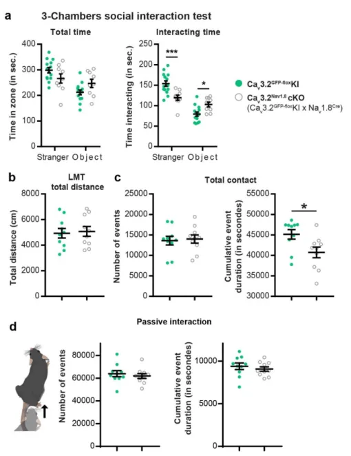
535
old Nav1.8cre mice (injected with rAAV
PHPs-CAG-mCherry or rAAVPHPs-Cav
3.2-FLEx-HA-536
hM3Dq). During recording, the animals were kept under the same condition as in the housing 537
facility (12hour Daylight, 500 lumens, food and water ad libidum) and the experimenter came 538
once per day to perform injection if necessary or to check the water and food level. 539
We used Python scripts provided to analyse all the data acquired by the system. 540
(www.livemousetracker.com; https://github.com/fdechaumont/lmt-analysis). We only
541
performed analyse during the activity phase (night cycle) as during the day mice nest together 542
which impaired the tracking and provide unreliable annotation. 543
von Frey
544
8 weeks after intrathecal injections, mice were first habituated for half an hour to the 545
experimental room in their home cage. Each mouse was then placed individually into small 546
arena (8*8cm) over a von Frey mesh for 45 minutes for habituation. Then, animals were 547
injected peritoneallly with 1mg/kg CNO diluted in sterile saline (prepared fresh each time). 30 548
to 45 minutes after the injections, von Frey filaments (0.07g, 0.6g, 2g) and the brush was 549
applied 5 time on each hind paw and withdrawal was then scored. Experiments were 550
performed in the morning (from 8 to 11 a.m.) under 500 lumens. The experimenter was blinded 551
to the animal conditions. 552
Gradient
17 8 weeks after intrathecal injections, mice were first habituated for half an hour to the 554
experimental room in their home cage and then injected with 1mg/kg CNO diluted in sterile 555
saline (prepared fresh each time). 30 minutes later, mice were placed into the bioseb thermal 556
gradient (2 corridors of 1m50 with one extremity cooled down to 5°C and the other to 50°C, 557
creating a thermal gradient spited into 20 thermal zone). Mice were free to explore for 90 558
minutes during which animal’s positions were tracked and annotated accordingly to the 559
temperature zones. For each run, a C-LTMRshM3Dq mouse and a control mouse were tested
560
simultaneously. However, the groups were labelled as such as he experimenter was still blind 561
to the animal conditions. Mice corridor distribution was alternated between each run to avoid 562
any corridor biased. Experiments were performed in the morning (from 8 to 12 a.m.) under 180 563
lumens. 564
Conditioned place preference (CPP)
565
8 weeks after intrathecal injections, mice were subjected to the conditioned place preference 566
protocol. This protocol consisted into 6 experimental days: the first day consisted into an 567
exploration and habituation phase of 30 minutes per mouse where mice were free to explore 568
the CPP arena (compartments: 25cm*20cm, corridor: 5cm*20cm . From the second to the fifth 569
day, animals were conditioned to associate a saline injection in the morning to their preferred 570
compartment (defined during the first day), and a CNO injection (1mg/kg) to the other 571
comportment in the afternoon. During this conditioning, animals received the injection and 572
stayed in their homecage for 30 minutes and then were placed into the designated 573
compartment for 30 minutes, to allow the CNO to reach its molecular target. To avoid that the 574
CNO was still active between two conditioning, we choose to stretch as much as possible the 575
period of time between the CNO injection from one given day and the next day saline injection. 576
On the sixth and last day animals were free to explore the all arena again for 30 minutes. The 577
position of the animal was detected by a network of infrared beams. The CPP rack allowed us 578
to perform 4 experiments at a time (Imetronic place preference setup), thus each run was 579
balanced to test 2 C-LTMRshM3Dq and 2 control mice, however the groups were labelled as
580
such as the experimenter was blinded to the animal conditions. Experiments were performed 581
in the morning (from 8 to 12 a.m.) under 180 lumens. 582
Immunohistology
583
Tissue collection and processing. Animals were transcardially perfused with phosphate-584
buffered saline (PBS) followed by 10% formaldehyde in PBS. Brain and spinal cord were 585
dissected, post-fixed in 10% formaldehyde for 24 hours, and cryoprotected in 30% sucrose in 586
PBS. DRGs were cryoprotected directly after formaldehyde perfusion. Tissues were then 587
frozen in Optimum Cutting Temperature (OCT, Tissue Tek) and sectioned using a cryostat 588
18 (Leica). Spinal cord and brain were sectioned at 40μm and stored in PBS + 0.05% azide at 589
4°C. For DRGs, tissues were sectioned at 18 μm, collected on Superfrost Plus slides (Fisher 590
Scientific), and stored at -80°C. 591
Immunofluorescence. Tissues were incubated for 1 hour and blocked in a solution consisting 592
of 0.1 M PBS with 0.3% Triton X-100 (Sigma) plus 5% normal donkey serum. Primary and 593
secondary antibodies were diluted in 0.1 M PBS with 0.3% Triton X-100 plus 1% normal 594
donkey serum. Sections were then incubated overnight at 4°C in primary antibody solution, 595
washed in 0.1 M PBS with 0.3% Triton X-100 for 40 min, incubated for 2 hrs in secondary 596
antibody at room temperature (RT), and washed again in 0.1 M PB for 40 min. Sections were 597
then mounted using Dako fluorescence mounting medium. Images were acquired with a Leica 598
SP8 confocal microscope. 599
Primary antibodies: anti-TH: Millipore (sheep; 1:500), anti-HAtag: Covenant (mouse, 1:1000); 600
Anti-Vglut3 : Synaptic system (Rabbit, 1:500). To identify IB4-binding cells, Fluorophore-601
conjugated IB4 (VectorLab, 1:500) was used in place of primary and secondary antibodies. 602
Secondary antibodies: Alexa Fluor®-conjugated secondary antibodies were acquired from 603
Invitrogen and Jackson Immunoresearch Labs. 604
Statistics
605
Statistics were performed with Graphpad Prism 8. Data are represented as mean ± s.e.m. For 606
bar graphs, each individual data points were superimposed under mean mean ± s.e.m. 607
Statistical tests used to compare values are indicated in each figure legends. 608
609 610
19
References
611
1. Kaiser, M. D. et al. Cereb Cortex 26, 2705–2714 (2016).
6122. Geerlings, S. W., Twisk, J. W. R., Beekman, A. T. F., Deeg, D. J. H. & van Tilburg, W.
613Soc Psychiatry Psychiatr Epidemiol 37, 23–30 (2002).
6143. Liljencrantz, J., Marshall, A., Ackerley, R. & Olausson, H. 563, 75–9 (2014).
6154. Abraira, V. E. & Ginty, D. D. 79, 618–39 (2013).
6165. Löken, L. S., Wessberg, J., Morrison, I., McGlone, F. & Olausson, H. Nat. Neurosci. 12,
617547–548 (2009).
6186. Perini, I., Olausson, H. & Morrison, I. Front Behav Neurosci 9, (2015).
6197. Olausson, H. et al. Nature Neuroscience 5, 900–904 (2002).
6208. Kumazawa, T. & Perl, E. R. Journal of Neurophysiology 40, 1325–1338 (1977).
6219. McGlone, F., Wessberg, J. & Olausson, H. Neuron 82, 737–755 (2014).
62210. Fairhurst, M. T., Löken, L. & Grossmann, T. Psychol Sci 25, 1124–1131 (2014).
62311. Croy, I. et al. Behavioural Brain Research SreeTestContent1 297, 37–40 (2016).
62412. Ishiyama, S. & Brecht, M. Science 354, 757 (2016).
62513. Orefice, L. L. et al. Cell 166, 299–313 (2016).
62614. Orefice, L. L. et al. Cell 178, 867-886.e24 (2019).
62715. Delfini, M.-C. et al. Cell Reports 5, 378–388 (2013).
62816. François, A. et al. Cell Reports 10, 370–382 (2015).
62917. Li, L. et al. 147, 1615–27 (2011).
63018. Reynders, A. et al. Cell Rep 10, 1007–1019 (2015).
63119. Seal, R. P. et al. Nature 462, 651–655 (2009).
63220. Usoskin, D. et al. Nat Neurosci 18, nn.3881 (2014).
63321. Chaumont, F. de et al. Nature Biomedical Engineering 1–13 (2019)
doi:10.1038/s41551-634019-0396-1.
63520
22. Akopian, A. N., Sivilotti, L. & Wood, J. N. Nature 379, 257–262 (1996).
636
23. Candelas, M. et al. Sci Rep 9, 1–18 (2019).
63724. Chan, K. Y. et al. 20, 1172–1179 (2017).
63825. Loo, K. M. J. van et al. J. Biol. Chem. 287, 15489–15501 (2012).
63926. Brumovsky, P., Villar, M. J. & Hökfelt, T. Exp Neurol 200, 153–165 (2006).
64027. Bohic, M. et al. Cell Reports 30, 602-610.e6 (2020).
64128. Bender, D., Holschbach, M. & Stöcklin, G. Nuclear Medicine and Biology 21, 921–925
642(1994).
64329. Benn, S. C., Costigan, M., Tate, S., Fitzgerald, M. & Woolf, C. J. J. Neurosci. 21, 6077–
6446085 (2001).
64530. Samad, O. A. et al. Mol Pain 6, 45 (2010).
64631. Francois, A. et al. 154, 283–93 (2013).
64732. Wang, R. & Lewin, G. R. J Physiol 589, 2229–2243 (2011).
64833. White, G., Lovinger, D. M. & Weight, F. F. Proc. Natl. Acad. Sci. U.S.A. 86, 6802–6806
649(1989).
65034. Cascio, C. J., Moore, D. & McGlone, F. Developmental Cognitive Neuroscience 35, 5–11
651(2019).
65235. Harlow, H. F., Dodsworth, R. O. & Harlow, M. K. PNAS 54, 90–97 (1965).
65336. Sheridan, M. A., Fox, N. A., Zeanah, C. H., McLaughlin, K. A. & Nelson, C. A. PNAS
654109, 12927–12932 (2012).
65537. Tanaka, S., Young, J. W., Halberstadt, A. L., Masten, V. L. & Geyer, M. A. Behav Brain
656Res 233, (2012).
65738. Voos, A. C., Pelphrey, K. A. & Kaiser, M. D. Soc Cogn Affect Neurosci 8, 378–386
658(2013).
65939. Crane, L., Goddard, L. & Pring, L. Autism 13, 215–228 (2009).
66021
40. Splawski, I. et al. J Biol Chem 281, 22085–22091 (2006).
661
41. Ackerley, R. et al. 34, 2879–83 (2014).
66242. Kupari, J. et al. bioRxiv 2020.12.07.414193 (2020) doi:10.1101/2020.12.07.414193.
66343. Renthal, W. et al. Neuron 108, 128-144.e9 (2020).
66444. Vrontou, S., Wong, A. M., Rau, K. K., Koerber, H. & Anderson, D. J. 493, 669–73
665(2013).
66645. Sharma, N. et al. Nature 577, 392–398 (2020).
66746. Zheng, Y. et al. Neuron 103, 598-616.e7 (2019).
66847. Nagi, K., Charfi, I. & Pineyro, G. 72, 3543–57 (2015).
66948. Watkins, R. H. et al. Journal of Neurophysiology (2020) doi:10.1152/jn.00587.2020.
67049. Terranova, M. L., Laviola, G. & Alleva, E. Dev Psychobiol 26, 467–481 (1993).
67150. Wesson, D. W. Current Biology 23, 575–580 (2013).
672673 674 675 676
22 677
Figure 1: C-LTMRs impairment via Cav3.2 deletion reduces social behaviour.
23 a. In a three-chamber social interaction test, control Cav3.2GFP-floxKI littermates have a
679
significantly higher preference index toward a stranger mouse than Cav3.2Nav1.8cKO. Unpaired
680
t-Test p<0.001, n Cav3.2GFP-floxKI= 14 (light green circles), n Cav3.2Nav1.8cKO= 9(open grey
681
circles). 682
b. Live mouse tracker configuration description. Each animal is implanted with a RFID chip for 683
authentication and tracking. The system includes 16 RFID antennas that cover the 50 by 50 684
cm arena which allow, with the deep sensing camera and the machine learning algorithm, to 685
track and automatically annotated the behaviour of 4 mice. Each group of mice includes 2 686
Cav3.2GFP-floxKI and 2 Cav3.2Nav1.8cKO. the experiment was performed over 3 days and each
687
behaviour was analysed during the nocturnal activity phases. 688
c. LMT index for 28 behavioural traits. Sum of all the number of events for the three nights. 689
d. LMT index for 28 behavioural traits. Sum of the duration of events for the three nights. 690
For c. and d.: The index for each trait was compared to one using one-sample two-sided 691
Student’s t-tests (corrected for multiple testing, because 28 tests were conducted for each 692
strain). The p values were color-coded in shades of blue depending of the p value as indicated 693
on the left. n = 10 694
e. f. Raw values extracted from the LMT for three consecutive nights for Cav3.2GFP-floxKI (light
695
green circles; n =10) and Cav3.2Nav1.8cKO (open grey circles; n=10). e. number of events and
696
cumulative duration of reciprocal social interaction (* p=0.0372). f. number of events and 697
cumulative duration of unilateral social interaction (* p=0.0429). unpaired t-test 698
24 699
Figure Supplementary 1: C-LTMRs hypo excitability via Cav3.2 deletion reduces social
700
behaviour.
701
a. In a three-chamber social interaction test, control Cav3.2GFP-floxKI littermates spent more time
702
interacting with a stranger mouse and less time with an inanimate object than Cav3.2Nav1.8cKO.
703
2 way ANOVA with Bonferroni post hoc test, ***p=0.0007; * p=0.262, n Cav3.2 KI GFP-flox = 14
704
(light green circles), n Cav3.2 KI GFP-flox = 9 (open grey circles).
705
b. Sum of the total distance travelled by each mouse during the three nights for Cav3.2 KI
GFP-706
flox KI (light green circles; n =10) and Ca
v3.2Nav1.8cKO (open grey circles; n=10).
707
c. raw values extracted from the LMT for three consecutive nights. Left: number of total contact, 708
right, cumulative event duration of contacts (* p=0.0220). 709
d. number of events and cumulative duration of passive social interaction. Wilcoxon matched- 710
unpaired t-test 711
25 712
Figure 2: Viral expression of DREADD receptors HA-hM3Dq in C-LTMRs.
713
a. Diagrams representing primary sensory neurons expressing Nav1.8 and Cav3.2 and the
714
population defined by the expression of both ion channels. 715
b. Strategy to express HA-hM3Dq in C-LTMRs using intrathecal injection of AAVPHPs serotypes
716
in Nav1.8Cre mice.
717
c. Left: representative images of an Immunofluorescence of Tyrosine Hydroxylase (TH, C-718
LTMRs marker, green) and HA tag (HA-hM3Dq, red) in thoracic dorsal root ganglia (T13) of a 719
Nav1.8Cre mice injected with the AAVPHPs-pCav3.2-FLEx-HA-hM3Dq. Filled arrowheads
720
indicate examples of neurons considered positive for HA and TH. Empty arrowheads indicate 721
examples of neurons considered positive for HA only. Right: Bar graph of the percentage of 722