HAL Id: hal-01052681
https://hal.inria.fr/hal-01052681
Submitted on 28 Jul 2014
HAL is a multi-disciplinary open access
archive for the deposit and dissemination of
sci-entific research documents, whether they are
pub-lished or not. The documents may come from
teaching and research institutions in France or
abroad, or from public or private research centers.
L’archive ouverte pluridisciplinaire HAL, est
destinée au dépôt et à la diffusion de documents
scientifiques de niveau recherche, publiés ou non,
émanant des établissements d’enseignement et de
recherche français ou étrangers, des laboratoires
publics ou privés.
The Mind-Mirror: See Your Brain in Action in Your
Head Using EEG and Augmented Reality
Jonathan Mercier-Ganady, Fabien Lotte, Emilie Loup-Escande, Maud
Marchal, Anatole Lécuyer
To cite this version:
Jonathan Mercier-Ganady, Fabien Lotte, Emilie Loup-Escande, Maud Marchal, Anatole Lécuyer.
The Mind-Mirror: See Your Brain in Action in Your Head Using EEG and Augmented Reality. IEEE
Virtual Reality (VR), Mar 2014, Minneapolis, United States. �hal-01052681�
The Mind-Mirror: See Your Brain in Action in Your Head
Using EEG and Augmented Reality
Jonathan Mercier-Ganady∗ Inria/IRISA Fabien Lotte† Inria/LaBRI Emilie Loup-Escande‡ Inria/IRISA Maud Marchal§ INSA/Inria Anatole L ´ecuyer¶ Inria/IRISA ABSTRACT
Imagine you are facing a mirror, seeing at the same time both your real body and a virtual display of your brain in activity and per-fectly superimposed to your real image “inside your real skull”. In this paper, we introduce a novel augmented reality paradigm called “Mind-Mirror” which enables the experience of seeing “through your own head”, visualizing your brain “in action and in situ”. Our approach relies on the use of a semi-transparent mirror posi-tioned in front of a computer screen. A virtual brain is displayed on screen and automatically follows the head movements using an optical face-tracking system. The brain activity is extracted and processed in real-time with the help of an electroencephalography cap (EEG) worn by the user. A rear view is also proposed thanks to an additional webcam recording the rear of the user’s head. The use of EEG classification techniques enables to test a Neurofeed-back scenario in which the user can train and progressively learn how to control different mental states, such as “concentrated” ver-sus “relaxed”. The results of a user study comparing a standard visualization used in Neurofeedback to our approach showed that the Mind-Mirror could be successfully used and that the partici-pants have particularly appreciated its innovation and originality. We believe that, in addition to applications in Neurofeedback and Brain-Computer Interfaces, the Mind-Mirror could also be used as a novel visualization tool for education, training or entertainment applications.
Keywords: Augmented Reality, EEG, Brain Activity, Mirror, Vi-sualization, BCI.
Index Terms: H.5.1 [Information Interfaces and Presentation]: Multimedia Information Systems—Artificial, augmented, and vir-tual realities; H.5.2 [Information Interfaces and Presentation]: User Interfaces—Input devices and strategies; I.3.7 [Computer Graph-ics]: Three-Dimensional Graphics and Realism—Virtual reality
1 INTRODUCTION
Nowadays ElectroEncephaloGraphy (EEG) and Brain-Computer Interfaces (BCI) provide a unique access to brain activity in real-time. This enables new kinds of applications such as “Neurofeed-back” which consists in progressively learning to control a brain pattern while continuously observing a real-time feedback about this pattern [4]. Neurofeedback is considered today in the medi-cal field for treating different kinds of pathology, such as attention deficits [11]. In most Neurofeedback applications, the feedback used is visual and consists in basic graphics such as 2D gauges in-creasing or dein-creasing over time. More elaborated visual feedback and 3D graphics can be used to display brain activity, but they are
∗e-mail: [email protected] †e-mail: [email protected]
‡e-mail: [email protected] §e-mail: [email protected] ¶e-mail: [email protected]
generally used in off-line visualization software [22], for instance for medical diagnosis.
Figure 1: The Mind-Mirror prototype. Left: A virtual brain superim-posed to the real user’s image (photomontage). Right: Our mirror-based augmented reality setup.
In this paper we study the combination of Augmented Reality (AR), 3D Visualization, and ElectroEncephaloGraphy. We intro-duce a novel setup called “Mind-Mirror” which enables the visual-ization of our own brain activity “inside our own head” by superim-position (see Figure 1-left). The brain activity is extracted in real-time using an EEG acquisition machine and is displayed in a mirror-based AR setup in front of the user’s skull in semi-transparency. The Mind-Mirror could therefore be used for entertaining or edu-cational activities, or for Neurofeedback applications, i.e., training to control our brain while being in a more natural, engaging, and immersive environment.
The remainder of this paper is organized as follows. Section 2 describes related work on augmented reality and brain activity visu-alization. Section 3 introduces the Mind-Mirror system and details its main components. Section 4 presents a pilot study comparing the Mind-Mirror to a classical 2D visualization used in current Neuro-feedback applications, in the context of controlling brain patterns related to concentration versus relaxation mental activities. The pa-per ends with a general discussion and a conclusion.
2 RELATEDWORK
Since the beginning of augmented reality the properties of half-silvered screens have been used to superimpose various graphics on top of real objects. These objects could be buttons [7] or vir-tual enemies in a video game [19]. In both cases, displayed objects would seem very realistic to the user since they are superimposed over real objects. These virtual objects are not directly dependent on the mental state of the user. A recent paper has shown that a high quality display of flames and smoke on a user’s hand in AR could induce a heat sensation [21]. Thus the realistic display of a virtual stimulation on a body part seems to have a strong impact on the sensations of users.
Mirror-based systems have already been used to see virtual ob-jects mapped on users in so-called “magic mirror” augmented
re-ality [12]. For instance Maes et al. conceived the “Artificial Life Interactive Video Environment” (ALIVE) that allowed interaction between an autonomous virtual agent, e.g. a dog, and the real user. Users could perform gestures and see themselves in a magic mirror while the virtual agent would perform some actions depending on the gesture. In some cases, the mirror can be used to display virtual objects “inside” the user’s real body. For instance Blum et al. used a half-silvered mirror to show various organs “in situ” [3]. However, the displayed organs were static and not dependent on the state of the real organs, since no device was used to capture their activity.
The visualization of brain activity has been extensively studied, often by using recorded data and viewing offline the most active ar-eas of the brain [2]. These active arar-eas can be represented through a flat 2D map of the brain (using changing colors to show high- and low-activity zones) or a topography of surface activity [13]. How-ever the existing approaches do not currently incorporate a direct user interaction.
In a recent study on Neurofeedback Hwang et al. used a real-time visualization of brain activity to train the user to BCI control [6]. Participants could visualize their cortical mu (8-12 Hz) rhythm activity in real-time, improving the BCI training and thus the per-formance when using the motor imagery paradigm. In this desktop setup participants were able to have a direct view of their brain ac-tivity using a simple 2D colored image of a virtual brain.
In the following section we introduce a novel approach relying on AR and 3D interaction for brain activity visualization in a magic-mirror setup.
3 THEMIND-MIRRORSYSTEM
3.1 Concept
The Mind-Mirror is an augmented reality paradigm that enables users to see in a mirror both their real head and a virtual display of their brain in activity and perfectly superimposed to their real head. In other words, when using the Mind-Mirror, users can see “through their own head”, visualizing their brain “in action and in situ”.
Our approach relies on the use of a semi-transparent mirror po-sitioned in front of a computer screen. A virtual brain is displayed on-screen and automatically follows the head movements using an optical face-tracking system. The brain activity is extracted and processed in real-time thanks to an EEG cap worn by the user. Since a mirror alone can only show the frontal parts of the brain, we have also added a rear-view. This view uses a webcam placed behind and above the head of the user.
3.2 System Description
The Mind-Mirror system is composed of several components which are displayed in Figure 2:
• EEG recording and amplification: EEG acquisition and amplification is performed using a set of electrodes mounted on a cap (1) and an EEG amplifier (g.tec, g.USBamp) (4). • Webcam: a webcam is used and positioned on a wall behind
the user (2) in order to capture the image of the back of the head of the user and display it in a rear-view window on the Mind-Mirror.
• Face and head tracking: Face and head tracking are achieved using a Microsoft Kinect camera (3) and its devel-opment kit [14]. It is used to overprint the virtual brain at the position of the user’s head in real-time. The Kinect devel-opment kit uses here depth and color sensing of the camera to track the human face using proprietary algorithms.
• Signal-processing: Signal processing is done on a computer (5) using the OpenViBE software platform [20].
Figure 2: Mind-Mirror system overview. Components: (1) EEG cap, (2) webcam, (3) Microsoft Kinect camera, (4) g.tec g.USBamp EEG amplifier, (5) laptop PC, (6) computer screen, (7) half-silvered foil.
• Display: The display system consists in a computer screen (6) that supports a half-silvered foil (7) applied onto a thin plastic plate. Due to the half-silvered foil only the screen parts that are bright can be seen. The rest of the screen surface appears opaque and reflective, as with a classical mirror. Using a real mirror enables users to see themselves directly, which would not be possible with a camera-based AR setup.
EEG data acquired with OpenViBE software [20] and head movement data from the Kinect are retrieved and processed within a Unity3D-based application that simulates and displays the virtual brain. Virtual brain activity visualizations are displayed in real-time (framerates between 25 and 80 Hz depending on the visualization type). Face-tracking and head movements are detected and pro-cessed with a latency of less than 500 ms.
3.3 Visualization Tools
As mentioned previously, the Mind-Mirror can be used to display the brain in action and “in situ”. Different visualization techniques can be used. We have explored the following ones for the Mind-Mirror.
3.3.1 Brain Topography
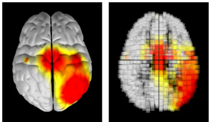
The brain topography visualization enables the display of the EEG signal power over the brain surface, as shown in Figure 3-left. This representation allows for a quick overview of the most active brain areas, i.e., the areas with the largest EEG signal power.
Figure 3: Brain visualization techniques. Left: topography display of surface brain activity. Right: volumetric display of brain activity using voxels. Red and yellow colors correspond to the high-activity spots.
3.3.2 3D Volumetric Representation Using Inverse Solution EEG signals correspond to brain activity at the surface of the scalp. Fortunately, it is possible to estimate the brain activity in the whole brain volume using algorithms known as inverse solutions [17]. The volumetric brain activity hence estimated can then be displayed with a 3D model of the brain using voxels. This visualization al-lows for a comprehensive 3D view of the brain activity, as shown in Figure 3-right.
3.3.3 Visualization Integrating Mental State Classification One of the main goals of BCI or Neurofeedback training applica-tions is to help users gain control of their own brain activity, in order to be able to modulate it and reach a specific mental state, e.g., a relaxed or a concentrated mental state. Therefore, in such applications, it is important for users to be aware of the mental state identified by the system, so that they can identify the relevant fea-tures of their brain activity and learn how to control them.
Current brain activity visualization tools, e.g., the ones men-tioned above, are not well suited for such purposes. Indeed, they do not show the mental state identified by the system, and display with equal focus the activity in the entire brain, making it difficult for the user to identify the relevant features in all the displayed in-formation. In contrast, classical BCI feedback and Neurofeedback only show simple 2D graphics such as gauges [15], only indicat-ing the mental state identified, without givindicat-ing information about the underlying brain activity despite this being recommended for a successful learning [10].
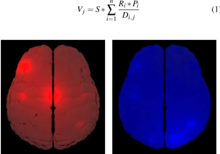
Therefore, in this paper, we propose a new brain activity visual-ization technique that aims at using the best of both worlds. Indeed, our approach 1) gives information about the underlying brain ac-tivity, using surface topography, and highlights some specific brain areas that are relevant for the targeted mental state classification, and 2) displays simultaneously the mental state currently identified by the BCI, which altogether is expected to help users identifying the relevant features of their brain activity in order to control more efficiently the targeted brain activity. With this new representation, the whole virtual brain is colorized according to the mental state of the user, as identified by the BCI system. Currently, we have explored this approach with a BCI that estimates the concentration and relaxation level of the user (see Section 4). When the system detects that the user is relaxed, then the whole virtual brain is col-ored in blue, otherwise it is colcol-ored in red. Moreover, the more blue the brain is displayed, the more confident the BCI system is in the corresponding mental state estimation. This confidence is computed using the output of the linear regression used in the BCI system (see Section 4.1): the larger the absolute value of the linear regression output, the more confident the estimation [5]. Then, in order to help the user pay attention to the most relevant information and not overwhelm him, the most important brain areas for the es-timation of the mental states are shown with a more intense color, as shown in Figure 4.
The most important brain areas were chosen using machine learning tools, as exposed in Section 4.1. In particular, the surface topography only displays the EEG signals in the frequency band that is the most relevant for classification (i.e., the most discrimina-tive frequency band in the Filter Bank Common Spatial Pattern al-gorithm [1]), and only the most relevant channels (i.e., the channels with the largest absolute weights in the Common Spatial Patterns filters for this frequency band) are displayed with more intense col-ors.
Surface topography is displayed on a 3D mesh of a brain using vertex coloration. In our current simple implementation the sur-face topography is computed as follows. The color of vertex j (Vj)
is defined using Equation (1). A state color S is defined as red if the detected mental state is concentrated, blue otherwise. Elec-trode Relevance (Ri) represents the relevance of the electrode i for
one mental state, computed during the initial calibration phase (see Section 4.3). Pirepresents the EEG band power at electrode i and
Di,jthe distance between electrode i and vertex j. The electrode
positions used are those of the international 10-20 system. Note that other computations could be used such as Surface Laplacians [20]. Vj= S ∗ n
∑
i=1 Ri∗ Pi Di,j (1)Figure 4: Brain activity visualization integrating concentra-tion/relaxation classification. The left image corresponds to a con-centrated mental state (red color) while the right image shows a re-laxed state (blue color). A brighter color represents a more intense electrical activity.
3.3.4 Rear-View
The rear-view, which allows users to see what is happening on the rear parts of their brain, is displayed on top of our mirror (see Fig-ure 5). This rear-view makes use of a standard computer webcam placed above and behind the user. Another virtual brain is used here and also superimposed over the image recorded by the webcam.
Figure 5: Rear-view window: a virtual brain is superimposed with the back of the user’s head. It follows the orientation of the real user’s head and enables the perception of what happens at the back of the head (brain).
4 PILOTSTUDY
In order to assess our Mind-Mirror system, we performed a pilot study in which the participants used it as feedback in a BCI based on the concentration and relaxation levels. More precisely, with this BCI, participants had to put themselves in a relaxed or in a concen-trated state, such mental states being analyzed and identified by the
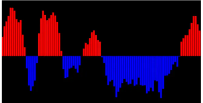
BCI in real-time. These two mental states could then be assigned to two different commands (see, e.g., [5]), although this was not the case in this study. Here, we studied both the performance (in terms of successfully recognized mental state) and preference (using sub-jective questionnaires) of the participants using the Mind-Mirror to visualize their brain activity in real-time while using the BCI. We also compared the Mind-Mirror to a temporal gauge, a typical rep-resentation used within the BCI and neurofeedback community (see Figure 6). This indeed enabled us to study the impact of the more complex but also more immersive and informative brain activity representation that is the Mind-Mirror on participants’ experience and BCI performance.
Figure 6: The temporal gauge display. The red and blue bars re-spectively represent the evolution of the concentration and relaxation levels in time.
4.1 Experimental Apparatus
The experiment was conducted in a room without environmental noise or other source of distraction. The participants were comfort-ably seated in front of a computer screen.
EEG signals were acquired with a sampling frequency of 512Hz using a 16-channels g.USBamp acquisition system. EEG were recorded at channel locations T7, T8, F7, F8, Fp1, Fp2, C3, Cz, C4, O1, O2, F3, F4, P3, Pz and P4 according to the international 10-20 system. The ground electrode was placed on position FCz, and the reference electrode on the participant’s left earlobe.
The concentration level of each participant was estimated using a subject-specific model obtained with machine learning [5]. More precisely, the Filter-Bank Common Spatial Pattern (FBCSP) algo-rithms [1] was used to identify the most relevant EEG frequency bands and channels to discriminate the concentrated from the re-laxed mental state [5]. Only frequency bands within the theta (4-7Hz) and alpha (8-14Hz) rhythms range were explored. Indeed, from a neurophysiological point of view, these rhythms are ex-pected to be correlated with concentration [16], and are less likely than higher frequency bands to be contaminated by muscle artifacts [5]. The FBCSP algorithm was optimized on training EEG signals collected during the initial calibration phase (see Section 4.3 for details on this phase). Then, FBCSP features were used to train a linear regression algorithm to estimate the participant’s concentra-tion level. Once the BCI is calibrated in this way, it can be used online. To do so, the FBCSP features were extracted from the EEG signals over the last 2s (using a sliding window scheme, with a 0.1s overlap between consecutive windows) and used as input for the linear regression, which output indicates the participant’s con-centration level. More precisely, a negative (respectively positive) output value means that the participant is in a relaxed (respectively concentrated) mental state.
4.2 Population
Twelve participants (aged from 21 to 30, mean=25, sd=2.8) took part in the experiment.
4.3 Experimental Plan
A calibration phase took place before the experiment. Participants were asked to concentrate for 60 seconds and then relax for 60 sec-onds without having any visual feedback. Participants were free to choose any cognitive activity for the concentration task. Sev-eral suggestions were made such as a “mental computation” task. The EEG data collected during this phase were used to calibrate the signal processing pipeline.
Two different representations were then compared: the Mind-Mirror and the temporal gauge. As the participants were concen-trating or relaxing, the brain activity representations would show the current level of concentration or relaxation, respectively.
The experiment was divided into 12 trials. Each trial comprised a relaxation phase followed by a concentration phase, each of them lasting 25 seconds. The experiment lasted 10 minutes per repre-sentation and so 20 minutes for the whole experiment. The two brain activity representations were used by each participant. The half-silvered foil was manually applied on the monitor screen for the Mind-Mirror condition, and taken away for the gauge condi-tion. Participants were divided into two groups. The first group started with the Mind-Mirror and the second group started with the temporal gauge.
4.4 Collected Data
For each trial and each participant, we recorded the BCI classifier output values resulting from the signal-processing pipeline. At the end of the experiment and for each representation, participants had to fill out a subjective Likert-scale questionnaire (1: strongly dis-agree to 7: strongly dis-agree). Questions were: “Do you think that the representation is x”, where x was one of the following criterion: (1) Comprehensible, (2) Motivating, (3) Simple, (4) Clear, (5) Innova-tive, (6) Original.
5 RESULTS
5.1 Classification performance results
We conducted statistical analysis in order to assess the BCI clas-sification performance for each brain activity representation. To do so, we first compared the linear regression output between the relaxed state and the concentrated state for each of the two visu-alizations separately, in order to assess whether participants could control the BCI with them. A paired t-test showed significant dif-ferences in BCI output between the concentrated and relaxed states for the gauge visualization (t(11)=-4.82, p=0.0005) as well as for the Mind-Mirror visualization (t(11)=-2.73, p=0.02). We also com-pared the classification performances obtained with the two visu-alizations using a paired t-test. Such classification performances were assessed using the average difference between the linear re-gression output during the concentration and relaxation conditions, as in [5]. The larger the difference, the better the discrimination be-tween the two mental states. We found no significant difference (p=0.19) between the classification performance with the Mind-Mirror condition (average difference: 0.31 ± 0.39) and the gauge condition (average difference: 0.46 ± 0.33), although there might be a slight trend towards better performances with the gauge. The average values of the linear regression output for each mental state are provided in Table 1.
5.2 Questionnaire Results
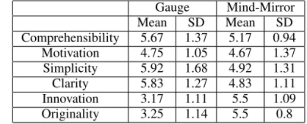
We performed a Wilcoxon test on the visual representations for the different criteria. We found a significant effect for 4 criteria: Sim-plicity (z=-1.981, p=0.048), Clarity (z=-2.089, p=0.037), Innova-tion (z=-2.890, p=0.004) and Originality (z=-2.924, p=0.003). The participants found the Mind-Mirror less simple, less clear but more innovative and more original than the gauge representation. Table 2 summarizes the questionnaire results for the 6 criteria.
Table 1: Classification performance results. Average values of the linear regression output and standard deviation are provided for each representation (Gauge; Mind-Mirror) and each state (Concentration; Relaxation), as well as the difference between the two states (Diff.)
Gauge Mind-Mirror Con. Relax. Diff. Con. Relax. Diff. Mean 0.21 -0.25 0.46 -0.004 -0.31 0.31 SD 1.44 1.50 0.33 1.1 1.22 0.39
Table 2: Subjective questionnaire results for the 6 criteria (1: worst to 7: best). Average values of mark and standard deviation were provided for each representation.
Gauge Mind-Mirror Mean SD Mean SD Comprehensibility 5.67 1.37 5.17 0.94 Motivation 4.75 1.05 4.67 1.37 Simplicity 5.92 1.68 4.92 1.31 Clarity 5.83 1.27 4.83 1.11 Innovation 3.17 1.11 5.5 1.09 Originality 3.25 1.14 5.5 0.8 6 GENERALDISCUSSION
The results obtained from the classification analysis and from the questionnaires enabled us to assess the pros and cons of the Mind-Mirror.
Concerning the pros, results from the classification analysis sug-gested that the participants were able to put themselves in a con-centrated or in a relaxed state with the Mind-Mirror representa-tion, hence showing that participants can successfully use the Mind-Mirror as a feedback for a neurofeedback/BCI application. Results also suggested that the Mind-Mirror, even if more complex than a classical gauge, does not seem to lead to substantially lower BCI performance, although there might be a trend towards slightly bet-ter performance with the gauge. Further studies with a larger pop-ulation would be required to confirm this point. Results from the questionnaire suggested that participants found the Mind-Mirror to be more original and innovative than traditional BCI feedback (i.e., the gauge). In other words, this means that the Mind-Mirror gives a higher quality experience than a gauge, with a more engaging feed-back environment. Moreover, as compared to the gauge, the Mind-Mirror provides more information, since it indicates in real-time which brain areas are active. The participants quoted the Mind-Mirror as “clear and easy to understand”, “quite precise”, “gives a good indication of the activated zones, without being too complex”. The Mind-Mirror thus provides an explanatory feedback, informing about what is going on in the user’s brain, whereas the gauge only provides a corrective feedback, only indicating whether the user is correctly relaxed or concentrated. These two feedback properties, namely, being engaging and explanatory, are recommended prop-erties for the efficient learning of a skill such as here learning how to control brain activity [10]. The evaluation reported in this paper is a short-term one, focused on feedback perception and usability. As such longer-term learning effects are unlikely to occur. In future works, it would be interesting to compare the classical gauge to the Mind-Mirror during multiple neurofeedback sessions, over multi-ple days. This would enable us to assess the users’ learning curves (i.e., performance improvements over time), and confirm whether the Mind-Mirror - due to its properties - can make learning faster and better as theoretically expected [10].
Concerning the Mind-Mirror cons, the user study highlighted that it was significantly more difficult to understand and to use than the classical gauge, at least for first-time users who qualified it as having a “slight lack of readability”. This result is not unexpected
since the Mind-mirror is a rich multidimensional feedback, thus a more complex one than the mono-dimensional gauge feedback. Users therefore need more time to get used to the Mind-Mirror feed-back, to understand how it works and how to handle it. This may explain why the classification analysis revealed no increase in BCI performance with the Mind-Mirror. An alternative interpretation that could explain such performance is that the Mind-Mirror dis-plays rich instantaneous information, but no temporal information, whereas the gauge displays an instantaneous information, more pre-cisely the regression output, but also the previous outputs, hence showing a short history describing temporal variations. In the fu-ture it would be interesting to add this temporal information to the Mind-Mirror. This could be done, e.g., by displaying the last few surface topographies as well as the current one, with the older ones displayed in dimmer colors or steam-like appearance, and slowly fading away as time passes. It would also be interesting to perform a follow-up longer-term study, in order to assess users’ performance and preference once they got used to the Mind-Mirror principle. Still regarding possible improvements to the Mind-Mirror design, some participants reported that they enjoyed the mirror principle with AR (“easier to control the movement without interfering with the task”, “nice to look at and entertaining”), but that it made vi-sually focusing on the brain or the user’s reflection more difficult (“the virtual brain appears on the foreground whereas the reflection seems to be on the background”). In the future, it could be relevant to compare this mirror AR approach to a more classical AR design with a webcam filming the user and overlaying the brain, without the use of a mirror. This would prevent users from looking at them-selves into the eyes as in a real mirror but could make the brain activity display easier to watch and to focus on.
As mentioned earlier, the Mind-Mirror has the potential to be used for multiple other applications, notably education, entertain-ment and neurofeedback training. Indeed, the Mind-Mirror could have educational purposes, as a support tool to teach brain biology and anatomy. For instance, users could select the brain area they are interested in by pointing to it using their own hands. Hand tracking could be done using the Kinect, and much meaningful information could be displayed about the selected brain area. This could be an engaging and effective way to learn, for instance, the different brain lobes names, their locations as well as how the brain works.
The Mind-Mirror could also be used for entertainment, as a mo-tivating feedback in BCI-based video games for instance [8]. It could also be used in serious games involving the brain, such as video games for brain fitness or for treating Attention Deficit Hy-per Activity Disorders [9].
Finally, and maybe more importantly, the Mind-Mirror could be a very powerful tool for Neurofeedback training, either for medi-cal applications or for BCI control. Actually, it could be used to train users to learn how to modulate the brain activity in some ar-eas of their brain. Indeed, as we have exposed before, the visu-alization tools offered by the Mind-Mirror are engaging, rich and explanatory which, from an instructional design point of view, is expected to lead to efficient skill learning, e.g., a BCI skill [10]. The Mind-Mirror is thus expected to improve learning in BCI or neurofeedback applications in the long-term. We performed here a first short-term study with concentration and relaxation. In the fu-ture, it would be interesting to explore it with other BCI paradigms, e.g., with BCI based on imagination of movements [18].
7 CONCLUSION
In this paper we introduced the Mind-Mirror, a system which com-bines augmented reality and EEG in order to enable users to vi-sualize their own brain operating inside their head. We proposed various brain activity visualization tools for this system, and iden-tified multiple possible use-cases, including education, entertain-ment, neurofeedback and BCI. We also conducted a user study to
compare feedback provided using the Mind-Mirror to a classical gauge feedback in a BCI experiment. Results suggested that partic-ipants found the Mind-Mirror more innovative and engaging than the gauge although more complex to use and understand. This com-plexity did not lead to a statistically significant drop in BCI perfor-mance. Overall, the Mind-Mirror seems to be a promising tool for a variety of applications, which would benefit from being validated by further studies.
Future work will concern more evaluations of the Mind-Mirror for BCI and neurofeedback applications. This implies formal ex-periments in other application contexts (e.g., game or education), as well as further studies to identify the best display parameters (e.g., color maps, contrasts, use of the mirror, etc.) for an optimal visualization and learning experience.
ACKNOWLEDGEMENTS
We would like to thank Jozef Leg´eny for his help as photographer and cameraman. This work was supported by the administrative region of Brittany and the French National Research Agency within the Homo Textilus project (grant ANR-11-SOIN-007).
REFERENCES
[1] K. K. Ang, Z. Y. Chin, C. Wang, C. Guan, and H. Zhang. Filter bank common spatial pattern algorithm on BCI competition IV datasets 2a and 2b. Frontiers in Neuroscience, 6(39), 2012.
[2] S. Baillet, K. Friston, and R. Oostenveld. Academic software applica-tions for electromagnetic brain mapping using MEG and EEG. Com-putational Intelligence and Neuroscience, 2011.
[3] T. Blum, V. Kleeberger, C. Bichlmeier, and N. Navab. mirracle: An augmented reality magic mirror system for anatomy education. In Proceedings of IEEE Virtual Reality Short Papers and Posters, pages 115–116, 2012.
[4] J. N. Demos. Getting Started with Neurofeedback. W. W. Norton & Company, 2005.
[5] L. George, F. Lotte, R. V. Abad, and A. L´ecuyer. Using scalp elec-trical biosignals to control an object by concentration and relaxation tasks: design and evaluation. In Proceedings of the IEEE International Conference EMBS, pages 6299–6302, 2011.
[6] H.-J. Hwang, K. Kwon, and C.-H. Im. Neurofeedback-based mo-tor imagery training for brain-computer interface. Journal of Neu-roscience Methods, 179(1):150–156, 2009.
[7] K. Knowlton. Computer displays optically superimposed on input devices. Bell System Technical Journal, 56(3):367–383, 1977. [8] A. L´ecuyer, F. Lotte, R. Reilly, R. Leeb, M. Hirose, and M. Slater.
Brain-computer interfaces, virtual reality, and videogames. IEEE Computer, 41(10):66–72, 2008.
[9] C. Lim, T. Lee, C. Guan, D. S. Fung, Y. Cheung, S. Teng, H. Zhang, and K. Krishnan. Effectiveness of a brain-computer interface based programme for the treatment of ADHD: A pilot study. Psychophar-macology Bulletin, 43(1):73–82, 2010.
[10] F. Lotte, F. Larrue, and C. M¨uhl. Flaws in current human training protocols for spontaneous brain-computer interfaces: lessons learned from instructional design. Frontiers in Human Neurosciences, 7(568), 2013.
[11] J. F. Lubar, M. O. Swartwood, J. N. Swartwood, and P. H. O’Donnell. Evaluation of the effectiveness of EEG neurofeedback training for ADHD in a clinical setting as measured by changes in TOVA scores, behavioral ratings, and WISC-R performance. Biofeedback and Self-regulation, 20(1):83–99, 1995.
[12] P. Maes, T. Darrell, B. Blumberg, and A. Pentland. The ALIVE sys-tem: Full-body interaction with autonomous agents. In Proceedings of IEEE Computer Animation, pages 11–18, 1995.
[13] J. Mellinger, G. Schalk, C. Braun, H. Preissl, W. Rosenstiel, N. Bir-baumer, and A. K¨ubler. An MEG-based brain-computer interface. Neuroimage, 36(3):581–593, 2007.
[14] Microsoft. Kinect face tracking SDK. http://msdn.microsoft.com/en-us/library/jj130970.aspx. Accessed December 5, 2013.
[15] C. Neuper and G. Pfurtscheller. Brain-Computer Interfaces, chapter Neurofeedback Training for BCI Control, pages 65–78. The Frontiers Collection, 2010.
[16] E. Niedermeyer. Electroencephalography: Basic principles, clinical applications, and related fields, chapter The Normal EEG of the Wak-ing Adult, page 167. Lippincott Williams & Wilkins, 2005. [17] R. Pascual-Marqui. Review of methods for solving the EEG inverse
problem. International Journal of Bioelectromagnetism, 1:75–86, 1999.
[18] G. Pfurtscheller and C. Neuper. Motor imagery and direct brain-computer communication. In Proceedings of the IEEE, volume 89, pages 1123–1134, 2001.
[19] W. Piekarski and B. Thomas. ARQuake: the outdoor augmented real-ity gaming system. Communications of the ACM, 45(1):3638, 2002. [20] Y. Renard, F. Lotte, G. Gibert, M. Congedo, E. Maby, V. Delannoy,
O. Bertrand, and A. L´ecuyer. OpenViBE: An open-source software platform to design, test, and use brain-computer interfaces in real and virtual environments. Presence: Teleoperators and Virtual Environ-ments, 19:3–53, 2010.
[21] P. Weir, C. Sandor, M. Swoboda, T. Nguyen, U. Eck, G. Reitmayr, and A. Dey. BurnAR: Feel the heat. In Proceedings of IEEE International Symposium on Mixed and Augmented Reality, pages 331–332, 2012. [22] C. Wolters, A. Anwander, X. Tricoche, D. Weinstein, M. Koch,
and R. MacLeod. Influence of tissue conductivity anisotropy on EEG/MEG field and return current computation in a realistic head model: a simulation and visualization study using high-resolution fi-nite element modeling. NeuroImage, 30(3):813–826, 2006.