HAL Id: hal-02043711
https://hal.archives-ouvertes.fr/hal-02043711
Submitted on 21 Feb 2019
HAL is a multi-disciplinary open access
archive for the deposit and dissemination of
sci-entific research documents, whether they are
pub-lished or not. The documents may come from
teaching and research institutions in France or
abroad, or from public or private research centers.
L’archive ouverte pluridisciplinaire HAL, est
destinée au dépôt et à la diffusion de documents
scientifiques de niveau recherche, publiés ou non,
émanant des établissements d’enseignement et de
recherche français ou étrangers, des laboratoires
publics ou privés.
fabrication of transparent electrodes based on silver
nanowires
Djadidi Toybou, Caroline Celle, Catherine Aude-Garcia, Thierry Rabilloud,
Jean-Pierre Simonato
To cite this version:
Djadidi Toybou, Caroline Celle, Catherine Aude-Garcia, Thierry Rabilloud, Jean-Pierre Simonato. A
toxicology-informed, safer by design approach for the fabrication of transparent electrodes based on
silver nanowires. Environmental science .Nano, Royal Society of Chemistry, 2019, 6 (2), pp.684-694.
�10.1039/c8en00890f�. �hal-02043711�
Science
Nano
PAPER
Cite this: Environ. Sci.: Nano, 2019, 6, 684
Received 14th August 2018, Accepted 10th October 2018 DOI: 10.1039/c8en00890f rsc.li/es-nano
A toxicology-informed, safer by design approach
for the fabrication of transparent electrodes based
on silver nanowires
Djadidi Toybou,
abCaroline Celle,
aCatherine Aude-Garcia,
cThierry Rabilloud
*
cand Jean-Pierre Simonato
*
aFabrication of silver nanowires (AgNWs) with fine and independent control of both the diameter (from 30 to 120 nm) and length (from 5 to 120μm) by concomitant addition of co-nucleants and temperature con-trol is demonstrated, and used for the preparation of size standards. Percolating random networks were fabricated using these standards and their optoelectronic properties were measured and compared with regard to the nanowire dimensions. The transparent electrodes appear suitable for various applications and exhibit excellent performances (e.g. 16 ohm sq−1at 93% transparency), with haze values varying from 1.6 to 26.2%. Besides, in vitro toxicological studies carried out on murine macrophages with the same size stan-dards revealed that AgNWs are weakly toxic (no toxicity observed below 50μg mL−1Ag), in particular com-pared to other silver nanoparticles. Short AgNWs (4μm) appeared to be slightly more toxic than longer AgNWs (10 and 20μm). Conversely, long AgNWs (20 μm) induced a more prolonged pro-inflammatory re-sponse in murine macrophages. These results contribute, in a safer by design approach, to promoting the use of short AgNWs. The global knowledge dealing with the combination of nanowire dimensions associ-ated with optoelectronic performances and relassoci-ated toxicity should encourage the rational use of AgNWs, and guide the choice of the most adequate AgNW dimensions in an integrated approach.
Introduction
The recent development of nanomaterials has generated a wave of hope in many fields. The potential of nanomaterials appears boundless, either due to their intrinsic properties (e.g. quantum dots and fullerenes), their additional proper-ties for bulk materials (e.g. nanocomposites), or when they are used in the form of assemblies (e.g. carbon nanotube-based cables). A characteristic and high-potential example of nanomaterial assembly is the fabrication of random networks
of metallic nanowires, in particular, silver nanowires
(AgNWs).1–3 Simply put, it is possible to generate random networks of metallic nanowires, above the percolation thresh-old, which conduct electricity through the nanowire-based metallic lattice and concurrently let the light pass through the empty spaces between the nanowires. This has been widely studied during the last decade and has raised the ma-turity of this technology up to the industrial level. Many ap-plications using AgNWs have been investigated, including
touch screens, light-emitting devices, transparent film
heaters and others.4–12 This system has many advantages
such as very low sheet resistance with high transparency, flex-ibility, and low-cost processing under ambient conditions.
Though many important advances have been realized in this field, some key points remain to be tackled, in particular, toxicity issues for safe industrial use. Since the expected
aUniv. Grenoble Alpes, LITEN DTNM, CEA, F-38054 Grenoble, France.
E-mail: jean-pierre.simonato@cea.fr
bUniv. Grenoble Alpes, ISTERRE, CNRS, F-38000, Grenoble, France
cUniv. Grenoble Alpes, Chemistry and Biology of Metals, CEA BIG, CNRS UMR
5249, F-38054 Grenoble, France. E-mail: thierry.rabilloud@cea.fr
Environmental significance
The use of silver nanowires is a promising alternative for the fabrication of transparent electrodes. They will be increasingly introduced in consumer devices. The relationship between the nanowires' dimensions and their toxicity is still insufficiently studied. We demonstrate a straightforward method to prepare silver nanowires with controlled dimensions. We propose a global approach, starting from tailor-made nanowires, based on a study of both their physical properties when used as random percolating networks, which is of interest from the technological point of view, and their toxicological behaviour toward murine macrophages, which is of interest from the health, safety & environment point of view. These results contribute, in a“safer by design ap-proach”, to promoting the use of short AgNWs.
Open Access Article. Published on 20 December 2018. Downloaded on 2/21/2019 9:52:12 AM.
This article is licensed under a
Creative Commons Attribution 3.0 Unported Licence.
View Article Online
properties are application-dependent and rely on the AgNWs' morphology,13,14it would be of great interest to be able to tai-lor the shape of the nanowires in order to obtain optimal opto-electronic performances for each use. Conventional parametric studies15,16 as well as other more specific methods17,18 have demonstrated efficient routes for modifying both the length and diameter, but not independently. An ultrasonic method was also proposed to select the desired length by cutting AgNWs; however, this method leads to poor control over size distribution and generates large amounts of by-products.19
The fabrication of AgNW size standards would also allow toxicological studies to be performed on definite species, in particular to determine whether the two main dimensions (i.e. diameter and length) play a critical role in toxicity. This is of tremendous importance since AgNWs will increasingly be introduced in technological devices and consumer prod-ucts, and thus data are awaited to assess the real toxicity of these nanomaterials. Although several reports have begun to tackle this topic,20–26much more remains to be done.
In this article, we propose a straightforward method to modulate independently the diameter and the length of nanowires in a wide range, allowing us to fabricate size stan-dards, i.e. AgNW samples with definite lengths and diame-ters. It has permitted us to perform a direct comparison of the optoelectronic properties of transparent electrodes made with these different nanowires, and to realize comparative ex-periments on toxicity with macrophages.
Materials and methods
Synthesis and purification of AgNWs
In a typical synthesis, AgNO3(0.68 g) was dissolved in EG (40
mL) at a slow stirring rate in a round flask. In another flask, PVP (average mol. wt 40 000, 1.77 g), NaCl, and the co-nucleant were dissolved in EG (80 mL) at 120 °C. The solu-tion was cooled to room temperature and then slowly added to the first flask within 8 min. The mixture was finally heated at the reaction temperature and cooled down at ambient tem-perature. The purification of the AgNWs was realized by de-cantation according to a published procedure.27
Characterization of the mean dimensions of AgNWs
The morphology analysis of AgNWs was performed using a Leo 1530 SEM. Measurement of the diameter and length di-mensions was realized with the ImageJ software. The statisti-cal studies were performed on 100 to 200 nanowires for each sample.
Preparation of AgNW electrodes
The AgNW-based solutions were used for the fabrication of electrodes after dilution in methanol at 0.2 mg mL−1 concen-tration. The silver concentration was measured using an atomic absorption spectrometer (Perkin Elmer AAnalyst 400). The deposition was performed on 2.5 × 2.5 cm2 heated sub-strates (70°C) with an automatic SonoTek spray-coater. The
performances of the electrodes were measured after cooling, without any post-treatment.
Performance measurements
Total transmittance and haze values were measured with a UV-visible spectrometer (Varian Cary 5000) equipped with an integrating sphere. The sheet resistance was set as the mean value of at least 5 measurements by using a four-pin probe with a Loresta resistivity meter (EP MCP-T360).
Toxicological assays
Nanomaterials. As control nanomaterials, spherical silver nanoparticles (<100 nm) coated with PVP40 were purchased from Sigma-Aldrich (catalogue number 758329) as a 5 wt% dispersion in ethylene glycol. Working solutions were pre-pared by dilution in deionized water. The characterization of these nanoparticles has been published previously by some authors of this article.28 Additional controls consisted of silica-based nanomaterials, either colloidal silica29or crystal-line silica (reference materials BCR66 and BCR70, purchased from Sigma-Aldrich).
Cell lines. The mouse macrophage cell line RAW 264.7 was obtained from the European Cell Culture Collection (Salisbury, UK). The cells were cultured in RPMI 1640 me-dium + 10% fetal bovine serum. The cells were seeded at 200 000 cells per ml and harvested at 1 000 000 cells per ml. For treatment with nanomaterials, the cells were seeded at 500 000 cells per ml. They were treated with nanomaterials on the following day and harvested after a further 24 hours in culture.
Neutral red uptake assay. This assay was performed according to a published protocol.30Cells grown in 12-well plates and treated or not with nanomaterials were incubated for 1 h with 40 μg mL−1 neutral red (final concentration, added from a 100× stock solution in 50% ethanol–water). At the end of the incubation period, the medium was discarded and the cell layer was rinsed twice with PBS (5 min per rinse). The PBS was removed, and 1 mL of elution solution (50% ethanol and 1% acetic acid in water) was added per well. The dye was eluted for 15 min under constant agitation, and the dye concentration was read spectrophotometrically at 540 nm. The results were expressed as % of the control cells (untreated).
Cytokine production and persistence experiments. The tu-mour necrosis factor (TNF-α), interleukin 6 (IL-6) and mono-cyte chemoattractant protein-1 (MCP-1) concentrations in the culture supernatants of cells exposed for 24 h to AgNWs and control nanomaterials were measured using the cytometric bead array (CBA) mouse inflammation kit (BD Pharmingen, France) according to the manufacturer's instructions. The measurements were performed on a FACSCalibur flow cytometer and the data were analysed using CellQuest soft-ware (Becton Dickinson). Bacterial lipopolysaccharide (LPS) was used as a positive control (200 ng mL−1, 24 h). At the end of the exposure period, the medium was removed and saved
Open Access Article. Published on 20 December 2018. Downloaded on 2/21/2019 9:52:12 AM.
This article is licensed under a
for cytokine measurements. Fresh complete culture medium (without nanomaterials) was added to the cells and left for 36 h. The medium was then removed and saved, and fresh com-plete culture medium was added for a final 36 h period. Thus for each nanomaterial, 3 cytokine assays were performed, covering the 24 h exposure period and two post-exposure 36 h time windows.
Results and discussion
Controlled synthesis of AgNWs: key factors for independent tuning of length and diameter
The synthesis of AgNWs has been extensively studied since the first report of Xia et al.31Among different routes to fabri-cate these nanowires, the most studied process has been the in situ reduction of silver salts by the reductive polyol method. This solvothermal process usually includes the use of ethylene glycol as the main solvent, silver nitrate as a cheap silver source, a nucleant with or without co-nucleantIJs)
to initiate the growth of nanowires and PVP
(poly-vinylpyrrolidone) as the capping agent which allows orienta-tion of the uniaxial growth.32 It has already been demon-strated that AgNWs with different lengths and diameters can be obtained“on demand” thanks to the conditions extracted from parametric studies; however, it has not been demon-strated so far that both the diameter and the length could be tuned independently. In other words, modification of the process to adjust one dimension has concomitantly a strong effect on the other dimension (e.g. protocol adjustment to modify the mean diameter will strongly impact the mean length of the AgNWs).
It has been previously reported in the literature that the use of a co-nucleant besides NaCl, the molar mass of PVP and synthesis temperature may strongly affect NW morphol-ogies.33 In particular, halide ions are known to modify the nanowire diameter.34–36 Bromide ions proved to be efficient in reducing the diameter, but at the same time they also
al-tered the length of AgNWs.37–39To begin this study, the influ-ence of F−, Cl−, Br− and I− as co-nucleants was investigated and compared to a standard synthesis protocol.40,41
The impact of each halide ion on the morphology of as-synthesized AgNWs is presented in Fig. 1. Compared to the standard synthesis without a co-nucleant (Fig. 1a, pink cross, 63 ± 10 nm diameter/8 ± 3μm length), each added halide ion (molar ratio, 1 : 1 NaCl : KX) has a noticeable effect on the fi-nal morphology of AgNWs. An exception is the case of iodide-modulated synthesis which leads only to irregular nano-particles. The results obtained with KBr or NaBr as a co-nucleant appeared very close. To confirm this point, NaCl and KCl were also used as nucleants (standard conditions, 0.7 mmol L−1), and as shown in Fig. 1(a), AgNWs with similar morphologies (51 ± 7 nm diameter and 6 ± 1μm length) were produced. This means that using KCl as a co-nucleant in an equimolar ratio with NaCl gives the same result as doubling the amount of NaCl; thus alkali counter-cations do not play a significant role in the reaction mechanism. Nevertheless, it must be noticed that both the diameter and length are af-fected in the same way when the chloride quantity changes, as already reported elsewhere.42Fluoride ions (Fig. 1a– blue triangle) increased the length from 8 ± 3 μm to 12 ± 5 μm, and the diameter to 71 ± 10 nm. Whereas the effects of fluo-ride and iodide ions have not been reported extensively, di-ameter shrinkage with the use of a bromide-based co-nucleant is in fair agreement with the literature.37–39,43
To further investigate the effect of bromide as a co-nucleant, we performed experiments with varying
concentra-tions of KBr, from 0 to 0.7 mmol L−1. The results are
presented in Fig. 1b. It appears that the diameter decreases from 63 ± 10 nm down to 20 ± 3 nm with increasing amount of bromide ions, while the length increases from 6 ± 3 to 12 ± 5μm. Nanowires with 15 nm diameter can also be obtained at higher concentrations of bromide, but the amount of me-tallic by-products becomes excessive. As shown here, and in agreement with literature reports, adjustment of the KBr
Fig. 1 Influence of several halide ions on the morphology of AgNWs. (a) Lengths of AgNWs synthesized by a standard protocol41(pink dot, NaCl as the only nucleant) and modified procedures with additional halide salts as co-nucleants (KBr, NaBr, KCl, NaCl, and KF). (b) Effect of KBr concen-tration on diameters and lengths of AgNWs.
Open Access Article. Published on 20 December 2018. Downloaded on 2/21/2019 9:52:12 AM.
This article is licensed under a
quantity leads to a fine control of the diameters but changes significantly the lengths.
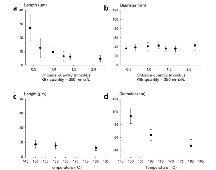
Thus, in our goal to fabricate “on demand” AgNW size
standards with defined lengths and diameters, we carried out experiments by setting the KBr amount at 350 mmol L−1 to control the diameter, and we introduced NaCl in various amounts in order to modulate the length. The results plotted in Fig. 2a show that when the chloride concentration was var-ied between 0.5 and 2.5 mmol L−1, we obtained a collection of AgNWs with lengths ranging from 28 ± 9 down to 4 ± 2 μm. For higher concentrations of chloride, nanoparticles were obtained as the sole product. These results demonstrate that by adequate choice of the nature of co-nucleants and by fine adjustment of their concentrations, the variation of length is actually possible while keeping the diameter almost unchanged (Fig. 2b). This is a straightforward route to select the desired length at constant diameter. The mean length modification can be ascribed to the increased (or decreased) number of seeds, which depends on the molar ratio between
chlorine and silver. This is consistent with the report from Buhro and colleague44who demonstrated, in the early stage of the polyol synthesis, the impact of chloride concentration on the generation of nucleation sites.
Whereas diameters below 50 nm are desirable for most optoelectronic applications requiring a low haze value, in some specific uses like photovoltaics, larger diameters are expected.5In our effort to modulate length and diameter in-dependently, we looked at a protocol to modify the diameter while keeping the length mostly unchanged. It was previously demonstrated by Unalan and colleagues that polyol synthesis carried out at low temperatures generates thick wires (with diameters higher than 300 nm) and that temperature plays a crucial role in the nanowire formation.15We used our stan-dard protocol at various temperatures. The results are presented in Fig. 2c and d. We observed that the mean diam-eter decreased drastically, from 90 ± 10 nm down to 50 ± 8 nm, when the temperature was raised from 150 to 180 °C. At the same time, the mean length of the AgNWs remained
Fig. 2 Tuning of the length of thin AgNWs by NaCl concentration and reaction temperature. (a) Length as a function of the chloride concentration, with a constant KBr concentration of 350 mmol L−1. Increasing the quantity of chloride decreases the mean length of AgNWs. (b) The diameter of AgNWs remains constant around 40 nm while the length decreases from 28μm to 3 μm. The KBr concentration is set at 350 mmol L−1. (c) The length remains almost constant while the reaction temperature is modified. (d) Diameter tuning by reaction temperature modification, where increasing the temperature induces a diameter decrease.
Open Access Article. Published on 20 December 2018. Downloaded on 2/21/2019 9:52:12 AM.
This article is licensed under a
almost constant. This demonstrates that it is also possible to change the diameter of AgNWs while keeping the length mostly unchanged by selecting the adequate reaction temperature.
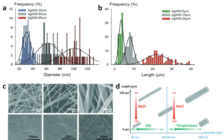
Fabrication of AgNW size standards. Thanks to the proto-cols presented hereinbefore, we were able to prepare different AgNW size standards, as shown in Fig. 3. Combinations of any lengths or diameters in the range of 5–120 μm and 30– 120 nm, respectively, were achievable. This gives access to calibrated samples of nanowires. Histograms showing the distribution in diameters of three different samples of 10μm long AgNWs with various mean diameters are presented in Fig. 3a, and histograms of three different populations with different lengths of 40 nm diameter AgNWs are shown in Fig. 3b. In each case, the overlap between two adjacent statis-tical populations was calculated to be below 15%. SEM im-ages are also presented to illustrate the major differences obtained between the different samples in Fig. 3c; they show undoubtedly how both diameters and lengths can be tuned according to specific synthetic protocols. The main criteria to tune the synthesis of the AgNW standards are schematized in Fig. 3d. We also verified that the proposed conditions are scalable. We performed experiments using up to 5 L semi-batch reactors, which allowed us to confirm that the synthe-sis of AgNWs with pre-determined dimensions can be carried
out“on demand”.
Optoelectrical performances of transparent electrodes: im-pact of AgNWs' dimensions on haziness. When assembled in the form of random networks above the percolation thresh-old, it is known that these materials can provide a relevant al-ternative to TCOs (transparent conductive oxides) for the fab-rication of transparent conductive materials. In this case, the properties of interest (e.g. transparency and sheet resistance) are measured at the macroscopic scale, but depend strongly on the nanosized building blocks. We thus fabricated trans-parent electrodes using various AgNW standards with aspect ratios (i.e. length/diameter ratios) ranging from 55 to 1000. For the sake of clarity, the different AgNW mean dimensions are expressed as DaLb, with a and b indicating the mean di-ameter in nm and the mean length in μm, respectively. The transparency, conductivity and haze factor (i.e. the diffused part of the transmitted light) of the electrodes were measured and compared.
A typical plot of the transmittance as a function of the sheet resistance is presented in Fig. 4a. This series of dots was measured using D30L10 AgNWs. As expected, for these dimensions and for highly purified nanowires, excellent optoelectrical performances were obtained, with for instance 97% transparency for a 50 ohm sq−1 sheet resistance. This compares very well with the state-of-the-art for metallic nanowire-based technology, and also with ITO or any other
Fig. 3 Independent tuning of the diameter and length of silver nanowires. (a) Histograms of three different mean diameters for 10μm long AgNWs. (b) Histograms of three different mean lengths for 40 nm diameter AgNWs (c) SEM pictures of various lengths and diameters. Top, from left to right: mean diameters of 30 nm, 60 nm, and>100 nm; bottom, from left to right: mean lengths of >100 μm, 10 μm, and 5 μm. (d) Schematic diagram to control the morphology of AgNWs.
Open Access Article. Published on 20 December 2018. Downloaded on 2/21/2019 9:52:12 AM.
This article is licensed under a
kind of transparent electrode. We chose to limit the field of study to nanowires with at most 20μm length because longer AgNWs are much more difficult to synthesize, to handle, to formulate and to deposit with high homogeneity on a large substrate area using low-cost solution deposition techniques. In Fig. 4b, the haze values are presented as a function of the total transmittance for AgNWs with different diameters and
for different lengths (5, 10 and 20 μm long). This graph
shows a linear relationship between the haze value and the total transmittance regardless of the length. The dimension effect is very important since the haze values vary from 16% down to less than 2%. Very low haziness, compatible with op-toelectronic applications, is demonstrated for small diame-ters, which is consistent with previous studies,45–47 and very interestingly, it is also shown that even with 90 nm diameter AgNWs, it is possible to achieve low haze values for long nanowires. The haze factor reported in the literature for sub-15 nm diameter silver nanowires reached very low values be-low 1% at 94.5% transmittance.48However, reducing the di-ameter of AgNWs below the mean free path of electrons (∼40
nm) is risky due to charge carrier scattering at the interface and to material instability under operational stress. The his-togram in Fig. 4c shows haze values at given sheet resistances for AgNWs with various lengths and diameters. When the densities of AgNWs are decreased (i.e. increasing sheet resis-tances), the haze value is lowered drastically regardless of the size standards. The impact of the length is obvious since 20 μm long nanowires are much less light diffusive than 10 μm long nanospecies. For instance, in the case of 40 nm diame-ter AgNWs, the haze value is more than doubled when the
length is reduced from 20 μm down to 10 μm. This is
expected by percolation theory. Indeed, for finite size scaling in stick percolation dealing with large size systems, the criti-cal number density (Nc) of sticks required for percolation is
given by Nc× L2= 5.637 where L is the length of nanowires.49
This indicates that for a two-fold increase of the length of the AgNWs, the required Nc to reach percolation is divided by
four.17,50As shown by De et al. and by Lagrange et al., perco-lation theory dominates the electrical behaviour of networks
associated with low density.51,52 However, it should be
Fig. 4 Optoelectrical properties of transparent electrodes fabricated with various AgNW size standards. (a) Total transmittance as a function of the sheet resistance for D30L10 AgNWs. (b) Haze values for 30, 60 and 90 nm diameter AgNWs as a function of the total transmittance. The green part represents measurements made on 5μm long AgNWs, the white part for 10 μm length and the pink part for 20 μm length. The solid lines correspond to linear regressions for each diameter. (c) Comparison of the haze level on given sheet resistances for standard AgNWs (D60L10), thin and short (D40L10), thick and long (D90L20) and thin and long nanowires (D40L20). (d) Left, a 96% transparent electrode with a very low haze value (1.6%); right, a 60% transparent electrode with high haziness (13.0%).
Open Access Article. Published on 20 December 2018. Downloaded on 2/21/2019 9:52:12 AM.
This article is licensed under a
emphasized that in our examples, the electrodes are fabri-cated with densities far above Nc, but the length effect is still
clearly perceptible. The diameter effect is also visible for both
10μm and 20 μm long AgNWs, and as expected, small
diam-eters lower the haziness. Pictures of two electrodes are in-cluded in Fig. 4d to illustrate the pronounced optical differ-ence (transparency and haziness) for different densities of AgNWs.
Toxicity of AgNW size standards. Even if silver toxicology is well known,53,54the toxicity of silver-based nanomaterials is difficult to predict because of their different shapes, which can contribute to different biological responses regarding the plurality of microorganisms. It is described in the literature that low-aspect ratio nanosilver species such as nanoparticles exhibit toxicity mainly relying on silver ion release, either ex-tracellularly (for bacteria) or due to internalization and disso-lution in the lysosomes for animal cells.28,55,56 For high as-pect ratio silver nanomaterials, like AgNWs, the toxicity effect may be driven by dissolution on the one hand; on the other hand, the form factor may contribute largely, as AgNWs ex-hibit a fibre shape having similarities with asbestos. Different toxicity approaches are currently ongoing to decipher possi-ble mechanisms in order to avoid toxicity caused by the as-pect ratio, and some research studies are carried out to estab-lish safety thresholds like for asbestos.57–59
In this study, we investigated the basic responses of mu-rine macrophages in the presence of AgNWs. We chose macrophages because they are in charge of removing nano-materials, and because they are also the main actors at play in insoluble fibre toxicity. Thanks to the fabrication of AgNW size standards described above, toxicological experiments were performed on macrophages to discriminate the effects of the diameter and length.
Previous studies dealing with the potential toxic effect of AgNWs on lung and macrophage models revealed a length-dependent toxicity for both in vitro and in vivo studies, with short nanowires causing less inflammatory responses.20,57,60–62 These different studies compared various lengths; however, a possible diameter effect was not directly tackled by compara-tive experiments, or both diameters and lengths were changed at the same time.
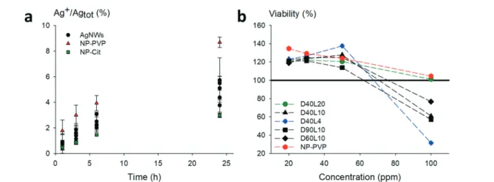
Prior to toxicity experiments on macrophages, we evalu-ated the contribution of dissolved species. The dissolution of relevant morphologies of nanowires was monitored over a pe-riod of 24 h in medium only under working conditions. The AgNWs were compared to silver nanoparticles coated with
PVP with a mean diameter close to standard AgNWs (≈60
nm) and citrate-coated silver nanoparticles which are known to dissolve quickly. The results are shown in Fig. 5a.
Regardless of their dimensions, AgNWs exhibit similar dissolution rates. Silver nanoparticles coated with PVP dissolved slightly faster than the nanowires. For all the AgNWs, the solubilized part of silver was found to be low (<10%), even after 24 h. The corresponding Ag+ concentra-tion was lower than the known silver ion cytotoxicity (1 μg mL−1); hence subsequent toxicity measurements on macro-phages were considered relevant to evaluate the potential in-trinsic toxicity of AgNWs.
AgNWs show low and length-dependent toxicity for macro-phages. Cytotoxicity assays were performed on RAW264.7 mu-rine cells, and the neutral red uptake assay was used to deter-mine the potential adverse effects for each morphology of AgNWs. Various morphologies were chosen to check indepen-dently the potential diameter or length effects. For instance, we used AgNWs with the diameter dimension set at 40 nm
with various lengths (4, 10 and 20 μm), and 10 μm long
Fig. 5 Dissolution rate of the studied nanomaterials and cell viability of RAW264.7 murine cells treated with the nanomaterials. (a) Dissolution monitored for 24 h at 37°C in RPMI medium containing only the nanomaterials at 2.5 μg mL−1concentration except for less concentrated citrate-coated nanoparticles (0.4μg mL−1). PVP-coated nanoparticles (NP-PVP, red triangle) dissolve faster than nanowires (AgNWs, black dot). The total dissolution of all the nanomaterials is less than 10% in the medium. (b) The cells were incubated for 24 h with different concentrations of the nano-materials (from 20 to 100 ppm). The shortest nanowires (D40L4) exhibit a higher toxicity, AgNWs with 10 μm length (D40L10, D60L10, and D90L10) exhibit a similar toxicity regardless of the diameter dimension, and long nanowires with 20μm length (D40L20) and PVP-coated nano-particles (NP-PVP) show a very low toxicity. The LC50 of the short nanowires, which exhibit the highest toxicity, is higher than 80 ppm.
Open Access Article. Published on 20 December 2018. Downloaded on 2/21/2019 9:52:12 AM.
This article is licensed under a
AgNWs with various diameters (40, 60 and 90 nm). No de-crease of cellular activity up to high doses (50 ppm nano-materials) was observed, as evidenced in Fig. 5b. The shortest
AgNWs (length of 4 μm) presented the lowest IC50 at 100
ppm concentration, followed by the three other nanowires
with 10 μm length. Longer AgNWs (20 μm length) did not
present a cellular activity decrease under our experimental conditions. This length effect can be related to the uptake ability of macrophages. Phagocytosis of short nanowires (4 μm) can be easily completed, leading to silver ion release in-side the cells, as with the well-described silver nanoparticle toxicity. The 10μm long nanowires, with different diameters, show a similar inhibition behaviour. The quasi non-toxicity
of long AgNWs (20 μm length) can probably be ascribed to
the fact that the nanowires were not internalized, as the threshold length of phagocytosis was exceeded. These results suggest that AgNWs induce cytotoxicity in a length-type-dependent manner, which is consistent with a reported study on RAW264.7 treated with carbon nanotubes of different lengths.63These results are in accordance with the literature, with cytotoxicity being correlated with the degree of cell up-take and the amount of ionic silver due to intracellular dissolution.62,64
Long AgNWs induce a slight but persistent
pro-inflammatory profile. Macrophages are known for their ca-pacity of phagocytizing particulate substances, such as patho-genic agents but also particulate chemicals (e.g. oxidized lipo-protein particles in the case of atherosclerotic foam cells).
They also play a strong role in immune reaction modulation through their ability to release various signalling molecules such as pro- or anti-inflammatory cytokines, depending on the nature of the phagocytized particles. They also trigger these mechanisms upon frustrated phagocytosis (e.g. in the asbestos case), which makes them an attractive choice to study possible pro-inflammatory responses. To evaluate this response, we measured two major inflammatory cytokines: interleukin 6 (IL-6) and tumour necrosis factor alpha (TNF-α) at the highest non-toxic concentration (50 ppm). The inflam-matory responses due to the nanowires were compared, on the one hand, to that of amorphous and crystalline silica par-ticles (LS30, BCR66, and BCR70) known to induce a low but
well-documented inflammatory response65–68 and on the
other hand, to bacterial lipopolysaccharide (LPS) as a positive control known to induce very strong pro-inflammatory responses.
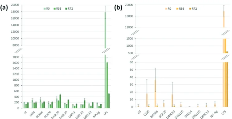
Fig. 6a shows the relative amount of secreted TNF-α by macrophages in response to the different nanowires. After 24 h, we can notice a similar production rate for all the mor-phologies of AgNWs. However, D40L20 is the only AgNW that induces a slightly higher TNF-α production than silica. For IL-6 (Fig. 6b), the production induced by AgNWs was very low and always lower than those induced by silica. Overall, the bar charts for IL-6 and TNF-α show a globally low secretion for each cytokine after a 24 h exposure to nanomaterials, representing less than 1% of the LPS-induced production. These results show that the AgNWs induce a weak increase
Fig. 6 Pro-inflammatory cytokine secretion after treatment with nanomaterials (R0) and persistence results at 36 h (R36) and 72 h (R72). The secretion level was compared to LPS as a positive control and silica-based nanomaterials (LS30, BCR66, and BCR70) as the nanomaterial control. (a) TNF-α secretion is very low (the same level as the control) for all AgNWs, and lower than the silica control (BCR66), except for D40L20. (b) The IL-6 secretion of cells treated with AgNWs is similar regardless of the length or diameter, and at the same level as the negative control. For both IL-6 secretion and TNF-α secretion, the comparative secretion with LPS is very low (<1%).
Open Access Article. Published on 20 December 2018. Downloaded on 2/21/2019 9:52:12 AM.
This article is licensed under a
in cytokine release, which correlated positively with the length of the wires, as expected for fibre-like materials.
One of the major mechanisms at play in persistent fibre/ particle toxicity is the presence of a prolonged pro-inflammatory response over time. In order to test this possi-bility by our in vitro system, we studied the evolution of the production of TNF-α and IL-6 over time after a 24 h exposure to nanomaterials (Fig. 6). LPS-induced TNF-α release was both acute and very transient. Compared to the control, none of the short nanowires, the amorphous silica or the silver nanoparticles induced any significantly higher TNF-α release. Only the small crystalline silica (BCR66) and the long nano-wires (20 μm) induced a weak but sustained TNF-α release. Although the shape of the curves is different for IL-6, with a prolonged LPS-induced release, the same trend can be ob-served, with only the small crystalline silica (BCR66) and the long nanowires inducing a prolonged IL-6 release compared to the control. These results suggest that the long nanowires have an inflammatory effect that is sustained over time. To the best of our knowledge, this is the first reported study on the persistence of the inflammatory response in vitro.
Our results are in line with those obtained using in vivo models20,26,60and suggest that simple in vitro models can be predictive of the inflammatory potential of nanomaterials. In addition, these in vitro systems are much cheaper and ethi-cally more acceptable than in vivo experiments.
Taken collectively, all these data allow us to propose a “safer by design” approach for the use of AgNWs, depending on the applications. Different performances for both electri-cal and optielectri-cal aspects (including haziness) can be achieved, depending on the density and on the dimensions of AgNWs. According to the toxicological results, a rational approach would be to foster the use of nanowires up to 10μm length since they can combine high optoelectrical performances and minimal biological effects.
Conclusions
By careful modifications of the polyol process, in particular, precisely defined halide ion concentrations and temperature setting, independent fine tuning of the diameter and length of AgNWs has been successfully demonstrated. Size standards were prepared accordingly, and used for the fabrication of transparent electrodes. Excellent optoelectrical performances were obtained and the haze value was varied from 1.6 to 26.2%. In the meantime, we performed in vitro toxicological assays on murine macrophages. The AgNWs were found to be weakly toxic for macrophages and showed a length-dependent toxicity, with the toxicity decreasing with length. Conversely, the cytokine release assays showed a weak but significant pro-inflammatory potential for long nanowires (20μm), and the observed persistence of the response, although at weak levels, calls for attention when devising the rules for the use of AgNWs in consumer products and in the recycling of such products. This study set out to take a holistic view of AgNWs, with a clear relationship between AgNW dimensions and both
their optoelectronic performances, when used in the form of random percolating networks, and their toxicity and pro-inflammatory potential. Since AgNWs will certainly be in-creasingly introduced in commercial devices in the short term, it seems important to have a global vision of these nanomaterials, which will ineluctably encourage the rational use of AgNWs in a“safer by design” approach.
Conflicts of interest
There are no conflicts of interest to declare.
Acknowledgements
This work was supported by the Labex Serenade (no. ANR-11-LABX-0064) funded by the “Investissements d'Avenir” French Government program of the French National Research Agency (ANR) through the AMIDEX project (no. ANR-11-IDEX-0001-02). In particular, the authors thank Pr L. Charlet for coordi-nating the “nanosilver topic”, and they are grateful for the funding of D. Toybou's PhD grant.
References
1 D. S. Hecht, L. Hu and G. Irvin, Adv. Mater., 2011, 23, 1482–1513.
2 S. Ye, A. R. Rathmell, Z. Chen, I. E. Stewart and B. J. Wiley, Adv. Mater., 2014, 26, 6670–6687.
3 D. Langley, G. Giusti, C. Mayousse, C. Celle, D. Bellet and J.-P. Simonato, Nanotechnology, 2013, 24, 452001.
4 H. Hosseinzadeh Khaligh, K. Liew, Y. Han, N. M. Abukhdeir and I. A. Goldthorpe, Sol. Energy Mater. Sol. Cells, 2015, 132, 337–341.
5 T. Sannicolo, M. Lagrange, A. Cabos, C. Celle, J.-P. Simonato and D. Bellet, Small, 2016, 12, 6052–6075.
6 T. Sannicolo, D. Muñoz-Rojas, N. D. Nguyen, S. Moreau, C. Celle, J.-P. Simonato, Y. Bréchet and D. Bellet, Nano Lett., 2016, 16, 6052–6075.
7 S.-Y. Chou, R. Ma, Y. Li, F. Zhao, K. Tong, Z. Yu and Q. Pei, ACS Nano, 2017, 11, 11368–11375.
8 W. Lan, Y. Chen, Z. Yang, W. Han, J. Zhou, Y. Zhang, J. Wang, G. Tang, Y. Wei, W. Dou, Q. Su and E. Xie, ACS Appl. Mater. Interfaces, 2017, 9, 6644–6651.
9 T. Sannicolo, N. Charvin, L. Flandin, S. Kraus, D. T. Papanastasiou, C. Celle, J.-P. Simonato, D. Muñoz-Rojas, C. Jiménez and D. Bellet, ACS Nano, 2018, 12, 4648–4659. 10 S. Choi, J. Park, W. Hyun, J. Kim, J. Kim, Y. B. Lee, C. Song,
H. J. Hwang, J. H. Kim, T. Hyeon and D.-H. Kim, ACS Nano, 2015, 9, 6626–6633.
11 Y. Atwa, N. Maheshwari and I. A. Goldthorpe, J. Mater. Chem. C, 2015, 3, 3908–3912.
12 D. Doganay, S. Coskun, S. P. Genlik and H. E. Unalan, Nanotechnology, 2016, 27, 435201.
13 C. Preston, Y. Xu, X. Han, J. N. Munday and L. Hu, Nano Res., 2013, 6, 461–468.
Open Access Article. Published on 20 December 2018. Downloaded on 2/21/2019 9:52:12 AM.
This article is licensed under a
14 C. Preston, Z. Fang, J. Murray, H. Zhu, J. Dai, J. N. Munday and L. Hu, J. Mater. Chem. C, 2014, 2, 1248–1254.
15 S. Coskun, B. Aksoy and H. E. Unalan, Cryst. Growth Des., 2011, 11, 4963–4969.
16 N. V. Nghia, N. N. K. Truong, N. M. Thong and N. P. Hung, Int. J. Mater. Chem., 2012, 2, 75–78.
17 S. M. Bergin, Y.-H. Chen, A. R. Rathmell, P. Charbonneau, Z.-Y. Li and B. J. Wiley, Nanoscale, 2012, 4, 1996.
18 Y. Shi, H. Li, L. Chen and X. Huang, Sci. Technol. Adv. Mater., 2005, 6, 761–765.
19 M. J. Large, J. Burn, A. A. King, S. P. Ogilvie, I. Jurewicz and A. B. Dalton, Sci. Rep., 2016, 6, 25365.
20 K. F. Chung, J. Seiffert, S. Chen, I. G. Theodorou, A. E. Goode, B. F. Leo, C. M. McGilvery, F. Hussain, C. Wiegman, C. Rossios, J. Zhu, J. Gong, F. Tariq, V. Yufit, A. J. Monteith, T. Hashimoto, J. N. Skepper, M. P. Ryan, J. Zhang, T. D. Tetley and A. E. Porter, ACS Nano, 2017, 11, 2652–2664. 21 L. D. Scanlan, R. B. Reed, A. V. Loguinov, P. Antczak, A.
Tagmount, S. Aloni, D. T. Nowinski, P. Luong, C. Tran, N. Karunaratne, D. Pham, X. X. Lin, F. Falciani, C. P. Higgins, J. F. Ranville, C. D. Vulpe and B. Gilbert, ACS Nano, 2013, 7, 10681–10694.
22 R. M. Silva, J. Xu, C. Saiki, D. S. Anderson, L. M. Franzi, C. D. Vulpe, B. Gilbert, L. S. Van Winkle and K. E. Pinkerton, Part. Fibre Toxicol., 2014, 11, 52.
23 M. Charehsaz, S. Coskun, H. E. Unalan, R. Reis, S. Helvacioglu, A. K. Giri and A. Aydin, Toxicol. Environ. Chem., 2017, 99, 837–847.
24 S. Sweeney, I. G. Theodorou, M. Zambianchi, S. Chen, A. Gow, S. Schwander, J. Zhang, K. F. Chung, M. S. P. Shaffer, M. P. Ryan, A. E. Porter and T. D. Tetley, Nanoscale, 2015, 7, 10398–10409.
25 R. Cui, Y. Chae and Y.-J. An, Chemosphere, 2017, 185, 205–212.
26 L. D. Scanlan, R. B. Reed, A. V. Loguinov, P. Antczak, A. Tagmount, S. Aloni, D. T. Nowinski, P. Luong, C. Tran, N. Karunaratne, D. Pham, X. X. Lin, F. Falciani, C. P. Higgins, J. F. Ranville, C. D. Vulpe and B. Gilbert, ACS Nano, 2013, 7, 10681–10694.
27 C. Mayousse, C. Celle, E. Moreau, J.-F. Mainguet, A. Carella and J.-P. Simonato, Nanotechnology, 2013, 24, 215501. 28 C. Aude-Garcia, F. Villiers, V. Collin-Faure, K. Pernet-Gallay,
P.-H. Jouneau, S. Sorieul, G. Mure, A. Gerdil, N. Herlin-Boime, M. Carrière and T. Rabilloud, Nanotoxicology, 2016, 10, 586–596.
29 B. Dalzon, C. Aude-Garcia, V. Collin-Faure, H. Diemer, D. Béal, F. Dussert, D. Fenel, G. Schoehn, S. Cianférani, M. Carrière and T. Rabilloud, Nanoscale, 2017, 9, 9641–9658. 30 G. Repetto, A. del Peso and J. L. Zurita, Nat. Protoc., 2008, 3,
1125–1131.
31 Y. Sun, Y. Yin, B. T. Mayers, T. Herricks and Y. Xia, Chem. Mater., 2002, 14, 4736–4745.
32 Y. Sun, B. Mayers, T. Herricks and Y. Xia, Nano Lett., 2003, 3, 955–960.
33 P. Zhang, I. Wyman, J. Hu, S. Lin, Z. Zhong, Y. Tu, Z. Huang and Y. Wei, Mater. Sci. Eng., B, 2017, 223, 1–23.
34 P. Zhang, Y. Wei, M. Ou, Z. Huang, S. Lin, Y. Tu and J. Hu, Mater. Lett., 2018, 213, 23–26.
35 L. Hu, H. S. Kim, J.-Y. Lee, P. Peumans and Y. Cui, ACS Nano, 2010, 4, 2955–2963.
36 C. Chen, L. Wang, G. Jiang, J. Zhou, X. Chen, H. Yu and Q. Yang, Nanotechnology, 2006, 17, 3933.
37 K. Zhang, Y. Du and S. Chen, Org. Electron., 2015, 26, 380–385.
38 B. Li, S. Ye, I. E. Stewart, S. Alvarez and B. J. Wiley, Nano Lett., 2015, 15, 6722–6726.
39 E.-J. Lee, Y.-H. Kim, D. K. Hwang, W. K. Choi and J.-Y. Kim, RSC Adv., 2016, 6, 11702–11710.
40 C. Celle, C. Mayousse, E. Moreau, H. Basti, A. Carella and J.-P. Simonato, Nano Res., 2012, 5, 427–433.
41 C. Mayousse, C. Celle, E. Moreau, J.-F. Mainguet, A. Carella and J.-P. Simonato, Nanotechnology, 2013, 24, 215501. 42 W. M. Schuette and W. E. Buhro, Chem. Mater., 2014, 26,
6410–6417.
43 R. R. da Silva, M. Yang, S.-I. Choi, M. Chi, M. Luo, C. Zhang, Z.-Y. Li, P. H. C. Camargo, S. J. L. Ribeiro and Y. Xia, ACS Nano, 2016, 10, 7892–7900.
44 W. M. Schuette and W. E. Buhro, ACS Nano, 2013, 7, 3844–3853.
45 Y. Liu, Y. Chen, R. Shi, L. Cao, Z. Wang, T. Sun, J. Lin, J. Liu and W. Huang, RSC Adv., 2017, 7, 4891–4895.
46 J. Lee, P. Lee, H. Lee, D. Lee, S. S. Lee and S. H. Ko, Nanoscale, 2012, 4, 6408.
47 X. Yu, X. Yu, J. Zhang, L. Chen, Y. Long and D. Zhang, Nano Res., 2017, 10, 3706–3714.
48 H.-W. Jang, Y.-H. Kim, K.-W. Lee, Y.-M. Kim and J.-Y. Kim, APL Mater., 2017, 5, 080701.
49 J. Li and S.-L. Zhang, Phys. Rev. E, 2009, 80, 040104.
50 J. Han, S. Yuan, L. Liu, X. Qiu, H. Gong, X. Yang, C. Li, Y. Hao and B. Cao, J. Mater. Chem. A, 2015, 3, 5375–5384. 51 S. De, T. M. Higgins, P. E. Lyons, E. M. Doherty, P. N.
Nirmalraj, W. J. Blau, J. J. Boland and J. N. Coleman, ACS Nano, 2009, 3, 1767–1774.
52 M. Lagrange, D. P. Langley, G. Giusti, C. Jiménez, Y. Bréchet and D. Bellet, Nanoscale, 2015, 7, 17410–17423.
53 K. M. Newton, H. L. Puppala, C. L. Kitchens, V. L. Colvin and S. J. Klaine, Environ. Toxicol. Chem., 2013, 32, 2356–2364.
54 C. Hogstrand, F. Galvez and C. M. Wood, Environ. Toxicol. Chem., 1996, 15, 1102–1108.
55 A. Pratsinis, P. Hervella, J.-C. Leroux, S. E. Pratsinis and G. A. Sotiriou, Small, 2013, 9, 2576–2584.
56 M. van der Zande, R. J. Vandebriel, E. Van Doren, E. Kramer, Z. Herrera Rivera, C. S. Serrano-Rojero, E. R. Gremmer, J. Mast, R. J. B. Peters, P. C. H. Hollman, P. J. M. Hendriksen, H. J. P. Marvin, A. A. C. M. Peijnenburg and H. Bouwmeester, ACS Nano, 2012, 6, 7427–7442.
57 R. M. Silva, J. Xu, C. Saiki, D. S. Anderson, L. M. Franzi, C. D. Vulpe, B. Gilbert, L. S. Van Winkle and K. E. Pinkerton, Part. Fibre Toxicol., 2014, 11, 52.
58 A. Schinwald, T. Chernova and K. Donaldson, Part. Fibre Toxicol., 2012, 9, 47–47.
Open Access Article. Published on 20 December 2018. Downloaded on 2/21/2019 9:52:12 AM.
This article is licensed under a
59 A. Schinwald, F. A. Murphy, A. Prina-Mello, C. A. Poland, F. Byrne, D. Movia, J. R. Glass, J. C. Dickerson, D. A. Schultz, C. E. Jeffree, W. MacNee and K. Donaldson, Toxicol. Sci., 2012, 128, 461–470.
60 A. Schinwald and K. Donaldson, Part. Fibre Toxicol., 2012, 9, 34.
61 A. Schinwald, T. Chernova and K. Donaldson, Part. Fibre Toxicol., 2012, 9, 47–47.
62 I. G. Theodorou, K. H. Müller, S. Chen, A. E. Goode, V. Yufit, M. P. Ryan and A. E. Porter, ACS Biomater. Sci. Eng., 2017, 3, 2336–2347.
63 D. Liu, L. Wang, Z. Wang and A. Cuschieri, Nanoscale Res. Lett., 2012, 7, 361.
64 X. Wang, Z. Ji, C. H. Chang, H. Zhang, M. Wang, Y.-P. Liao, S. Lin, H. Meng, R. Li, B. Sun, L. V. Winkle, K. E. Pinkerton, J. I. Zink, T. Xia and A. E. Nel, Small, 2014, 10, 385–398. 65 C. M. Sayes, K. L. Reed and D. B. Warheit, Toxicol. Sci.,
2007, 97, 163–180.
66 D. Warheit, T. McHugh and M. Hartsky, Scand. J. Work, Environ. Health, 1995, 19–21.
67 M. V. D. Z. Park, I. Lynch, S. Ramírez-García, K. A. Dawson, L. de la Fonteyne, E. Gremmer, W. Slob, J. J. Briedé, A. Elsaesser, C. V. Howard, H. van Loveren and W. H. de Jong, J. Nanopart. Res., 2011, 13, 6775–6787.
68 E.-J. Park, J. Roh, Y. Kim and K. Choi, J. Health Sci., 2011, 57, 60–71.
Open Access Article. Published on 20 December 2018. Downloaded on 2/21/2019 9:52:12 AM.
This article is licensed under a