0022-3360/93/0067-1047$03.00
EVOLUTION IN YODERIMYINAE (EOMYIDAE: RODENTIA),
WITH NEW MATERIAL FROM THE WHITE RIVER FORMATION
(CHADRONIAN) AT FLAGSTAFF RIM, WYOMING
ROBERT J. EMRY1 AND WILLIAM W. KORTH2
1 Department of Paleobiology, National Museum of Natural History, Smithsonian Institution, Washington, D.C. 20560, and 2Department of Geological Sciences, University of Rochester, Rochester, New York 14625, and
Department of Geosciences, Hobart and William Smith Colleges, Geneva, New York 14456
ABSTRACT—Three species of Yoderimyinae (Eomyidae: Rodentia) are recognized from the lower part of the White River Formation (early to medial Chadronian) in the Flagstaff Rim area, Wyoming. The new material allows an improved diagnosis for the subfamily. The enamel microstructure of Yoderimyinae supports its inclusion in the Eomyidae.
A new genus, Zemiodontomys, is established for Yoderimys burkei Black, and new material, including upper dentition, is referred to this species. This genus differs from Yoderimys in having higher crowned and more lophodont teeth and in lacking P3. A second new genus, Litoyoderimys, is established for Yoderimys lustrorum Wood, and a new species, L. auogoleus, is referred to the genus. This genus has lower crowned, more cuspate teeth than Yoderimys.
Through early and medial Chadronian time, evolution in yoderimyines includes the following morphologic transformations: increase in size; increase in crown height and lophodonty of cheek teeth; reduction of P3 (from double-rooted, to single-rooted, to absent); increase in relative size of P4 and p4; and increased longitudinal torsion of the mandible.
INTRODUCTION
7
'ODERIMYS BUMPI was described by Wood (1955) from the"Yoder formation" of eastern Wyoming, considered by Wood (1955) and most subsequent authors as representing early Chadronian time. Wood (1955) discussed several possible al locations of his new genus (Sciuravidae, Zapodidae, Eomyidae, new family), but resolved the question by erecting a new sub family of Eomyidae, the Yoderimyinae, to receive two genera, his new genus Yoderimys and another "undescribed form from the Vieja of Texas" (Wood, 1955, p. 519). Wood's arrangement has been followed, and progressively corroborated, by the au thors of four additional species: Y. burkei Black, 1965, from Pipestone Springs, Montana; Y. steward (Russell, 1972) from Saskatchewan (also discussed by Storer, 1978); Y. lustrorum Wood, 1974, the form that Wood had previously (1955) men tioned as an undescribed new genus from the Vieja of Texas; and Y. yarmeri Wilson and Runkel, 1991, also from Trans-Pecos Texas. In their study of the incisor enamel microstructure in eomyids, Wahlert and von Koenigswald (1985) included Yo
derimys in the Eomyidae, but did not illustrate or describe its
enamel specifically.
New specimens in the collections of the National Museum of Natural History (Smithsonian Institution) demonstrate a greater diversity of Chadronian Yoderimyinae than previously known. The yoderimyine material was among abundant microverte-brates obtained by screen washing large volumes of matrix from three sites in the White River Formation in the Flagstaff Rim area of Wyoming (Emry, 1973). These three sites, all fossil quar ries that also produced large samples of macro vertebrates, occur along the south side of the Little Lone Tree Gulch drainage (Emry, 1973, figs. 17, 19). Names of these sites, used in field and museum records, are Low Red Quarry, Dry Hole Quarry, and B minus 44' Quarry. The Low Red Quarry, stratigraphically lowest of the three, occurs at 18.3 m (60 ft) above the base of the generalized zonation section (Emry, 1973, p. 29) in the "Lower Banded Zone," in the NE'A, SW1/*, NE»/4, sec. 24, T3 IN, R82W. Dry Hole Quarry is slightly higher stratigraphically, at about 21.3 m (70 ft) above the base of the section (for location see Emry, 1978, p. 1,007). B minus 44' Quarry occurs, not surprisingly, at 44 ft (13.4 m) below Ash B, or about 40 m (131 ft) above the base of the generalized section (for location, see Emry, 1978, p. 1,005). The diverse fauna recovered from the
Low Red and Dry Hole Quarries indicates an early Chadronian age; both have many elements in common with the Yoder Local Fauna, with which they are considered essentially coeval. The diverse fauna of B minus 44' Quarry is typical of medial Chad ronian time (Emry et al., 1987); it is substantially homotaxial with the Pipestone Springs local fauna of Montana.
Abbreviations used: CM, Carnegie Museum of Natural His tory; FMNH, Field Museum of Natural History; SDSM, South Dakota School of Mines, Rapid City; USNM, National Museum of Natural History (Smithsonian Institution).
Dental terms are based on those of Wood and Wilson (1936).
SYSTEMATIC PALEONTOLOGY
Order RODENTIA Bowdich, 1821 Family EOMYIDAE Deperet and Douxami, 1902
Subfamily YODERIMYINAE Wood, 1955
Emended diagnosis.—Skull sciuromorphous, lacking
inter-orbital foramen of eomyines; anterior end of masseteric fossa expanded laterally on mandible; ascending ramus widely sep arated from tooth row; plane of angular process inclined me-dioventrally making medial surface of mandible strongly con cave; dP3 present in all species; P4 and p4 progressively larger relative to Ml and ml; cheek teeth brachydont to mesodont, progressively dominated by lophs, cusps progressively less dis tinct; wide anterior cingula on all molars; distinct anteroconid on p4; incisor enamel with two-part portio interna, but inner part thinner than in other eomyids, and outer part thicker.
Included genera. — Yoderimys Wood, 1955; Zemiodontomys
n. gen.; Litoyoderimys n. gen.
Range. —Chadronian of Trans-Pecos Texas, Wyoming, Mon
tana, and Saskatchewan and possibly Duchesnean of Saskatch ewan. Ostrander (1985, table 1, appendix II) discussed several Chadronian local faunas in northwestern Nebraska; Yoderimys species appear only in his faunal lists, without further discussion, with no illustrations, and with no specimens cited as vouchers. It is not certain that a yoderimyine is represented by the unusual teeth from the Lac Pelletier Lower Fauna of Saskatchewan, referred by Storer (1987) to "Yoderimys sp. indet."
Discussion.— Wood's diagnosis of the subfamily Yoderimyi
nae, based on Yoderimys bumpi and the Vieja form that he later (1974) named Y. lustrorum, included only dental characters. 1047
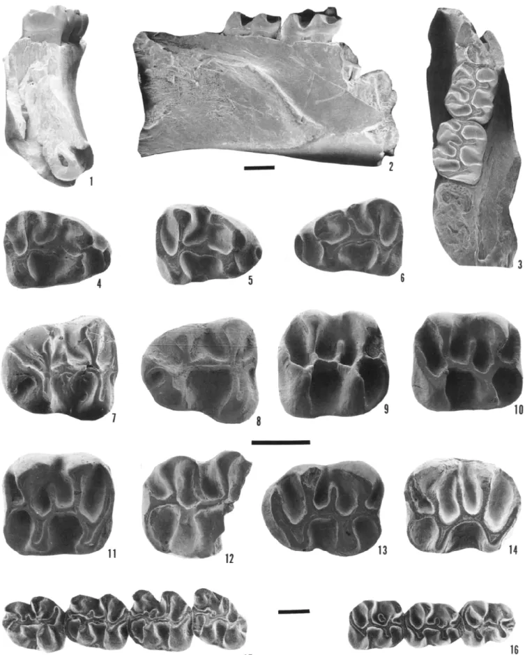
FIGURE 1 —Yoderimys species. 1-14, Yoderimys stewarti (Russell), referred specimens from Flagstaff Rim area, Wyoming. 1-3, USNM 455769, right partial mandibular ramus with ml-2, in /, anterior, 2, lateral, and 3, occlusal views; 4, USNM 455780, right dp4; 5, USNM 455820
Two distinctive characters of the mandible, not previously not ed, that also separate yoderimyines from other eomyids are the morphology of the masseteric fossa and the attitude of the an gular process and ascending ramus. In eomyines, the masseteric fossa is shallow, bounded ventrally and dorsally by low, rounded ridges, and typically terminates anteriorly beneath p4 or m 1 in a shallow U- or V-shape. In yoderimyines, the anterior limit of the fossa is also beneath p4 or m 1, but here the anterior end of the fossa is built up much thicker. A depression in the side of the jaw below ml, more prominent in Zemiodontomys, results in a small shelf above the dorsal border of the fossa. The dorsal masseteric crest is at a much steeper angle than is typically the case in eomyines. The ventral border of the masseteric fossa is less prominent than the dorsal, and continues forward past its junction with the dorsal crest to form a semicircular depression ahead of the greater part of the fossa.
In typical eomyines, the ascending ramus and angular process lie in the same plane as the horizontal ramus and tooth row; the ascending ramus is separated from the tooth row by just a narrow, shallow valley, and its anterior edge passes the tooth row at a shallow angle. In yoderimyines, the anterior edge of the ascending ramus passes the alveolar border at a much steeper angle, and the plane of the ascending ramus and angular process is progressively (i.e., more so in Zemiodontomys) rotated me-dioventrally about the long axis of the horizontal ramus. This rotation moves the ascending ramus laterally away from the horizontal ramus, leaving a broad, deep valley between the as cending ramus and the tooth row. In his description of " Y."
burkei, Black (1965, p. 31) noted that the ascending ramus "ap
pears to arise much farther down the side of the mandible than in other eomyids, with its anterior border well below m2." The rotation of the angle also affects the ventral part of the horizontal ramus and the incisor, so that the medial surface of the mandible becomes progressively more concave, and the incisor is rotated linguad relative to the cheek teeth.
Because the permanent dentition of " Yoderimys" burkei ap pears to lack P3, the presence of P3 can no longer be considered diagnostic for the subfamily. In yoderimyines, the cheek teeth range from cuspate in "y." lustrorum and the new species de scribed below to highly lophate in "y." burkei, where the cusps are so completely submerged into the lophs that their limits cannot be recognized except perhaps as bulges within the base of the lophs. Thus, "cusps plump and not reduced to narrow crests" (Wood, 1955, p. 519) is no longer diagnostic of the subfamily.
Genus YODERIMYS Wood, 1955
Type species. — Yoderimys bumpi Wood, 1955.
Included species. — Yoderimys steward (Russell, 1972); Y.
yar-meri Wilson and Runkel, 1991.
Emended diagnosis.—Chtek teeth lophate, with distinct cusps;
P3 present; mesolophs and mesolophids variable in length; ec-toloph complete from paracone to metacone; metacone oblique ly compressed.
Range. — Chadronian of Saskatchewan, Wyoming, and
Trans-Pecos Texas. Possibly Duchesnean of Saskatchewan.
Discussion. — Yoderimys is morphologically intermediate be
tween the other Chadronian genera of Yoderimyinae named below. The cheek teeth are more lophate than those referred to
Litoyoderimys and are lower crowned and less lophate than
those of Zemiodontomys. In the Flagstaff Rim section, Yoder
imys and Litoyoderimys occur together in the lower, early Chad
ronian, part of the section. Zemiodontomys is from a higher stratigraphic level, in the medial Chadronian part of the section, considered approximately coeval with the Pipestone Springs local fauna of Montana.
YODERIMYS STEWARTI (Russell, 1972)
Figures 1.1-1.4, 2, 3
Holotype. —ROM 6317, left maxillary fragment with M l - 2 . Referred specimens.— Material from Saskatchewan is listed
by Russell (1972) and Storer (1978). Material from Ragstaff Rim area of Wyoming is as follows. From Dry Hole Quarry, USNM numbers: 455769, right partial ramus with ml and m2; 455773, 776, 780, Rdp4; 455772, 774, 775, 777, 779 Ldp4; 455787 Rp4; 455786, 789-791 Lp4; 455770, 792, 793, 796, 798-803, Rm 1 or m2; 455794, 795, 797, 804, 805, Lm 1 or m2; 455782, 785, Rm3; 455783, 784, Lm3; 455756, 759, 761, RdP4; 455758, 760, LdP4; 455764-765, RP4; 455762-763, LP4; 455742, 746, 747, 749, 751, 754, RM1 or M2; 455743-745, 748, 750, 752, 753, 771, LM1 or M2; 455766, RM3; 455767, 768, LM3. From Low Red Quarry, USNM numbers: 455820, Rdp4; 455819, Ldp4; 455821, Lp4; 455822-824, Rml or m2; 455814-815, RP4; 455812, LP4; 455816, RM1 or M2; 455811, 813, LM1 or M2; 455818, RM3; 455817, LM3.
Emended diagnosis. —Cheek teeth more lophate and with cusps
less distinct than in Y. bumpi; mesolophid of m2 and m3 same as in m 1, originating from ectolophid and extending lingually. Smaller than Y. yarmeri Wilson and Runkel, 1991.
Morphology.— Of the Ragstaff Rim sample, only one speci
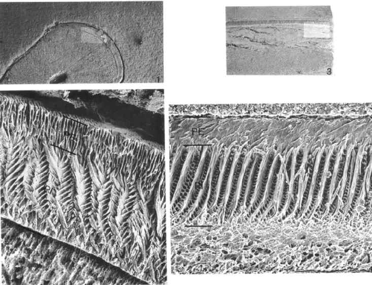
men (USNM 455769) has an incisor associated with cheek teeth. This is a partial mandibular ramus with m 1 and m2; the anterior end of the jaw is missing, but the broken incisor can be observed in cross section (Figure 1.1). Although identification of rodent taxa on the basis of isolated incisors is a dubious exercise at best, isolated incisors in the washed samples can be allocated to the species if associations with cheek teeth are established in single specimens in the same sample. In this instance, allocations of isolated incisors could also be made on the basis of size, because the samples contain no other rodent cheek teeth of appropriate size. These incisors do not differ in any important way from those of Y. bumpi described and illustrated by Wood (1955, p. 523 and fig. 1). The enamel of these incisors (Figure 2) has the three-layered Schmelzmuster characteristic of eomyids (Wahlert and von Koenigswald, 1985), but differs from that of eomyines principally in the relative thickness of the layers; the inner part of the portio interna is much thinner relative to the outer part, compared to that in eomyines, and the portio externa is relatively thicker.
All tooth positions are represented in the Ragstaff Rim sam ples. Of these, only the deciduous fourth premolars have not been described previously for the species. DP4 is smaller and more cuspate than P4, its four primary cusps more distinct, the lophs lower, and the basins shallower (Figure 3.5). The meso-loph is variably developed; in 455756 and 455759 it consists of a short spur projecting inward from the ectoloph, and in 455758 (Figure 3.5) and 455761 it is longer but lower. The paracone and metacone are anteroposteriorly compressed. The
right dp4; 6, USNM 455722, left dp4; 7, USNM 455790, left p4; 8, USNM 455821, left p4; 9, USNM 455801, right ml or m2; 10, USNM 455822, right ml or m2; 11, USNM 455800, left ml or m2; 12, USNM 455794, partial left ml or m2; 13, USNM 455782, right m3; 14, USNM 455783, left m3. 15, 16, Yoderimys bumpi Wood from Yoder local fauna of Goshen Hole of Wyoming. 15, SDSM 5330, holotype, left p4-m3; 16, SDSM 5325, right p4-m2. 1-3, 15, 16, approximately x 10, 4-14 approximately x20; scale bars = 1 mm.
FIGURE 2—Enamel microstructure of Yoderimys stewarti (Russell), referred lower incisor from Flagstaff Rim area. /, 2, transverse section and 3,
4, sagittal section. Lighter rectangles on 1 and 3 show positions of 2 and 4, respectively. PE, portio externa; PI, portio interna; change in
direction of enamel prisms within portio interna is typical of eomyids. Approximate magnifications are 1, x 35.2; 2, x 717; 3, x 32.0; 4, x 500. protocone and hypocone are obliquely compressed. The
par-astylar area is expanded anteriorly. The entoloph (mure) is a short, low, anteroposteriorly directed crest near the midline of the tooth, connecting the lingual ends of paracone and metacone. The ectoloph is complete, but very weak, providing minimal closure of the trigon basin. The buccal part of the anterior cin-gulum (the part anterior to the paracone) is strongly developed, but it is weakly developed lingual to its junction with the anterior arm of the protocone. The posterior cingulum is complete from the hypocone to the posterior crest of the metacone.
The permanent cheek teeth of Yoderimys have been described by Wood (1955), Russell (1972), Storer (1978), Kihm (1987), and Wilson and Runkel (1991). P4, Ml, and M2 all have the same basic pattern, and as isolated teeth are difficult to separate. Kihm (1987, p. 32), for example, was apparently unable to separate isolated examples of P4, Ml, and M2. It therefore seems worthwhile to mention some of the characters that dis tinguish P4 and allow it to be confidently identified. P4 is larger than the molars, although with isolated teeth this may be in sufficient to permit identification. The anterior half (protoloph) of P4 is noticeably wider than the posterior (metaloph) half (see Figure 3.1-3.4), which is not the case with the molars. The part of the anterior cingulum (anteroloph) buccal to its junction with
the anterior arm of the protoconid is anterobuccally expanded, encloses a basin that is larger than that of the molars, and is crescentic rather than straight. The paracone is relatively larger than the metacone, and the paracone and metacone are closer together. The basin bounded by protoloph, metaloph, ectoloph and mure is smaller than in the molars and is more or less equally divided by a short, complete mesoloph that connects the mure and ectoloph. P4 typically has a small, concave in terdental wear facet for P3 on its anterior surface, just where the anterior arm of the protocone meets the anterior cingulum. On Ml and M2, interdental wear facets are typically developed across most of the width of the anterior cingulum, where these teeth contact P4 and M1, respectively. The tooth illustrated by Storer (1978, fig. 8A) as an Ml or M2 is almost certainly a P4, based on its size, shape, and morphology; the size and position of its anterior interdental wear facet should confirm this iden tification.
Ml and M2 are so nearly identical in size and morphology that when isolated they cannot be distinguished; they are lumped in one category in the statistical summaries presented here (Ta ble 1). As Wood (1955) noted in the original sample from the Yoder Fauna, the mesoloph is extremely variably developed. Figure 3.6-3.10 shows the amount of variation represented in
FIGURE 5—Upper teeth of Yoderimys stewarti (Russell), referred, from Flagstaff Rim area, Wyoming, in occlusal view. 1, USNM 455814, right P4; 2, USNM 455764, right P4; 3, USNM 455762, left P4; 4, USNM 455812, left P4; 5, USNM 455758, left dP4; 6, USNM 455746, right Ml or M2; 7, USNM 455749, right Ml or M2; 8, USNM 455753, left Ml or M2; 9, USNM 455754, right Ml or M2; 10, USNM 455750, left Ml or M2; 11, USNM 455817, left M3; 12, USNM 455766, right M3. All approximately x20; scale bar = 1 mm.
the Flagstaff Rim samples; in some teeth the mesoloph is but a small spur directed buccally from the entoloph (mure) (Figure 3.6), in others (Figure 3.8) it extends anterobuccally to fuse with the posterior part of the paracone, and in still others (Figure 3.9, 3.10) it reaches the ectoloph.
M3 in the Flagstaff Rim samples is essentially as described by Wood for the Yoder sample of Y. bumpi. Minor variation is seen in the metaloph, which may or may not have a small anterobuccally directed spur that meets the ectoloph, cutting off a small basin (compare Figure 3.11 and 3.12).
Of the mandibular dentition, Dp4 has not been described previously. It is roughly triangular in occlusal outline, with the metalophid much narrower than the hypolophid (Figure 1.4-1.6). Compared to p4, it is smaller, relatively narrower, and has more distinct cusps and less prominent lophs. The ectolophid is low, centrally positioned, and anteroposteriorly directed. The protoconid and metaconid are close together. The protoconid is connected to a small anteroconid by a low loph that arises buccally and curves anterolingually. A strong crest runs poste
riorly from the metaconid, ending at the midpoint of the tooth. The mesolophid is variably developed (compare Figure 1.9,
1.10, 1.11, 1.12); where most complete it connects lingually with the posterior crest of the metaconid. The posterior cingulum continues from the hypoconid to the lingual border of the tooth, where a lower connection to the entoconid closes a basin be tween metalophid and posterior cingulum.
Measurements.—Measurements of the Yoderimys teeth from
Low Red Quarry and Dry Hole Quarry are statistically sum marized in Table 1. Figures 4 and 5 show sizes of all upper and lower first and second molars.
Discussion.—Wood (1980) questioned the validity of Yoderi mys stewarti (Russell, 1972), noting that it appeared to differ
only in its slightly larger size from Y. bumpi. He refrained from synonymizing the species, however, suggesting that better ma terial might justify the recognition of two species. The material from Flagstaff Rim seems only to complicate the issue of size distinction. When the length and maximum width of M1 and M2 (Figure 4) and m 1 and m2 (Figure 5) of the Flagstaff Rim
TABLE 7—Statistical summary of measurements (in mm) of teeth of
Yoderimys stewarti (Russell), referred, from Flagstaff Rim area, Wy
oming. AP, maximum anteroposterior; TR, maximum transverse; TR tri, transverse at trigonid (metalophid); TR tal, transverse at tal-onid (hypolophid). Measurement dp4, AP dp4, TR tri dp4, TR ta p4, AP p4, TR tri p4, TR tal ml or m2, m 1 or m2, ml or m2, m3, AP m3, TR tri m3, TR tal dP4, AP dP4, TR P4, AP P4, TR Ml orM2, Ml orM2, M3, AP M3, TR AP TRtri TRtal AP TR N 9 10 9 6 6 6 19 19 18 3 4 4 4 5 7 7 15 13 5 3 Range 1.79-2.05 1.03-1.27 1.25-1.52 2.25-2.39 1.43-1.65 1.85-2.03 1.91-2.33 1.49-2.07 1.76-2.12 2.16-2.23 1.49-1.63 1.69-1.92 1.73-1.99 1.57-1.80 2.06-2.23 2.34-2.50 1.82-2.14 2.06-2.36 1.45-1.78 1.73-2.07 Mean 1.94 1.11 1.40 2.29 1.58 1.96 2.13 1.77 1.88 2.20 1.56 1.81 1.82 1.69 2.17 2.39 1.97 2.17 1.66 1.91 SD 0.081 0.072 0.088 0.051 0.078 0.072 0.115 0.125 0.083 0.038 0.060 0.097 0.114 0.097 0.055 0.058 0.087 0.087 0.130 0.176
cv
4.14 6.47 6.28 2.23 4.96 3.65 5.40 7.05 4.39 1.72 3.85 5.33 6.24 5.72 2.54 2.43 4.44 4.00 7.82 9.19 samples are plotted, along with the ranges of the same mea surements for the Y. bumpi sample from the Yoder fauna (Wy oming) and Y. stewarti sample from the Calf Creek local fauna (Saskatchewan), the ranges of measurements are seen to overlap extensively; in fact, the range of length of m 1 and m2 of Y.stewarti completely overlaps the range of Y. bumpi. Moreover,
the mean values for Y. stewarti are not invariably larger; the
i
2.5
2.3
2.1
1.9
1.7h
1.5
h
i-T
T^
.®HP
Typ» J I I I I I I L1.5 1.7 1.9 2.1
2.3 2.5
L e n g t h
FIGURE 4—Bivariate plot of lengths and widths of Ml and/or M2 of yoderimyines. Solid squares represent Flagstaff Rim Yoderimys, re ferred here to Y. stewarti (Russell). Solid circles represent
Litoyoderi-mys auogoleus n. sp., including holotype (marked type). Solid lines
indicate ranges of measurements of topotypic Yoderimys bumpi given by Wood (1955) and Kihm (1987). Dashed lines indicate ranges of measurements of topotypic Y. stewarti given by Russell (1972) and Storer (1978). Open circles with 1 and 2 indicate Ml and M2, re spectively, of holotype of Y. stewarti. Lines indicating ranges cross each other at their respective mean values.
2.4 r
2.2 h
JC 4 - » ■ D£
2.0
1.8
1.6
1.4
* ■-^fe
P> Ji
1.4 1.6 1.8 2.0 2.2 2.4
Length
FIGURE 5—Bivariate plot of lengths and widths of ml and/or m2 of yoderimyines. Solid squares represent Flagstaff Rim Yoderimys, re ferred here to Y. stewarti (Russell). Solid circles represent
Litoyoderi-mys auogoleus n. sp. Solid lines indicate ranges of measurements of
topotypic Yoderimys bumpi given by Wood (1955) and Kihm (1987). Dashed lines indicate ranges of measurements of topotypic Y. stewarti given by Russell (1972) and Storer (1978). Open circles with 1 and 2 indicate m 1 and m2, respectively, of the holotype of Y. bumpi. Lines indicating ranges cross each other at their respective mean values.
mean value for the length of M1 and M2 of Y. stewarti is less than that of Y. bumpi (and Storer's statistics also mistakenly include some P4 measurements, which, because P4 is larger than the molars, surely artificially extends the upper end of this range and increases the mean value). Furthermore, the length and width values for the type Ml and M2 of Y. stewarti fall closer to the mean values of Y. bumpi than to the mean values of Y. stewarti, and, conversely, the width and length values of the type m 1 and m2 of Y. bumpi fall closer to the mean values of Y. stewarti than of Y. bumpi. With these considerations, and the fact that the sample sizes are very small, the larger size of
Y. stewarti is unproven, at the least.
There is some suggestion that the fourth premolars of Y.
stewarti may be wider, relative to the width of the first molars,
than in Y. bumpi. Statistically, however, the difference is very slight, and again the sample size of Y. bumpi is too small to permit confidence in a conclusion. The original sample of Y.
bumpi includes the only two specimens in which p4 and m 1 of
single individuals are associated; according to Wood's (1955, p. 523) measurements, p4 is slightly narrower than ml in one specimen and slightly wider in the other.
In five of the six examples of p4 in the Flagstaff Rim samples, a short crest links the anteroconid to the protoconid (Figure 1.7,
1.8); in the remaining specimen, USNM 455789, this crest unites with the metalophid midway between the metaconid and pro toconid. Wood (1955, p. 521) noted that in the holotype of Y.
bumpi this short crest from the anteroconid runs to the meta
conid, whereas in his two referred specimens it runs to the protoconid. In Y. stewarti from Saskatchewan, Storer (1978, p. 35) described the anteroconid of p4 (sample size = 6) as "large, usually connected to both protoconid and metaconid," although in the four specimens illustrated by Storer (1978, fig. 8), the
most prominent connection appears to be to the protoconid. The apparent inconsistency of the primary anteroconid linkage leaves it unreliable as a species-defining character.
The Flagstaff Rim material does have consistent characters that distinguish it from Y. bumpi and seem to be consistent with
Y. stewarti. These are the slightly more lophodont, less cuspate
cheek teeth, and the condition of the mesolophid. In the Flagstaff Rim samples, the mesolophid appears to be similar in ml, m2, and m3: the length of the mesolophid varies, but it is invariably connected to the ectolophid (mure) behind the junction of the ectolophid and protoconid. In the one jaw in which ml and m2 of a single individual are preserved together (Figure 1.3) the mesolophid is virtually identical in ml and m2, and in all of the 22 isolated molars identified as ml, m2, or m3, the con nection to the ectolophid is the same. Yoderimys bumpi and Y.
yarmeri differ from this condition; the mesolophid of m 1 is as
described above, but m2 and m3 lack a true mesolophid, having instead a short lophid directed posterolingually from the me-talophid, ahead of the ectolophid-protoconid junction (see Wood,
1955, fig. 1A, C, and Wilson and Runkel, 1991, fig. 7). It appears therefore that the Flagstaff Rim material differs from the type species, Y. bumpi, in having slightly higher crowned, more lophate cheek teeth with this consistent metalophid con nection. Yoderimys yarmeri is larger than the Flagstaff Rim material and has the metalophid connections seen in Y. bumpi. The characters of the Flagstaff Rim material seem to be con sistent with Y. steward, so we make this referral while recog nizing that the similarities may be due in part to the fact that the sample of Y. stewarti is not large enough to establish con fidently the consistency of its characters.
ZEMIODONTOMYS n. gen.
Type and only species. — Yoderimys burkei Black, 1965 = Ze miodontomys burkei n. comb.
Emended diagnosis.— Cheek teeth mesodont, highly lophate,
with cusps completely submerged into lophs; DP3 present, P3 absent; P4 substantially larger than Ml and M2; mesoloph of DP4 and P4 bifurcated at buccal end; buccal end of mesoloph turns forward to join buccal end of protoloph, and buccal end of posterior cingulum turns forward to join buccal end of me-taloph, forming two closed basins on DP4, P4, Ml, M2; valley between mesoloph and metaloph open buccally (i.e., no ecto loph) on Ml and M2; M3 more complex than in Yoderimys, with complete mesoloph joining complete ectoloph midway be tween protoloph and metaloph; mesolophid long on lower cheek teeth.
Range.— Medial Chadronian of Montana and Wyoming. Etymology.— Greek Zemia, loss; odontos, tooth; plus mys,
mouse; alluding to the loss of P3, which is present in other known yoderimyines.
Discussion.—Zemiodontomys is easily distinguished from Yoderimys and Litoyoderimys (described below) by its more
highly lophodont cheek teeth, heavier mandible, and absence of P3. The similarity of its cheek tooth occlusal pattern to that of Yoderimys and the presence of dP3 (which is unknown in Eomyinae) clearly place Zemiodontomys in the Yoderimyinae. The very high lophs, with complete submergence of cusps, in the cheek teeth of Zemiodontomys make it the most derived of known yoderimyine genera. The occurrences of Z. burkei at Pipestone Springs and Flagstaff Rim are the latest of the Chad ronian yoderimyines.
ZEMIODONTOMYS BURKEI n. comb.
Figure 6
Holotype. —CM 9782, partial left mandibular ramus with
p4-m2, lacking ascending ramus and angle.
Referred specimens.—USNM 455810, right ramal fragment
with m l - 2 ; 455806, left maxillary fragment with dP3 and dP4; 455807, right maxillary fragment with dP4 and Ml; 455735, RdP4; 455808, left maxillary fragment with P4-M2; 455736, RM1 or M2, 455755, LM1 or M2; 455809, left maxillary frag ment with M2 and M3.
Horizon and locality. —Holotype from Pipestone Springs lo
cality in the Renova Formation, Jefferson County, Montana. All referred specimens are from "B minus 44' Quarry" in the White River Formation, Natrona County, Wyoming (location given above, in introduction).
Diagnosis.— As for the genus.
Morphology. —The body of the mandible is heavier than in Yoderimys. The angular process is rotated even more
medioven-trally than in Yoderimys, making the medial surface of the ramus even more concave. The lower incisor is also rotated much more than in Yoderimys (compare Figure 6.4 with Figure 1.1); when viewed anteriorly (Figure 6.4) the right incisor appears to have been rotated linguad so that long axis of its cross section is about 80 degrees from the plane of the cheek teeth.
The lower molars were described by Black (1965), although USNM 455810 shows that these teeth are more lophate than could be discerned from the holotype, which is highly worn.
DP3 is peg-like with a single, central cusp (Figure 6.2). A low posterior cingulum encloses a shallow basin posterior to the main cusp.
DP4 is considerably smaller than its permanent replacement, and is somewhat smaller than M1; it has lower crowns and more distinct cusps than any of the permanent cheek teeth (Figure 6.4, 6.5). The buccal margin is longer than the lingual margin. The paracone and metacone are anteroposteriorly compressed; the lingual cusps are less clearly distinguishable within the pos terolingually directed lophs. The anterior cingulum arises from the anterobuccal part of the protocone and continues to the anterobuccal coiner of the tooth; it does not unite with the paracone. The lingual extension of the anterior cingulum is very short. The mesoloph splits at its buccal end; the anterior branch fuses with the posterior edge of the paracone, and the shorter posterior branch terminates in the valley formed between the metaloph and mesoloph. The posterior cingulum continues from the hypocone to the posterobuccal edge of the metacone, en closing a basin.
In the one specimen that has P4 (a maxilla with P4-M2, USNM 455808, Figure 6.7), there is no indication that P3 was present. The bone surface anterior to P4 appears very porous, as if an alveolus had been filled with spongy bone.
P4 is noticeably larger than the molars, and is much larger than DP4. Morphologically it resembles DP4 more closely than it does the molars; the primary cusps remain more distinct than in the molars. The anterior cingulum is relatively shorter than in DP4, so that the valley between the anterior cingulum and the protoloph opens more anteriorly than buccally. The me soloph divides buccally, as in DP4.
M1 is slightly smaller than M2, but otherwise these molars are so nearly identical that the following description applies to both (Figure 6.5-6.7). The paracone appears as a slight swelling at the buccal end of the protoloph, but the other cusps are completely submerged into the lophs. The buccal part of the anterior cingulum is as high as the primary lophs, and continues to the anterobuccal corner of the tooth, but does not continue to the paracone. The lingual part of the anterior cingulum is much lower, and forms the anterolingual margin of the tooth to the base of the protocone. The mesoloph is as high as the primary lophs, and nearly as long. It does not quite reach the buccal margin of the tooth; its buccal end curves anteriorly to unite with the protoloph (at the paracone), enclosing a small
deep basin. The posterior cingulum is also as high as the primary lophs; it extends buccally from the hypocone, and turns ante riorly at its buccal end to unite with the buccal end of the metaloph, very similar to the mesoloph-protoloph union, giving the tooth a double-loop.
M3 is similar to the other molars in its crown height and degree of loph development, but is smaller, lacks a hypocone and has a continuous ectoloph connecting the buccal ends of protoloph, mesoloph, metaloph, and posterior cingulum, closing three basins (Figure 6.6). The buccal end of the anterior cin gulum has a lower connection to the protoloph. The M3 of
Zemiodontomys is more complex than that of Yoderimys (com
pare Figures 6.6 and 3.11, 3.12).
Measurements. — Measurements, in millimeters, of all B Mi
nus 44' Quarry specimens are as follows. USNM 445806: dP4, AP = 2.11, TR = 1.85. USNM 445807: dP4, AP = 1.98, TR = 1.87; Ml, AP = 1.87, TR = 2.19. USNM 455808: P4, AP = 2.38, TR = 2.51; Ml, AP = 2.04, TR = 2.27; M2, AP = 2.05, TR = 2.46. USNM 455809: M2, AP = 2.18, TR = 2.16; M3, AP = 1.71, TR = 2.04. USNM 455735: dP4, AP = 1.66, TR = 1.64. USNM 455736: Ml or M2, AP = 2.06, TR = 2.14. USNM 455755: Ml or M2, AP = 1.86, TR = 2.15. USNM 455810: ml, AP = 2.12.
Discussion.—-The type, and previously the only known spec
imen, of Z. burkei is a mandible with heavily worn teeth (CM 9782) from Pipestone Springs, Montana. Due to its advanced wear, it was not possible for Black (1965) to recognize the degree of lophodonty of this species, compared to Y. bumpi, the type species of Yoderimys. However, Black did note that the meso lophids appeared longer and that the teeth were larger than those of Y. bumpi, the only other species known at the time. Contrary to the measurements given in his tables (Black, 1965, p. 32), the p4 of CM 9782 is larger than ml (Black's illustration, 1965, fig. 5i, also shows p4 noticeably larger than ml). The only lower teeth of Z. burkei from FlagstafFRim are the somewhat damaged ml and m2 in a partial mandible (USNM 455810, Figure 6.1-6.3). These teeth are less worn than those of the holotype, but it is evident that the mesolophids are long, and the alveoli for p4 suggest that it was larger than m 1. As mentioned above, the upper teeth referred to this species show a considerably enlarged P4.
The mandible of Z. burkei is much heavier than in Yoderimys (as noted by Black, 1965), and the angle is rotated much more medioventrally. The close similarity of USNM 455810 and CM 9782, in characters of the mandible, as well as of the teeth, supports the allocation of the Flagstaff Rim specimens to Z.
burkei.
LITOYODERIMYS n. gen.
Type species. —Yoderimys lustrorum Wood, 1974 = Litoyo-derimys lustrorum n. comb.
Diagnosis. — Yoderimyine with cusps still distinct within lophs;
teeth more cuspate and less lophate than in Yoderimys; P3 pres ent; mesolophs and mesolophids short; no ectoloph on upper cheek teeth; p4 smaller than m 1; anterior cingula on molars not as prominent as in Yoderimys; trigonid of p4 a small, enclosed basin.
Referred species.—Litoyoderimys auogoleus n. sp.
Range.— Early Chadronian of Wyoming and Trans-Pecos
Texas.
Etymology.— Greek, litos, simple; Yoderimys, subfamilialref
erence.
Discussion.—Wood (1955, p. 519) erected the subfamily
Yo-derimyinae for "two genera of rodents," his new genus Yoder
imys and "an undescribed form from the Vieja of Texas." In
his description of Y. bumpi, Wood (1955) mentioned several characters of the Vieja form, but did not describe and name it at that time. Wood later (1974) described the Vieja species and named it Yoderimys lustrorum, noting that he believed the dif ferences between it and Y. bumpi were not great enough to warrant establishing a new genus. We now recognize a new species from Wyoming that is near Y. lustrorum in dental mor phology. Their lesser degree of lophodonty, more prominent cusps, and absence of an ectoloph on the cheek teeth are suffi cient to warrant a new genus for these more primitive yoderi-myines. LITOYODERIMYS AUOGOLEUS n. sp. Figure 7.1-7.4 Holotype.-USNM 455737, Right Ml or M2. Referred specimens.-USNM 455778 Rdp4; 455739, Rml or m2; 455741, Rml or m2; 455740, Lml or m2; 455738 LM1 or M2.
Horizon and locality. — Holotype and referred specimens all
from Dry Hole Quarry (see introduction, above).
Diagnosis.— Slightly smaller than Y. lustrorum; cheek teeth
slightly more lophate; longer mesoloph and distinctive meso-style on upper molars; anterior cingulum on upper molars con tinuous onto lingual side of tooth, ending posteriorly at hypo cone; anterior cingulum strongly attached to metalophid; mesolophid stronger on lower molars than in L. lustrorum.
Etymology. —Greek, auos, dry; goleus, hole. Alluding to the
type locality.
Description.—Upper molars quadrate, slightly wider than long.
Lophs well developed, but cusps nevertheless distinct. Paracone and metacone anteroposteriorly compressed; protocone and hy pocone obliquely compressed (Figure 7.1, 7.2). Anterior cin gulum strong; its buccal part higher and appears to be contin uation of crest that connects protocone to anterior cingulum. Lingual part of anterior cingulum lower; at anterolingual corner of tooth it turns posteriorly and continues faintly across lingual margin of protocone to base of hypocone, lingually blocking valley between protocone and hypocone. Anterior loph from hypocone turns abruptly lingually to become mesoloph, but has very weak connection to protoloph. Strong mesoloph nearly reaches buccal margin, but is separated by narrow notch from small but distinct mesostyle. Posterior cingulum continuous from hypocone to posterior part of metacone, closing posterior basin. Metacone does not show oblique compression seen in Yoderi
mys.
Lower molars longer than wide (Figure 7.3, 7.4). Prominent anterior cingulum extends across most of anterior edge of tooth; connected to the metalophid by high anteroposteriorly directed lophid. Primary cusps show slight amount of compression that becomes more pronounced in Yoderimys. Metaconid slightly farther forward than protoconid. Ectolophid straight; on one specimen (USNM 455741, Figure 7.4) fails to join hypolophid (on this same specimen, a narrow, deep notch interrupts hy polophid, separating entoconid and hypoconid). Mesoconid is indicated by slight swelling near the center of ectolophid. Me-FIGURE 6—Zemiodontomys burkei (Black), referred specimens from Flagstaff Rim area, Wyoming. 1-3, USNM 455810, right mandibular ramus
with ml-2, in 1, lateral, 2, occlusal, and 3, anterior views. 4, occlusal view of USNM 455806, left maxillary fragment with dP3-dP4; 5, occlusal view of USNM 455807, right maxillary fragment with dP4-Ml; 6, occlusal view of USNM 455809, maxillary fragment with M2-3; 7, occlusal view of USNM 455808, maxillary fragment with P4, Ml-2. 1-3 approximately x 10, 4-7 approximately x20; scale bars = 1 mm.
FIGURE 7—Litoyoderimys species. 1-4, Litoyoderimys auogoleus n. sp., from Flagstaff Rim area, Wyoming. 1, USNM 455737, holotype, right Ml or M2, in occlusal view; 2, USNM 455738, left Ml or M2; 3, USNM 455740, left ml or m2; 4, USNM 455741, partial right ml or m2.
5, Litoyoderimys lustrorum (Wood), FMNH PM 432, right p4-m2, from Vieja area of Trans-Pecos Texas. 1-4 approximately x20, 5 approx
imately x 18; scale bars = 1 mm.
solophid distinct, short spur directed toward base of metaconid. In 455741 (Figure 7.4), small cuspule, separated from ectolo-phid, appears in mesolophid position. Posterior cingulid con tinuous from hypoconid to posterolingual base of entoconid.
One dp4 with anterior extremity missing (USNM 455778) referred to this new species. Crown of this tooth lower than in lower molars described above, and cusps and crests not as prom inent, but pattern of these cusps and lophs essentially as in lower molars.
Measurements.—Measurements, in millimeters, of teeth of Litoyoderimys auogoleus are as follows. USNM 455737 (ho
lotype): Ml or M2, AP = 1.69, TR = 1.83. USNM 455738: Ml or M2, AP = 1.67, TR = 1.75. USNM 455739: ml or m2, AP = 1.62, TR tri = 1.45, TR tal = 1.63. USNM 455740: ml or m2, AP = 1.74, TR tri = 1.42, TR tal = 1.55. USNM 455741: ml or m2, AP = 1.607, TR tri = 1.40, TR tal = 1.46. USNM 455778: dp4, TR tri = 0.90, TR tal = 1.22. Size of these teeth is also indicated by solid circles on Figures 4 and 5.
Discussion.— The cheek teeth referred here to Litoyoderimys auogoleus are distinguished from those of Yoderimys and Ze-miodontomys by their distinct, plump cusps and lesser degree
of lophodonty. As in L. lustrorum, no ectoloph is present on the upper molars and the metacone is not obliquely compressed as it is in the more advanced yoderimyines.
Litoyoderimys auogoleus differs from L. lustrorum in its slightly
smaller size, more lophate teeth, anterior cingulum on upper molars continuous to base of hypocone, longer and higher me-solophs and mesolophids, and anterior cingulid more strongly connected to the metalophid of the lower molars (in L. lustrorum the anterior cingulid is essentially isolated).
The cheek teeth of L. lustrorum are more primitive than those of the other known yoderimyines, and the species occurs earlier. This species appears to have no derived characters that would preclude its being a structural ancestor to the later yoderimyines.
Litoyoderimys auogoleus has a specialized lingual cingulum not
seen in later yoderimyines, and was a contemporary of Yoder
imys bumpi, removing it from an ancestral position with respect
to the more derived and/or later yoderimyines.
CONCLUSIONS
With the recognition of three genera, and at least six species, of yoderimyines in the Chadronian, the subfamily now begins to approach the level of diversity seen in North American eomyines of the same time (Fahlbusch, 1979).
From early to medial Chadronian time the yoderimyines show distinct trends toward larger size, higher crowned and more lophate cheek teeth, and progressively larger relative size of P4 and p4. In Litoyoderimys, the smallest and earliest genus, cusps are the most prominent features of the cheek teeth, and the crests that link the cusps are less elevated than in Yoderimys. In Yoderimys, which is geologically younger but overlaps the range of Litoyoderimys, the teeth are larger, their crests more elevated into lophs, and the cusps recognizable only as swellings within these lophs. In the geologically youngest species,
Zemio-dontomys burkei, the cheek teeth are even larger and higher
crowned, and have become so lophate that the limits of the cusps can no longer be recognized within the lophs. In Z. burkei, P4 is noticeably larger than M1, and appears to take the place of both dP3 and dP4, with P3 being absent.
Similarly, the mandible shows progressive changes in mor phology through the early and medial Chadronian. The man dible of Litoyoderimys lustrorum (Wood, 1974, fig. 34) is slightly stouter but otherwise quite similar to that of its contemporary eomyines. The mandible of Yoderimys is relatively heavier than that of Litoyoderimys, and shows the beginning of longitudinal torsion (medial side of the horizontal ramus concave, ascending ramus laterally rotated and displaced, and tip of the incisor rotated laterally). The mandible of Zemiodontomys is even more derived; the body of the horizontal ramus is clearly relatively deeper and heavier than in all other yoderimyines, the masse-teric fossa is even more laterally displaced, and the longitudinal
torsion of the incisor has realigned it well outside of the plane of the cheek teeth.
These relatively rapid morphological changes seen in the yo-derimyines are not paralleled in other Chadronian eomyid ro dents. Though the diversity is even greater within Chadronian Eomyinae, no morphologic transformations have been observed that would even permit the recognition of evolutionary lineages within the subfamily.
The distinctive (divergent) characters of the dentition and skull of Yoderimys led Wood (1955) to establish a subfamily for it, separate from all other eomyids known at the time. In his cladogram of the geomyoid rodents, Wahlert (1978, fig. 8) placed Yoderimys as the sister group of all other eomyids, noting its distinctive characters. With the increased diversity now rec ognized, and the amount of morphologic divergence seen in
Zemiodontomys, with its advanced character states, it becomes
tempting to erect a new family to contain Litoyoderimys, Yoderi
mys, and Zemiodontomys. However, the similarities in dental
pattern (see Black, 1965) and the similarities in incisor enamel microstructure between these genera and other eomyids argue for the more conservative alternative of retaining the Yoderi-myinae as a subfamily of Eomyidae.
ACKNOWLEDGMENTS
The FlagstaffRim specimens that form the basis for this report were collected over the course of several summers of field work, during which many tons of matrix were washed to recover the samples. At various times the following have assisted with wash ing and sorting: Dan Chaney, Michael Cohen, Nancy David, Jennifer Emry, Constance Gawne, Frederick Grady, Laura Holmes, Elizabeth Hunter, Arnold Lewis, Michael Pechacek, and Donald Prothero. Dan Chaney took the SEM photographs and otherwise assisted with the illustrations. Korth's contri bution to this report was supported in part by a Smithsonian Institution Short Term Study Grant. We thank Philip Bjork of the South Dakota School of Mines for loan of the topotypic material of Yoderimys bumpi. We thank John Rensberger and John Storer for improvements to this report resulting from their careful critical reviews.
REFERENCES
BLACK, C. C. 1965. Fossil mammals from Montana, Pt. 2, Rodents from the early Oligocene Pipestone Springs Local Fauna. Annals of Carnegie Museum, 38:1-48.
BOWDICH, T. E. 1821. An Analysis of the Natural Classifications of Mammalia for the Use of Students and Travellers. J. Smith, Paris, 115 p.
DEPERET, C, AND A. DOUXAMI. 1902. Les vertebres oligocenes de Pyrimont-Chalonges (Savoie). Abhandlungen der Schweizerischen Palaeontologischen Gesellschaft, 29:1-91.
EMRY, R. J. 1973. Stratigraphy and preliminary biostratigraphy of the Flagstaff Rim Area, Natrona County, Wyoming. Smithsonian Con tributions to Paleobiology, 18:1-43.
. 1978. A new hypertragulid (Mammalia, Ruminantia) from the early Chadronian of Wyoming and Texas. Journal of Paleontology, 52:1,004-1,014.
, P. R. BJORK, AND L. S. RUSSELL. 1987. The Chadronian, Orellan, and Whitneyan North American Land Mammal Ages, p. 118-152.
In M. O. Woodburne (ed.), Cenozoic Mammals of North America:
Geochronology and Biostratigraphy. University of California Press, Berkeley.
FAHLBUSCH, V. 1979. Eomyidae—Geschichte einer Saugetierfamilie. Palaeontologische Zeitschrift, 53:88-97.
KIHM, A. J. 1987. Mammalian paleontology and geology of the Yoder Member, Chadron Formation, east-central Wyoming, p. 28-45. In J. E. Martin and G. E. Ostrander (eds.), Papers in Vertebrate Paleon tology in Honor of Morton Green. Dakoterra, 3.
OSTRANDER, G. E. 1985. Correlation of the early Oligocene (Chad ronian) in northwestern Nebraska, p. 205-231. In J. E. Martin (ed.), Fossiliferous Cenozoic Deposits of Southwestern South Dakota and Northwestern Nebraska. Dakoterra, 2.
RUSSELL, L. S. 1972. Tertiary mammals of Saskatchewan. Part III: The Oligocene fauna, non-ungulate orders. Life Sciences Contribu tions of the Royal Ontario Museum, 84:1-97.
STORER, J. E. 1978. Rodents of the Calf Creek local fauna (Cypress Hills Formation, Oligocene, Chadronian) Saskatchewan. Natural His tory Contributions of the Saskatchewan Museum of Natural History, 9:1-61.
. 1987. Dental evolution and radiation of Eocene and early Oli gocene Eomyidae (Mammalia, Rodentia) of North America, with new material from the Duchesnean of Saskatchewan, p. 108-117. In J. E. Martin and G. E. Ostrander (eds.), Papers in Vertebrate Paleontology in Honor of Morton Green. Dakoterra, 3.
WAHLERT, J. H. 1978. Cranial foramina and relationships of the Eo-myoidea (Rodentia, Geomorpha). Skull and upper teeth of
Kansasi-mys. American Museum Novitates, 2645:1-16.
, AND W. VON KOENIGSWALD. 1985. Specialized enamel in incisors of eomyid rodents. American Museum Novitates, 2832:1-12. WILSON, J. A., AND A. C. RUNKEL. 1991. Prolapsus, a large sciuravid
rodent, and new eomyids from the late Eocene of Trans-Pecos Texas. Pearce-Sellards Series, 48:1-30.
WOOD, A. E. 1955. Rodents from the lower Oligocene Yoder For mation of Wyoming. Journal of Paleontology, 29:519-524.
. 1974. Early Tertiary vertebrate fauna, Vieja Group, Trans-Pecos Texas: Rodentia. Bulletin of the Texas Memorial Museum, 21:1-112.
. 1980. The Oligocene rodents of North America. Transactions of the American Philosophical Society, 70:1-68.
, AND R. W. WILSON. 1936. A suggested nomenclature for the cusps of the cheek teeth of rodents. Journal of Paleontology, 10:388-391.