HAL Id: inserm-02368647
https://www.hal.inserm.fr/inserm-02368647
Submitted on 18 Nov 2019
HAL is a multi-disciplinary open access archive for the deposit and dissemination of sci-entific research documents, whether they are pub-lished or not. The documents may come from teaching and research institutions in France or abroad, or from public or private research centers.
L’archive ouverte pluridisciplinaire HAL, est destinée au dépôt et à la diffusion de documents scientifiques de niveau recherche, publiés ou non, émanant des établissements d’enseignement et de recherche français ou étrangers, des laboratoires publics ou privés.
a tumorigenic transcription factor signature driving
glioblastoma cell aggressiveness
Alexandra Bogeas, Ghislaine Morvan-Dubois, Elías El-Habr, François-Xavier
Lejeune, Matthieu Defrance, Ashwin Narayanan, Klaudia Kuranda, Fanny
Burel-Vandenbos, Salwa Sayd, Virgile Delaunay, et al.
To cite this version:
Alexandra Bogeas, Ghislaine Morvan-Dubois, Elías El-Habr, François-Xavier Lejeune, Matthieu De-france, et al.. Changes in chromatin state reveal ARNT2 at a node of a tumorigenic transcription factor signature driving glioblastoma cell aggressiveness. Acta Neuropathologica, Springer Verlag, 2018, 135 (2), pp.267-283. �10.1007/s00401-017-1783-x�. �inserm-02368647�
https://doi.org/10.1007/s00401-017-1783-x
ORIGINAL PAPER
Changes in chromatin state reveal ARNT2 at a node of a tumorigenic
transcription factor signature driving glioblastoma cell aggressiveness
Alexandra Bogeas1 · Ghislaine Morvan‑Dubois1 · Elias A. El‑Habr1 · François‑Xavier Lejeune2 · Matthieu Defrance3 ·
Ashwin Narayanan1 · Klaudia Kuranda4 · Fanny Burel‑Vandenbos5,6 · Salwa Sayd1 · Virgile Delaunay1 ·
Luiz G. Dubois1 · Hugues Parrinello7 · Stéphanie Rialle7 · Sylvie Fabrega8 · Ahmed Idbaih9 · Jacques Haiech10 ·
Ivan Bièche11 · Thierry Virolle5 · Michele Goodhardt4 · Hervé Chneiweiss1 · Marie‑Pierre Junier1
Received: 26 May 2017 / Revised: 25 October 2017 / Accepted: 25 October 2017 / Published online: 17 November 2017 © The Author(s) 2017. This article is an open access publication
Abstract
Although a growing body of evidence indicates that phenotypic plasticity exhibited by glioblastoma cells plays a central role in tumor development and post-therapy recurrence, the master drivers of their aggressiveness remain elusive. Here we mapped the changes in active (H3K4me3) and repressive (H3K27me3) histone modifications accompanying the repression of glioblastoma stem-like cells tumorigenicity. Genes with changing histone marks delineated a network of transcription factors related to cancerous behavior, stem state, and neural development, highlighting a previously unsuspected association between repression of ARNT2 and loss of cell tumorigenicity. Immunohistochemistry confirmed ARNT2 expression in cell sub-populations within proliferative zones of patients’ glioblastoma. Decreased ARNT2 expression was consistently observed in non-tumorigenic glioblastoma cells, compared to tumorigenic cells. Moreover, ARNT2 expression correlated with a tumo-rigenic molecular signature at both the tissue level within the tumor core and at the single cell level in the patients’ tumors. We found that ARNT2 knockdown decreased the expression of SOX9, POU3F2 and OLIG2, transcription factors implicated in glioblastoma cell tumorigenicity, and repressed glioblastoma stem-like cell tumorigenic properties in vivo. Our results reveal ARNT2 as a pivotal component of the glioblastoma cell tumorigenic signature, located at a node of a transcription factor network controlling glioblastoma cell aggressiveness.
Keywords Brain cancer · Glioma · Xenograft · ChIP
Introduction
De novo glioblastoma, the most common and malignant primary brain tumor in adults, is a paradigmatic example
of heterogeneous tumors [11, 49, 54, 64]. This
heterogene-ity stems from clonal selection of genomic and phenotypic variants, which arises not only from the accumulation of
mutations but also from dynamic changes in cell states [27,
28]. As a result, cells with different functional properties
co-exist such as proliferative versus non-proliferative, migra-tory versus static, stem-like versus non-stem, pro-angiogenic versus non-pro-angiogenic. Understanding the basis for this heterogeneity is of importance to efficiently target pivotal tumor cells, especially in glioblastoma that exhibits a dismal prognosis despite aggressive therapies.
Studies of glioblastoma cells endowed with stem-like and tumor-initiating properties (GBM stem-like cells) have shown that aside from the heterogeneity linked to distinct mutational loads, cancer cell diversification can be achieved at the functional level within an unchanged
genomic background [16]. Glioblastoma cells have been
shown to adopt distinct transcriptomic profiles combined Hervé Chneiweiss and Marie-Pierre Junier are co-seniors.
Ghislaine Morvan-Dubois and Elias A. El-Habr equally contributed.
Electronic supplementary material The online version of this article (https://doi.org/10.1007/s00401-017-1783-x) contains supplementary material, which is available to authorized users. * Hervé Chneiweiss
herve.chneiweiss@inserm.fr * Marie-Pierre Junier
marie-pierre.junier@inserm.fr
with potentially distinct phenotypes and functional behav-iors in response to environmental cues, which either favor
acquisition of stem-like and tumorigenic properties [3, 24,
52] or in contrast induce their loss [41, 57]. Epigenetic
plasticity has been shown to accompany GBM stem-like
cell adaptations to their changing microenvironment [21,
22, 52, 71].
An important source of epigenetic plasticity is brought by post-translational histone modifications, such as meth-ylation, acetmeth-ylation, phosphorylation or ubiquitinylation of
histone lysine (K) and arginine (R) residues [45]. These
histone modifications alter either the affinity between DNA and histones or create binding sites for chromatin remodeling factors, thereby controlling DNA compac-tion and accessibility, subsequent transcripcompac-tion and hence
ultimately functional outcomes [7, 65]. Pioneer studies in
embryonic stem cells (ESC) first revealed the link between histone H3 K4 and K27 trimethylation (H3K4me3 and H3K27me3) with transcriptional expression and
repres-sion, respectively [50, 53, 78], the importance of which
has been confirmed by large scales epigenomic
stud-ies notably in the brain [12]. In addition, these studies
reported the existence of bivalent genes bearing both
H3K4me3 and H3K27me3 histone marks in ESC [4, 6] as
well as in adult multipotent/somatic stem cells [13, 51].
These bivalent genes are associated with RNA polymerase II at their transcription start sites and are thought to be in a “poised” state ready to be fully activated or repressed
during differentiation [1, 10, 37, 50].
Here, we focused on the H3K4me3 and H3K27me3 marks to gain insights into the transcription factor net-work that sustains glioblastoma cell tumorigenic
proper-ties through a bottom-up approach schematized in Fig. 1a.
We used as a starting paradigm human glioblastoma cells expressing or not expressing the micro-RNA cluster miR-302–367. Indeed our previous studies have shown that the expression of miR-302–367 represses the stem-like, and most importantly, tumor-initiating properties of human
glioblastoma cells [22]. Mapping the genes
epigeneti-cally modified in glioblastoma cells following repression of their tumorigenic properties, allowed the modeling of an interrelated array of transcription factors implicated in pathways important for malignancy, stem cell state, and neural development. Most importantly, our results pinpointed a previously unsuspected involvement of the hypoxia-inducing factor (HIF) family member aryl hydro-carbon receptor nuclear translocator 2 (ARNT2) in the control of glioblastoma cell aggressiveness. We then veri-fied and extended our findings using a combination of bio-informatics analysis of independent glioblastoma datasets, analysis of patients’ tumor tissues, genetic manipulations of independent additional glioblastoma cell cultures and in vivo experiments. Our results demonstrate that ARNT2
controls the expression of several transcription factors associated with the stem-like properties of glioblastoma cells, and is essential for full tumorigenicity of glioblas-toma cells.
Materials and methods
Cell cultures
GBM stem-like cells with mesenchymal (TG1), and clas-sical transcriptome profiles (6240** and 5706**) were isolated from neurosurgical biopsy samples of human pri-mary glioblastoma affecting 62–68-year-old patients, with a IDH wild-type status, and characterized for their
stem-like and tumor-initiating properties as described [2, 25, 56,
62, 63, 67]. TG1-miR was derived from TG1 as described
[22]. GBM stem-like cells 6240** and 5706** were stably
transduced with a lentiviral construct encoding the firefly luciferase (6240**) or the firefly luciferase and the
fluores-cent protein GFP (5706**) [62]. All cells were cultured in
defined medium containing bFGF and EGF. TG1, 6240**, and 5706** stem-like cells were transduced with lentiviral vectors encoding a control or an ARNT2 shRNA construct (pLKO.1-HPGK-puro-U6-non mammalian shRNA con-trol, and pLKO.1-HPGK-puro-CMV-TGFP-U6-shARNT2, Sigma, France). All non-transduced cells were eliminated following puromycin treatment (2 µg/ml) for 10 days. Len-tivirus was produced by the Plateforme vecteurs viraux et transfert de gènes (Necker Federative structure of research, University Paris Descartes, France).
Viable cell counting
Trypan blue exclusion test was used to determine the num-bers of viable cells (Trypan blue solution, ThermoFisher, 0.4% v/v, 3 min incubation at room temperature). Blue and white cells (dead and alive, respectively) were counted with the Countess automated cell counter (Thermo Fisher, France).
Extreme limiting dilution assays (ELDA)
Cells were plated in 96-well plates at 1, 5, and 10 cells/
well/100 μl as previously described [2]. The percentage of
wells with cell spheres was determined after 7 days. The analysis of the frequency of sphere-forming cells, a
surro-gate property of brain cancer stem-like cells [24] was
per-formed with software available at http://bioinf.wehi.edu.au/
ChIP‑seq sample preparation and analysis
ChIP assays were performed using ChIP-IT Express Magnetic Chromatin Immunoprecipitation kit following the manufacturer’s protocol (Active motif, France) and
2 × 10⁶ cells per sample and per epitope. Briefly, TG1 and
TG1-miR-302–367 cells were cross-linked in 0.5% formal-dehyde/PBS for 10 min at room temperature and then treated with 0.125 M glycine in PBS pH 7.4 for 5 min at room tem-perature. Samples were subsequently washed twice with ice-cold PBS and once with ice-ice-cold PBS supplemented with protease inhibitors cocktail prior to be lysed. Chromatin a b ChIP Anti-H3K4me3 Anti-H3K27me3 TG1 TG1-miR TG1 TG1-miR H3K4me3 H3K27me3 H3 total 150 0 50 100 H3K4me3 H3K27me3 TG1 TG1-miR
Protein expression level
s (arbitrary units ) Total = 23252 TG1 TG1-miR H3K4me3 H3K27me3
H3K4me3 + H3K27me3 none
d
All pairwise comparisons p<0.001
TG1 TG1-miR
Gene numbers
ALL 9628 1985 1772 4239 ALL 9767 2058 1751 4048 H3K4me3 H3K27me3 H3K4me3 + H3K27me3 none
2 4 14 10 8 6 12 2 4 14 10 8 6 12 tumorigenic non-tumorigenic ChIP Anti-H3K4me3 Anti-H3K27me3 Comparative analysis Gene numbers All pairwise comparisons
p<0.001 c Transcriptome levels Transcription factors with changes in histone marks Functional relevance analysis Patients’
tumors modelsAnimal Modeling
transcription factor network
Fig. 1 Global maintenance of histone marks in differentiated GBM stem-like cells. a Schematic overview of the strategy of the study. See text for details. b Global distribution of histone marks is simi-lar across the genome in TG1 and in TG1-miR (TG1 overexpress-ing miR-302–367 cluster). None = genes non-detected followoverexpress-ing ChIP-seq analysis with H3K4me3 or H3K27me3 antibodies. Their numbers were calculated using the human reference genome hg19.
c miR-302–367 expression does not change the overall proportion
of H3 bearing a trimethylation of K4 or K27. Left panel: example of Western blot detection of H3K4me3 and H3K27me3. Right panel: densitometry analysis of relative levels of H3K4me3 and H3K27me3 forms normalized to the total levels of H3. Mean ± SD, n = 4
inde-pendent experiments. d Positive correlation between chromatin state changes and gene expression levels in TG1 and TG1-miR deter-mined with DNA microarrays. Box plots show the level of transcripts according to the histone marks associated to the corresponding gene. White boxes: all genes regardless of the histone mark (ALL). Green boxes: genes associated with the active mark H3K4me3. Yellow boxes: genes associated with both marks (bivalent mark). Red boxes: genes associated with the repressive mark H3K27me3. Gray boxes: genes non associated with either histone mark (none). The dotted line represents the median level of all genes analyzed (white box). All pairwise differences among group means are statistically significant (p < 0.001, pairwise t test)
fragments ranging from 200 to 500 bp were obtained by sonication (10 pulses at 40% of amplitude, 20 s ON, 50 s OFF, Sonics Vibracell VCX 130 sonicator, Sonics and mate-rials, USA). Chromatin was then incubated overnight at 4 °C on a rotor with anti-H3K4me3 (Millipore, 07-473, France) or anti-H3K27me3 (Millipore, 07-449, France). The chro-matin–antibody complexes were then washed, eluted and reverse cross-linked at 65 °C for 5 h. The eluted DNA was treated sequentially with Proteinase K and RNase A, and purified with the MinElute Reaction Cleanup Kit (Qiagen, 28204, France). The amount of DNA obtained was measured with a Qubit fluorometer (ThermoFisher, France). Library preparation was performed using the ChIP-Seq Sample Preparation kit (Illumina) on 10 ng of purified ChIP DNA samples. Libraries were sequenced on a Hiseq 2000, 1 library per lane, following standard procedures (Sequenc-ing Platform of Montpellier GenomiX, MGX, France). An input control was sequenced for each cell type, and used for normalization. Alignments of the reads to the hg19 human reference genome were performed with CASAVA (1.8.2 version, Illumina). Alignments with more than two mismatching bases within the 32 first bases of the read were discarded. Visualization was performed with the Integrative
Genomics Viewer (www.broadinstitute.org/igv/home). Peak
detection was performed using the MACS software version
1.4.2 (http://liulab.dfci.harvard.edu/MACS/) [76] with input
control libraries from the corresponding cell types. Peaks were then annotated using a window of ± 20 kb with respect to the coordinates of the beginning and end of RefSeq tran-scripts. More than 150 million short reads were obtained for all samples. These short reads were uniquely aligned to the human genome, resulting in a 77 and 76% of the genome covered in TG1 and TG1-miR, respectively. The data have
been deposited in NCBI’s Gene Expression Omnibus [20]
and are accessible through GEO Series accession number
GSE98330 (https://www.ncbi.nlm.nih.gov/geo/query/acc.
cgi?acc=GSE98330). ARNT2 ChIP was performed as described above using anti-ARNT2 antibodies (Santa Cruz, Cliniscience sc-5581X, France) and 100–1000 bp 5706** chromatin fragments. QPCR analysis was performed on total (input) and immunoprecipitated chromatin, and results nor-malized over the corresponding input signal. Enhanced rep-resentation of the regions of interest was compared to TBP promoter negative control. Sequences of all primers used for ChIP-qPCR are listed in Online Resource 1.
Gene expression analysis
Total RNA was prepared using the RNeasy Plus Univer-sal kit (Qiagen, France) according to the manufacturer’s instruction. An on-column DNase digestion was performed during the extraction to yield a pure RNA fraction (RNase-Free DNase Set, Qiagen). cDNA was prepared using the
QuantiTect Reverse Transcription Kit (Qiagen) according to manufacturer’s instructions. Expression profiles of TG1 and TG1-miR-302–367 were determined using Affymetrix 1.0 Human Exon ST arrays according to the manufacturer’s instructions in three successive cell passages (Strasbourg France Génomique platform, France). The signals obtained were normalized to a series of housekeeping genes (30 in total), and log2 transformed. RT-QPCR assays were per-formed using a Quantstudio6 (Applied Biosystems, France). PCR was performed using the SYBR Green PCR Core Rea-gents kit (Applied Biosystems, France). The thermal cycling conditions comprised an initial denaturation step at 95 °C for 10 min and 45 cycles at 95 °C for 15 s and 60 °C for 1 min. Transcripts of the TBP gene encoding the TATA box-binding protein (a component of the DNA- box-binding protein complex TFIID) were quantified as an endogenous RNA control. Quantitative values were obtained from the cycle number (Ct value), according to the manufacturer’s manu-als (Applied Biosystems). Sequences of all primers used for QPCR are listed in Online Resource 2.
Expression profiling
Statistical and graphical analyses of ChIP-seq and microar-ray data were performed using the R software version 3.2.3 (http://cran.r-project.org/). Gene ontology (GO) analysis was
performed with DAVID software (version 6.8, http://david.
abcc.ncifcrf.gov/). GO analysis of all genes changing histone marks in TG1-miR compared to TG1 was achieved using all human genes as background (Homo Sapiens from DAVID). GO analyses of genes exchanging an active for a repressive histone mark and vice versa between TG1 and TG1-miR were achieved using as background all the genes with dif-fering histone marks in TG1-miR and TG1. Genes encoding
transcription factors were retrieved using the KEGG (http://
www.genome.jp/kegg/), and Genomatix databases (Geno-matix, Germany). Interactions between the retrieved set of 202 transcription factors were analyzed with the STRING
database (version 10.0, http://string-db.org/). Heat maps
and z scores were downloaded from the IVY dataset (http://
glioblastoma.alleninstitute.org), and analyzed with XLSTAT version 1.2. z-score graphs were generated with Prism 6.0 software (GraphPad). ARNT2 mRNA expression was ana-lyzed using publicly available data using the R2
Genom-ics Analysis and Visualization Platform (http://r2.amc.nl)
(Lee, mixed glioblastoma dataset, GEO ID: GSE4536) and
the HGGC website (http://130.238.55.17/hgcc/). TCGA
transcriptome dataset of 481 surgical tissue samples of untreated primary glioblastoma (tcga 540 glioblastoma) was analyzed using the R2 Genomics Analysis and Visu-alization Platform. Single glioblastoma cell transcriptomes
acc.cgi?acc=GSE57872, and analyzed with XLSTAT ver-sion 1.2.
Immunoblotting
Cells were harvested, washed with PBS and cell lysis was performed in 50 mM Tris–HCl pH 7.4 buffer containing 1% Triton X-100, 150 mM NaCl, 0.5 mM EGTA, 0,5 mM EDTA and anti-protease cocktail (Complete Protease inhibi-tor Cocktail Tablets, Roche, France). Protein extracts (30 μg) were separated by SDS-PAGE and transferred to Hybond-C Extra nitrocellulose membranes (GE Healthcare, USA)
as described [70]. The following antibodies were used for
immunoblotting: anti-actin (Millipore Chemicon, 1:10000), anti-ARNT2 (Santa Cruz, 1:2000), anti-histone H3 (Abcam, 1:50000), anti-trimethyl-histone H3 (Lys 4) (Cosmobio, 1:500), and anti-trimethyl-histone H3 (Lys 27) (Upstate-Millipore, 1:3000). The secondary antibodies were mouse IgG (Santa Cruz Biotechnology, 1:10000) and anti-rabbit IgG (GE Healthcare, 1:10000). Signal detection was performed with the ECL + chemiluminescence detection system (PerkinElmer, France). Densitometric analysis was achieved using ImageJ software.
Immunohistochemistry
Morphologic examination of patients’ glioblastoma retions was performed on Hematoxylin and Eosin stained sec-tions (3–4 μm). Immunolabeling was performed using an automated system (Autostainer Dako, Glostrup Denmark). Deparaffinization, rehydration and antigen retrieval were performed using the pretreatment module PTlink (Dako). ARNT2 immunostaining was achieved using anti-ARNT2 (Santa Cruz, 1:50) and anti-Ki67 antibodies (MIB-1, Dako, prediluted). Immunostaining was scored by a pathologist (FBV).
Xenografted mouse brains were dissected after killing of the mice at 45 days graft of 6240** or 42 days post-graft of 5706** GBM stem-like cells expressing shControl or shARNT2. The brains were fixed in 4% paraformaldehyde in PBS for 48 h at 4 °C, cryoprotected in 30% sucrose in PBS at 4 °C until the tissue sank, frozen in isopentane at – 40 °C, and stored at – 80 °C. Cryostat sections of 30-µm-thickness were cut in the frontal plane. Thirteen sections, from the olfactory bubs to the posterior end of cerebellum were selected for the analysis. Sections were incubated with DAPI (Sigma, France) for 10 min at room temperature. Sections staining was analyzed with a fluorescent microscope (Axi-oplan 2, Zeiss). Images were acquired on digital camera (DXM 1200, Nikon, USA) using Zen 2 software (Zeiss) and prepared using Adobe Photoshop software (Adobe Systems, San José, USA).
Intracranial xenografts
The animal maintenance, handling, surveillance, and experi-mentation were performed in accordance with and approval from the Comité d’éthique en expérimentation animale Charles Darwin No. 5 (Protocol #3113). 6240** and 5706** GBM stem-like cells transduced with a luciferase encoding lentivirus and either a shControl or a shARNT2, were used. 140,000 (6240** and 5706**), 40,000 (6240**, 5706**), 20,000 (6240**) or 10,000 (6240**) cells were injected ster-eotaxically into the striatum of anesthetized 8- to 9-week-old nude mice (Envigo Laboratories, France), using the follow-ing coordinates: 0 mm posterior and 2.5 mm lateral to the bregma, and 3 mm deep with respect to the surface of the skull. Luminescent imaging was performed on a photonIm-ager Biospace (Biospace Lab, France), after intra-peritoneal injection of 150 μl luciferin 20 mM (Thermo Fisher, 88293). Tumor formation was monitored by bioluminescence until all mice of the control group showed a signal. Biolumi-nescent signals were visualized with M3 Vision software (Biospacelab).
Statistical analysis
R version 3.2.3, XLSTAT version 1.2 or Prism 6.0 software (GraphPad) were used for statistical analyses. The level of significance was set at p < 0.05. The type of statistical test used is provided in the figure legends. All experiments were performed using independent biological samples with the exception of the ChIP-seq. All experiments were repeated at least three times in an independent manner with the excep-tion of the microarray experiment. PCA analysis was per-formed on XLSTAT version 1.2, based on a Pearson correla-tion matrix. First and second component (F1 and F2 axis) were used to generate a correlation circle where the variables (genes) were plotted as vectors according to their correlation with F1 and F2 axis.
The figures were prepared using Adobe Illustrator (Adobe Systems).
Results
Repression of GBM stem‑like cell properties is accompanied by discrete changes in epigenetic profiles
Lentiviral expression of miR-302–367 in the TG1 human GBM stem-like cell line (referred to as TG1-miR) resulted
in loss of their stem-like and tumorigenic properties [22].
H3K4me3 and H3K27me3 profiling of TG1 and TG1-miR was performed by ChIP followed by deep sequencing (data
cgi?acc=GSE98330). For each cell type analyzed, approx-imately 16,000 genes (~ 70% of the complete human exome) were found to be associated with the H3K4me3 and/or H3K27me3 mark (Online Resource 3). This analy-sis revealed a predominance of genes (~ 48%) associated with the active H3K4me3 mark in TG1 and in TG1-miR
(Fig. 1b). Only ~ 10% were associated with the
repres-sive H3K27me3 mark. An equivalent proportion (~ 10%) was associated with the bivalent mark (H3K4me3 and
H3K27me3) (Fig. 1b). Western blot assays further
dem-onstrated similar H3K27me3 and H3K4me3 protein levels
in TG1 and TG1-miR (Fig. 1c). As described in other cell
types [5, 78], both marks were enriched in TG1 and
TG1-miR at the level of the TSS, with the H3K27me3 mark being in addition spread along the gene bodies (Online Resource 4). Furthermore, as expected, the highest tran-script levels were observed in the group of genes associ-ated with the H3K4me3 mark, the lowest transcript lev-els in the group of genes associated with the H3K27me3 mark, whereas genes associated with the bivalent mark
had intermediate expression levels (Fig. 1d). The mean
transcript level of the group of genes associated with none of the marks was slightly above the mean expression level of the genes carrying the H3K27me3 mark, suggesting that these genes tended to be repressed. Altogether, these results show that miR-302–367 does not alter global levels of H3K4me3 and H3K27me3, or the proportion of genes associated with either modification or the repartition of the histone marks along the genes.
While the proportion of genes associated with each his-tone mark was globally unchanged, further analysis revealed a set of 5151 genes exhibiting a change in histone modi-fications between TG1 and TG1-miR. This number corre-sponded to 22% of the total number of sequenced genes. The overlap of genes differentially expressed between TG1
and TG1-miR is depicted with a Venn diagram (Fig. 2a).
Detailed analysis pointed to the H3K4me3 mark as the most conserved mark following miR-302–367-induced repression
of the cells’ properties (Fig. 2b, Online Resource 3). Of the
11,080 genes enriched for H3K4me3 mark in TG1, only 92 (~ 0.8%) switched to H3K27me3, whereas 945 (~ 8.5%) acquired a bivalent chromatin state and 391 (~ 3.5%) lost the mark. Of the 2512 genes associated with H3K27me3, 112 genes (~ 4.5%) switched to H3K4me3 marks. Of the 2336 bivalent genes, 788 (34%) turned into H3K4me3 only, whereas 272 (~ 11.5%) retained only the H3K27me3 mark. In summary, close to half of the repressed (H3K27me3, 44%) and poised (bivalent, 49%) genes underwent a change in their epigenetic marks, whereas only a minority of active genes (H3K4me3, 13%) underwent epigenetic modifica-tions in TG1-miR. Altogether, these results show that the repressive effects of miR-302–367 are accompanied by changes in the chromatin state of a subset of genes while
the proportional repartition of each histone mark across the genome is conserved.
Changes in histone modifications highlights ARNT2 as a core member of a transcription factor network associated with maintenance of GBM stem‑like cell properties
To identify the function of the genes whose chromatin state is modified following repression of the properties of GBM stem-like cells, we performed functional enrichment analysis
using DAVID toolbox [14, 15]. In a first step, we performed
a gene ontology (GO) analysis using the whole set of the 5151 genes associated with different histone modifications
in TG1 and TG1-miR (Fig. 2c, Online Resource 5). Several
terms related to the central nervous system were signifi-cantly enriched as expected for cells derived from the brain. Consistent with the drastic change in cell functional state
induced by the miR-302–367 cluster [22], we also found
terms grouping genes located at the core of cell behavior (such as transcription, metabolism), and related to develop-ment and differentiation. We also found categories associ-ated with cell motility (cell adhesion, differentiation, and chemotaxis) consistent with the propensity of TG1-miR to adhere to a permissive plastic support and with the loss of
their invasive capacity [22]. Functional enrichment analysis
restricted to genes that permute from an active to a repres-sive histone mark showed enrichments in terms related to the maintenance of the undifferentiated features of the cells (Notch and Wnt signaling pathways, negative regulation of
neuron differentiation, Fig. 2d, Online Resource 5).
Con-versely, genes that changed from the repressive H3K27me3 to the active H3K4me3 mark showed enrichments in
terms related to neural cell differentiation (Fig. 2e, Online
Resource 5).
TG1 cells overexpressing miR-302–367 cluster exhibit a drastic change in their functional state, from tumorigenic cells with stem cell-like features to non-tumorigenic cells
lacking stem-like properties [22]. To further decipher the
molecular basis of this phenotypic change, we focused our analysis on transcription factors, which are likely to play a pivotal role in driving the changes in functional state. Genes encoding transcription factors were retrieved using
the KEGG (http://www.genome.jp/kegg/), and Genomatix
databases (Genomatix, Germany). Interactions between the set of 202 transcription factors retrieved from the 5151 genes presenting a change in epigenetic mark (Online Resource
6) were then analyzed using the STRING software (http://
string-db.org/ version 10.0, [69]. Only interactions with a high confidence level (0.7) were selected. Addition of three supplementary transcription factors, which were associated with the active H3K4me3 mark in both TG1 and TG1-miR (hypoxia inducible factor 1α/HIF1A, hypoxia inducible
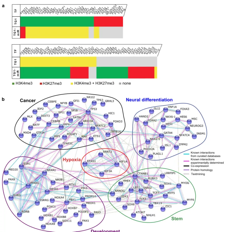
factor 2α/EPAS1 and beta-catenin/CTNNB1), allowed opti-mization of the modeling of a network including a maximal number of elements. This analysis yielded a densely con-nected network gathering 91 transcription factors whose
genes are associated with changing histone modifications
following expression of the miR-302–367 cluster (Fig. 3a
and b). The network included five nodes grouping transcrip-tion factors not only involved in cancer, but also in stemness
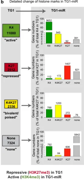
a b "active" "bivalent/ poised" "repressed" "none" K4 11080 K27 2512 K4K27 2336 None 7324 K4 K4 K4 K4 K4K27 K4K27 K4K27 K4K27 K27 K27 K27 K27 none none none none 2 4 8 6 0 10 60 80 100 40 60 80 100 0 20 40 60 80 100 0 20 2 4 8 6 0 10 60 80 100 12 TG1 TG1-miR 9652 945 92 391 112 172 1407 821 788 1200 272 76 792 84 5842 606
Detailed change of histone marks in TG1-miR
Gene numbers (% of total K4 in TG1) Gene numbers (% of total K27 in TG1) Gene numbers (% of total K4K27 in TG1) Gene numbers (% of total none in TG1)
All genes with differing marks in TG1 and TG1-miR
Cell-cell signaling Cell adhesion
Growth Transcriptional activator activity
Regulation of angiogenesis Cell differentiation
Cell fate commitment c
Response to oxydative stress Intracellular signal transduction
Protein dimerization activity Negative regulation of neuron differentiation Response to hypoxia
Wnt signaling pathway Transcriptional activator activity Tissue regeneration Regulation of cell proliferation
Protein homodimerization activity Glutamatergic synapse Synapse Protein autophosphorylation Cell projection Cell junction e d Active (H3K4me3) in TG1
Repressive (H3K27me3) in TG1-miR Repressive (Active (H3K4me3)H3K27me3) in TG1-miR in TG1
0 10 20 30 40 50
-log10 (P-value) Notch signaling pathway
0 2 4 6 8 10
-log10 (P-value) Postsynaptic cell membrane
GABAergic synapse Metabolic pathways Transcritpion regulation -log10 (P-value)
0 2 4 6 8 10
Nervous system development Regulation of MAPK cascade Cell surface receptor signaling pathway Chemical synaptic transmissionOxidation-reduction process Multicellular organism developmentEnergy reserve metabolic process Telencephalon development Drug transmembrane transport Chemokine-mediated signaling pathwayAdenylate cyclase-activating G-protein Cell chemotaxis Ion transmembrane transport Neuropeptide signaling pathway Extracellular matrix organization Proteinaceous extracellular matrix
TG1 K4
TG1-miR K4 TG1-miR K27
TG1 K27
Fig. 2 Loss of tumorigenic properties is associated with rearrange-ment of H3K4me3 and H3K27me3 marks in discrete subsets of genes. a Venn diagram illustrating genes groups with differing his-tone marks in TG1 and TG1-miR. Numbers of genes in each cat-egory are indicated. Numbers highlighted with colors correspond to gene with marks changing following the expression of miR-302– 367, colors representing the final epigenetic status. K4 = K4me3.
K27 = K27me3. b Detailed representation of histone mark changes observed in the TG1-miR. K4 = K4me3. K27 = K27me3. c Gene ontology analysis of all genes with changes in H3K4me3 and H3K27me3 marks between TG1 and TG1-miR. d Gene ontology analysis of genes undergoing a transition from H3K4me3 in TG1 to H3K27me3 in TG1-miR. e Gene ontology analysis of genes undergo-ing a transition from H3K27me3 in TG1 to H3K4me3 in TG1-miR
(e.g., NANOG, LEF1), in neural differentiation (e.g., FOXA2/3, NKXs, NEUROG3) and in development (e.g., HOXs, PAXs) that could all be related to a node regrouping
transcription factors of the hypoxia pathway. Interestingly, this network included two of the three transcription factors that exchanged the active H3K4me3 mark for the repressive b a Stem Neural differentiation Cancer Development Hypoxia Known interactions from curated databases Known interactions experimentally determined Textmining
Co-expression Protein homology
H3K4me3 H3K27me3 H3K4me3 + H3K27me3 none
TF ARNT2 LEARF1ID 3A ATOH 7 ESRR G FOXC 1 GFGTI1F3 A HAND 2 HES1HEINS6SM 1 IRF5JDLRP2RF IP1 MAFMXD 4 NR4A 1 PBX1PITX 1 PLAG L1 POU3 F3 RORBSOX6TCZFF7PM 2 HOXB 7 ZIC1GATA 4 NR5A 1 FOXA 2 HOXB 1 ISNEL1UR OG3 NKX6 -1 PHOX 2A POU4 F1 SOX1SPIB TG 1 TG 1-mi R T F CEBP B CEBP E ESR1ETS1FOXO 3 HHHIEXF3 A IRNFF4AT C4 NFYBPAPPX3AR G PRDM 14 RUNX 3 SATB 1 SNAI 2 SPI1TCZBF3TB 16 MEO X1 NFAT C2 NKX2 -5 PAX1RXSORGX1 1 TFAP 2C MYF 6 NR0B 1 TP63DDIT 3 HIC1HOXA 4 HOXA 9 HOXB 8 MED 20 MLXMSCNHLH 1 PAX8TBX1 9 TP53BATFCDFOX1XA 3 GRHL 3 HNF1 A MYF 5 MYO G NANO G NR1I 2 ZIC3GLI2 T G 1 T G 1-m iR
Fig. 3 Epigenetic regulation of transcription factors highlights ARNT2 as a new actor in the maintenance of GBM stem-like cells properties. a Overview of the transcription factors from the STRING network undergoing transition in epigenetic marks in TG1-miR. b STRING analysis of transcription factors changing H3K4me3 and H3K27me3 marks in TG1-miR. Edges between proteins symbolize the confidence index of the interaction probability. Edges are colored
based on the source of information: known interactions from curated databases (light blue), known interactions experimentally deter-mined (pink), co-expression (black), protein homology (purple) and text mining (gray). Note that HIF1A, EPAS1 (HIF2A) and CTNNB1 (beta-catenin) have the same histone mark (H3K4me3) in TG1 and TG1-miR
H3K27me3 mark, namely LEF1 and ARNT2. LEF1 is a key component of the Wnt/β-catenin signaling pathway, a pathway known to contribute to the maintenance of the
properties of GBM stem-like cells [38, 77, 79]. ARNT2 is
considered, like its paralog ARNT, as an accessory partner required for full transcriptional activity of several proteins
including HIF1α, HIF2α, AHR, NPAS4, and SIM1 [17, 29,
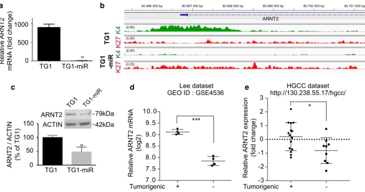
61, 66]. Analysis of mRNA levels showed that ARNT2 was
the only transcription factor in the hypoxia pathway (ARNT, ARNT2, HIF1α/HIF1A, HIF2α/EPAS1, HIF3α/HIF3A, and HIF1α inhibitor/HIF1AN), to exhibit a drastic reduction of
its mRNA levels in TG1-miR compared to TG1 (Fig. 4a,
Online Resource 7). This finding was coherent with changes in histone modifications at the ARNT2 locus from H3K4me3
in TG1 to H3K27me3 in TG1-miR (Fig. 4b). We did not find
miR-302–367 target sites within the ARNT2 mRNA
(MIR-Base, http://www.mirbase.org/), indicating that decreased
ARNT2 expression in TG1-miR does not stem from direct targeting by miR-302–367. Immunoblot analysis showed that reduced transcription of ARNT2 was associated with a
decrease in ARNT2 protein levels (Fig. 4c). These results
together with the scant information currently available on the role of ARNT2 in cancer, led us to investigate further
the possible implication of ARNT2 in the regulation of glio-blastoma cell properties.
ARNT2 is functionally associated with a molecular signature linked to glioblastoma cell tumorigenicity within the patients’ tumors
We first analyzed ARNT2 expression in two published independent transcriptome datasets of glioblastoma cells either devoid of or endowed with tumor-initiating
prop-erties [41, 73]. In agreement with our observations, we
found that ARNT2 expression was downregulated in non-tumorigenic cells compared to non-tumorigenic cells in both
datasets (Fig. 4d, e). Further, the analysis of the TCGA
transcriptome dataset of 481 surgical tissue samples of untreated primary glioblastoma using the GlioVis Platform
[9] showed lower ARNT2 mRNA levels in glioblastoma
tissues than in non-tumoral brain tissues (Online Resource 8A). The finding of higher ARNT2 mRNA levels in nor-mal brain tissues than in GBM tissues from which neurons are absent is coherent with the high ARNT2 expression in
mature neurons [18, 19, 32]. Analysis of the TCGA
glio-blastoma dataset and the French glioma dataset gse16011 a c d 0 500 1000 TG1 TG1-miR Relative ARNT 2
mRNA (fold change)
TG1 TG1-mi R ARNT2 ACTIN 79kDa 42kDa TG1 TG1-miR 0 50 100 150 ARNT2 / ACTIN (% of TG1) e K4 K4 K2 7 TG 1 b Lee dataset GEO ID : GSE4536 Tumorigenic K2 7 TG 1 -miR ARNT2 (0.95) (0.95) (0.25) (0.25) 80 696 000 bp 80 697 000 bp 80 698 000 bp 80 699 000 bp 80 700 000 bp 80 701 000 bp Tumorigenic
Relative ARNT2 expression
(fold change
)
Relative ARNT2 mRNA
(log2) HGCC dataset + -7.0 7.5 8.0 8.5 9.0 9.5 10.0 *** + --3 -2 -1 0 1 2 3 * http://130.238.55.17/hgcc/
Fig. 4 Decreased ARNT2 expression is associated with non-tumori-genic glioblastoma cells. a Decreased ARNT2 mRNA levels in TG1-miR compared to TG1. QPCR assay. **p < 0.01, unpaired t test with Welch’s correction, mean ± SD, n = 3 independent biological sam-ples. b Loss of the active H3K4me3 mark and gain of the repressive H3K27me3 mark around the ARNT2 transcription start site in TG1-miR. c Decreased ARNT2 protein levels in TG1-miR compared to TG1. Western blot analysis. **p < 0.01, unpaired t test with Welch’s correction, mean ± SD, n = 3 independent biological samples. d
Analysis of published transcriptome dataset of early passage (P3) glioblastoma cells isolated from four human tumors, either endowed with self-renewing and tumor-initiating properties or devoid of them following serum-treatment [41]. ***p < 0.001, unpaired t test with Welch’s correction, mean ± SD, n = 4. e Analysis of the publicly available HGCC transcriptome dataset of glioblastoma cells isolated from distinct patients’ tumors and characterized for their ability to initiate tumors [73]. *p < 0.05, unpaired t test with Welch’s correc-tion, mean ± SD, n = 15 (Tumorigenic), n = 9 (non-Tumorigenic)
[26], showed no variation in ARNT2 expression according to MGMT status, IDH1 mutation or EGFR amplification (not shown). In accordance with the narrow distribution of ARNT2 expression levels across glioblastoma samples (Online Resource 8A), no correlation could be disclosed using the R2 Genomics Analysis and Visualization
Plat-form (http://r2.amc.nl) between variations in ARNT2
mRNA levels and the overall survival of patients (Online Resource 8B).
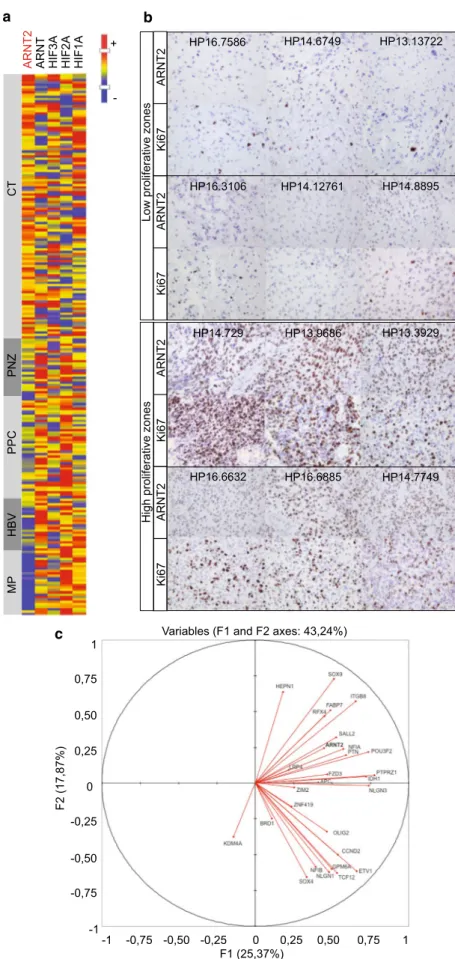
Glioblastoma are characterized by the intermingling of differing tumor tissues including dense tumor areas where cancer cells predominate (“cellular tumor areas”), necrotic and perinecrotic areas with sparser tumor cells, areas more or less angiogenic, and infiltrated areas where tumor cells are distributed through the brain parenchyma. To refine the analysis of ARNT2 and its family kin in glioblastoma, we explored its expression using the IVY dataset, which provides gene mRNA levels in distinct glioblastoma zones (http://glioblastoma.alleninstitute.org). The results of this analysis singled out ARNT2 among the other HIF family members. ARNT2 mRNA levels were higher in the cellular areas of the tumors than in the perinecrotic zones and barely detectable in tumor blood vessels. HIF3A expression was evenly distributed in the different tumor zones, while HIF1A, HIF2A, and ARNT expressions were enriched in the
perine-crotic zones and/or in blood vessels of the tumor (Fig. 5a,
Online Resource 9). Coherent with the profile of ARNT2 mRNA distribution across glioblastoma areas, immunohis-tochemical analysis of neurosurgical samples of patient’s tumors revealed enrichment in ARNT2-expressing cells with increased distance from necrosis (Online Resource 9B). Importantly, this analysis revealed that ARNT2-expressing cells were especially enriched in the proliferative zones of
the tumor (Fig. 5b).
To further explore the relevance of ARNT2 expression in the context of the human tumors, we compared its expres-sion in the tumor core areas of glioblastomas (IVY dataset) with the expression of genes associated with glioblastoma cells endowed with tumorigenic and stem-like properties. We used a 28 molecules’ signature delineated by the Bern-stein laboratory from the combined analysis of single cell transcriptome profiles of cultured GBM stem-like cells and
of 254 cells sampled from five different glioblastomas [55].
We retrieved expression data from 27 of the 28 components of the signature from the IVY dataset (Online Resource 10). Principal component analysis (PCA) showed that ARNT2 expression co-varied with the tumorigenic/stem signature
along the first principal component axis (F1 axis) (Fig. 5c).
We verified whether this co-variation occurred also at the single cell level using the published transcriptome profiles
of 254 glioblastoma cells [55]. This dataset contains 25 of
the 28 signature’s components (Online Resource 10). Prin-cipal component analysis showed that ARNT2 expression
co-varied also with the stem signature at the single cell level (Online Resource 11).
To probe the functional relevance of the co-variations disclosed by analysis of glioblastoma tissues and single cells transcriptomes, we determined the effect of ARNT2 knockdown on the expression of SOX9, POU3F2 and OLIG2 in independent GBM stem-like cell cultures (6240** and 5706**) distinct from TG1 and TG1-miR. These three genes were selected with respect to their previously
dem-onstrated role in glioblastoma cell tumorigenicity [31, 44,
68]. An 80 to 90% decrease in ARNT2 mRNA level was
observed in cells expressing shARNT2 (Fig. 6a, b, Online
Resource 13). We observed decreased SOX9, POU3F2 and OLIG2 mRNA levels following ARNT2 knockdown using lentiviral transduction of small hairpin RNA (shControl or
shARNT2, Fig. 6a, b, Online Resource 12). Similar results
were obtained in TG1 cells expressing shARNT2 (Online Resource13). In addition, we verified that ARNT2, OLIG2, POU3F2 and SOX9 expressions did not change in conditions of reduced oxygen levels (Online Resource 14). Finally, we verified whether ARNT2 binding sites are present within OLIG2, POU3F2 and SOX9 regulatory regions by ChIP-qPCR experiments using ARNT2 antibodies. This analysis showed an enrichment in ARNT2 binding sites in one or more of the SOX9, POU3F2 and OLIG2 regulatory regions tested, compared to the house keeping gene TBP (Online Resource 15), indicating that OLIG2, POU3F2 and SOX9 can be directly regulated through ARNT2 binding to their regulatory regions. Altogether these results confirmed the functional relevance of the co-variation of ARNT2 expres-sion with the tumorigenic/stem signature identified by tumor tissue and single cell transcriptome analysis. Our analysis further showed that ARNT2 down-regulation was
accompa-nied by a decrease in LEF1 mRNA levels (Fig. 6a, b, Online
Resource 13A), whereas the expression of the HIF family members HIF1A, HIF2A and ARNT varied from one cell
line to another (Fig. 6a, b, Online Resource 13A).
Taken together, these results demonstrate that ARNT2 is expressed at the mRNA and protein level within the tumors of patients with glioblastoma. Further, they show that ARNT2 is part of a tumorigenic/stem signature of glio-blastoma cells, and regulates the expression of transcription factors previously shown to be involved in the control of glioblastoma cell tumorigenicity.
ARNT2 is essential for the maintenance of glioblastoma cell tumorigenic properties
The above results, associated with our initial finding of ARNT2 down-regulation in glioblastoma cells deprived of tumorigenic properties, led us to evaluate the role of ARNT2 in the control of glioblastoma cell tumorigenicity using orthotopic xenografts of GBM stem-like cells (6240**,
Fig. 5 ARNT2 is expressed in patients’ glioblastoma and is associated with a tumorigenic/ stem signature. a ARNT2 expression prevails in glio-blastoma areas of high tumor cell density (CT). Note the absence of clear-cut correlation between ARNT2 and other HIF family members in glioblas-toma. Heat map representation of mRNA levels evaluated in distinct glioblastoma zones (IVY dataset). Glioblastoma zones defined according to IVY gap white paper (May 2015 v.1) as follows. CT: cellular tumor zone of glioblastoma constituting the major part of core, with 100/1 to 500/1 tumor cell to normal cell ratio. PNZ: perinecrotic zone corresponding to a 10-30 cells’ boundary along a necrotic zone but lacking a clear demarcation. PPC: pseu-dopalisading cells aggregates around a necrotic area. HBV: hyperplastic blood vessels with thickened walls and endothelial cell proliferation. MP: areas of microvascular proliferation characterized by two or more vessels sharing a common wall.
b ARNT2 positive cells are
enriched in high proliferative zones of the tumor, as shown by immunohistochemical staining of ARNT2 and Ki67 in sister sections of patients’ glioblas-toma. Magnification ×400. c ARNT2 expression in patients’ glioblastoma tumor core cor-relates with expression of genes composing the glioblastoma stem-like cells signature (listed in Online Resource 6). Cor-relation circle with F1 and F2 principal components. Analysis performed with transcriptome data from IVY dataset glioblas-toma core zones (http://glioblas-toma.alleninstitute.org) HP13.13722 HP14.8895 HP14.6749 HP14.12761 HP16.7586 HP16.3106 a CT PN Z PPC HB V MP HIF1 A HIF2 A HIF3 A ARNT ARNT 2 -+ b
c Variables (F1 and F2 axes: 43,24%)
0 -0,25 -0,75 -0,50 -1 0,25 0,75 0,50 1 F2 (17,87%) F1 (25,37%)0 -0,25 -0,75 -0,50 -1 0,25 0,50 0,75 1
Low proliferative zone
s
High proliferative zone
s HP14.7749 HP13.3929 HP16.6885 HP13.9686 HP14.729 HP16.6632 ARNT 2 Ki67 ARNT 2 Ki67 ARNT 2 Ki67 ARNT 2 Ki67
5706**) expressing either a shControl or a shARNT2. ARNT2 knockdown inhibited the proliferation and the
clonality of the cells in vitro (Fig. 6c–e, Online Resource
13B and C). Of note, our observation of increased ARNT
mRNA levels upon ARNT2 knockdown (Fig. 6a, b) indicates
that ARNT cannot compensate for ARNT2 knockdown. h −3.5 −3.0 −2.5 −2.0 −1.5 −1.0 −0.5 0.0
log fraction nonresponding
dose (number of cells)
6240** −2.5 −2.0 −1.5 −1.0 −0.5 0.0
dose (number of cells) shCTL shARNT2 5706** 0 2 4 6 8 10 0 2 4 6 8 10 shCTL shARNT2 g 6240** 5706** (0/7) (0/6) (6/7) (6/6) 0 10 20 30 40 50 0 20 40 60 80 100 Tum or in ci denc e( %) shCTL shARNT2 0 10 20 30 40 50 60 0 20 40 60 80 100 Tum or in ci denc e( %) shCTL shARNT2 a 6240** 5706** -1.0 -0.8 -0.6 -0.4 -0.2 0.0 0.2 0.4 -1.0 -0.8 -0.6 -0.4 -0.2 0.0 0.2 0.4 ARNT 2 ARNT HI FA HIF2 A OLIG2 SOX9 POU3F2 LEF1 ARNT 2 ARNT HI FA HIF2 A OLIG2 SOX9 POU3F2 LEF1 mRNA fold change in 6240* * (shARNT2 over shCTL) mRNA fold change in 5706** (shARNT2 over shCTL)
Proliferation (fold change
) c e 6240** 5706** -1.0 -0.8 -0.6 -0.4 -0.2 0.0 * *** 0 20 40 60 80 100 120 140 0 20 40 60 80 100 shCTL shAR NT2 Percent surviva l Days elapsed 6240** survival Percent surviva l Days elapsed shCTL shAR NT2 b d f
log fraction nonresponding
j k i P = 0.0174 Time (Days) Time (Days) 5706** survival *** * ** ** ** * * ** * * * * ** * shCTL shARNT2
Tumor flux values (relative to shCTL)
0.0 0.5 1.0 1.5 2.0 2.5 6240** * shCTL shARNT2
Tumor flux values (relative to shCTL)
0.0 0.5 1.0 1.5 2.0 ** 5706** 00 100 150 200 250 20 40 60 80 100 P = 0.0062 P = 2.52 10-18 P = 6.9 10-19
Orthotopic xenografts of 1×, 2×, 4× or 14 × 104 6240** or
5706** GBM stem-like cells stably expressing luciferase and either shControl or shARNT2 were used to follow tumor development with bioluminescent imaging. Results showed a striking reduction in tumor incidence in mice grafted with 6240**- and 5706**-shARNT2 compared to mice grafted
with 6240**- and 5706**-shCTL cells (Fig. 6f, i, Online
Resource 16A–D). Bioluminescence imaging 42 days post-graft revealed tumor formation in six out of seven, and in
six out of six mice engrafted with 14 × 104
6240**-shCon-trol and 5706**-shCon6240**-shCon-trol, respectively (Fig. 6f, i). In
contrast, no bioluminescent signal was detected in the mice grafted with 6240**-shARNT2 or 5706**-shARNT2
(Fig. 6f, i). Similar results were obtained when grafting
smaller numbers of cells (Online Resource 13). The reduced tumor development in the mice grafted with 6240** and 5706**-shARNT2 was confirmed by immunohistochemistry (Online Resource 16E-F). Survival assays revealed differing long-term consequences of ARNT2 knockdown according to the GBM stem-like cells grafted. Although we observed a significant improvement in the survival of the mice grafted with either 6240** or 5706** cells expressing shARNT2
(Kaplan–Meier analysis, Fig. 6h, k), only mice grafted with
6240**-shARNT2 eventually developed tumors. Determi-nation of human ARNT2 mRNA levels by QPCR in these tumors showed ARNT2 as well as OLIG2, POU3F2 and SOX9 transcripts levels similar to tumors of the shCTL group (Online Resource 16G). This result indicates that the 6240** cells that formed the tumors escaped ARNT2 inhibi-tion, further pointing to an essential role of this transcription factor for glioblastoma cell aggressiveness. Taken together, these results show that ARNT2 participates in the control of the tumorigenicity of glioblastoma cells.
Discussion
Understanding the molecular basis of the varying functional cell states that co-exist within glioblastoma and participate in tumor resistance to treatments is of great importance to improve current therapeutic management. Differences in the ability of glioblastoma cells from the same tumor to initiate neoplasms has notably been highlighted by graft-ing cells sorted from glioblastoma surgical resections in
immune-deficient mouse brains [35, 58, 73]. Recent
stud-ies have also shown the striking phenotypic plasticity of glioblastoma cells, which can adopt more or less aggressive states during the course of the tumor evolution and treatment
[3, 33]. Here, we identified changes in the chromatin state
of transcription factors, which accompany the passage of GBM stem-like cells from a highly aggressive to a poorly tumorigenic state. We uncovered a novel transcription factor controlling glioblastoma cell tumorigenicity, which is local-ized at a node of a transcription factor network controlling glioblastoma cell aggressiveness, and which clusters with a tumorigenic/stem signature of glioblastoma cells at both tissue and single cell levels.
Using the human glioblastoma cell line TG1 expressing or not expressing the micro-RNA cluster miR-302–367 as a
model system [22], we profiled histone modifications. The
results of our analyses uncovered a subset of genes show-ing changes in H3K4me3 or H3K27me3 between TG1 cells and TG1-miR cells in which the stem-like and tumorigenic properties have been repressed by expression of the miR-302–367. In agreement with the previously reported associa-tion of miR-302–367 with differentiaassocia-tion of GBM stem-like
cells [22], [23], ontological pathway analysis of the subset of
genes with changes in histone marks showed enrichment in ontological gene groups related to development and engage-ment in differentiation pathways. Enrichengage-ments in terms related to nervous system were also obtained (neuron, neu-rogenesis, synapse, forebrain), indicating conservation in the tumor cells of an imprint of their tissue of origin. Retrieval of the 202 transcription factors undergoing a change in his-tone marks further highlighted molecular pathways already
Fig. 6 ARNT2 down-regulation impairs tumor initiation and devel-opment. a, b Consequences of ARNT2 down-regulation on the expression of HIF family members (HIF1A, HIF2A, ARNT), on core components of the tumorigenic/stem signature of glioblastoma cells (OLIG2, SOX9, POU3F2), and on the effector of the Wnt sign-aling pathway, LEF1. QPCR assay. Results are presented as fold changes in mRNA levels detected in GBM stem-like cells expressing shARNT2 compared to shControl (shCTL). *p < 0.05, **p < 0.01, ***p < 0.001, unpaired t test with Welch’s correction, mean ± SD,
n = 3 independent biological samples. c Down-regulation of ARNT2
is accompanied with decreased cell proliferation. shARNT2 versus shControl. *p < 0.05, ***p < 0.001, unpaired t test with Welch’s correction, mean ± SD, n = 3 independent biological samples. d, e Knocking-down of ARNT2 inhibits the sphere‐forming capability of GBM stem-like cells. Extreme limiting dilution assays. Sphere for-mation was scored 7 days after seeding 6240** (d) and 5706** (e) GBM stem-like cells expressing shControl or shARNT2. Frequency of sphere‐forming cells: 6240** shCTL = 1/3.32 (lower 4.68, upper 2.41); 6240** shARNT2 1/267.47 (lower 1887.52, upper 38.27),
n = 16, p = 2.52 10−18. 5706** shCTL = 1/3.84 (lower 5.42, upper
2.77); 5706** shARNT2 1/Inf (lower Inf, upper 91.30), n = 16, p = 6.9 10−19. f, i Knocking-down of ARNT2 inhibits tumor
inci-dence. Bioluminescent analyses of tumor growth initiated by graft-ing 6240** (f) and 5706** (i) GBM stem-like cells transduced with a luciferase construct and either a shControl or a shARNT2 construct. The percentage of tumor incidence was monitored for 6 (6240**) and 8 (5706**) weeks. g, j Bioluminescent analyses of tumor growth initiated by grafting 6240** (g) and 5706** GBM stem-like cells transduced with a luciferase construct and either a shControl or a shARNT2 construct. 28 days post-graft. Quantification of the biolumi-nescent signals. Mean ± SD, n = 7 mice per group for 6240** (g) and
n = 6 per group for 5706** (j). h, k Kaplan–Meier survival curves
demonstrating a significant survival benefit of mice grafted with GBM stem-like cells expressing shARNT2 compared to mice grafted with GBM stem-like cell expressing shControl. 6240** shCTL and shARNT2, each n = 6 (h). 5706*** shCTL, n = 5, 5706** shARNT2, n = 4 (k). Log-rank Mantel–Cox test
identified as important players in the regulation of neural stem/progenitor cell but also of ESC and GBM stem-like cell behaviors, illustrating the pertinence of mapping his-tone epigenetic marks for identifying regulators of glioblas-toma cell properties. For example, we observed an increased H3K27me3 associated with the gene encoding Nanog, a key factor in ESC pluripotency, and which has also been impli-cated in the maintenance of GBM stem-like cell properties
[22, 52, 75]. Similarly, changes were observed for LEF1, an
effector of the Wnt signaling pathway known to be involved
in neurogenesis [8] and maintenance of GBM stem-like cell
[38, 77, 79], TCF3 and TCF7 that are transcriptional
regula-tors of the Wnt pathway in neural stem cells and ESC [40, 74]
and the HES bHLH genes and FOXCs transcription factors implicated in the Notch signaling pathway, which is activated
in neural stem cells and GBM stem-like cells [36, 72].
Mapping known and predicted protein–protein interac-tions between transcription factors exhibiting changes in histone marks in GBM stem-like cell lacking tumorigenic properties generated a network articulated around ARNT2. ARNT2, like its paralog ARNT, is considered to act as a dimerization partner of HIF1/2α, the heterodimers triggering
the expression of hypoxia-related genes [46, 61]. ARNT2
expression is especially abundant in the central nervous
sys-tem and kidney, while that of ARNT is ubiquitous [18, 32].
In the central nervous system, ARNT2 mRNA and protein
are enriched in neurons [18]. Although ARNT2/HIFs and
ARNT/HIFs heterodimers are equally efficient to ensure
neuronal responses to hypoxia [46], ARNT2 protein levels
do not increase under hypoxic conditions unlike those of
ARNT and HIF1/2α [42, 47]. The role of ARNT2 in
can-cer is poorly explored. ARNT2 has been associated with increased as well as decreased growth of non-cerebral
can-cers [39, 43, 46, 48, 59, 60]. In glioblastoma HIF1 and 2α,
but not ARNT2, have been associated to adaptation of
can-cer cells to hypoxic conditions [30, 42]. We found that the
profile of ARNT2 expression in glioblastoma does not cor-respond with that expected for a hypoxia-related molecule. ARNT2 expression was highest in glioblastoma core zones rather than in the hypoxic necrotic and pseudopalisading zones. This observation favors a hypoxia-independent tran-scriptional role for ARNT2.
Of note, ARNT2 is one of the few transcription fac-tors switching from the active H3K4me3 to the repres-sive H3K27me3 mark in GBM stem-like cells expressing miR-302–367. Analysis of publically available data sets indicated that down-regulation of ARNT2 mRNA occurs not only in the TG1-miR cell line, but importantly is also observed in non-tumorigenic glioblastoma cells either
directly sorted from patients’ tumors [73] or following
serum-induced differentiation of GBM stem-like cell [41].
This was confirmed at the protein level by immunohis-tochemistry of ARNT2 expression in sub-populations of
cells within proliferative zones of patients’ glioblastoma. Furthermore, we demonstrated that ARNT2 knockdown inhibits tumor-initiating properties in vivo, supporting a role of ARNT2 in the tumorigenicity of glioblastoma cells.
Examination of tumor patients’ transcriptome datasets further associated ARNT2 with a tumorigenic/stem
sig-nature of glioblastoma cells [55] at both the tissue and
single cell levels. To ascertain the functional relevance of this association, we focused on three transcription fac-tors members of this stem signature SOX9, POU3F2 and OLIG2, since the knockdown of these factors has previ-ously been reported to inhibit glioblastoma cell
tumori-genicity in vivo [31, 44, 68]. We found that ARNT2
knock-down not only impaired the cell tumorigenicity in vivo but also resulted in decreased expression of SOX9, POU3F2 and OLIG2, hence placing ARNT2 at the core of tran-scriptional regulations of glioblastoma cell tumorigenicity. In conclusion, our results uncover a novel transcription factor essential for glioblastoma cell tumorigenic proper-ties, show its functional relevance within the context of the patients’ tumor, and shed new lights on the combinato-rial organization of the transcription factor networks that regulate glioblastoma cell aggressiveness.
Acknowledgements We are obligated to the members of the Cancer
stem cell network of Ile-de-France headed by Dr. Christine Chomienne, for their continuous support and helpful discussions. We are grateful to A. Dias-Morais for technical assistance, and Dr. Tomohiro Yamaki (Chiba Ryogo Center, Japan) for helpful discussions. We thank for their precious help A. Borderie, S. Destree and C. Hagnere (Hôpital Pasteur, CHU Nice).
Compliance with ethical standards
Ethical approval All the procedures performed in studies involving human participants were in accordance with the 1964 Helsinki dec-laration and its later amendments and to the French laws. The institu-tional review board of the Sainte-Anne Hospital Center—University Paris Descartes (Comité de protection des personnes Ile de France III) approved the study protocol (Protocol Number DC-2008-323). All samples were obtained with informed consent of patients. The animal maintenance, handling, surveillance, and experimentation were per-formed in accordance with and approval from the Comité d’éthique en expérimentation animale Charles Darwin No. 5 (Protocol #3113).
Funding This work was supported by La Ligue nationale contre le cancer (Equipe Labellisée LIGUE 2013, Equipe Labelisée LIGUE 2016 HC/MPJ), Institut National du Cancer (INCa 2012-1-PLBIO-07-INSERM-1, INCa-AAP Epigénétique et cancer 2014, HC/MPJ), Fondation pour la recherche sur le cerveau (FRC), Agence Nationale de la Recherche (ANR-13-1SV1-0004-03, JH/HC), Cancéropole Région Ile-de-France (EAE and AB fellowships), CAPES/COFECUB (Coor-dination pour le perfectionnement du personnel de l’enseignement supérieur/Comité français d’evaluation de la coopération universitaire et scientifique avec le Brésil, LGD fellowship), and Fundação Ary Frauzino para o Câncer (LGD fellowship).
Conflict of interest The authors declare that they have no conflict of interest.
Open Access This article is distributed under the terms of the Creative Commons Attribution 4.0 International License (http://creativecom-mons.org/licenses/by/4.0/), which permits unrestricted use, distribu-tion, and reproduction in any medium, provided you give appropriate credit to the original author(s) and the source, provide a link to the Creative Commons license, and indicate if changes were made.
References
1. Alder O, Lavial F, Helness A, Brookes E, Pinho S, Chandrashekran A et al (2010) Ring1B and Suv39h1 delineate distinct chromatin states at bivalent genes during early mouse lineage commitment. Development 137:2483–2492. https://doi.org/10.1242/dev.048363 2. Assad Kahn S, Costa SL, Gholamin S, Nitta RT, Dubois LG,
Feve M et al (2016) The anti-hypertensive drug prazosin inhib-its glioblastoma growth via the PKCdelta-dependent inhibition of the AKT pathway. EMBO Mol Med 8:511–526. https://doi. org/10.15252/emmm.201505421
3. Auffinger B, Tobias AL, Han Y, Lee G, Guo D, Dey M et al (2014) Conversion of differentiated cancer cells into cancer stem-like cells in a glioblastoma model after primary chemotherapy. Cell Death Differ 21:1119–1131. https://doi.org/10.1038/cdd.2014.31 4. Azuara V, Perry P, Sauer S, Spivakov M, Jorgensen HF, John RM
et al (2006) Chromatin signatures of pluripotent cell lines. Nat Cell Biol 8:532–538. https://doi.org/10.1038/ncb1403
5. Barski A, Cuddapah S, Cui K, Roh TY, Schones DE, Wang Z et al (2007) High-resolution profiling of histone methylations in the human genome. Cell 129:823–837. https://doi.org/10.1016/j. cell.2007.05.009
6. Bernstein BE, Mikkelsen TS, Xie X, Kamal M, Huebert DJ, Cuff J et al (2006) A bivalent chromatin structure marks key develop-mental genes in embryonic stem cells. Cell 125:315–326. https:// doi.org/10.1016/j.cell.2006.02.041
7. Bernstein BE, Meissner A, Lander ES (2007) The mammalian epig-enome. Cell 128:669–681. https://doi.org/10.1016/j.cell.2007.01.033 8. Bielen H, Houart C (2014) The Wnt cries many: Wnt regula-tion of neurogenesis through tissue patterning, proliferaregula-tion, and asymmetric cell division. Dev Neurobiol 74:772–780. https://doi. org/10.1002/dneu.22168
9. Bowman RL, Wang Q, Carro A, Verhaak RG, Squatrito M (2017) GlioVis data portal for visualization and analysis of brain tumor expression datasets. Neuro Oncol 19:139–141. https://doi. org/10.1093/neuonc/now247
10. Brookes E, de Santiago I, Hebenstreit D, Morris KJ, Carroll T, Xie SQ et al (2012) Polycomb associates genome-wide with a specific RNA polymerase II variant, and regulates metabolic genes in ESCs. Cell Stem Cell 10:157–170. https://doi.org/10.1016/j. stem.2011.12.017
11. Chiesa-Vottero AG, Rybicki LA, Prayson RA (2003) Comparison of proliferation indices in glioblastoma multiforme by whole tis-sue section vs tistis-sue microarray. Am J Clin Pathol 120:902–908. https://doi.org/10.1309/8UAU-KFK3-NBDM-VTNU
12. Consortium RE, Kundaje A, Meuleman W, Ernst J, Bilenky M, Yen A et al (2015) Integrative analysis of 111 reference human epig-enomes. Nature 518:317–330. https://doi.org/10.1038/nature14248 13. Cui K, Zang C, Roh TY, Schones DE, Childs RW, Peng W et al
(2009) Chromatin signatures in multipotent human hematopoietic stem cells indicate the fate of bivalent genes during differentiation. Cell Stem Cell 4:80–93. https://doi.org/10.1016/j.stem.2008.11.011 14. da Huang W, Sherman BT, Lempicki RA (2009) Bioinformat-ics enrichment tools: paths toward the comprehensive functional analysis of large gene lists. Nucleic Acids Res 37:1–13. https:// doi.org/10.1093/nar/gkn923
15. da Huang W, Sherman BT, Lempicki RA (2009) Systematic and integrative analysis of large gene lists using DAVID bioinfor-matics resources. Nat Protoc 4:44–57. https://doi.org/10.1038/ nprot.2008.211
16. Debruyne DN, Turchi L, Burel-Vandenbos F, Fareh M, Almairac F, Virolle V et al (2017) DOCK4 promotes loss of proliferation in glioblastoma progenitor cells through nuclear beta-catenin accumulation and subsequent miR-302–367 cluster expression. Oncogene. https://doi.org/10.1038/onc.2017.323
17. Dougherty EJ, Pollenz RS (2008) Analysis of Ah receptor-ARNT and Ah receptor-ARNT2 complexes in vitro and in cell culture. Toxicol Sci 103:191–206. https://doi.org/10.1093/toxsci/kfm300 18. Drutel G, Heron A, Kathmann M, Gros C, Mace S, Plotkine M
et al (1999) ARNT2, a transcription factor for brain neuron sur-vival? Eur J Neurosci 11:1545–1553
19. Drutel G, Kathmann M, Heron A, Gros C, Mace S, Schwartz JC et al (2000) Two splice variants of the hypoxia-inducible factor HIF-1alpha as potential dimerization partners of ARNT2 in neu-rons. Eur J Neurosci 12:3701–3708
20. Edgar R, Domrachev M, Lash AE (2002) Gene expression omni-bus: NCBI gene expression and hybridization array data reposi-tory. Nucleic Acids Res 30:207–210
21. El-Habr EA, Dubois LG, Burel-Vandenbos F, Bogeas A, Lipecka J, Turchi L et al (2017) A driver role for GABA metabolism in controlling stem and proliferative cell state through GHB pro-duction in glioma. Acta Neuropathol 133:645–660. https://doi. org/10.1007/s00401-016-1659-5
22. Fareh M, Turchi L, Virolle V, Debruyne D, Almairac F, de-la-Forest Divonne S et al (2012) The miR 302–367 cluster drastically affects self-renewal and infiltration properties of glioma-initiating cells through CXCR4 repression and consequent disruption of the SHH-GLI-NANOG network. Cell Death Differ 19:232–244. https://doi.org/10.1038/cdd.2011.89
23. Fareh M, Almairac F, Turchi L, Burel-Vandenbos F, Paquis P, Fontaine D et al (2017) Cell-based therapy using miR-302–367 expressing cells represses glioblastoma growth. Cell Death Dis 8:e2713. https://doi.org/10.1038/cddis.2017.117
24. Flavahan WA, Wu Q, Hitomi M, Rahim N, Kim Y, Sloan AE et al (2013) Brain tumor initiating cells adapt to restricted nutrition through preferential glucose uptake. Nat Neurosci 16:1373–1382. https://doi.org/10.1038/nn.3510
25. Galan-Moya EM, Le Guelte A, Lima Fernandes E, Thirant C, Dwyer J, Bidere N et al (2011) Secreted factors from brain endothelial cells maintain glioblastoma stem-like cell expansion through the mTOR pathway. EMBO Rep 12:470–476. https://doi. org/10.1038/embor.2011.39
26. Gravendeel LA, Kloosterhof NK, Bralten LB, van Marion R, Dub-bink HJ, Dinjens W et al (2010) Segregation of non-p.R132H mutations in IDH1 in distinct molecular subtypes of glioma. Hum Mutat 31:E1186–1199. https://doi.org/10.1002/humu.21201 27. Gupta PB, Fillmore CM, Jiang G, Shapira SD, Tao K,
Kuperwas-ser C et al (2011) Stochastic state transitions give rise to pheno-typic equilibrium in populations of cancer cells. Cell 146:633– 644. https://doi.org/10.1016/j.cell.2011.07.026
28. Hanahan D, Weinberg RA (2011) Hallmarks of cancer: the next gen-eration. Cell 144:646–674. https://doi.org/10.1016/j.cell.2011.02.013 29. Hao N, Whitelaw ML, Shearwin KE, Dodd IB, Chapman-Smith
A (2011) Identification of residues in the N-terminal PAS domains important for dimerization of Arnt and AhR. Nucleic Acids Res 39:3695–3709. https://doi.org/10.1093/nar/gkq1336 30. Heddleston JM, Wu Q, Rivera M, Minhas S, Lathia JD, Sloan
AE et al (2012) Hypoxia-induced mixed-lineage leukemia 1 regulates glioma stem cell tumorigenic potential. Cell Death Differ 19:428–439. https://doi.org/10.1038/cdd.2011.109 31. Hiraoka K, Hayashi T, Kaneko R, Nasu-Nishimura Y,