[i]Lactobacillaceae[/i] and cell adhesion: genomic and functional screening
Texte intégral
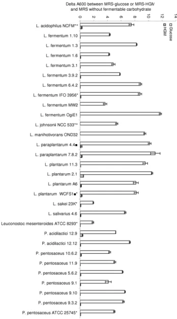
Figure

Documents relatifs
CFAO:Programmations ciblées 46 CFAO : Lapreuvepar 2 49 Moulistes portugais: runion fait la force 55 International: rembellie retrouvée 57 Glossaire:Le dico du pro 59 Mesure:
Soient premier et dernier les extrémités gauche et droite de l'intervalle dans lequel on cherche la valeur x, on calcule m, l'indice de l'élément médian :. m(premier + dernier) div
et à Brienz. Certainement, sans qu'elles fussent jolies, il faisait beau les voir ramer d'un bras vi- goureux, en chantant les rondes alpestres. li tues
[r]
[r]
le hall d’entrée met en scène la continuité entre l’extérieur et l’intérieur avec le mouve- ment du voile de béton courbe scandé par le rythme des potelets métalliques et ses
Réalisé en béton prêt à l’emploi coulé en place, le dallage d’un bâtiment industriel ou commercial est l’élément de structure de l’ouvrage sans doute le plus
The above claim is a modern interpretation of the Dulac’s classification [Dul08] of such Morse centers in a complex domain, see Lins Neto [LN14, Theorem 1.1]. Indeed, it is easier
