HAL Id: hal-02362225
https://hal.archives-ouvertes.fr/hal-02362225
Submitted on 13 Nov 2019
HAL is a multi-disciplinary open access
archive for the deposit and dissemination of sci-entific research documents, whether they are pub-lished or not. The documents may come from teaching and research institutions in France or abroad, or from public or private research centers.
L’archive ouverte pluridisciplinaire HAL, est destinée au dépôt et à la diffusion de documents scientifiques de niveau recherche, publiés ou non, émanant des établissements d’enseignement et de recherche français ou étrangers, des laboratoires publics ou privés.
Genetic analyses led to the discovery of a super-active
mutant of the RNA polymerase I
Tommy Darrière, Michael Pilsl, Marie-Kerguelen Sarthou, Adrien Chauvier,
Titouan Genty, Sylvain Audibert, Christophe Dez, Isabelle Léger-Silvestre,
Christophe Normand, Anthony Henras, et al.
To cite this version:
Tommy Darrière, Michael Pilsl, Marie-Kerguelen Sarthou, Adrien Chauvier, Titouan Genty, et al.. Genetic analyses led to the discovery of a super-active mutant of the RNA polymerase I. PLoS Genetics, Public Library of Science, 2019, 15 (5), pp.e1008157. �10.1371/journal.pgen.1008157�. �hal-02362225�
Genetic analyses led to the discovery of a super-active mutant of the RNA
1polymerase I
23
Tommy Darrière1, Michael Pilsl2, Marie-Kerguelen Sarthou1, Adrien Chauvier1, Titouan 4
Genty1, Sylvain Audibert1, Christophe Dez1, Isabelle Léger-Silvestre1, Christophe Normand1, 5
Anthony K. Henras1, Marta Kwapisz1, Olga Calvo3, Carlos Fernández-Tornero4, Herbert 6
Tschochner2 and Olivier Gadal1*
7 8 9
1 Laboratoire de Biologie Moléculaire Eucaryote, Centre de Biologie Intégrative (CBI),
10
Université de Toulouse, CNRS, UPS, Toulouse, France 11
2Universität Regensburg, Biochemie-Zentrum Regensburg (BZR), Lehrstuhl Biochemie III,
12
Regensburg, Germany 13
3Instituto de Biología Funcional y Genómica, IBFG-CSIC, Universidad de Salamanca,
14
Salamanca, Spain 15
4Centro de Investigaciones Biológicas, CSIC, Madrid, Spain
16 17 18 19 20 21 22 23 * Corresponding author 24
Email: olivier.gadal@ibcg.biotoul.fr (OG) 25
Manuscript Click here to
Abstract:
1Most transcriptional activity of exponentially growing cells is carried out by the RNA 2
Polymerase I (Pol I), which produces a ribosomal RNA (rRNA) precursor. In budding yeast, 3
Pol I is a multimeric enzyme with 14 subunits. Among them, Rpa49 forms with Rpa34 a Pol 4
I-specific heterodimer (homologous to PAF53/CAST heterodimer in human Pol I), which 5
might be responsible for the specific functions of the Pol I. Previous studies provided insight 6
in the involvement of Rpa49 in initiation, elongation, docking and releasing of Rrn3, an 7
essential Pol I transcription factor. Here, we took advantage of the spontaneous occurrence of 8
extragenic suppressors of the growth defect of the rpa49 null mutant to better understand the 9
activity of Pol I. Combining genetic approaches, biochemical analysis of rRNA synthesis and 10
investigation of the transcription rate at the individual gene scale, we characterized mutated 11
residues of the Pol I as novel extragenic suppressors of the growth defect caused by the 12
absence of Rpa49. When mapped on the Pol I structure, most of these mutations cluster 13
within the jaw-lobe module, at an interface formed by the lobe in Rpa135 and the jaw made 14
up of regions of Rpa190 and Rpa12. In vivo, the suppressor allele RPA135-F301S restores 15
normal rRNA synthesis and increases Pol I density on rDNA genes when Rpa49 is absent. 16
Growth of the Rpa135-F301S mutant is impaired when combined with exosome mutation 17
rrp6∆ and it massively accumulates pre-rRNA. Moreover, Pol I bearing Rpa135-F301S is a 18
hyper-active RNA polymerase in an in vitro tailed-template assay. We conclude that wild-19
type RNA polymerase I can be engineered to produce more rRNA in vivo and in vitro. We 20
propose that the mutated area undergoes a conformational change that supports the DNA 21
insertion into the cleft of the enzyme resulting in a super-active form of Pol I. 22
Author summary:
1The nuclear genome of eukaryotic cells is transcribed by three RNA polymerases. RNA 2
polymerase I (Pol I) is a multimeric enzyme specialized in the synthesis of ribosomal RNA. 3
Deregulation of the Pol I function is linked to the etiology of a broad range of human 4
diseases. Understanding the Pol I activity and regulation represents therefore a major 5
challenge. We chose the budding yeast Saccharomyces cerevisiae as a model, because Pol I 6
transcription apparatus is genetically amenable in this organism. Analyses of phenotypic 7
consequences of deletion/truncation of Pol I subunits-coding genes in yeast indeed provided 8
insights into the activity and regulation of the enzyme. Here, we characterized mutations in 9
Pol I that can alleviate the growth defect caused by the absence of Rpa49, one of the subunits 10
composing this multi-protein enzyme. We mapped these mutations on the Pol I structure and 11
found that they all cluster in a well-described structural element, the jaw-lobe module. 12
Combining genetic and biochemical approaches, we showed that Pol I bearing one of these 13
mutations in the Rpa135 subunit is able to produce more ribosomal RNA in vivo and in vitro. 14
We propose that this super-activity is explained by structural rearrangement of the Pol I 15
jaw/lobe interface. 16
Introduction
1The nuclear genome of eukaryotic cells is transcribed by three RNA polymerases [1]. RNA 2
polymerase II (Pol II) transcribes most of the genome and is responsible for all messenger 3
RNA production. RNA polymerases III and I are specialized in the synthesis of a limited 4
number of transcripts. RNA polymerase III (Pol III) produces small structured RNAs, 5
including tRNAs and the 5S ribosomal RNA. RNA polymerase I (Pol I) produces a single 6
transcript, the large polycistronic precursor (35S pre-rRNA in yeast; 47S in human), which 7
constitutes the first step of ribosome biogenesis. Pre-rRNA is then processed by multiple 8
successive steps into the mature rRNAs (25S, 18S, and 5.8S in yeast; 28S, 18S and 5.8S in 9
human). Despite producing a single transcript, Pol I is by far the most active eukaryotic RNA 10
polymerase, responsible for up to 60% of the total transcriptional activity in exponentially 11
growing cells [2]. The strongly transcribed rRNA genes can be visualized using the DNA 12
spread method developed by Miller et al, 1969, in which the 35S rRNA genes (rDNA) exhibit 13
a “Christmas tree” configuration, with up to 120 polymerases per transcribed gene [3]. 14
The full subunit composition and structural data are now available for the three nuclear RNA 15
polymerases of the budding yeast Saccharomyces cerevisiae [4,5][6,7]. Pol I contains a core 16
of shared or homologous subunits that are largely conserved in eukaryotes and archaea, as for 17
the other two nuclear RNA polymerases [8]. The two largest subunits (Rpa190 and Rpa135) 18
form the DNA-binding cleft that carries the catalytic center. Rpb5, Rpb6, Rpb8, Rpb10, and 19
Rpb12 are shared with Pol II and Pol III, whereas Rpc40 and Rpc19 are only shared with Pol 20
III. This nine-subunit core is associated with the stalk, a structure formed in Pol I by the 21
heterodimeric complex Rpa43/Rpa14, which is involved in docking the essential Rrn3 22
initiation transcription factor to the enzyme [9–12]. The Pol I-Rrn3 complex interacts with 23
promoter bound factors, the core factor (CF), forming the initially transcribing complex (ITC) 24
[13–16]. Additionally, Pol I and Pol III contain subunits that are functionally and structurally 25
related to Pol II-specific basal transcription factors, called the "Built-in Transcription Factors" 1
[17–19]. Their presence in Pol I and Pol III results in a higher number of subunits, from 12 2
subunits in Pol II, to 14 and 17 for Pol I and Pol III respectively, and correlates with 3
substantial transcript production from a few genes [8]. The heterodimer formed by Rpa34 and 4
the N-terminal domain of Rpa49 (Rpa49Nt) in Pol I (equivalent to Rpc53 and Rpc37 in Pol 5
III) is related to the basal transcription factor TFIIF, and stimulates endogenous transcript 6
cleavage activity [18,20,21]. Rpc34 in Pol III and the Rpa49 C-terminal domain (Rpa49Ct) 7
bear a tandem winged helix domain similar to TFIIE, also named A49tWH [18,20]. Rpa49Ct 8
binds upstream DNA [15,22] and is involved in initiation and elongation [18,23]. Finally, 9
Rpa12 in Pol I and Rpc11 in Pol III harbour a C-terminal domain involved in stimulating 10
endogenous transcript cleavage activity, similar to that of TFIIS for Pol II [17,24]. 11
Yeast genetic studies of Pol III and Pol I “Built-in Transcription Factors” have revealed 12
striking differences, despite their clear similarities. Each Pol III subunit is essential, but none 13
of the Pol I “Built-in Transcription Factors" is required for cell growth. Lack of Rpa34 or 14
invalidation of Rpa49Nt, by removing the TFIIF-like heterodimer, has no growth effect in 15
vivo [18,25,26]. In contrast, full or C-terminal deletion of RPA49 leads to a strong growth 16
defect at all temperatures, which is more severe below 25°C [18,26,27]. Full deletion of 17
RPA12 leads to a strong growth defect at 25 and 30°C, and is lethal at higher temperatures 18
[28]. Lack of the C-terminal extension of Rpa12 abolishes stimulation of intrinsic cleavage, 19
without any detectable growth defect [17,24]. Finally, yeast strains carrying the triple deletion 20
of RPA49, RPA34 and RPA12 are viable, but accumulate the growth defects associated with 21
each of the single mutants [25]. 22
Pol I is functional in the absence of Rpa49, but shows well-documented initiation and 23
elongation defects, both in vivo and in vitro [23,26,27,29–31]. Restoration of active rRNA 24
synthesis, in the absence of Rpa49, has been used to identify factors involved in initiation and 25
elongation, such as Hmo1 and Spt5 [26,30,32]. Here, we made use of the spontaneous 1
occurrence of extragenic suppressors of the growth defect of the rpa49 null mutant [27] to 2
better understand the activity of Pol I. We showed that the suppressing phenotype was caused 3
by specific point mutations in the two largest Pol I subunits, Rpa190 and Rpa135. We 4
identified a small area around Phe301 in subunit Rpa135, at an interface formed by the lobe in 5
Rpa135 and the jaw made up of regions of Rpa190 and Rpa12, where most mutations cluster. 6
Characterizing the Rpa135-F301S allele, we showed in an in vitro assay, that such Pol I 7
mutant is more active than the wild-type enzyme. In vivo, overproduction of rRNA by Pol I 8
bearing the Rpa135-F301S mutation was observed in backgrounds where the nuclear 9
exosome activity is impaired by RRP6 deletion. 10
Results
1Isolation of extragenic suppressor mutants of the growth defect in absence of Rpa49
2
We characterized extragenic suppressors of the RPA49 deletion to better understand how cell 3
growth is achieved in the absence of Rpa49. RPA49 full-deletion mutants show a strong 4
growth defect at 30°C and are unable to grow at 25°C. However, spontaneous suppressors 5
have been previously observed [27]. We quantified the frequency of occurrence of individual 6
clones able to grow at 25°C. There was a low frequency of colony occurrence, comparable 7
with the spontaneous mutation rate of a single control gene (CAN1; < 5.10-6). We isolated 8
more suppressors after irradiating the cells with UV light. UV irradiation, resulting in a 9
survival rate of about 50%, increased the frequency of suppressor mutations by approximately 10
10-fold. We identified clones that grew at 25°C after three days and selected individual 11
colonies, called SGR for Suppressor of Growth Defect of RPA49 deletion, with various 12
growth rates. We ranked SGR from 1 to 186 based on their growth rates at 25°C; SGR1 had a 13
growth rate comparable to the wild-type (WT) condition (Fig. 1A). We crossed the 186 SGR 14
clones with a strain of the opposite mating type bearing the deletion of RPA49 to obtain 15
diploid cells homozygous for the RPA49 deletion and heterozygous for each suppressor. The 16
restoration of growth of the diploids at 25°C showed that all suppressor phenotypes obtained 17
were fully or partially dominant. We focused on the most efficient suppressor clones, SGR1 18
and SGR2, and performed tetrad analysis to follow segregation of the observed suppression 19
phenotype. Each suppressor phenotype was linked to a single locus in the genome and SGR 20
mutants had no strong growth defect (SGR1 in Fig. 1A). We used global genomic mapping of 21
SGR1 and SGR2, derived from "genetic interaction mapping" (GIM) methods [33] (Materials 22
and Methods; S1 Fig), and found a genomic linkage with genes encoding the two largest Pol I 23
subunits: RPA135 for SGR1 and RPA190 for SGR2 (S1 Fig). Sequencing of the genomic 24
DNA revealed that SGR1 bears a double mutation, whereas SGR2 bears a single one 25
(RPA135-I218T/R379K and RPA190-A1557V alleles, respectively). Furthermore, we 1
identified an additional mutant, SGR3, in RPA135 (RPA135-R305L). The heterogeneity of the 2
growth induced by strong UV mutagenesis prevented suppressor cloning from the 183 other 3
SGR clones. 4
We next used the dominant phenotype of these suppressors to isolate more alleles, which 5
suppress the deletion phenotype of RPA49 in RPA190 and RPA135. We constructed a library 6
of randomly generated mutants (see Materials and Methods) by propagating plasmids bearing 7
WT RPA135 or RPA190 in a mutagenic E. coli strain. After phenotypic selection of rpa49∆ 8
mutants bearing a mutagenized Rpa190 or Rpa135 subunit at 25°C, each plasmid bearing a 9
suppressor allele was extracted, sequenced, and re-transformed into yeast to confirm the 10
suppressor phenotype. We thus isolated nine novel alleles of RPA190 and thirteen of RPA135 11
that were able to restore growth of rpa49 deletion mutant at 25°C (S1 Table). We evaluated 12
the suppression strength based on growth restoration relative to WT at 25°C, as for the SGR 13
strains. Suppressor alleles obtained by mutagenesis of RPA190 or RPA135, more effective 14
than SGR1, 2, or 3 were identified (S1 Table). In conclusion, we identified 22 novel alleles of 15
genes coding for the two largest Pol I subunits as extragenic suppressors of the rpa49∆-16
associated growth defect. 17
rpa190 and rpa135 mutant alleles can bypass the need of RPA49 for optimal growth
18
The growth of the strains bearing one of six suppressor alleles (E1274K, RPA190-19
C1493R, RPA190-L1262P, RPA135-R379G, RPA135-Y252H, and RPA135-F301S) was 20
evaluated by a 10-fold dilution test (Fig. 1B), showing significant suppression by all in the 21
absence of Rpa49. In previous genetic studies, other genetic backgrounds that alleviate the 22
growth defect of rpa49∆ at 25°C were isolated: rpa43-35,326 [26], decreased rDNA copy 23
number [29], Hmo1 over-expression [30], or Spt5 truncations [32]. For all these mutants, 24
rRNA synthesis was only partially restored in the absence of Rpa49 and significant 25
transcription defects remained. Here, we focused on the RPA135-F301S allele, the most 1
effective growth suppressor of the RPA49 deletion: the rpa49∆ RPA135-F301S double mutant 2
grew almost as well at 25°C as the WT strain (Fig. 1B). 3
We sought further insight into the effect of the suppressors by integrating the RPA135-F301S 4
point mutation into the endogenous gene in three genetic backgrounds: WT, rpa49∆ (full 5
deletion), or rpa49∆Ct. Note that yeast bearing rpa49∆Ct or rpa49∆ full-deletion have a 6
similar growth defect, but have different Pol I subunits composition [26,27]. In the absence of 7
Rpa49, Rpa34 does not associate with transcribing Pol I while in strains bearing the rpa49∆Ct 8
allele, Rpa34 and Rpa49Nt remain associated with the polymerase [26,27]. The growth rate 9
was determined in each of these yeast strains at 30°C, in the presence or absence of RPA135-10
F301S. The suppressor allele RPA135-F301S had no effect on growth in the WT strain 11
(doubling time of 102 min). The doubling time was 180 min for the rpa49∆ strain and 12
RPA135-F301S restored growth to a doubling time of 135 min. We observed similar 13
suppression on the rpa49∆Ct background. 14
Most suppressors mutations are clustered in the jaw-lobe module
15
Structural data are now available for Pol I in an inactive form [4,5], in complex with Rrn3 16
[12,23,34], associated with other initiation factors [13–15], in elongating forms [22,35], and 17
in the paused state [36](Fig. 2A). We mapped Rpa135 and Rpa190 residues that suppress the 18
growth defect of the mutant strain rpa49∆ onto the structure of WT Pol I in which the full 19
structure of Rpa49 was determined [15] (Fig. 2B). Most of the suppressor mutations, which 20
provided growth recovery (S1 table), appeared to be clustered at a specific interface between 21
the two largest subunits, Rpa190 and Rpa135 (Fig. 2B), between the lobe (Rpa135 - salmon) 22
and the jaw (Rpa190 - blue). In RPA135, we found five suppressor mutations, which modify a 23
small region of 60 residues within the lobe domain (S2 Fig). Note that in this region, three 24
amino acids "DSF" (D299, S300, F301), which are conserved among eukaryotic species, are 25
all mutated in suppressors (S2 Fig). Substituted residues likely result in destabilization of this 1
interface, suggesting a specific rearrangement of the interface lobe/jaw in each mutant. The 2
jaw is also characterized by the presence of a β-strand in the structure of Rpa12 (Rpa12 - 3
yellow: residues 46-51, Fig. 2B) that, along with four β-strands in Rpa190, forms a five-4
stranded anti-parallel β-sheet. The Rpa12 β-strand faces the Rpa135 lobe domain (residue 252 5
to 315 of Rpa135 - salmon), in which six independent mutations were found, including 6
RPA135-F301S. 7
To evaluate the implication of Rpa12 in suppression, we then tested whether mutated alleles 8
of RPA12 could behave as suppressors. We generated a library of randomly mutagenized 9
RPA12, and screen for RPA12 alleles able to correct rpa49∆ growth defect at 25°C. Two 10
dominant alleles (RPA12-S6L and RPA12-T49A) indeed efficiently suppressed the growth 11
defect of rpa49∆ and of rpa49∆Ct to a similar extend (Fig. 2D, just shown for rpa49∆Ct). 12
RPA12-S6L and RPA12-T49A obtained by random mutagenesis are specifically located in the 13
"hotspot" at the jaw/lobe interface. Threonine 49 of Rpa12 is located on the β-strand (Fig. 14
2C), facing residues D299, S300, and F301 of Rpa135, and Rpa190-E1274 (Fig. 2B). The 15
second mutation, RPA12-S6L, is located in the N-terminal domain of Rpa12 (Fig. 2C). 16
In conclusion, all point mutations in Rpa190, Rpa135, and Rpa12 detected in the hotspot 17
domain of the jaw/lobe interface substitute for RPA49 in vivo. 18
19
Pol I bearing Rpa135-F301S or Rpa12-S6L restores efficient rRNA synthesis and Pol I
20
occupancy on rRNA genes in the absence of Rpa49Ct in vivo
21
We used yeast mutant cells with a low (about 25 copies, +/- 3 copies) and stabilized (fob1∆) 22
number of rDNA repeats to better associate the growth phenotype of RPA135-F301S or 23
RPA12-S6L allele to rRNA synthesis activity and Pol I density on transcribed genes in vivo. 1
This genetic background is the best suited to study variations in the number of polymerase 2
molecules per rRNA gene because it has a low number of rDNA copies, almost all in the 3
active state with a very high Pol I loading rate [29,37,38]. We generated five strains in this 4
low copy background (bearing single mutations; rpa49∆Ct, RPA135-F301S, RPA12-S6L and 5
double mutants combining rpa49∆Ct with RPA135-F301S or RPA12-S6L alleles) and 6
determined their doubling time (Fig. 3A) and de novo synthesis of rRNA (Fig. 3B). Note that 7
rDNA copies number is similar between strains, as indicated by chromosome XII size in 8
pulse-field gel electrophoresis (S3 Fig). The presence of the RPA135-F301S allele in Pol I 9
effectively compensated the growth defect caused by the absence of C-terminal part of Rpa49 10
in this background. 11
Labelling of the nascent rRNA was performed using a 2-min pulse with 3H adenine. We 12
performed the labelling in three independent cultures because of heterogeneity due to random 13
occurrence of suppressors in cell cultures of the rpa49∆Ct mutant. When compared to a WT 14
strain, RNA precursors synthesis was reduced approximately five-fold for rpa49ΔCter, even 15
under permissive conditions (30°C) (compare Fig. 3B, lane 1 to lanes 2 to 4). Pol I activity in 16
the presence of RPA135-F301S, with or without Rpa49Ct, was similar to that of the WT 17
enzyme (Fig. 3B, lane 5 and 6). Similarly, RPA12-S6L partially restored Pol I activity in the 18
absence of Rpa49Ct (compare Fig. 3B, lanes 7 and 8). Thus, RPA135-F301S and RPA12-S6L 19
appeared to largely restore rRNA production in the absence of C-terminal part of Rpa49. 20
To get insight in Pol I activity in suppressors strains, we combined the rRNA synthesis 21
quantification with the analysis of the Pol I distribution along the rRNA genes. We evaluated 22
Pol I density on transcribed genes by performing Miller spreads, the only technique that 23
currently allows the counting of individual Pol I molecules on a single rRNA genes [3,29]. 24
Using Miller spreads, we previously showed that full deletion of rpa49 resulted in a three-fold 25
decrease of Pol I density per gene [3,29]. We show here that strain expressing the rpa49∆Ct 1
allele results in a four-fold decrease of Pol I density per gene, with about 21 Pol I detected per 2
gene, as compared to about 91 detectable in WT condition (Fig. 2C). Expression of the 3
RPA135-F301S, WT for RPA49, had no detectable influence on Pol I density (Fig. 3C, 4
RPA135-F301S). In contrast to strain rpa49∆Ct, double mutant rpa49∆Ct RPA135-F301S or 5
rpa49∆Ct RPA12-S6L showed significantly higher Pol I occupancy (46 and 43 respectively 6
instead of 21 Pol I molecules per gene). Using ChIP, we reproduced that Pol I occupancy in 7
absence of Rpa49Ct is drastically reduced [26]. We confirmed Miller spread quantification, in 8
which RPA135-F301S significantly increased Pol I occupancy in absence of Rpa49Ct, 9
although not to WT level (Fig. 3D). 10
11
Overall, these results show that the presence of the RPA135-F301S, or to a lesser extend 12
RPA12-S6L allele, in a strain lacking C-terminal part of Rpa49 restores rRNA synthesis to 13
WT levels. However, Pol I density on rRNA genes is only partly restored, indicative of an 14
improved transcription initiation, or increased stability of elongating Pol I in absence of 15
Rpa49Ct. 16
Genetic interplay between suppressors alleles and Pol I domains
17
Extensive genetic characterization of Pol I subunits together with recent structural analysis 18
have provided insight in their involvement in catalytic steps (initiation, pause release or 19
termination). To investigate suppression mechanism, we then decided to explore which 20
domains or subunits of Pol I are required for the suppression to occur. We tested deletion of 21
RPA14, RPA34 or RPA12, and of RPA190 alleles (rpa190∆loop) coding for Rpa190 lacking 22
specific domain. The structure of Rpa190 revealed the presence of an extended loop inside the 23
DNA-binding cleft folded in a "expander/DNA mimicking loop" conformation when Pol I is 24
in an inactive, dimeric form [4,5]. This loop is inserted in the jaw domain of Rpa190 (Fig. 25
2B), in the vicinity of the mutation hotspot. A small deletion of this Rpa190 domain (1361-1
1390) resulted in a slight slow-growth phenotype [4]. We generated a larger deletion allele, 2
rpa190∆loop (deletion of residues 1342-1411 of Rpa190), and observed no associated growth 3
defect (Fig. 4A). We were unable to generate a viable double mutant when combining this 4
mutation with the rpa49 full deletion. Thus, the DNA-mimicking loop is required for Pol I 5
activity in the absence of Rpa49. We next tested whether deletion of this loop influences the 6
suppression by the F301S allele. Note that the rpa190∆loop combined with RPA135-7
F301S had no growth phenotype. There was no difference in the growth of the rpa49∆ 8
RPA135-F301S double mutant and that of the triple mutant rpa49∆ RPA135-F301S 9
rpa190∆loop (Fig. 4A). Thus, the expander/DNA mimicking loop of Rpa190 is not required 10
for suppression, but is required for the viability of the rpa49 deletion mutant. 11
Rpa34 forms a heterodimer with Rpa49Nt, and Rpa14 is essential in absence of Rpa49. We 12
then introduced RPA135-F301S in yeast strains lacking either Rpa34 or Rpa14 (Fig. 4B and 13
C). Growth of RPA135-F301S/rpa34∆ and RPA135-F301S/rpa14∆ double mutants were not 14
different from that of the single mutants. However, RPA135-F301S suppressed the growth 15
defect of the viable double mutant, rpa34∆ rpa49∆ (Fig. 4B). The double deletion mutant 16
lacking both Rpa49 and Rpa14 was not viable [25]. Introduction of the suppressor RPA135-17
F301S, by genetic crossing, resulted in a triple mutant (rpa14∆ rpa49∆ RPA135-F301S) that 18
could grow, but slower than WT (Fig. 4C). We conclude that RPA135-F301S does not require 19
Rpa14 or Rpa34 for the suppression to occur. 20
We next evaluated which part of Rpa12 subunit was required for the suppression to occur 21
(Fig. 5). First, we evaluated growth of rpa12 alleles when combined with rpa49 deletion. The 22
C-terminal region of Rpa12 (TFIIS-like) is inserted towards the active center of Pol I to 23
stimulate intrinsic cleavage activity but is displaced during productive initiation and 24
elongation steps. C-terminal deletion of Rpa12 resulted in normal growth [24] (Fig. 5A- lane 25
2), although the rpa12∆Ct allele is unable to stimulate cleavage activity in vitro [17]. Full 1
deletion of RPA12 led to a heterogeneous growth phenotype when propagated at 30°C. To 2
overcome this heterogeneity, we constructed a strain with RPA12 under the control of the 3
regulatable pGAL promoter. Depletion of Rpa12 on glucose containing medium, like full 4
RPA12 deletion, resulted in a slight growth defect at 25°C, which was stronger at 30°C [28] 5
(Fig. 5A-lane 3). In contrast, RPA49 deletion resulted in a growth defect at 30°C, which was 6
stronger at 25°C [27] (Fig. 5A-lane 4). Combining rpa12∆Ct with rpa49∆ resulted in a mild 7
synergistic phenotype, with a stronger growth defect at both 25°C and 30°C (Fig. 5A - lane 8
5). The double mutant lacking both full Rpa12 and Rpa49 subunits was viable, but had a 9
major growth defect [25] (Fig. 5A - lane 6). 10
Secondly, in double rpa12/rpa49 mutants, we tested the expression of the suppressors alleles, 11
which were isolated (RPA12-S6L, RPA12-T49A or RPA135-F301S). We explored whether the 12
C-terminal extension of Rpa12 was necessary for suppression of the rpa49∆ phenotype. We 13
introduced the Rpa12 C-terminal truncation into the strain bearing both rpa49∆ and 14
suppressor allele RPA12-S6L. RPA12-S6L resulted in similar suppression of the rpa49∆ 15
growth defect (Fig. 5B). We then introduced the RPA135-F301S allele in strains lacking 16
Rpa49 with or without the entire Rpa12 subunit and assessed the suppression phenotype at 17
25°C (Fig. 5C). The growth defect of rpa49∆ was completely suppressed by the RPA135-18
F301S allele when Rpa12 was expressed (Fig. 5C, left panel), whereas suppression mediated 19
by RPA135-F301S was not detected in the absence of Rpa12 (Fig. 5C, middle panel). After 10 20
days, rpa49∆ rpa12∆ double mutant behaved exactly the same with or without the RPA135-21
F301S allele, demonstrating that suppressor allele has no effect in absence of Rpa12 (Fig. 5C, 22
right panel). 23
In conclusion, we show that RPA135-F301S suppression of rpa49∆-associated growth defect 24
does not require Rpa190 DNA mimicking loop, Rpa34, or Rpa14. Rpa12 C-terminal portion 25
involved in stimulating cleavage activity is also not required for suppression. In contrast, 1
Rpa12 N-terminal domain is required for the suppression to occur. 2
3
In vitro characterization of RNA polymerase I bearing RPA135-F301S
4
In vitro, C-terminal part of Rpa49 is essential in promoter-dependent transcription assay [23]. 5
Our in vivo analysis suggests that in RPA135-F301S mutant background, C-terminal part of 6
Rpa49 is not required for rRNA synthesis. Our hypothesis is that Rpa135-F301S partly 7
compensates the requirement for C-terminal part of Rpa49 in initiation. We used the 8
promoter-dependent in vitro transcription system and tailed-template system to assess this 9
hypothesis (Fig. 6A and B). Results were strikingly different when using promoter-dependent 10
or tailed-template systems. After depletion of Rpa49, purified Pol I lacks both Rpa34 and 11
Rpa49 subunits and is the so-called Pol A* complex [23,25,31]. Pol I lacking subunits 12
Rpa49/Rpa34 (Pol A*, Fig. 6A lane 2) was almost inactive in promoter-dependent assay 13
when compared to wild-type Pol I (WT) or Pol I bearing Rpa135-F301S (Fig. 6A, lane 1 and 14
3). Note that addition of recombinant Rpa34/Rpa49, Rpa49Ct alone or Rpa34/Rpa49N-ter 15
stimulated transcription by Pol A* [23]; similarly, recombinant Rpa34/Rpa49 stimulated 16
transcription of Pol A* bearing Rpa135-F301S (S4 Fig). Ruling out our hypothesis, Pol I 17
bearing Rpa135-F301S did not restore promoter-dependent activity of RNA Pol I lacking 18
Rpa49 (Fig. 6A lane 4). We next tested RNA synthesis in a tailed-template system. Pol I 19
lacking Rpa34/Rpa49 was partly deficient in tailed template assay (Fig. 6B, lane 2) [23]. 20
Interestingly, in this promoter-independent assay, we clearly observed that the RNA synthesis 21
by the polymerase bearing Rpa135-F301S was increased compared to one with the WT 22
polymerase (Fig. 6B compare lane 1 to 3). Moreover, Pol I bearing Rpa135-F301S fully 23
restored tailed template production of RNA Pol I lacking Rpa49 (Fig. 6B, lane 4). We 24
conclude that in the in vitro promoter-dependent transcription assay, RPA135-F301S 25
suppressor does not correct initiation defect due to the absence of Rpa49. However, in 1
presence of Rpa135-F301S, a more efficient polymerase is engineered, able to produce more 2
ribosomal RNAs from DNA tailed template. 3
4
In vivo characterization of RNA polymerase I bearing Rpa135-F301S
5
In vitro, Pol I bearing Rpa135-F301S is over-producing RNA compared to WT. However in 6
vivo, we could not reveal increased production of rRNA (2 min pulse labelling, see Fig. 3B). 7
Pre-rRNAs which are not properly folded into pre-ribosome are targeted to degradation by the 8
3' to 5' exoribonucleolytic activity of the exosome [39]. We hypothesized that overproduced 9
rRNAs in RPA135-F301S mutant background could be targeted by the nuclear exosome. 10
Rrp6, part of the nuclear exosome complex, was deleted in a WT strain and in a strain bearing 11
RPA135-F301S mutation. We observed a strong synergistic growth defect in strain bearing 12
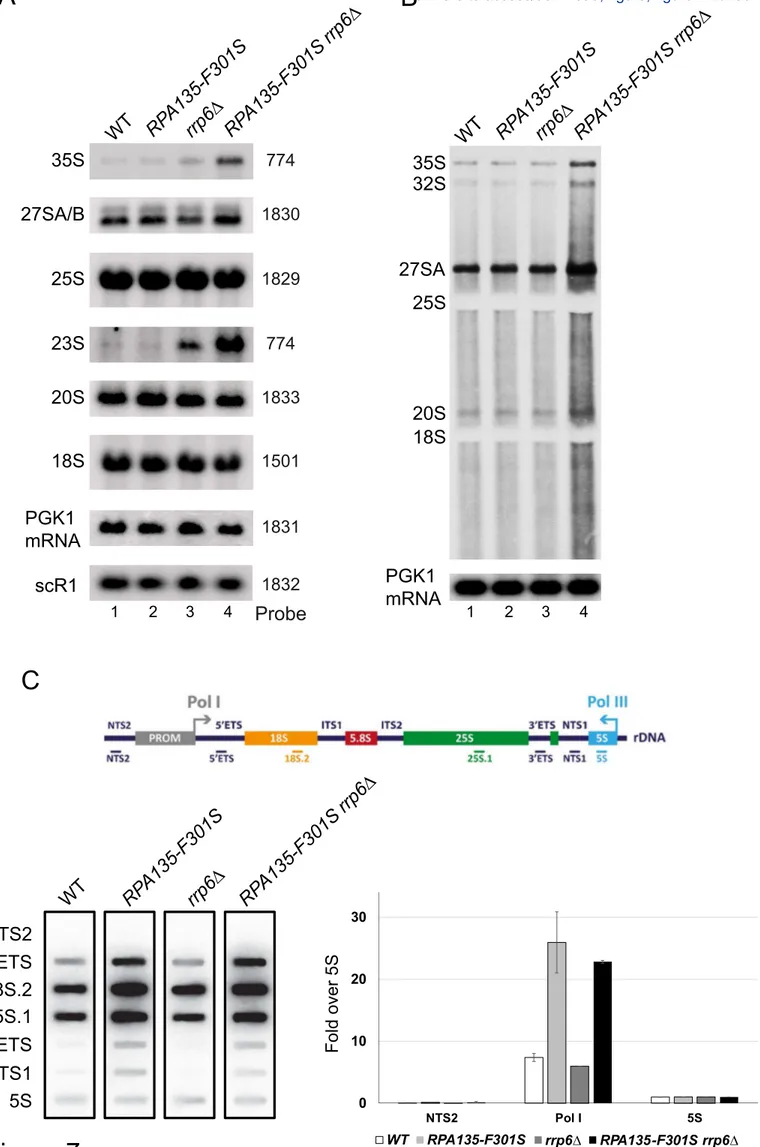
both RPA135-F301S and the deletion of RRP6 (S5 Fig). Northern blot analysis (Fig. 7A) 13
showed that accumulation in RPA135-F301S single mutant was indistinguishable from the 14
WT for all RNA probed. rrp6∆ single mutant accumulates 23S and 35S (pre-)rRNA [40,41] 15
and in correlation with the growth defect of the double mutant RPA135-F301S rrp6∆, we 16
could observe a 2-fold increase in 35S and 23S (pre-)rRNA accumulation as compared to 17
rrp6Δ. Accumulation of 35S and 23S could indicate an increase of RNA production, or a 18
defect in early maturation steps. We decided to directly assess over-expression of (pre-)rRNA 19
using short in vivo labelling experiments (40 seconds). As previously reported with very short 20
pulse labelling, accumulation of 20S rRNA is barely detectable, while 27SA and 35S are 21
already accumulated [42]. We could detect a strong accumulation of newly synthesized (pre-) 22
rRNA in the double mutant RPA135-F301S rrp6∆ compared to WT, RPA135-F301S or rrp6∆ 23
strains. Note that increased background signal could indicate an accumulation of partially 24
degraded, abortive transcripts or elongating rRNA transcript of various sizes. These results 25
suggests that rRNA are over-expressed in strain bearing RPA135-F301S, but are quickly 1
decayed by Rrp6. We confirmed this observation by evaluating ongoing transcription 2
independently of decay machinery using high-resolution transcriptional run-on (TRO) 3
analysis (Fig. 7C). Indeed, TRO assay make use of 10% sarkosyl, which permeabilizes cell 4
membranes, reversibly blocks elongating polymerases and inhibits RNAse activity [43–46]. 5
Permeabilized cells are then incubated with [α32P]-UTP to resume transcription. 6
Neosynthesized radiolabeled RNAs are extracted, and used to probe slot-blots loaded with 7
single strand DNA fragments complementary to rDNA locus. Using incorporation of [α32 P]-8
UTP in the 5S rRNA transcribed by RNA polymerase III as internal control, TRO revealed a 9
three-fold increase of rRNA transcription in cells bearing RPA135-F301S allele, irrespective 10
of Rrp6 presence (Fig. 7C). These results confirmed that Pol I bearing Rpa135-F301S is over-11
producing RNA compared to WT and that over-produced RNAs are targeted for degradation 12
by the exosome. All together, we concluded that Pol I bearing Rpa135-F301S is a hyper-13
active RNA polymerase in vivo. 14
Discussion
1Here, we characterized extragenic suppressors of the growth defect of the rpa49 null mutant 2
to better understand the activity of Pol I. We showed that altering a very specific area of Pol I 3
resulted in an enzyme with modified catalytic properties sufficient to restore wild-type growth 4
in absence of Rpa49. 5
6
Suppressor mutations are not at the Rrn3-Pol I stalk interface
7
Our previous studies suggested the specific involvement of Rpa49 in the association and 8
dissociation of initiation factor Rrn3 from the Pol I stalk [26,29]). Here, we show that 9
genetically modified polymerases lacking Rpa49 or Rpa49Ct, with a single modified residue 10
in Rpa190, Rpa135, or Rpa12, at a position diametrically to the position that binds to Rrn3, 11
can initiate transcription and that strains harbouring them grow normally. Moreover, mutant 12
Pol I with Rpa135-F301S does not restore promoter dependent activity in absence of Rpa49. 13
We propose that, independently of the important interplay between Rpa49 and Rrn3 during 14
initiation, Rpa135-F301S can stimulate Pol I activity. 15
16
A novel role of Rpa12 subunit
17
The Rpa12 subunit is involved in stimulating the intrinsic cleavage activity of Pol I through a 18
TFIIS-like domain at its C-terminus. Purified Pol I with Rpa12 lacking the C-terminal domain 19
has no cleavage activity [17]. Furthermore, the C-terminal domain of Rpa12 can contact the 20
active site of the polymerase in the inactive conformation and is retrieved in both initiation 21
competent and elongating forms of the polymerase. However, the cleavage activity is re-22
activated when Pol I is paused [36]. Direct evidence that cleavage is not involved in 23
suppression of the growth defect came from the experiments showing a fully functional 24
suppressor phenotype for RPA12∆Ct-S6L, which lacks the domain required for stimulating 1
cleavage. 2
The N-terminal domain of Rpa12, at the surface of Pol I, is involved in the recruitment of the 3
largest subunit, Rpa190 [24] and is required for docking this subunit to the enzyme. A linker 4
region of Rpa12 connects its N-terminal module (equivalent to the N-terminal domain in the 5
Pol II subunit Rpb9) at the surface of Pol I to its mobile C-terminal region (TFIIS-like) and is 6
therefore indirectly required for cleavage. In vitro, purified Pol I that lacks Rpa12 has less 7
activity than WT Pol I in promoter-dependent transcription assays (S6 Fig). Mutations in 8
other Pol I domains, such as deletions in the Rpa190-DNA mimicking loop, Rpa34 or Rpa14, 9
did not influence suppression of the rpa49 deletion growth defect by the RPA135-F301S 10
allele. In contrast, the Rpa12 linker was absolutely required for efficient suppression. 11
Accordingly, RPA135-F301S allele was unable to restore efficient growth when Rpa12 was 12
absent. Thus, Rpa12 and RPA135-F301S likely cooperate in the super-active Pol I enzyme. 13
Note that rearrangement of Rpa12 have recently be shown to correlate with dissociation of 14
Rpa49/Rpa34 heterodimer from Pol I, confirming the tight interplay between those subunits 15
[47]. 16
Modification of the jaw/lobe interface may facilitate DNA cleft closure
17
Pol I undergoes major conformational changes during the transcription cycle, mainly affecting 18
the width of the DNA-binding cleft [48]. During the initiation of transcription, the cleft 19
aperture narrows from a semi-open configuration, as seen in cryo-EM structures of the 20
enzyme bound to Rrn3 [12,23,34], to a fully closed conformation observed in transcribing 21
complexes [13,15] (Fig. 8). This allows gripping of the transcription bubble inside the cleft 22
(Fig. 8A-B). Following Rrn3 release, DNA binding is further secured, by the Rpa49-linker, 23
which crosses the cleft from the lobe to the clamp, passing over the downstream DNA, and by 24
the Rpa49Ct, which binds the upstream DNA in the vicinity of the clamp [15,22]. Therefore, 1
Rpa49Ct is anchored in a position securing cleft closure (Fig. 8B). 2
Cleft closure is achieved by the relative movement of two structural units, located on opposite 3
sides of the cleft, pivot with respect to each other using five hinges [4]. The unit consisting of 4
the shelf and clamp modules appears to be rigid, whereas the unit comprising the core and 5
lobe modules, which is in the vicinity of the mutated residues involved in suppression of the 6
rpa4∆growth defect, undergoes internal rearrangements (S1 Movie). The most prominent 7
reorganization within this latter unit affects the Rpa190 jaw domain, the outer rim of which 8
shifts away from the DNA by approximately 3.7 Å, using the lobe/jaw interface as a hinge 9
(Fig. 8B; blue arrow). This movement also involves the linker region of Rpa12, which 10
contains a β-strand (residues 46-50) that completes a four-stranded β-sheet in the Rpa190 jaw 11
domain. As a result, a short α-helix within the Rpa12 linker region shifts by approximately 12
3.0 Å (Fig. 8B; yellow arrow). 13
Rearrangements in the jaw made up of regions of Rpa190 and Rpa12 are likely essential to 14
allow pivoting of the shelf-clamp unit against the core-lobe unit. Without such motion, cleft 15
closure would be impossible (S1 Movie). Structural analysis suggests that the C-terminal 16
domain of Rpa49 and its linker domain are involved in securing the closed cleft conformation 17
[15,22]. Cleft closure is likely destabilized in the rpa49∆ mutant (Fig. 8C). We propose that 18
Rpa135-F301S or Rpa12-S6L favors DNA capture by increasing the flexibility of the 19
lobe/jaw/Rpa12 interface of Pol I relative to that of the WT polymerase, facilitating cleft 20
closure in the absence of Rpa49 (Fig. 8D, red arrow). As shown in vitro, mutant Pol I with 21
Rpa135-F301S is super-active. In vivo, the increased accumulation of pre-rRNA, detectable in 22
absence of the nuclear exosome (rrp6∆), is in agreement with the in vitro super-activity. We 23
propose that Pol I bearing Rpa135-F301S might facilitate cleft closure, in addition to secure 24
cleft closure by Rpa49. Such mutant enzyme could capture DNA more efficiently than WT 1
polymerase (Fig. 8E). Alternatively, the mutations could also favour a 'closed cleft' 2
conformation in the presence of DNA with little impact on flexibility. 3
4
Pol I cleft closure is a limiting step in catalytic cycle
5
Here, we show that Pol I can be engineered to generate more rRNA. A super-active mutant of 6
Pol II was already characterized: point mutation in trigger loop (Rpb1-E1103G) leads to 7
increase RNA polymerization rate, affecting pausing and transcriptional fidelity [49,50]. 8
Despite a very similar active site organization, an analogous mutation in the Pol I trigger loop 9
resulted in a very different outcome, as it reduces the elongation rate [51]. Such observations 10
led to the conclusion that Pol I catalytic cycle is very different than the one of Pol II, with 11
different rate limiting steps [51–53]. We show here that mutations away of the active center 12
can lead to a super-active form of Pol I. This difference can stem from wide-open 13
configuration of the DNA-binding cleft in Pol I compared to other polymerases. We propose 14
that Pol I cleft opening and closure is a limiting step in the Pol I catalytic cycle. 15
Materials and Methods
1Construction of plasmids and yeast strains
2
The oligonucleotides used in this study are listed in S4 Table. Plasmids and details of the 3
cloning steps are described in S3 Table. Randomly mutagenized RPA190 and RPA135 4
libraries were obtained by transformation and amplification of pVV190 and pNOY80, 5
respectively, into XL1-red strains according to the manufacturer’s guidelines (XL1-Red 6
Competent Cells, from Agilent Technologies). Yeast strains are listed in S2 Table, and were 7
constructed by meiotic crossing and DNA transformation [54][55]. The yeast media and 8
genetic techniques were described previously [56]. yCNOD226-1a was obtained from 9
yCNOD223-2a by switching the KAN-MX to the NAT-MX marker under the control of the 10
MF(ALPHA)2/YGL089C promoter (alphaNAT-MX4), which allowed selection of MATα 11
haploid cells [33]. OGT9-6a is an offspring of yCNOD226-1a crossed with BY4741. OGT8-12
11a is an offspring of BY4742 crossed with Y1196. Strains OGT9-6a and OGT8-11a were 13
plated on rich media and UV irradiated (5W/m2 during 5 second), resulting in 50% survival. 14
LH514D and LH11D are suppressor clones of the growth defect selected from UV-irradiated 15
OGT9-6a grown at 25°C. AH29R is a suppressor clone of the growth defect selected from 16
UV-irradiated OGT8-11a grown at 25°C. Genetic interaction mapping (GIM) analysis of the 17
RPA49 deletion mutant was performed as described previously [33]. Microarray data were 18
normalized using MATLAB (MathWorks, Inc., Natick, MA) as previously described [29]. 19
OGT15-7b is an offspring of LH514D with BY4741, followed by homologous recombination 20
using PCR-amplified fragments generated with oligos 1716 and 1717 and pCR4-HIS3 as 21
template. yTD16-1a was first transformed with plasmid pCJPF4-GAL49-1. Then, RPA135 22
was tagged by homologous recombination using PCR-amplified fragments generated with 23
oligos 835 and 836 and genomic DNA of strain RPA135-TAP or yTD6-6c, generating yTD27-24
1 and yTD28-1a, respectively. Strain yTD25-1a bears a C-terminal deletion of RPA49 25
generated by homologous recombination using PCR-amplified fragments generated with 1
oligos 208 and 1515 and pFA6-KAN-MX6 as template. yTD11-1a was derived from strain 2
yTD25-1a after switching to HPH-MX by homologous recombination using pUC19-HPH cut 3
by BamHI. C-terminal deletion of RPA49 in yTD29-1a and yTD30-1a were generated by 4
homologous recombination using PCR-amplified fragments generated with oligos 649 and 5
650 and yTD11-1 genomic DNA as template, transformed into yTD27-1 and yTD28-1a, 6
respectively. Genomic allelic insertion of RPA12-S6L in yTD31-1a and yTD23-1a was 7
performed by homologous recombination using PCR-amplified fragments generated with 8
oligos 1556 and 1557 and pRS316-A12-S6L-KAN as template, transformed into yTD27-1 9
and yTD29-1a, respectively. 10
TGT135-3b was obtained after sporulation of y27138 transformed by pNOY80. TGT135-3b 11
and OGT15-9d were mated to generate TGT12. yTD2-3b and yTD2-3d are offspring of 12
TGT12 transformed with pGL135_33. yTD6-6c and yTD6-6b were generated by homologous 13
recombination using pTD2_6c_135TAP cut with XhoI-NsiI, transformed into yTD2-3b and 14
yTD2-3d, respectively. yTD48-1a was generated by deletion of the Rpa190 DNA-mimicking 15
loop using homologous recombination with PCR-amplified fragments generated using oligos 16
1189 and 1194 and genomic DNA of strain SCOC2260 as template, transformed into 17
BY4741. 18
yTD51-2c, yTD51-8a, and yTD51-5a are offspring of yTD48-1a mated with yTD37-3a. 19
yTD36-2b is an offspring of yCN224-1a mated with yTD40-1a. yTD37-3d and yTD37-7d are 20
offspring of yCN224-1a mated with yTD41-1a. yTD38-3d is an offspring of yCN225-1a 21
mated with yTD40-1a and yTD39-8a is an offspring of yCN225-1a mated with yTD41-1a. 22
Strain yTD53-1a was constructed by homologous recombination using a PCR-amplified 23
fragment generated with oligos 1634 and 1635 and pFA6a-KanMX6-GAL::3HA as template. 24
OGT30-1a and OGT30-3a are offspring of yTD53-1a mated with yTD6-6b. yTD40-1a and 1
yTD41-1a were generated by homologous recombination using PCR-amplified fragments 2
generated with oligos 700 and 1679 and pFA6a-HA-KlURA3 as template, transformed into 3
OGT30-3a and OGT30-1a respectively, switching RPA135-TAP-tag to untagged RPA135. 4
Strain yMKS8-1a was constructed by homologous recombination using a PCR-amplified 5
fragment generated with oligos 1711 and 1713 and pFA6a-KanMX6-GAL::3HA as template, 6
transformed into strain yCD2-2a. yMKS9-9d is offspring of yMKS8-1a mated with yTD6-6c. 7
Mapping extragenic suppressors allele by genetic linkage
8
Extragenic suppressors allele SGR were mapped using GIM methods, in which a strain is 9
crossed with the entire pool of haploid deletion strains [57]. Here, deletions are used to 10
evaluate linkage to SGR locus. Due to the genetic suppression, Individual deletions 11
genetically linked to SGR are counter-selected in rpa49∆ background. Genetic mapping of 12
SGR locus is based on strong genetic linkage of counter-selected deletion alleles (see S1 Fig), 13
evaluated using micro-array of deletion bar-code (ArrayExpress accession E-MTAB-7831) 14
[58]. 15
16
In vivo labelling and RNA extraction and analysis
17
Metabolic labelling of pre-rRNA was performed as previously described [59] with the 18
following modifications. Strains were pre-grown in synthetic glucose-containing medium 19
lacking adenine at 30°C to an OD600 of 0.8 at. One-milliliter cultures were labeled with 50
20
µCi [8-3H] adenine (NET06300 PerkinElmer) for 2 min. Cells were collected by 21
centrifugation and the pellets were frozen in liquid nitrogen. RNA was then extracted as 22
previously described [60] and precipitated with ethanol. For high molecular weight RNA 23
analysis, 20% of the RNA was glyoxal denatured and resolved on a 1.2% agarose gel. Low 24
molecular weight RNAs were resolved on 8% polyacrylamide/8.3 M urea gels. 25
Miller spreads experiments and analysis
1
Chromatin spreading was mainly performed as described previously with minor modifications 2
[61]. Carbon-coated grids were rendered hydrophilic by glow discharge instead of ethanol 3
treatment. Negatively stained chromatin was obtained by short incubation with heavy metal 4
followed by quick drying of the sample. Images were obtained using a JEOL JEM-1400 HC 5
electron microscope (40 to 120 kV) with an Orius camera (11Mpixels). The position of the 6
RNA polymerase I molecules and the rDNA fiber were determined by visual inspection of 7
micrographs using Image J (http://rsb.info.nih.gov/ij/). Digital images were processed by 8
software programs Image J and Adobe Photoshop® (v. CS6). 9
10
In vitro promoter-dependent and tailed template transcription assays
11
In vitro promoter-dependent transcription reactions were performed as previously described 12
[23,62] with some modifications. Briefly, 1.5 ml reaction tubes (Sarstedt safety seal) were 13
placed on ice. Template (0.5–1 µl; 50–100 ng DNA) was added, corresponding to a final 14
concentration of 5–10 nM per transcription reaction (25-µl reaction volume). Core factor (1–2 15
µl; 0.5 to 1 pmol/µl; final concentration 20–40 nM) and 1–3 µl Pol I (final concentration 4–12 16
nM) were added to each tube. Then, 20 mM HEPES/KOH pH 7.8 was added to a final 17
volume of 12.5 µl. Transcription was started by adding 12.5 µl 2X transcription buffer. The 18
samples were incubated at 24°C for 30 min at 400 rpm in a thermomixer. 19
25 nM of tailed templates were used in a total volume of 25 µl. The transcription was 20
performed as described in promoter-dependent transcription reactions: Tailed templates are 21
PCR-amplified fragments generated with oligos 1834 and 1835 and “pUC19tail_g-22
_601_elongated” as PCR-template, treated with Nb.BsmI (NEB) to generate a nick. Nicked 23
site allows non-specific initiation of Pol I without the addition of initiation factors. 24
Transcription was stopped by adding 200 µl Proteinase K buffer (0.5 mg/ml Proteinase K in 1
0.3 M NaCl, 10 mM Tris/HCl pH 7.5, 5 mM EDTA, and 0.6% SDS) to the supernatant. The 2
samples were incubated at 30°C for 15 min at 400 rpm in a thermomixer. Ethanol (700 µl) 3
p.a. was added and the tubes mixed. Nucleic acids were precipitated at -20°C overnight or for 4
30 min at -80°C. The samples were centrifuged for 10 min at 12,000g and the supernatant 5
removed. The precipitate was washed with 0.15 ml 70% ethanol. After centrifugation, the 6
supernatant was removed and the pellets dried at 95°C for 2 min. RNA in the pellet was 7
dissolved in 12 µl 80% formamide, 0.1 M TRIS-Borate-EDTA (TBE), 0.02% bromophenol 8
blue, and 0.02% xylene cyanol. Samples were heated for 2 min with vigorous shaking at 95°C 9
and briefly centrifuged. After loading on a 6% polyacrylamide gel containing 7M urea and 1X 10
TBE, RNAs were separated by applying 25 watts for 30–40 min. The gel was rinsed in water 11
for 10 min and dried for 30 min at 80°C using a vacuum dryer. Radiolabelled transcripts were 12
visualised using a PhosphoImager. 13
14
RNA extractions and Northern Hybridizations.
15
RNA extractions and Northern hybridizations were performed as previously described [60]. 16
For high molecular weight RNA analysis, 3µg of total RNA were glyoxal denatured, resolved 17
on a 1.2% agarose gel and transferred to a nylon membrane. The sequences of 18
oligonucleotides used to detect the RNA species: 35S rRNA, 25S rRNA, 20S rRNA, 18S 19
rRNA, 27S rRNA, PGK1 mRNA and SCR1 ncRNA are respectively 774, 1829, 1833, 892, 20
1830, 1831 and 1832 are reported in S4 Table. 21
Chromatin immunoprecipitation (ChIP). ChIPs were performed essentially as described
22
previously [63]. At least three independent cultures of each yeast strain were grown to 23
exponential phase OD600 = 0.4-0.8 in YPD (yeast extract-peptone-glucose) at 30°C, and
cross-24
linked for 10 min at RT by the addition of formaldehyde (F1635, SIGMA) to a final 25
concentration of 1.2%. Adding glycine quenched the cross-linking reaction. Cells were 1
broken in lysis buffer (50 mM HEPES [pH 7.5], 150 mM NaCl, 1 mM EDTA, 1% Triton X-2
100, 0.1% deoxycholate Na, 0.1% SDS, 1 mM AEBSF [Euromedex] and cOmplete EDTA-3
free [Roche]) with glass beads (diameter, 0.425 to 0.6 mm; SIGMA) in a Precellys 24 4
homogenizer (bertin technologies) for 3 min, 6000 rpm at 4°C. Extracted chromatin was 5
sonicated to obtain 300-500 bp DNA fragments. 100 μg of sonicated chromatin (protein 6
content measured using BCA Protein Assay kit, ref# 23225, Thermo Scientific) was 7
immunoprecipitated for 4h at 21°C on Pan mouse Dynabeads (Invitrogen, 200 µl). 8
Immunoprecipitated DNA was purified and quantified by real-time qPCR using iTaq 9
universal SYBR®Green Supermix (Bio-Rad) and the ViiA7 AB Applied Biosystems (Life 10
Technologies). Primer pairs used for amplification are listed in S4 table. Signals were 11
analysed with Quant Studio Real Time PCR Software v1.1 and are expressed as percentage of 12
input DNA. Bars on the graph show the median of values normalised to wild-type value. 13
Error bars correspond to standard deviations of at least three independent cultures for each 14
strain. 15
Pulse-field gel electrophoresis (PFGE). PFGE is a technique that resolves
chromosome-16
sized DNA molecules in an agarose gel. Over-night YPD cultures at 30°C were harvested in 17
0.1% azide Na and kept on ice. Cells were washed twice in cold 0.05 M EDTA [pH 8.0]. 18
~2.108 cells were resuspended in 1ml of freshly prepared NZ buffer (Citrate Phosphate buffer
19
33 mM, ETDA 0.05 M [pH 8.0], Sorbitol 1.2 M). Cells were pellets (8000 rpm, 1 min, 4°C) 20
and resuspended in 500 µL of Zymolyase buffer (NZ buffer with 20T zymolyase) and 500 µL 21
LMP 2% (SeaKem LE agarose, Lonza, ref# 50001). Homogenized pellets were poured into 22
the plug molds (~100 µl/plug) and incubated for 30 min in humid chamber at 37°C then 10 23
min at 4°C. Solidified plugs were transferred in 14 ml tubes with round bottom and incubate 24
with 400 µl/plug of PK buffer (EDTA 0.125 M [pH 9.5], Sarkosyl 1%, PK 1 mg/ml (Roche, 25
ref# 03115801001) and incubated 1-2 hours at 37°C with gentle agitation. PK buffer was 1
changed and plugs were incubated O/N with gentle agitation. De-proteinated plugs were 2
washed with 10 ml TE followed by washing with 10 ml TE + 1 mM AEBSF (Euromedex) for 3
2 hours. AEBSF was washed out twice with 10 ml of 0.05 M EDTA [pH 8.0] for 30 minutes. 4
Plugs were stored at 4°C till usage in TE or 0.05 M EDTA [pH 8.0]. Plugs were loaded on 5
0.8% agarose gel in 1×TAE buffer (Certified Megabase agarose, Bio-Rad, ref# 161-3109) and 6
run in 1×TAE buffer for 50h in CHEF-DRIII System with chiller unit (Bio-Rad) with 7
following parameters: 3V/cm, initial S/time 250 sec, final S/time 900 sec, angle 120. Gel was 8
stained with ethidium bromide (10 mg/ml in water). Gel was processed for Southern blotting: 9
DNA was transferred on nylon membrane by passive transfer in 10×SSC buffer (Amersham 10
Hybond XL, GE Healthcare). Membranes were UV-cross-linked and hybridized overnight 11
with 32P-labeled single-stranded DNA probes at 42 °C. Blots were washed twice with 2×SSC 12
and 1% SDS for 20 min and once with 0.5×SSC and 1% SDS for 20 min at 37 °C. rDNA 13
locus (chromosome XII) was detected with 18S specific probe 14
(CATGGCTTAATCTTTGAGAC). This protocol was kindly provided by B. Pardo (IGH, 15
Montpellier, France), and is extensively described in [64]. 16
17
Transcriptional run-on analysis 18
TRO was performed as previously described [39,43]. Slot blots were loaded with single-19
stranded 80-mers DNA oligonucleotides: 1855 (NTS2), 1857 (5’ETS), 1859 (18S.2), 1860 20
(25S.1), 1861 (3’ETS), 1862 (NTS1), 1863 (5S US) and 1864 (5S DS). 21
22 23 24
Acknowledgements
1We would like to thank the imaging platform of Toulouse TRI for their assistance. We thank 2
Margaux Cescato, Lilian Guillot and Jorge Perez-Fernandez for strain and plasmid 3
constructions. YCp50-26 carrying RPA49 was kindly provided by P. Thuriaux. We also thank 4
Melanie Panarotto for the Miller spreads analysis. This work benefited from exchanges with 5
all unit members, including Anthony Henras, Frederic Beckouët and Lise Dauban. We thank 6
Stephanie Balor and Vanessa Soldan from the METI platform for the TEM acquisitions. 7
References
12
1. Chambon P (1975) Eukaryotic nuclear RNA polymerases. Annu Rev Biochem 44: 613– 3
638. doi:10.1146/annurev.bi.44.070175.003145. 4
2. Warner JR (1999) The economics of ribosome biosynthesis in yeast. Trends Biochem 5
Sci 24: 437–440. 6
3. Miller OL, Beatty BR (1969) Visualization of nucleolar genes. Science 164: 955–957. 7
4. Fernández-Tornero C, Moreno-Morcillo M, Rashid UJ, Taylor NMI, Ruiz FM, et al. 8
(2013) Crystal structure of the 14-subunit RNA polymerase I. Nature 502: 644–649. 9
doi:10.1038/nature12636. 10
5. Engel C, Sainsbury S, Cheung AC, Kostrewa D, Cramer P (2013) RNA polymerase I 11
structure and transcription regulation. Nature 502: 650–655. doi:10.1038/nature12712. 12
6. Hoffmann NA, Jakobi AJ, Moreno-Morcillo M, Glatt S, Kosinski J, et al. (2015) 13
Molecular structures of unbound and transcribing RNA polymerase III. Nature 528: 14
231–236. doi:10.1038/nature16143. 15
7. Cramer P, Bushnell DA, Kornberg RD (2001) Structural basis of transcription: RNA 16
polymerase II at 2.8 angstrom resolution. Science 292: 1863–1876. 17
doi:10.1126/science.1059493. 18
8. Werner F (2008) Structural evolution of multisubunit RNA polymerases. Trends 19
Microbiol 16: 247–250. doi:10.1016/j.tim.2008.03.008. 20
9. Peyroche G, Milkereit P, Bischler N, Tschochner H, Schultz P, et al. (2000) The 21
recruitment of RNA polymerase I on rDNA is mediated by the interaction of the A43 22
subunit with Rrn3. EMBO J 19: 5473–5482. doi:10.1093/emboj/19.20.5473. 23
10. Yamamoto RT, Nogi Y, Dodd JA, Nomura M (1996) RRN3 gene of Saccharomyces 24
cerevisiae encodes an essential RNA polymerase I transcription factor which interacts 25
with the polymerase independently of DNA template. EMBO J 15: 3964–3973. 26
11. Blattner C, Jennebach S, Herzog F, Mayer A, Cheung ACM, et al. (2011) Molecular 27
basis of Rrn3-regulated RNA polymerase I initiation and cell growth. Genes Dev 25: 28
2093–2105. doi:10.1101/gad.17363311. 29
12. Torreira E, Louro JA, Pazos I, González-Polo N, Gil-Carton D, et al. (2017) The 30
dynamic assembly of distinct RNA polymerase I complexes modulates rDNA 31
transcription. Elife 6. doi:10.7554/eLife.20832. 32
13. Engel C, Gubbey T, Neyer S, Sainsbury S, Oberthuer C, et al. (2017) Structural basis of 33
RNA polymerase I transcription initiation. Cell 169: 120–131.e22. 34
doi:10.1016/j.cell.2017.03.003. 35
14. Sadian Y, Tafur L, Kosinski J, Jakobi AJ, Wetzel R, et al. (2017) Structural insights into 36
transcription initiation by yeast RNA polymerase I. EMBO J 36: 2698–2709. 37
doi:10.15252/embj.201796958. 38
15. Han Y, Yan C, Nguyen THD, Jackobel AJ, Ivanov I, et al. (2017) Structural mechanism 39
of ATP-independent transcription initiation by RNA polymerase I. Elife 6. 40
doi:10.7554/eLife.27414. 41
16. Keener J, Josaitis CA, Dodd JA, Nomura M (1998) Reconstitution of yeast RNA 42
polymerase I transcription in vitro from purified components. TATA-binding protein is 43
not required for basal transcription. J Biol Chem 273: 33795–33802. 44
17. Kuhn C-D, Geiger SR, Baumli S, Gartmann M, Gerber J, et al. (2007) Functional 45
architecture of RNA polymerase I. Cell 131: 1260–1272. 46
doi:10.1016/j.cell.2007.10.051. 47
18. Geiger SR, Lorenzen K, Schreieck A, Hanecker P, Kostrewa D, et al. (2010) RNA 48
polymerase I contains a TFIIF-related DNA-binding subcomplex. Mol Cell 39: 583– 49
594. doi:10.1016/j.molcel.2010.07.028. 1
19. Fernández-Tornero C, Böttcher B, Rashid UJ, Steuerwald U, Flörchinger B, et al. 2
(2010) Conformational flexibility of RNA polymerase III during transcriptional 3
elongation. EMBO J 29: 3762–3772. doi:10.1038/emboj.2010.266. 4
20. Landrieux E, Alic N, Ducrot C, Acker J, Riva M, et al. (2006) A subcomplex of RNA 5
polymerase III subunits involved in transcription termination and reinitiation. EMBO J 6
25: 118–128. doi:10.1038/sj.emboj.7600915. 7
21. Wu C-C, Lin Y-C, Chen H-T (2011) The TFIIF-like Rpc37/53 dimer lies at the center 8
of a protein network to connect TFIIIC, Bdp1, and the RNA polymerase III active 9
center. Mol Cell Biol 31: 2715–2728. doi:10.1128/MCB.05151-11. 10
22. Tafur L, Sadian Y, Hoffmann NA, Jakobi AJ, Wetzel R, et al. (2016) Molecular 11
structures of transcribing RNA polymerase I. Mol Cell 64: 1135–1143. 12
doi:10.1016/j.molcel.2016.11.013. 13
23. Pilsl M, Crucifix C, Papai G, Krupp F, Steinbauer R, et al. (2016) Structure of the 14
initiation-competent RNA polymerase I and its implication for transcription. Nat 15
Commun 7: 12126. doi:10.1038/ncomms12126. 16
24. Van Mullem V, Landrieux E, Vandenhaute J, Thuriaux P (2002) Rpa12p, a conserved 17
RNA polymerase I subunit with two functional domains. Mol Microbiol 43: 1105–1113. 18
25. Gadal O, Mariotte-Labarre S, Chedin S, Quemeneur E, Carles C, et al. (1997) A34.5, a 19
nonessential component of yeast RNA polymerase I, cooperates with subunit A14 and 20
DNA topoisomerase I to produce a functional rRNA synthesis machine. Mol Cell Biol 21
17: 1787–1795. 22
26. Beckouet F, Labarre-Mariotte S, Albert B, Imazawa Y, Werner M, et al. (2008) Two 23
RNA polymerase I subunits control the binding and release of Rrn3 during 24
transcription. Mol Cell Biol 28: 1596–1605. doi:10.1128/MCB.01464-07. 25
27. Liljelund P, Mariotte S, Buhler JM, Sentenac A (1992) Characterization and 26
mutagenesis of the gene encoding the A49 subunit of RNA polymerase A in 27
Saccharomyces cerevisiae. Proc Natl Acad Sci USA 89: 9302–9305. 28
28. Nogi Y, Yano R, Dodd J, Carles C, Nomura M (1993) Gene RRN4 in Saccharomyces 29
cerevisiae encodes the A12.2 subunit of RNA polymerase I and is essential only at high 30
temperatures. Mol Cell Biol 13: 114–122. 31
29. Albert B, Léger-Silvestre I, Normand C, Ostermaier MK, Pérez-Fernández J, et al. 32
(2011) RNA polymerase I-specific subunits promote polymerase clustering to enhance 33
the rRNA gene transcription cycle. J Cell Biol 192: 277–293. 34
doi:10.1083/jcb.201006040. 35
30. Gadal O, Labarre S, Boschiero C, Thuriaux P (2002) Hmo1, an HMG-box protein, 36
belongs to the yeast ribosomal DNA transcription system. EMBO J 21: 5498–5507. 37
31. Huet J, Buhler JM, Sentenac A, Fromageot P (1975) Dissociation of two polypeptide 38
chains from yeast RNA polymerase A. Proc Natl Acad Sci USA 72: 3034–3038. 39
32. Viktorovskaya OV, Appling FD, Schneider DA (2011) Yeast transcription elongation 40
factor Spt5 associates with RNA polymerase I and RNA polymerase II directly. J Biol 41
Chem 286: 18825–18833. doi:10.1074/jbc.M110.202119. 42
33. Decourty L, Saveanu C, Zemam K, Hantraye F, Frachon E, et al. (2008) Linking 43
functionally related genes by sensitive and quantitative characterization of genetic 44
interaction profiles. Proc Natl Acad Sci USA 105: 5821–5826. 45
doi:10.1073/pnas.0710533105. 46
34. Engel C, Plitzko J, Cramer P (2016) RNA polymerase I-Rrn3 complex at 4.8 Å 47
resolution. Nat Commun 7: 12129. doi:10.1038/ncomms12129. 48
35. Neyer S, Kunz M, Geiss C, Hantsche M, Hodirnau V-V, et al. (2016) Structure of RNA 49
polymerase I transcribing ribosomal DNA genes. Nature 540: 607–610. 50