HAL Id: tel-00826943
https://tel.archives-ouvertes.fr/tel-00826943
Submitted on 28 May 2013HAL is a multi-disciplinary open access archive for the deposit and dissemination of sci-entific research documents, whether they are pub-lished or not. The documents may come from teaching and research institutions in France or abroad, or from public or private research centers.
L’archive ouverte pluridisciplinaire HAL, est destinée au dépôt et à la diffusion de documents scientifiques de niveau recherche, publiés ou non, émanant des établissements d’enseignement et de recherche français ou étrangers, des laboratoires publics ou privés.
Role of Sema3A/Neuropilin1 signaling in GnRH system
development and study of the involvement of
NO-synthesizing neurons in the kisspeptin-dependent
preovulatory activation of adult GnRH neurons
Naresh Kumar Hanchate
To cite this version:
Naresh Kumar Hanchate. Role of Sema3A/Neuropilin1 signaling in GnRH system development and study of the involvement of NO-synthesizing neurons in the kisspeptin-dependent preovulatory activation of adult GnRH neurons. Human health and pathology. Université du Droit et de la Santé -Lille II, 2011. English. �NNT : 2011LIL2S046�. �tel-00826943�
UNIVERSITE DROIT ET SANTE DE LILLE II
Ecole Doctorale Biologie - SantéThese
Pour l'obtention du grade de
DOCTEUR DE L'UNIVERSITE DE LILLE II Spécialité : Neuroscience
Presentée par
Naresh Kumar HANCHATE
Role of Sema3A/Neuropilin1 signaling in GnRH system development and
Study of the involvement of NO-synthesizing neurons in the
kisspeptin-dependent preovulatory activation of adult GnRH neurons
Soutenue le 12 Decembre 2011 devant le Jury composé de:
Rapporteurs :
Jeroen PASTERKAMP Assistant Professor, UMC, Utrecht, Netherlands
Valerie SIMONNEAUX Directeur de Recherche, CNRS UPR-3212, Strasbourg
Examinateurs :
Ulrich BOEHM Directeur de Recherche, Hamburg, Allemagne
Philippe CIOFI Charge de recherche, INSERM U862, Bordeaux.
Jacques YOUNG PU/PH, INSERM U693, Le Kremlin-Bicêtre
Jean-Louis NAHON Directeur de Recherche, IPMC UMR 6097, Valbonne
Directeur de These :
Acknowledgement
The path of success is not straight, but riddled with temptations, impediments and pitfalls all along. All these difficulties make us realize the importance of success humbly.
I feel short of words to express my deep sense of gratitude towards my parents, family and friends for their moral support and motivation with whose grace present study has been completed.
I would like to greatly thank Dr. Jeroen Pasterkamp and Dr. Valerie Simonneaux, for kindly accepting our request as rapporteurs for my thesis and taking the precious time for giving evaluating and giving their valuable and critical comments on my thesis.
I would like to thank Dr. Jacques Young and Dr. Jean-Louis Nahon to kindly accepting our request as examinateurs for my thesis and for their honoured presence for my phd defense.
It gives me immense pleasure in expressing my sincere gratitude to my mentor Dr. Vincent Prevot for introducing me to the enthusiastic field of ‘Neuroscience’, his kind supervision, guidance and encouragement throughout the course of this study. I also appreciate his untiring efforts during the entire tenure of my research work. I earnestly believe that without his right suggestions, this work would not have been completed. He is more than a supervisor for me. Thanks Sir, for believing in me and standing by me over the years.
Special whole-hearted thanks to Dr. Jean-Claude Beauvillain and Dr. Pierre Poulain for their inspirational, highly intellect discussions and special moments spent together during my graduation.
Special thanks to Dr. Sebastien Bouret, for his valuable and crucial suggestions that help us to build my projects and also for introducing me to Dr.Takako Makita and eventually helping me to obtain a postdoc in her lab.
Special thanks to Dr. Paolo Giacobini for his unconditional, continuous and timely support for my projects. It was a great experience to work with him and to have all the long discussions and also to have good friendly moments together.
Special thanks to Dr. Jean-pierre Hardelin and Dr. Catherine Dode for their timely collaboration and their findings on human kallmann syndrome patients that strongly supports this study.
A special thanks to Jp, for his ever cheerful moments, all the important advices and great help. I am very happy to meet u in france and to keep this friendship forever.
A special thanks to Eglantine for the unforgettable cherishing moments together, building all the faith, trust and confidence in me, for embedding all the good values and most importantly to stay beside me even during the most difficult moments of life and during this thesis.
A special thanks to Sophie her cheerful, memorable moments and for the unconditional motivation and encouragement I needed throughout the thesis.
Un tres grand merci a Daniele pour tout l'aide, pour les tres bon moments, pour m'avoir accompagne tout au long de ma these.
A special thanks to Andrea for sharing his monstrous technical knowledge that had helped me to prepare my figures and writing the manuscripts in time.
I would like to thank the team members Ariane, Amandine, Benedicte, Odile, Celine, Anne, Fanny, Filippo, Guillaume, Sophie, Christelle, Cecile for all the help, discussions and good moments spent together.
Une tres grand merci a Anne Loyens, Karim, Joelle, Patrick, Lucien, toute l'equipe de l'animalerie, je voudrais remercier specialement Julien, François, Ingrid, les deux Delphine, Yann, pour toute l'aide que vous m'avez apporté durant quatre ans.
Une tres grand merci a tout l'equipe de la secretariat specialment Sophie, Michelle, Marie-Jeanne and Celine pour toute l'aide que vous m'avez apporté durant quatre ans.
Fianlly, I am thankful to INSERM for awarding me the research fellowship.
Resume
Reproduction in mammals is regulated by neurons that synthesize and secrete gonadotropin-releasing hormone (GnRH) and across the species these neurons are present in few numbers scattered in the hypothalamus. Due to limited neurogenesis of these neuronal cell types outside the brain in the olfactory placode, these neurons are subjected to tight regulation during embryonic development to reach their final targets in the hypothalamus, from birth until puberty for minimal secretion of hormone and during adults to achieve pulsatile secretion of the hormone. Deregulation in any of these mechanisms may lead to deleterious effects on adult reproduction and clinical pathologies like absence of puberty, hypogonadism, sterility, amenorrhea, etc. Kallmann syndrome (KS), one of these severe reproductive pathologies is an inherited disorder and patients affected with this syndrome display anosmia (inability to smell) and hypogonadotropic hypogonadism (HH). Genetic screening of molecules in these patients lead to identification of genes like KAL1, FGFR1,
FGF8, PROK2, PROKR2, WDR11 and CHD7 encoding proteins that play an important role in
migration and targeting of olfactory system during embryonic development however these genes account only for 30% of KS cases emphasizing the need for further characterization and identification of other genes. While these proteins are involved in ontogenesis olfactory and GnRH system, genetic screening of molecules in patients suffering from normosmic idiopathic HH lead to identification of genes encoding for Kisspeptin receptor-GPR54,
TAC-TACR3, LEP-LEPR, PCSK-1, GnRH receptor-GnRHR and GnRH-1 itself that play a crucial
role in occurrence of puberty or adult reproduction.
Here, for my PhD thesis, we focused on studying the role of guidance molecule Semaphorin3A (Sema3A)-Neuropilin1 (Nrp1) interactions in ontogenesis of GnRH neurons during embryonic development while in adults we first addressed the question if hypothalamic Kisspeptin neurons interact with neurons containing neuronal nitric oxide synthase (nNOS), the mutation of which causes HH in mice, and physiological significance of this interactions in regulation of GnRH neurons and neuroendocrine control of female reproduction.
Semaphorin3A, that belongs to highly conserved semaphorin family of guidance molecules has been known to be implicated in ontogenesis and targeting of olfactory system
through its receptor Neuropilin1 however its role in development of GnRH neurons and adult reproduction is not known. Using genetic mouse model Nrp1sema/sema that has a mutated Neuropilin1 receptor lacking functional semaphorin binding domain, we identified the crucial role of these interactions implied in ontogenesis of GnRH neurons. These mice had disrupted olfactory and vomeronasal projections to the olfactory bulb and to the ventral forebrain respectively. Disrupted olfactory projections lead to suckling dysfunction and early postnatal death while the disrupted vomeronasal projections lead to defective migratory behavior of GnRH neurons. The few mutant mice that grow until adults display reproductive deficits and KS like phenotype and interestingly genetic screening of human patients suffering from KS by our collaborators lead to the identification of inactivating mutations of SEMA3A gene in these patients.
Hypothalamic Kisspeptin and Nitric oxide synthesizing neurons have been recognized as key regulators of GnRH neurons modulating adult reproductive function. However, no efforts have been made to study if there is any cross-talk existing between the 2 neuronal cell types. Here, we show that kisspeptin neurons project to specific population of nNOS neurons in the preoptic region and the nNOS neurons in these region express kisspeptin receptor-Gpr54. Intraperitoneal injections of kisspeptin-10 readily induced posttranslational modification of nNOS protein in these neurons and these modifications by kisspeptin-10 require Gpr54 receptor and is mediated through PI3K-AKT pathway. Interestingly, we demonstrate that during the gonadal steroid negative feedback, a constitutive basal level of NO released by neurons in the preoptic region maintain tonic inhibition on GnRH neurons resulting in nadir levels of gonadotropin release whereas high activation of NO synthesizing neurons in the preoptic region induced by increased estrogen-kisspeptin signaling during the gonadal steroid positive feedback leads to high amount of NO release that eventually set GnRH neurons to release peak levels of GnRH hormone resulting in a surge of gonadotropins necessary to trigger ovulation.
Finally our results demonstrate that Sema3A-Nrp1 interactions are implicated in ontogenesis of olfactory and GnRH neurons during embryonic development and nNOS neurons are important mediators of peripheral estrogens-kisspeptin signaling onto GnRH neurons and adult reproduction and propose to further study the implication of nNOS neurons in reproductive pathologies.
List of Publications
Hanchate NK, Giacobini P, Lhuillier P, Espy C, Fouveaut C, Leroy C, Baron S, Parkash J,
Campagne C, Collier F, Garcia-Pineiro A, Dewailly D, Cortet-Rudelli C, Gersak K, Pugeat M, Young J, Hardelin JP, Prevot V, Dodé C. Semaphorin-3A signaling insufficiency in humans affected by Kallmann syndrome. (In preparation)
Hanchate NK, Parkash J, Leroy D, de Tassigny X, Colledge WH, Prevot V.
Kisspeptin-GPR54 signaling in mouse NO-synthesizing neurons participates in the hypothalamic control of ovulation. (Under Revision in J.Neuroscience)
Clasadonte J, Poulain P, Hanchate NK, Corfas G, Ojeda SR, Prevot V. Prostaglandin E2 release from astrocytes triggers gonadotropin-releasing hormone (GnRH) neuron firing via EP2 receptor activation. Proc Natl Acad Sci U S A. 2011 Sep 20; 108(38):16104-9
Campagne C, Giacobini P, Hanchate NK, Parkash J, Mazur D, Ciofi P, Bouret SG, Prevot V. Sema3A is an endothelial-secreted chemotropic factor that controls GnRH axon plasticity in the adult brain. (In preparation)
Prevot V, Hanchate NK, Bellefontaine N, Sharif A, Parkash J, Estrella C, Allet C, de Seranno S, Campagne C, de Tassigny X, Baroncini M. Function-related structural plasticity of the GnRH system: a role for neuronal-glial-endothelial interactions. Front Neuroendocrinol.
2010 Jul; 31(3):241-58.
Bellefontaine N, Hanchate NK, Parkash J, Campagne C, de Seranno S, Clasadonte J, de Tassigny X, Prevot V. Nitric oxide as key mediator of neuron-neuron and endothelia- to-glia communication involved in the neuroendocrine control of reproduction.
Neuroendocrinology. 2011 Feb; 93:74-89.
Prevot V, Bellefontaine N, Baroncini M, Sharif A, Hanchate NK, Parkash J, Campagne C, de Seranno S. Gonadotrophin-releasing hormone nerve terminals, tanycytes and neurohaemal junction remodelling in the adult median eminence: functional consequences for reproduction and dynamic role of vascular endothelial cells. J Neuroendocrinol. 2010 Jul; 22(7):639-49.
Poster and Oral Communications
Hanchate NK, Parkash J, Mazure D, Colledge WH, d'Anglemont de Tassigny X, Prévot V.
Nitric Oxide synthesizing neurons: Important mediators of Kisspeptin - GnRH neuron interactions during the estrous cycle. 41st Annual meeting of Neuroscience (SFN), November 2011, Washington, DC, USA. (Poster presentation)
Hanchate NK, Giacobini P, Mazure D, Prévot V. Neuropilin1-Sema3A interactions are
involved in development and function of GnRH neurons. 40th Annual meeting of Neuroscience (SFN), November 2010, San Diego, USA. (Poster presentation)
Hanchate NK, Giacobini P, Mazure D, Prévot V. Neuropilin1-Sema3A interactions are
involved in development and function of GnRH neurons. 14ème Journée LARC
Neurosciences, 29 October, 2010, Lille, France. (Oral communication)
Hanchate NK, Giacobini P, Mazure D, Prévot V. Role of Neuropilin1-Sema3A interactions
in development and function of GnRH neurons. The 7th International Congress of Neuroendocrinology (ICN), July 2010, Rouen, France. (Poster presentation)
Bellefontaine N, Hanchate NK, Leroy D, Colledge WH, d'Anglemont De Tassigny X, Prévot V. Morphological evidence for the interrelationship of kisspeptin and nitric oxide containing neurons in the GnRH neuronal network. The 7th International Congress of Neuroendocrinology (ICN), July 2010, Rouen, France. (Poster presentation)
Hanchate NK, Campagne C, Mazure D, Prévot V. Role of Neuropilin1-Sema3A interactions
in GnRH neurons function. 36th International Congress of Soceity of Neuroendocrinology (SNE) September 2009, Nice, France (Poster presentation)
Hanchate NK, Giacobini P, Mazure D, Prévot V. Neuropilin1-Sema3A interactions are
involved in development and function of GnRH neurons. 13ème Journée LARC
INDEX
LIST%OF%FIGURES%AND%TABLES... 4! % INTRODUCTION ... 6! 1.!THE!GNRH!SYSTEM...6! 1.1.#Brief#History... 6! 1.2.#Embryonic#origin#of#GnRH#cells ... 8! 1.3.#The#GnRH#neurons#migratory#pathway ... 8! 1.4.#Puberty...10! 1.5.#Pulsatile#and#cyclic#secretion#of#GnRH...10! ! 2.!HYPOGONADOTROPIC!HYPOGONADISM... 12! 2.1#Kallmann#syndrome ...14! 2.1.1.#Genes#implied#in#the#aetiology#of#Kallmann#syndrome...14! 2.1.2.!KAL1...14! 2.1.3.!FGFR1!and!FGF8 ...16! 2.1.4.!PROKR2!and!PROK2 ...17! 2.1.5.!CHD7...17! 2.1.6.!WDR11...18! 2.1.7.!!NELF...18! # 2.2.#Normosmic#idiopathic#hypogonadotropic#hypogonadism#(nIHH)...19! 2.2.1.!GNRHR ...19! 2.2.2.!GNRH1...20! 2.2.3.!KISS1R!/!GPR54 ...21! 2.2.4.!TAC3!and!TACR3 ...23! 2.2.5.!LEP!and!LEPR...24! ! 3.!EMERGING!CONCEPT!OF!DIGENIC!/!OLIGOGENIC!INHERITANCE!INVOLVED!IN!PATHOGENECITY!OF!!!KS.! MORE!KS/NIHH!GENES!TO!BE!DISCOVERED. ... 25!! 4.!FACTORS!INVOLVED!IN!ONTOGENESIS!OF!OLFACTORY/GNRH!SYSTEM!DURING!EMBRYONIC!DEVELOPMENT ... 26! 4.1.#PSANNCAM ...26! 4.2.#Hepatocyte#Growth#Factor#(HGF)#/#Met#signaling ...26! 4.3.#CholecystokininN8#/#CCKN1R...27! 4.4.#SDFN1#/#CXCR4 ...27! 4.5.#GABA ...28! 4.6.#OTX2...29! 4.7.#Axl...29! 4.8.#Reelin#/#ApoER2#/#Lrp8 ...30! 4.9.#Ephrin#/#Ephrin#receptor ...31! 4.10.#Netrin#/#DCC...32! # 4.11.#Semaphorins ...32! 4.11.1.!Semaphorin!and!their!receptors...33! 4.11.2.!Sema3A!signaling ...36! 4.11.3.!Semaphorins!identified!to!play!an!important!role!in!ontogogenesis!of!GnRH/olfactory!system ...39! 4.11.4.!Sema3F!/!Neuropilin2...39! 4.11.5.!Sema4D!/!Plexin!B1!/!Met ...39! 4.11.6.!Sema3A!/!Neuropilin1!/!Neuropilin2...40! ! 5.!FACTORS!INVOLVED!IN!MATURATION!AND!FUNCTION!OF!GNRH!SYSTEM... 41! # 5.1.#KisspeptinNGpr54#signaling#in#Reproduction ...41!
5.1.1.!Brief!history...41! 5.1.2.!Nomenclature ...41! 5.1.3.!Kisspeptin!gene!products!and!its!cleaved!forms...42! 5.1.4.!Gpr54!receptor!signaling ...42! 5.1.5.!Distribution!of!kisspeptin...44! 5.1.6.!Distribution!of!Gpr54!expressing!cells ...46! 5.1.7.!Direct!and!indirect!effects!of!kisspeptin!on!GnRH!neurons...48! 5.1.8.!Kisspeptin!Z!Gpr54!signaling!in!Puberty ...48! 5.1.9.!Differential!regulation!of!2!kisspeptin!population!by!estrogens...54! 5.1.10.!Negative!feedback!action!of!sex!steroids!on!Kiss1!gene!expression!in!ARC...54! 5.1.11.!Positive!feedback!action!of!estradiol!on!Kiss1!gene!expression!in!AVPV...54! 5.1.12.!Kisspeptin!signaling!in!seasonality...55! # 5.2.#Nitric#oxide#system...56! 5.2.1.!Introduction...56! 5.2.2.!Modulation!of!GnRH!neuronal!activity!by!neuronal!nitric!oxide!during!the!ovarian!cycle. ...59! 5.2.4.!Neuronal!NO!exerts!an!acute!postsynaptic!action!on!GnRH!neurons ...62! 5.2.5.!Regulation!of!neuronal!nitric!oxide!synthase!by!ovarian!steroids...63! # 5.3.#Jak2...66! 5.4.#Six6 ...66! 5.5.#TTF1...67! 5.6.#EAP1...68! 5.7.#Glial#factors...69! 5.7.1.!ErbB!receptor!signaling...69! 5.7.1.!SynCAM1...70! % OBJECTIVES ... 72! ! PART!I. ... 74!
TO!EVALUATE!THE!ROLE!OF!THE!GUIDANCE!MOLECULE!SEMAPHORIN3A!AND!ITS!OBLIGATORY!COZRECEPTOR! NEUROPILIN1!IN!THE!DEVELOPMENT!OF!THE!GNRH!SYSTEM!IN!MICE!AND!HUMANS!AND!DETERMINE! WHETHER!SEMAPHORIN!3A!INSUFFICIENCY!IS!ASSOCIATED!WITH!KALLMAN!SYNDROME... 74!
Results...76!
! PART!II... 100!
TO!STUDY!THE!ROLE!OF!THE!KISSPEPTINZGPR54!SIGNALING!IN!NO!PRODUCING!NEURONS!IN!THE! NEUROENDOCRINE!CONTROL!OF!REPRODUCTION!IN!MICE. ... 100! Results... 102! DISCUSSION...146! PERSPECTIVES ...154! ANNEXE ...160% %
List of figures and Tables
Figure 1. Location and forms of GnRH in representative brains of fish and mammals.
Figure 2. Scheme illustrating the embryonic origin and migratory pathway of GnRH neurons.
Figure 3. The Hypothalamic-pituitary-gonadal axis.
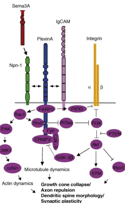
Figure 4. Schematic representation of semaphorins and their receptors (neuropilins and
plexins), divided into subfamilies on the basis of sequence similarity and structural features.
Figure 5. Sema3A binding to the Nrp–plexinA complex promotes FARP2 dissociation from
plexinA
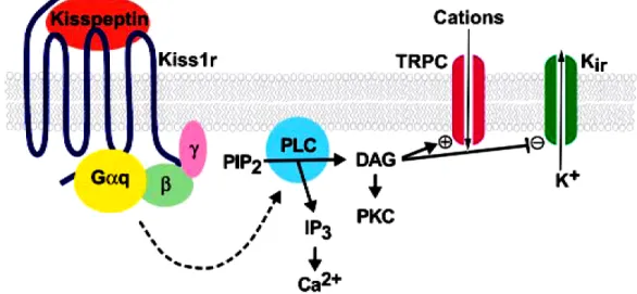
Figure 6. Proposed mechanism of neuronal depolarization by kisspeptin binding to its
receptor, Kiss1r.
Figure 7. Products of the Kiss1 gene.
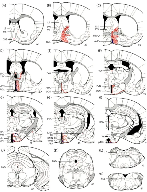
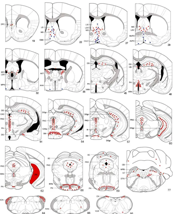
Figure 8. The distribution of kisspeptin immunoreactivity in the adult female mouse brain. Figure 9. Schematic diagrams showing distribution of Gpr54-expressing cells in adult female
mouse brain.
Figure 10. KiSS-1 mRNA is differentially regulated by estrogen in the forebrain of the
mouse.
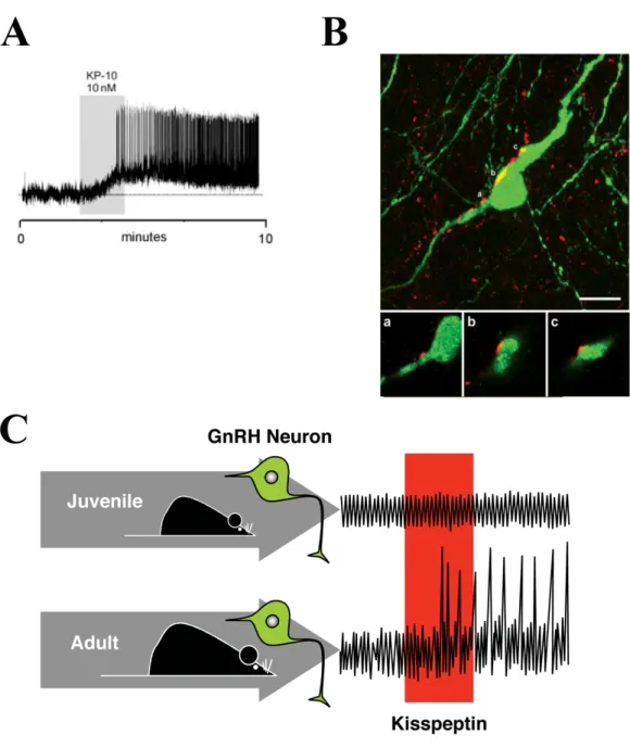
Figure 11. A. Kisspeptin exerts a direct potent activational effect on GnRH neurons in adult
female proestrous mice.
Figure 12. Kisspeptin-signaling in puberty
Figure 13. Within the preoptic region, NO release modulates GnRH neuronal activity and is required for estrous cyclicity.
Figure 14. Schematic representation of the possible estradiol-mediated changes in
protein-protein interactions involved in the control of nNOS activity in the preoptic region of the hypothalamus during the ovarian cycle
Table 1. Genes identified to cause Kallmann syndrome / normosmic HH in humans. Table 2. Prevalence of known genetic defects in hypogonadotropic hypogonadism Table 3. Examples of digenic interactions in hypogonadotrophic hypogonadism
Introduction
1. The GnRH systemMammalian reproduction is controlled by integrated sets of interactions between the hypothalamus, pituitary gland and gonads. Each component of the reproductive system is regulated by feedback mechanisms that coordinate the processes leading to gonadotropin secretion, gamete production and maintenance of the species (Conn and Crowley, 1994; S.R. Ojeda, 2006). In most mammalian species, GnRH neurons are distributed in the preoptic area and adjacent sites in the rostral region of the hypothalamus, rather than concentrated in a discrete nucleus. These scattered neurons are believed to form a diffuse neural network that functions coordinately as a GnRH pulse generator (Knobil, 1990). The generation of pulsatile GnRH release at the median eminence is the central and essential element governing reproductive function, and depends on the coordinated activities of the 1500 or so GnRH neurons that are located in the hypothalamus (Herbison et al., 2008; Wray, 2001; Wray, 2010). Studies using intrahypothalamic injection of immortalized GnRH neurons (GT1–7 cells) in hypogonadal mice revealed that regardless of the number of neurons injected, normal reproductive function could be restored (Silverman et al., 1992).
1.1. Brief History
The GnRH decapeptide from the hypothalamus of pigs and sheep was first isolated and characterized in the 1970s (Burgus et al., 1972; Matsuo et al., 1971). In the early 1980s, a second GnRH isoform from chickens was isolated and described (chicken GnRH-II) (Miyamoto et al., 1984) and a third isoform was found in fish (salmon GnRH) (Sherwood et al., 1983). As more forms of GnRH were isolated, initially they were named after the animals in which they were first found. Since the initial localization of GnRH1 to the hypothalamus, other forms of GnRH have been isolated and localized to the nervus terminalis of the forebrain (GnRH3) and to the midbrain (GnRH2) (Figure. 1). GnRH3 neurons of the nervus terminalis (a cranial nerve) have a neuromodulatory role in the forebrain and olfactory epithelium (Abe and Oka, 2002; Eisthen et al., 2000), and also act as modulators of olfactory mediated behaviors (Wirsig-Wiechmann et al., 2002). Similarly, the GnRH2 cells of the midbrain also appear to modulate sexual behaviors (Millar, 2003). Therefore, GnRH-containing cells of the nervus terminalis (GnRH3) and midbrain (GnRH2) appear to mediate
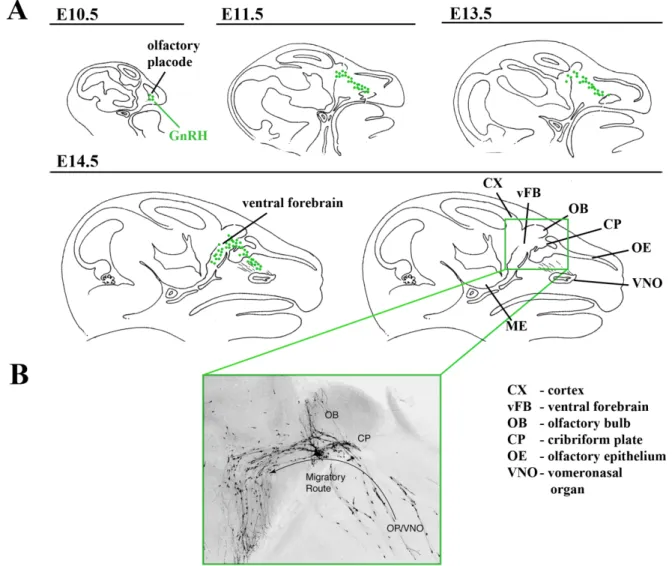
Figure 1. Location and forms of GnRH in representative brains of fish and mammals.
Sagittal sections of fish (a) and mouse (b) brain, anterior to the left. GnRH1 cells are depicted in orange, GnRH2 and GnRH3 cells are depicted in purple because they are proposed to have the same developmental origin. These are generalized locations in the brains and can vary among vertebrates. (c) Forms of GnRH for which cDNA has been sequenced and grouped, showing that representatives of all vertebrates have at least two forms of GnRH (GnRH1 and GnRH2), whereas forms having the structure of GnRH3 are so far specific to fishes. [Adapted from (Whitlock, 2005)]
reproductive behaviors, whereas GnRH cells of the hypothalamus (GnRH1) play an endocrine role.
1.2. Embryonic origin of GnRH cells
In 1989, two separate laboratories reported that the GnRH cells of the ventral forebrain of the mouse appeared to arise from the olfactory placode. Based on observations of staged fixed tissue using immunocytochemistry and in situ hybridization, GnRH cells appeared to migrate through the nasal septum into the forebrain along the terminal nerve–vomeronasal nerves (Schwanzel-Fukuda et al., 1989; Wray et al., 1989a; Wray et al., 1989b). In these studies, GnRH-positive cells were first seen in association with the olfactory placode, and then appeared in association with the vomeronasal nerve (VNN) and nervus terminalis at intermediate locations between the olfactory placode and forebrain. Once the GnRH cells entered the forebrain, they left the VNN and entered the hypothalamus, following no anatomically defined pathway. Thus, it was proposed that the olfactory placode generated not only olfactory sensory neurons and support cells, but also neuroendocrine cells containing GnRH.
1.3. The GnRH neurons migratory pathway
The GnRH neuronal migratory route can be divided into four specific stages (Figure. 2) (Tobet et al., 2001; Tobet and Schwarting, 2006; Wray, 2010). Analysis of this specific neuronal migratory process is limited by the fact that there is no marker of all GnRH neurons other than GnRH itself. No transcription factor or gene product has been demonstrated to mark all GnRH neurons early in development. The GnRH–GFP mice have green fluorescent protein expression driven by the GnRH promoter, but it is relatively weakly expressed early in development and while tracking GFP signal is useful, it does not reflect the total immunoreactive GnRH population (Spergel et al., 1999; Suter et al., 2000). Since many external inputs inhibit GnRH gene and protein expression, the exact number of neurons and their location during development is not absolute but reflective of detected GnRH mRNA or protein. Future research is needed to identify markers of GnRH neurons. With this caveat, four steps of neuronal migration can be distinguished: (1) After their birth in the area of the olfactory placode in the mouse at approximately E10.5, GnRH neurons migrate together with vomeronasal axons across the nasal mesenchyme into the forebrain (Schwanzel-Fukuda et al., 1989; Wray, 2010; Wray et al., 1989a). This initial step requires both the movement of GnRH
Figure 2. Scheme illustrating the embryonic origin and migratory pathway of GnRH neurons. GnRH neurons first appear in the olfactory placode at E10.5. (A) Series of sagittal
sections from mouse head at Embryonic day (E) 10.5, 11.5, 13.5 and 14.5 illustrating the migratory route of GnRH neurons from the olfactory placode to ventral forebrain areas. (B) Microphotograph of a mouse embryo at E15 in sagittal plane illustraing the migration of GnRH neurons from the olfactory placode (OP/VNO) to the olfactory bulb that eventually enter the ventral forebrain towards the preoptic / Hypothalamic areas. (Adapted from Wiermann et al., 2004)
neurons and the specific factors that promote the adherence of the neurons to axons of the vomeronasal nerve. (2) At the level of the cribriform plate, specific cues are needed as the vomeronasal nerve (VNN) divides with a branch that guides GnRH neurons turning caudally into the forebrain. (3) After crossing the cribriform plate and movement towards the ventral forebrain, specific factors promote the extension of long processes through the basal forebrain toward the hypothalamus. (4) Lastly, the neurons detach from their axonal guides and disperse further in the hypothalamus and stop migrating. The migratory process is usually completed by birth and the GnRH neurons as they reach the preoptic areas send their axons to the median eminence where they secrete and release the hormone. The exact steps may differ slightly across species, but most agree that a similar set of mechanistic steps are critical to target GnRH neurons to their appropriate destination in the hypothalamus so that the connection to the pituitary and ultimately reproductive competence can be achieved.
1.4. Puberty
Puberty is the transition to adulthood that culminates in the production of mature gametes and the initiation of reproductive activity (Ojeda, 2006). The process begins within the central nervous system, where gonadotropin-releasing hormone (GnRH) neurons are activated to release high-frequency pulses of the neurohormone, stimulating pituitary gonadotropic hormone secretions that in turn direct gonadal steroid hormone production and maturation of gametes (Plant, 2006; S.R. Ojeda, 2002; Sisk and Foster, 2004; Terasawa and Fernandez, 2001). Though GnRH neuronal axons achieve formation of neurovascular junctions at the median eminence by birth, GnRH neurons are maintained under continous inhibitory synaptic inputs to release minimum amounts of the hormone into the portal blood until puberty (Terasawa and Fernandez, 2001). An ill-defined developmental clock, as well as permissive somatic and environmental signals, govern the onset, progression, and completion of the pubertal acceleration of GnRH release.
1.5. Pulsatile and cyclic secretion of GnRH
GnRH neurons are distributed in the preoptic area and adjacent sites in the rostral region of the hypothalamus, rather than concentrated in a discrete nucleus. These scattered neurons are believed to form a diffuse neural network that functions coordinately as a GnRH pulse generator (Moenter et al., 2003; Van Goor et al., 1999a; Van Goor et al., 1999b). In mammals, the pattern of gonadotropin secretion includes both pulse and surge phases, which
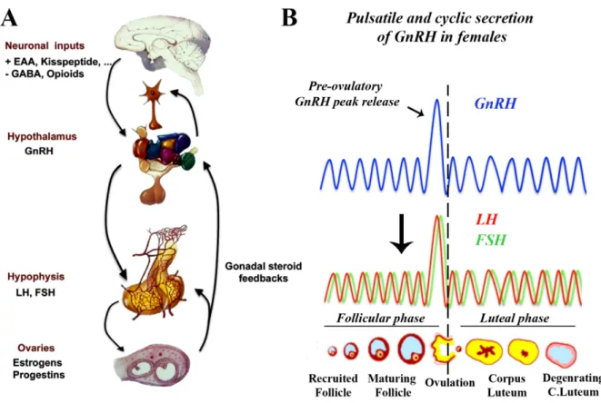
Figure 3. (A) The Hypothalamic-pituitary-gonadal axis. The hypothalamic GnRH neurons
are the final common pathway for central control of gonatropin secretion are subjected to a complex array of excitatory and inhibitory transsynaptic inputs that modulate their activity. GnRH neuroendocrine neurons project to the median eminence where they make contact with basal lamina and open into the pericapillary space of the primary hypophyseal portal plexus. Upon reaching the pituitary portal system, GnRH travels to the pituitary to stimulate the synthesis and secretion of pituitary gonadotropins: luteinizing hormone (LH) and follicle stimulating homone (FSH). Blood-borne LH and FSH act on target cells in the gonads (here the ovaries) to direct production of gametes, as well as the secretion of steroid hormones. Within the brain, gonadal steroids influence GnRH secretion via neuroendocrine feedback loops. EAA, excitatory amino acids and GABA, gammaaminobutyric acid. (Adapted from Prevot et al 2010). (B) In females, the GnRH is secreted in a pulsatile and cyclic manner characterized by the pre-ovulatory peak release that is necessary to induce peak secretion of gonadotropins - LH and FSH and trigger ovulation. During the ovarian cycle, follicular phase denotes the period during which the follicules mature, followed by ovulation (release of oocyte or female gamete). Luteal phase begins with the formation of corpus luteum that may end into pregnancy upon successful mating or luteolysis or degeneration of corpus luteum. In humans, the ovarian cycle is called menstrual cycle that lasts for 28 days while in rodents is called the estrous cycle that lasts for 4-5 days.
are regulated independently (Figure. 3) (Tsutsumi and Webster, 2009). Both the frequency and amplitude of the GnRH release in pulsatile manner are important for the development of sex function. GnRH secretion in pulsatile fashion is important in order to avoid the down regulation and sensitivity of GnRH receptor in the pituitary gonadotrophs (Belchetz et al., 1978; Schang et al., 2011). Many of the stimulatory and inhibitory signals may influence the pulse-generator by acting on secondary neurons.
In most of mammalian females, GnRH/LH is secreted in a cyclic manner i.e, called ovarian cycle or precisely estrous cycle in rodents and menstrual cycle in humans that has a duration of 4-5 days in rodents and 28 days in humans necessary for successful ovulation (Bakker and Baum, 2000; Bronson and Vom Saal, 1979; Christian and Moenter, 2010). In rats, the estrous cycle that has a duration of 4-5 days can be monitored by observing the cellular morphology of vaginal lavages that contain round lymphocytes during the diestrous phase I and II (low gonadotropins), round nucleated cells during the proestrous (LH surge) followed by cornified cells during the estrous phase (Freeman.M.E., 2006). Unlike rats, the estrous cycle in mice may vary between 4-7 days and usually do not follow a regular pattern of cycle. Throughout the estrous/menstrual cycle, GnRH and gonadotropin secretion are regulated by the negative feedback of ovarian steroids resulting in low level of secretion, but rising estrogens released by the developing follicles reach at a peak level that at acts a positive feedback resulting in a preovulatory GnRH/LH surge triggering ovulation.
2. Hypogonadotropic hypogonadism
Idiopathic hypogonadotropic hypogonadism (IHH) is characterized by delayed or absent sexual development associated with inappropriately low gonadotropin and sex steroid levels in the absence of anatomical or functional abnormalities of the hypothalamic–pituitary– gonadal axis (Layman, 2007). The major underlying cause of IHH is failure to activate pulsatile secretion of gonadotropin-releasing hormone (GnRH) during puberty, a developmental stage characterized by a substantial increase in the frequency and amplitude of pulses of this hormone. The development of GnRH neurons is unusual in that they originate outside the brain. Similar to the development of olfactory fibers, GnRH neurons are formed in the nasal placode, from which they migrate by use of the olfactory pathway to help guide them to their ultimate destination in the hypothalamus (Tobet et al., 2001). Other abnormal phenotypes that are commonly associated with and segregate with hypogonadism in affected families is anosmia, an inability to perceive smells, which is explained by the common
Table 1. Genes identified to cause Kallmann syndrome / normosmic HH in humans.
Gene Name Chromosomal Gene product OMIM no.
locus
KAL1 Xp22.3 Anosmin-1 300836
FGFR1 8p12 Fibroblast growth factor receptor-1 136350
FGF8 10q.24 Fibroblast growth factor 8 600483
PROKR2 20p12.3 Prokineticin Receptor 2 607123
PROK2 3p13 Prokineticin 2 607002
CHD7 8q12.2 Chromodomain helicase
DNA-binding protein 7 608892
NELF 9q34.3 Nasal embryonic LHRH factor 608137
WDR11 10q26 WD repeat-containing protein 11 606417
KISS1R 19p13.3 KISS1 receptor 604161
TAC3 12q13–q21 Tachykinin 3 162330
TACR3 4q25 Tachykinin receptor 3 162332
LEP 7q31.3 Leptin 164160
LEPR 1p31 Leptin Receptor 601007
GNRH1 8p21–p11.2 Gonadotropin-releasing hormone 152760
GNRHR 4q21.2 Gonadotropin-releasing hormone
receptor 138850
Table 2. Prevalence of known genetic defects in hypogonadotropic hypogonadism
Kallmann syndrome Normosmic HH
KAL1 10-20 % GNRHR 10-40 % FGFR1 10 % GNRH1 < 1 % FGF8 < 2 % TACR3 ? * PROKR2 5 % TAC3 ? * PROK2 2-5 % KISS1R < 3 % CHD7 5 % # PROKR2 1 % NELF 1 % PROK2 1 % FGFR1 3 % FGF8 < 2 % CHD7 5 % # Unkown HH (KS + nIHH) 70 %
* small studies reported to date found mutations in TACR3 in 40% and in TAC3 in 10% if familial cases of nIHH, but further reports are awaited .
# May usually be identified by accesory features including semicircular can hypoplasia. [Adapted from (Semple and Topaloglu, 2010)]
embryonic origins and developmental pathways of GnRH and olfactory neurons. Therefore HH can be divided into kallmann syndrome (IHH with anosmia) and normosmic IHH.
2.1 Kallmann syndrome
More than 150 years ago, Aureliano Maestre de San Juan described an adult male with testes of prepubertal size and absent olfactory bulbs (Maestre, 1856). In the 1940s, Franz Kallmann added to this by documenting hypogonadism co-segregating with anosmia in two families (Kallmann.F.J, 1944), convincingly establishing a genetic basis for the condition, while de Morsier later added neuropathological detail (de morsier G, 1963). The discovery of GnRH in the early 1970s (Guillemin, 1977; Guillemin, 2005) led quickly to the demonstration that the hypogonadism of Kallmann Syndrome (KS) – as the combination of HH and anosmia has come to be labelled in the English speaking world – was central in origin (Lieblich et al., 1982).
Kallmann Syndrome has a prevalence of around 1 in 8000, and is five times more common in men than women. X-linked recessive, autosomal dominant (AD) and autosomal recessive (AR) patterns of inheritance are observed; however, many cases are sporadic or do not appear to show a Mendelian inheritance pattern (Dode and Hardelin, 2009). Other associated neurological and somatic abnormalities, such as synkinesia (Conrad et al., 1978; Quinton et al., 1996), agenesis of the corpus callosum (Dode et al., 2003; Dode et al., 2006), sensorineural deafness (Coatesworth and Woodhead, 2002; Hill et al., 1992), abnormal eye movements (Schwankhaus et al., 1989; Soderlund et al., 2002), unilateral renal agenesis (Kirk et al., 1994; Wegenke et al., 1975) agenesis of one or several teeth (hypdontia) (de Zegher et al., 1995; Dode et al., 2003; Hardelin et al., 1993b; Molsted et al., 1997) and cleft palate (Molsted et al., 1997; Santen and Paulsen, 1973), may segregate with the Kallmann syndrome phenotype, which suggests a common genetic origin of these abnormalities (Tsai and Gill, 2006). The first underlying genetic defect was identified 47 years after Kallmann’s report (Ballabio et al., 1986), later there has been a dramatic increase in the rate of identification of other genes (Table. 1). described below that play an important role in the pathogenesis of KS.
2.1.1. Genes implied in the aetiology of Kallmann syndrome 2.1.2. KAL1
KAL1 gene encodes for anosmin-1, the first protein shown to be involved in X-linked form of
kallmann syndrome in humans (Franco et al., 1991; Legouis et al., 1991). The KAL1 gene comprises 14 exons and encodes an extracellular 680-amino-acid protein with complex structure that includes an N-terminal signal peptide followed by a cysteine-rich region, a whey acidic protein-like domain, four fibronectin-like type III repeats that are homologous to cell adhesion molecules, and several predicted heparin sulfate proteoglycan (HSPG) binding regions (Franco et al., 1991; Legouis et al., 1991). During the development, anosmin-1 is expressed in basement membranes of developing olfactory bulb, retina and kidney (Bribian et al., 2006; Hardelin et al., 1999; Lutz et al., 1994; Lutz et al., 1993; Rugarli et al., 1993). Anosmin-1 is involved in the control of different cell functions, including cell adhesion, neurite/axonal elongation and fasciculation, epithelial morphogenesis as well as in the migratory activity of GnRH neurons (Bulow et al., 2002; Cariboni et al., 2004; Hu et al., 2004; Schwanzel-Fukuda et al., 1989). Various types of KAL1 abnormalities have been reported distributed throughout the entire gene including missense, nonsense and splice site mutations, intragenic deletions and submicroscopic chromosomal deletions (Albuisson et al., 2005; Balasubramanian et al., 2010; Bianco and Kaiser, 2009; Bouloux et al., 1991; Hardelin, 1997; Hardelin et al., 1993a; Hardelin et al., 1992; Hardelin et al., 1993b; Izumi et al., 2001; Kim et al., 2008a; Legouis et al., 1991; Massin et al., 2003; Matsuo et al., 2000). Patients with
KAL1 mutations usually exhibit an almost uniformly severe and highly penetrant reproductive
phenotype. Most patients with X-linked Kallmann syndrome have micropenis and bilaterally undescended testes at birth, reflecting severe congenital GnRH and gonadotropin insufficiency. Bimanual synkinesia, characterized by involuntary upper body mirror movements, may be caused by an abnormal projection of the corticospinal tract connecting the motor cortex with the primary anterior motor neurons in the spinal cord (Conrad et al., 1978; Quinton et al., 1996; Schwankhaus et al., 1989). These KAL1 abnormalities have been identified in approximately 8–11% of the sporadic and in 14–50% of the familial cases of X-linked Kallmann (Bhagavath et al., 2007) however recently mutations in this gene have been identified along with mutations in other genes supporting the digenic/oligogenic pattern of inheritance (Albuisson et al., 2005; Crowley et al., 2008; Dode and Hardelin, 2009; Hardelin and Dode, 2008; Pitteloud et al., 2010; Pitteloud et al., 2007a; Salenave et al., 2008; Sykiotis et al., 2010a; Sykiotis et al., 2010b). Since rodent homologue of KAL1 gene has not been identified Kal1-/- mice does not exist to unravel exact mechanisms of this protein, however studies have shown that ansomin-1 co-localizes with fibroblast growth factor receptor-1, in the olfactory bulb during development indicating that it involves in the FGFR1 signaling
cascades (Bribian et al., 2006; Gonzalez-Martinez et al., 2004).
2.1.3. FGFR1 and FGF8
FGFR1 gene containing 21 exons encodes for fibroblast growth factor-1 protein. Human
FGFR1 is a member of the receptor tyrosine kinase superfamily (Mason, 2007; Tsai et al., 2011). The prototypical FGFR comprises three extracellular Ig-like domains (D1, D2 and D3), one acid box domain, one transmembrane helix domain, and two intracellular tyrosine-kinase domains (Beenken and Mohammadi, 2009). FGFR1 signaling is achieved by receptor conformational changes upon ligand binding in presence of heparan or heparan sulfate proteoglycan (HSPG), leading to dimerization and subsequent activation by autophosphorylation of the tyrosine-kinase intracellular domains. FGFR1 signaling majorly activates MAP Kinase pathway and regulates cell proliferation, migration, differentiation, and survival essential for various stages of development (Mason, 2007; Tsai et al., 2011). It is expressed in multiple embryonic tissues and organs such as skeletal tissues, inner ear, and rostral forebrain (Bachler and Neubuser, 2001; Ford-Perriss et al., 2001). Several lines of evidence hypothesized that FGF signaling is critical for the development, proper formation and maintenance of a functional GnRH system (Chung et al., 2008; Chung and Tsai, 2010; Falardeau et al., 2008; Gill et al., 2004; Gill and Tsai, 2006; Miraoui et al., 2011; Tsai et al., 2005; Tsai et al., 1995), and it can be modulated by anosmin-1 (Hu et al., 2009). The FGFR1/anosmin-1 connection is supported by the shared clinical findings of patients carrying
KAL1 and FGFR1 mutations (Pitteloud et al., 2007a).
Of the 22 identified FGF ligands, evidence from mouse studies indicate FGF8 is the most likely FGF candidate for FGFR1 involved in the early development of olfactory/vomeronasal and GnRH systems (Falardeau et al., 2008; Meyers et al., 1998). Fgf8 mRNA is found as early as E9.5 in the ectodermal region of ventral/lateral of the commisural plate and later in the ventro-medial olfactory placode (Kawauchi et al., 2005). Reduced mRNA levels (54%) in the homozygous hypomorphic Fgf8 mice leads to absence of olfactory bulb, vomeronasal organs and the GnRH neuronal system is completely eliminated in the newborn mutant mice while the heterozygous mice contains approximately 50 % fewer GnRH neurons (Falardeau et al., 2008). In addition, mutations in FGF8 gene were identified human patients suffering from rare adult onset hypogonadism strongly suggest involvement of FGF8-FGFR1 signaling in kallmann syndrome (Falardeau et al., 2008; Trarbach et al., 2010).
2.1.4. PROKR2 and PROK2
PROK2 gene encodes for 81 amino acid ligand prokineticin 2 (PK2) (also called Bv8), is a
secreted bioactive protein possessing 10 conserved cysteines that form five disulfide bonds.
PROKR2 encodes a 384 amino acid G-protein-coupled receptor, prokineticin receptor 2
(PK-R2) (Bullock et al., 2004; Martin et al., 2010). While other PROKR1 encodes prokineticin receptor 1 is preferentially expressed in the peripheral tissues, PROKR2 is predominantly expressed in the CNS (Masuda et al., 2002; Negri et al., 2007). The binding of PK2 to PK-R2 leads to their coupling to Gq protein, promoting intracellular Ca2+ mobilization (Lin et al., 2002; Martucci et al., 2006) and activation of signaling cascades that are important for development of olfactory system and GnRH neuronal progenitors (Martin et al., 2010). Initial studies on knockout mice lacking Pk2 (Pk2-/- mice) (Pitteloud et al., 2007b) and Prokr2 (Prokr2-/-) (Matsumoto et al., 2006; Ng et al., 2005) shed lights on their roles in the development of olfactory/GnRH neuronal systems while in contrast the knockout mice lacking Pkr1 (Pkr1-/-) (Matsumoto et al., 2006) had no adverse affects on the development of olfactory bulb and GnRH neurons. Both the null mice exhibit olfactory bulb hypoplasia due to altered neurogenesis and decreased GnRH neuron migration to the hypothalamus resulting in adult hypogonadotropic hypogonadism however it has been shown that PROKR2 is not expressed in GnRH neurons suggesting the effect is indirect (Pitteloud et al., 2007b). Later, several mutations in PROK2 and PROKR2 genes have been found in heterozygous, homozygous or compound heterozygous state in humans suffering from either kallmann syndrome or normosmic IHH (Balasubramanian et al., 2010; Bhagavath and Layman, 2007; Bhangoo and Jacobson-Dickman, 2009; Bianco and Kaiser, 2009; Cole et al., 2008; Dode et al., 2006; Hardelin and Dode, 2008; Martin et al., 2010; Monnier et al., 2009; Pitteloud et al., 2007b).
2.1.5. CHD7
CHD7 gene encodes the chromodomain helicase DNA-binding protein 7, belonging to a
family which shares a unique combination of functional domains consisting of two N-terminal chromodomains, followed by a SWI2/SNF2-like ATPase/-helicase domain and a DNA binding domain (Marfella and Imbalzano, 2007). CHD7 protein complexes that affect chromatin structure and gene expression is expressed in the olfactory epithelium, the hypothalamus and the pituitary gland, which suggests its important roles development of olfactory bulb and GnRH system during embryonic development (Bosman et al., 2005; Hurd et al., 2007; Sanlaville et al., 2006). Originally mutations in CHD7 were identified in patients
with CHARGE syndrome, a developmental multisystem autosomal-dominant disorder consisiting of eye coloboma, heart defects, choanal atresia, retardation of growth and development, genito-urinary anomalies, and ear abnormalities (Blustajn et al., 2008; Cortez et al., 1993; Lalani et al., 2006; Pinto et al., 2005; Tellier et al., 1998), but recently mutations in this gene were identified in KS/IHH patients without CHARGE syndrome thus representing a milder allelic variant of CHARGE syndrome (Bianco and Kaiser, 2009; Dode and Hardelin, 2009; Kim et al., 2008b).
2.1.6. WDR11
WDR11 gens encodes for 1,224 amino acid WD repeat-containing protein 11 that belongs to
the WD repeat-containing protein family. WD repeats that typically occur multiple times within a protein are approximately 30- to 40-amino acid domains containing several conserved residues, including a trp-asp at the C-terminal end (Chernova et al., 2001; Philipps et al., 2008). These domains are involved in protein-protein interactions and often WD repeats are found in heterotrimeric G proteins and regulatory proteins, such as those involved in cell division, cell-fate determination, gene transcription, mRNA modification, transmembrane signaling, and vesicle fusion (Stirnimann et al., 2010). Recently, (Kim et al., 2010) have found 6 different missense mutations in WDR11 in KS/IHH patients by deletion and linkage mapping of genes near the 10q26 chromosomal breakpoint. Characterization of Wdr11 gene expression in rats revealed that its highly expressed in the ovary, olfactory bulb, hypothalamic preoptic area and other regions of brain. Yeast two-hybrid screening to identify its putative binding partners revealed that it interacts with EMX1, a homeodomain transcription factor that participates in the development of olfactory neurons and in situ hybridization analysis in mouse embryo revealed a strong expression of Wdr11 throughout the developing central nervous system right from E10.5 - E14.5 when the GnRH neurons begin to originate and migrate from the olfactory placode. Though the exact function of this protein is unknown, the mutations identified in these patients lead to reduced binding of WDR11 to EMX1 and authors suggested it could be probably be involved in the Shh signaling pathway that terminates with Emx protein (Kim et al., 2010).
2.1.7. NELF
Nasal embryonic LHRH factor (NELF) was isolated from expression profiling of migrating and non-migrating primary rodent GnRH neurons (Kramer and Wray, 2000). High levels of mRNA and protein expression were found in the forebrain, olfactory epithelium, and olfactory
pit of embryos with maximal expression between E12.5 and E14.5 in the olfactory epithelium and olfactory pit. NELF protein expression was demonstrated in the cell soma and processes and appeared to be on the surface of the neurons. Since NELF is present on both olfactory and GnRH neurons, these authors suggested that NELF may serve as a common guidance cue for olfactory axon projections and subsequent migration of GnRH neurons. When nasal explants were treated with antisense oligonucleotides, NELF protein decreased by 60%, GnRH neuron fiber complexity and length decreased, and there was a reduction in the number of GnRH neurons in the periphery of the explant. These findings suggested that NELF may play an important role in GnRH neuron migration. The human ortholog was cloned by (Miura et al., 2004) and, although they identified one heterozygous missense mutation, no functional analysis was performed. Recently, the coexistence of a NELF mutation and an FGFR1 mutation was reported, which together produced IHH (Pitteloud et al., 2007a). No knockout mouse model has been analyzed to date and the exact physiologic role of this protein awaits further studies.
2.2. Normosmic idiopathic hypogonadotropic hypogonadism (nIHH)
While mutations in genes playing an important role in ontogenesis of olfactory / GnRH system leads to kallmann syndrome, mutations in genes playing an important role in pubertal activation, GnRH activation/secretion lead to hypogonadotropic hypogonadism leaving the olfactory system intact. First identification of loss-of-function mutations in GnRH receptor in 1997 by (de Roux et al., 1997) responsible for complete or partial gonodotropic deficiency lead to series of discovery of genes involved in normosmic IHH listed in table 1.
2.2.1. GNRHR
The human GnRH receptor (GNRHR) gene maps to chromosome 4q13.2–13.3 and is comprised of three exons that encode a 328-amino acid protein, with >85% homology within mammalian species, with near identity in the transmembrane domains (Cheng and Leung, 2005; Cheng and Leung, 2000). The GnRHR contains seven transmembrane (TM) domains, six of which alternate extra- and intracellular loops with an extracellular amino terminus. However, GnRHR is unique among the rhodopsin family of GPCRs in its lack of an intracellular carboxy terminus. The extracellular domains and superficial regions of the TMs are involved in binding of GnRH, and the TMs are believed to be involved in receptor
configuration and conformational change associated with signal propagation (receptor activation) (Millar et al., 2004; Stojilkovic et al., 1994). These changes are thought to propagate into conformational changes in the intracellular domains involved in interacting with G proteins and other proteins for intracellular signal transduction. GnRH binds to the GnRHR in a hairpin structure with the amino- and carboxy-terminal domains contributing mainly to receptor binding and activation (Karten and Rivier, 1986). Since the original reports of GNRHR mutations causing nIHH (de Roux et al., 1997; Karges et al., 2003), a variety of inactivating mutations have been described (Bedecarrats and Kaiser, 2007). Most are missense mutations and a significant number of compound heterozygous changes are seen. The most common mutations occur in the first extracellular and third intracellular loops, although they span across the receptor. The first extracellular loop mutations reduce ligand affinity and the third intracellular loop mutations reduce signal transduction. Recently, a cell-permeant small molecule that was a GnRH antagonist was shown to rescue most of the naturally occurring mutants by increasing their expression, presumably by stabilizing their intracellular processing and transport (Leanos-Miranda et al., 2002). These small molecular ‘chaperones’ offer future therapeutic options for patients with GNRHR and other G-protein coupled receptor mutations. GNRHR mutations can account for up to 40% of familial cases of nIHH and perhaps up to 17% of sporadic cases of nIHH (Balasubramanian et al., 2010). Patients with GNRHR mutations present with a wide spectrum of reproductive symptoms ranging from severe hypogonadotropism including microphallus and undescended testes in males at birth to failure of pubertal development in adolescence (Beranova et al., 2001) as well as infertility in adults (Balasubramanian et al., 2010; Beranova et al., 2001; Cerrato et al., 2006; de Roux, 2006; de Roux et al., 1997; Karges et al., 2003; Nimri et al.). However, partial defects are also seen with significant variations in phenotypes despite similar genetic functional defects. This variability of clinical phenotypes presumably reflects several issues including the severity of intrinsic disruption of the GNRHR processing and/or function, the dosing of genes involved (heterozygous, biallelic/homozygous or compound heterozygous mutations), the coexistence of mutations in other GnRH deficiency causing genes (oligogenicity) (Pitteloud et al., 2007a), and as yet unapparent epigenetic and environmental factors.
2.2.2. GNRH1
The human GNRH1 gene is located at 8p21–8p11.2, consists of 4 exons and encodes the preprohormone that is ultimately processed to produce GnRH decapeptide (Cheng and Leung,
2005). Mutation in GNRH1 is an obvious candidate as an etiology for human GnRH deficiency and in keeping with this, in the hypogonadal (hpg) mouse model that competely lacks detectable levels of GnRH leading to low levels of gonadotropins (LH and FSH) and failure of development of testes and ovaries (Cattanach et al., 1977; Mason et al., 1986a; Mason et al., 1986b). In situ hybridization studies show detectable mRNA transcripts of GnRH but immuncytochemical analysis fail to detect any GnRH antigen in these mice. Detailed analysis of the GnRH gene locus revealed a deletion of 33.5 kilobases encompassing the distal half of the Gnrh1 gene encoding the common biosynthetic precursor of GnRH and GnRH-associated peptide that results in active transcription of the gene but translationally incompetent mRNA resulting in the absence of decapeptide (Cattanach et al., 1977; Mason et al., 1986a; Mason et al., 1986b). Recent studies on homozygous hpg/ GFP-positive mice have shown normal distribution of GnRH neurons in the septopreoptic area, projections of GnRH neurosecretory axons to the median eminence and normal electrophysiological properties of GnRH-GFP neurons establishing that autocrine-paracrine GnRH-signaling is not required to the developmental migration of GnRH neurons into the brain and GnRH peptide itself is mandatory for gonadotropins releasing function and absence of GnRH as in the hpg mice results in abnormal reproductive function leading to hypogonadotropic hypogonadism (Gill et al., 2008). Although human GNRH1 mutations have been elusive for many years, two independent groups have recently described homozygous frameshift GNRH1 mutations in patients with GnRH deficiency (Bouligand et al., 2009a; Bouligand et al., 2009b; Chan et al., 2009b). In both studies, consistent with the critical role played by GnRH, subjects with homozygous mutations in GNRH1 showed severe hypogonadism with affected males having microphallus. In addition to homozygous frameshift mutations, heterozygous rare sequence variants in GNRH1 have also been described in one of these studies. As seen with other genes implicated in GnRH neuronal ontogeny, it is likely that oligogenicity and genotypic synergy with known/unknown genes may operate to produce the phenotype. No non-reproductive features were reported in these patients.
2.2.3. KISS1R / GPR54
In 2003, using linkage analysis in inbred families, two groups independently identified
GPR54, a G protein-coupled receptor, and its cognate ligand, kisspeptin, to be upstream
gatekeepers of GnRH neurons (de Roux et al., 2003; Seminara et al., 2003). Coupled with complementary mouse genetics and in vitro confirmation of loss of biological activity of the mutant receptor protein, GPR54 was implicated as a key regulator of puberty and a number of
mutations in this receptor have now been described in nIHH patients (Balasubramanian et al., 2010). Review of human and mouse KISS1R investigations have provided fascinating insights into the role of KISS1/ KISS1R biology in human reproduction that is described later in this section. Both Kiss1–/– and Kiss1r–/– mice are phenocopies of nIHH and interestingly show normal GnRH content in the hypothalamus, providing the first indication that mutations in
KISS1R do not affect GnRH neuronal migration or GnRH synthesis but rather GnRH release
(d'Anglemont de Tassigny et al., 2007b; Seminara et al., 2003). Neuroendocrine profiling of probands with KISS1R mutations has generally shown dampened but present low amplitude LH pulses suggesting some degree of endogenous GnRH secretion that is synchronized but reduced in pulse amplitude (Chan et al., 2009a; Tenenbaum-Rakover et al., 2007). An African-American proband with a compound heterozygous mutation in KISS1R also showed a striking leftward shift in his LH dose-response relationship to exogenous GnRH. This observation suggests some degree of endogenous pituitary priming by intact but dampened GnRH pulsatility. In one published report of a male with KISS1R mutation who presented with cryptorchidism and micropenis in infancy, neuroendocrine evaluation at 2 months of age revealed undetectable gonadotropins also suggesting a role for the KISS1/KISS1R system in the ‘mini-puberty’ of infancy (Semple et al., 2005). Some patients with KISS1R mutations are able to undergo folliculogenesis, spermatogenesis and successful pregnancies following therapy with exogenous GnRH, suggesting some degree of intact pituitary gonoadotropin function with no significant primary gonadal defects or defects in placentation in these individuals (Pallais et al., 2006). Although a variety of diverse phenotypes is often seen in human GnRH-deficient subjects, nonreproductive phentoypes have yet to be described in subjects with KISS1R mutations.
Although mutations in the KISS1/KISS1R pathway do not seem to be a significant contributor to human GnRH deficiency (<5%), their relative rarity probably suggests an evolutionarily critical role in reproduction and species propagation that might well have undergone negative selection. The recently reported documentation of a ligand independent gain-of-function mutation in GPR54 in a girl with idiopathic central precocious puberty demonstrates the crucial role of this system in the maturation of the reproductive axis (Teles et al., 2008). Thus, the discovery of this previously unsuspected pathway in human reproduction that has now been discovered to represent a key control point of GnRH at sexual maturation in all mammals studied to date has ignited a burst of basic and clinical research into the
therapeutically modulate human GnRH function and potentially translate the biology of this pathway into wider clinical applications across diverse reproductive phenotypes.
2.2.4. TAC3 and TACR3
TAC3 gene encodes for neurokinin B and TACR3 encodes for G protein-coupled receptor,
neurokinin B receptor (NK3R). The tachykinins (TAC) are a family of evolutionarily conserved peptides that are widely distributed within the mammalian peripheral and central nervous systems and play a well-recognized role as excitatory neurotransmitters (Almeida et al., 2004). The tachykinins are also present in non-neuronal cells and in non-innervated tissues, and they have been implicated in the regulation of many physiological and pathological processes. Neurokinin B belongs to a phylogenetically conserved family of proteins which also includes substance P, neurokinin A and hemokinin-1 (Pennefather et al., 2004). Tachykinins mediate their biological actions through three distinct G-protein-coupled receptors, denoted NK1, NK2 and NK3 (Almeida et al., 2004). The NK3R, mainly expressed in the CNS (central nervous system), is the most selective of the tachykinin receptors, with highly preferential binding and activation by NKB. Recently (Topaloglu et al., 2009) using autozygosity mapping through use of genome-wide SNP genotyping in familes with affected individuals with nIHH identified homozygous loss-of-function mutations in either TAC3 or
TACR3. This human genetic investigation highlights a hitherto unrecognized role of the
TAC3/TACR3 pathway in regulation of the GnRH pulse generator. Male patients with
TACR3 mutations characteristically have micropenis and fail to enter puberty during
adolescence. These reproductive phenotypes strongly suggest an important role of the TAC3/TACR3 pathway in both the ‘mini-puberty’ of infancy and gonadotropin activation at puberty. None of the patients reported in the first human study had KS, suggesting a primary role of this TAC3/TACR3 pathway in functional integrity of the GnRH pulse generator. The receptor TACR3 is expressed on the GnRH axons and the KNDy (kisspeptin, neurokinin and dynorphin) neuronal population and neurons coexpressing KISS1 and TAC3 have been described in the arcuate nucleus and postulated to be the primary pathway mediating the sex-steroid feedback to the GnRH neurons (Ciofi et al., 1994; Ciofi et al., 2007; Goodman et al., 2007; Goubillon et al., 2000). However, murine deletion of the murine ortholog of TACR3 has not been associated with reproductive abnormalities as seen in humans (Kung et al., 2004). Recently, Gianetti et al. have reported several TAC3/TACR3 mutations in a large outbred cohort of patients with nIHH (Gianetti et al., 2010). In this detailed report, micropenis was observed in a majority of male subjects. In addition, evidence of neuroendocrine recovery of
the hypothalamo-pituitary-gonadal axis was observed in a significant number of male and female subjects during adulthood. These observations strongly suggest that the TAC3/TACR3 signaling pathway is critical during the neonatal period and puberty, but is relatively less-critical in adulthood . No nonreproductive features have been reported in patients with TACR3 mutations, but TAC3 mutation have been associated with learning disabilities. In addition, the potential oligogenic interactions of the TAC3/TACR3 pathway with the other genes related to GnRH ontogeny will need to be determined. The hypothalamic interplay between KISS1 and NKB is already well recognized and this interaction is likely to be a subject of intense study in the coming years (Rance et al., 2010).
2.2.5. LEP and LEPR
Leptin, encoded by LEP, is a fat-derived hormone that regulates food intake, energy expenditure and reproduction at the hypothalamic level. Exogenous administration of leptin accelerates puberty in mice and normalizes reproductive deficiencies in leptin-null (ob/ob) mice, which suggests that leptin may be a link between body fat and reproductive capability (Chehab, 1996; Chehab et al., 1996). Accordingly, loss of body fat owing to starvation or excessive exercise is known to suppress reproduction and results in amenorrhea and infertility (Licinio, 1997). Likewise, inactivating mutations in LEP (Strobel et al., 1998) or the gene encoding its receptor, LEPR (Clement et al., 1998), have been found in patients with severe obesity and hypogonadotropic hypogonadism. These inactivating mutations account for less than 5% of normosmic IHH and have an autosomal recessive pattern of transmission (Table 1). The central role of leptin in these cases is highlighted by the recovery of gonadotropin secretion and menstrual cycles after treatment with recombinant leptin in females with amenorrhea due to congenital leptin deficiency (Licinio et al., 2004) or hypothalamic amenorrhea (Welt et al., 2004).
In humans, leptin has a permissive role in the control of reproduction, being necessary but not sufficient for the onset of puberty and maintenance of fertility. Evidence from studies of animal models points that GnRH neurons themselves do not express receptor for action of leptin and hence require other cell types to mediate the reproductive effects of leptin (Quennell et al., 2011); (Donato et al., 2011a; Donato et al., 2011b).
3. Emerging concept of Digenic / Oligogenic inheritance involved in pathogenecity of KS. More KS/nIHH genes to be discovered.
IHH has been classically considered a monogenic disorder with Mendelian inheritance pattern. In light of the report by Pitteloud et al., 2007, where two IHH families were described, one with Kallmann syndrome with FGFR1 and NELF mutations and another with normosmic IHH with GNRHR and FGFR1 mutations, a possible digenic model causing the IHH phenotype was proposed (Crowley et al., 2008; Pitteloud et al., 2010; Pitteloud et al., 2007a; Sykiotis et al., 2010b). Coexistence of mutations in PROKR2 and KAL1 genes and in PROKR2 and PROK2 genes has also been described in cases of Kallmann syndrome (Dode et al., 2006; Guimiot et al.; Hardelin and Dode, 2008; Leroy et al., 2008). Defects in different genes appear to act synergistically to modify the severity of the GnRH deficiency, partially explaining the wide phenotypic variability observed within and across families with IHH and Kallmann syndrome. Moreover, the genes identified so far account only for 30% of the KS/nIHH case indicating other genes could be involved.
Table 3. Examples of digenic interactions in hypogonadotrophic hypogonadism
Gene 1 Gene 2 Reference
PROK2 PROKR2 Cole et al.35
FGFR1 NELF Pitteloud et al.40
GNRHR FGFR1 Pitteloud et al.40
FGF8 FGFR1 Falardeau et al.25
KAL1 PROKR2 Canto et al.
[Adapted from (Semple and Topaloglu, 2010)]
Here we list probable candidate genes that have shown to play an important role in ontogenesis of olfactory / GnRH system or in regulation of HPG axis in adults in rodents.