HAL Id: tel-01413425
https://tel.archives-ouvertes.fr/tel-01413425
Submitted on 9 Dec 2016
HAL is a multi-disciplinary open access archive for the deposit and dissemination of sci-entific research documents, whether they are pub-lished or not. The documents may come from teaching and research institutions in France or abroad, or from public or private research centers.
L’archive ouverte pluridisciplinaire HAL, est destinée au dépôt et à la diffusion de documents scientifiques de niveau recherche, publiés ou non, émanant des établissements d’enseignement et de recherche français ou étrangers, des laboratoires publics ou privés.
The role of ROS signaling in adult regeneration and
development
Francesca Meda
To cite this version:
Francesca Meda. The role of ROS signaling in adult regeneration and development. Biochemistry, Molecular Biology. Université Paris sciences et lettres, 2016. English. �NNT : 2016PSLEE007�. �tel-01413425�
THÈSE DE DOCTORAT
de l’Université de recherche Paris Sciences et Lettres
PSL Research University
Préparée au Collège de France
The role of ROS signaling in adult regeneration
and development
COMPOSITION DU JURY :
M. BESSIS Alain
École Normale Supérieure (IBENS), Président
Mme. GALLIOT Brigitte
Université de Genève, Rapporteur M. MASSAAD Charbel
Université Paris 5, Rapporteur
Mme. SCHNEIDER-MAUNOURY Sylvie Université Paris 6, Membre du jury
Mme. VRIZ Sophie
Université Paris 7, Directrice de thèse
M. JOLIOT Alain
Collège de France, Co-directeur de thèse
Soutenue par FRANCESCA MEDA
le 06 Juillet 2016
h
Ecole doctorale
n°
515
COMPLEXITÉ DU VIVANT
Dirigée par Sophie VRIZ
et Alain JOLIOT
“Discovery consists in seeing what everyone else has seen
and thinking what no one else has thought”
Albert Szent-Györgyi de Nagyrápolt (Nobel Prize in Physiology or Medicine 1937)
MANY THANKS TO…
… all the members of my thesis defense committee, for having accepted to be here today and to evaluate my work: Prof. Brigitte Galliot and Prof. Charbel Massaad who revised this manuscript, the president Dr. Alain Bessis, and the examiner Dr. Sylvie Schneider-Maunoury.
… my thesis directors, Prof. Sophie Vriz and Dr. Alain Joliot, for having welcomed me in their newly-formed joined group and given me the opportunity to work in parallel with zebrafish and cell culture, learning from the expertise of both of them. Thank you for your supervision of my work and your support.
… all the present and past members of the “Homeoproteins and Plasticity” group, for the practical help and the time spent together at the bench, for the (not always) scientific discussions, for the “petit goûter” breaks and for all the nice time spent together.
… the members of the office C2.7, the “best office in the world”, for the daily laughs, the French lessons and the serious discussions. Special thanks to our fantastic printer: without that beautiful singing the office would not have been the same.
… Matthieu, for taking care of our aquatic little friends, but also for appreciating my tiramisù.
… the imaging facility, for the help with microscopes, acquisitions and image treatment.
… all the people of the neighboring labs, because even just a smile in the corridor could make each day a better day.
… the friends I met in Paris, for the relaxing time spent outside of the lab.
… the friends who are PhD students, for having shared with me the difficulties but especially the joys on the way to become a “Doctor”.
GRAZIE A…
… la mia famiglia, mamma Giuliana, papà Luigi e “la sore” Ilaria, per avermi sempre aiutato e supportato nelle mie scelte di studio, anche quando queste mi hanno portato a trasferirmi prima in un'altra città e poi addirittura in un altro Stato, e per essermi sempre stati vicini, anche quando eravamo fisicamente lontani.
… le mie coinquiline, le “padovane” e la “parigina”, per aver creato con me una piccola famiglia lontano da casa, per le spedizioni all’Ikea, per le serate karaoke e per le serate telefilm, per i turni di pulizia scritti sul calendario, per i nomi assurdi dati ad ogni oggetto della casa, per le chiacchierate in terrazzo, per gli esperimenti in cucina… e per tutti gli altri momenti passati insieme.
… gli amici che ho lasciato in Italia, perché rendono sempre più bello il tornare a casa, e quelli che sono in giro per il mondo, perché mi danno un motivo in più per fare la valigia e partire.
… “les italiens” del Master di Genetica, per i pranzi, le cene, gli aperitivi e le serate insieme, per il primo anno a Parigi passato a Porte de Clignancourt, per i pomeriggi al parco, per il supporto alla Nazionale Italiana anche in terra straniera, ma soprattutto per avermi fatto sentire “a casa” anche lontano da casa.
… Giacomo, per tante tante cose, ma soprattutto perché mi sopporti… e perché sei disposto a farlo per il resto della vita!
TABLE OF CONTENTS
TABLE OF FIGURES ... 6
General context and aims of the thesis project ... 7
1. INTRODUCTION ... 9
1.1. The regeneration process ... 10
1.1.1. The history of regeneration research ... 10
1.1.2. Regeneration among metazoans ... 12
1.1.3. Regeneration and asexual reproduction ... 17
1.1.4. Regeneration and cellular pluripotentiality ... 18
1.1.5. Regeneration and development ... 19
1.1.6. Regeneration and homeostasis ... 22
1.1.7. Regeneration and scarring ... 22
1.1.8. A general scheme for the regeneration process ... 23
1.2. Zebrafish and its caudal fin regeneration ability ... 25
1.2.1. The structure of the adult caudal fin ... 26
1.2.2. The regeneration process of the adult caudal fin ... 27
1.2.3. The signaling pathways involved in the tail regeneration process ... 29
1.3. The role of nerves during regeneration ... 33
1.3.1. The neurotrophic hypothesis and the factor X ... 35
1.4. Reactive Oxygen Species ... 39
1.4.1. ROS signaling: the mechanisms ... 42
1.4.2. ROS signaling targets: molecules, signaling pathways and cellular processes ... 45
1.4.3. ROS and regeneration ... 48
1.4.4. ROS as possible mediator between nerves and regeneration process ... 52
1.4.5. How to visualise ROS (and in particular H2O2): HyPer ... 53
1.5. Hedgehog (Hh) proteins and signaling pathway ... 56
1.5.1. Expression, processing and secretion of the Hh active peptide ... 58
1.5.2. Hh signaling pathway ... 61
1.5.3. Hh signaling during zebrafish fin regeneration ... 66
1.6. Palmitoylation ... 69
1.6.1. S-palmitoylation: features and targets ... 71
1.6.3. S-palmitoylation: the mechanisms ... 76
1.6.4. N-palmitoylation of Shh: mechanism and functions ... 80
1.6.5. Protein palmitoylation: regulation of axonal growth and synaptic plasticity ... 83
2. RESULTS ... 89
2.1. Nerves control redox levels in mature tissues through Schwann cells and Hedgehog signaling ... 90
2.2. Hydrogen peroxide (H2O2) controls axon pathfinding during zebrafish development . 110 2.3. H2O2 gradients control protein S-acylation during morphogenesis ... 127
3. DISCUSSION ... 141
3.1. Similarities and differences among different reactive oxygen species ... 142
3.2. Gradient formation: a different process between development and regeneration ... 143
3.3. The relationship between nerves and H2O2 (and Sonic Hedgehog) ... 146
3.3.1. nAG is a thioredoxin ... 147
3.4. Perspectives ... 149
TABLE OF FIGURES
Figure 1: Regeneration among metazoans. ... 12
Figure 2: Different regeneration strategies in metazoans. ... 14
Figure 3: Increasing regulation of cellular pluripotentiality with increasing morphological complexity. ... 18
Figure 4: Scaling differences in limb regeneration and development. ... 20
Figure 5: Scheme of the tri-modular composition of the regeneration process. ... 24
Figure 6: Different time scales in the regeneration process of different organisms. ... 26
Figure 7: Zebrafish caudal fin skeleton structure. ... 27
Figure 8: Schematic representation of the events occurring from fin amputation to blastema formation. ... 29
Figure 9: Nerve dependence of regeneration. ... 34
Figure 10: Nerve independence of regeneration in aneurogenic limbs. ... 36
Figure 11: Intracellular sources of ROS. ... 40
Figure 12: Hydrogen peroxide production and degradation within the cell. ... 41
Figure 13: H2O2-mediated cysteine oxidation of redox-sensitive proteins. ... 43
Figure 14: Possible mechanisms for H2O2-dependent signal transduction. ... 45
Figure 15: ROS and regeneration. ... 50
Figure 16: Genetically encoded fluorescent biosensor HyPer for H2O2 measuring. ... 54
Figure 17: Shh controls mouse development from an embryo to an adult. ... 57
Figure 18: Hedgehog protein maturation. ... 59
Figure 19: Hh signal transduction in Drosophila and mammalian systems. ... 62
Figure 20: Major lipid modifications of proteins. ... 69
Figure 21: Location of sites of S-acylation in transmembrane and peripheral-membrane proteins. ... 73
Figure 22: S-acylation regulates multiple steps in the life cycle of membrane and peripheral-membrane proteins. ... 75
Figure 23: S-palmitoylation process. ... 78
Figure 24: N-palmitoylation of Shh. ... 82
Figure 25: Sustained ROS production is essential for regeneration to proceed in adult zebrafish. ... 90
Figure 26: Gradients formation. ... 144
Figure 27: catalase expression in zebrafish embryos. ... 145
Figure 28: Schematic model for H2O2-nerves interactions during appendage regeneration.. ... 146
General context and aims of the thesis project
For long time the reactive oxygen species have been considered only as deleterious compounds for the living organisms, because of their capability to irreversibly damage all biological molecules (nucleic acids, lipids, proteins). However, in the last years it is becoming evident how different ROS possess different characteristics, which lead them to act differently on target biological compounds: whereas the more reactive superoxide anion (O2
-) and hydroxyl radical
(OH) are still considered potential dangerous molecules for the cells, the less reactive and more
diffusible hydrogen peroxide (H2O2) is thought to be a good candidate to act as a signaling
molecule and play a role in different cellular pathways.
The main aim of my thesis project was to study the role of hydrogen peroxide signaling during the processes of morphogenesis and adult regeneration.
In the recent years, hydrogen peroxide (H2O2) has been found to be a necessary signaling
molecule for the processes of regeneration in different species. Moreover, it has also been
demonstrated that H2O2 promotes axon growth in wounded caudal fin of zebrafish larvae. Since
also innervation is known to play a crucial role during the regeneration process, I first wondered if H2O2 and nerves could cooperate to promote a successful regeneration process; adult zebrafish
caudal fin regeneration appeared to be the good model to study their possible interplay.
I found that H2O2 and nerves interact during the regeneration process in a positive feedback loop
and that their interaction seems to be mediated by Schwann cells that express Shh signaling.
A similar interaction between nerves and H2O2 could take place also during development.
Indeed, we observed in the lab that H2O2 levels are very dynamic during zebrafish development
and, interestingly, relatively high during morphogenesis. In particular, H2O2 is present at
important concentrations in the optic tectum of the two days post fertilisation embryo. Since at this time of the zebrafish embryo development the retinal ganglion cells (RGCs) start to project
their axons precisely toward the optic tectum, we wondered if also in this case H2O2 and axonal
projections could interact each other.
We found that also during development this interaction takes place and, even in this case, Shh signaling seems to be play a role in the interplay between H2O2 and axonal projections.
Finally, another objective of the thesis was to try to understand how H2O2 could act specifically
It is generally thought that ROS signaling acts on proteins whose activity relies upon active cysteine residues, modifying the oxidation state of the sulphur group of this amino acid. Interestingly, the process of S-acylation, which is important for both the processes of axonal projections growth and Shh protein maturation, consists in the covalent attachment of a fatty acid, often a palmitate, onto a cysteine residue sulphur group. Moreover, similarly to the ROS signaling that induces very rapid and reversible modifications, the process of S-palmitoylation provides rapid and reversible responses to metabolic changes and to cellular environment modifications. For all these reasons, the S-palmitoylation process appears to be a possible target of the ROS signaling.
I then wondered if a relationship between H2O2 levels and S-palmitoylation could exist and I
found through cell culture experiments that the augmentation of H2O2 levels leads to the
inhibition of the S-palmitoylation process. This project is still ongoing and the relevance of this result is currently being tested also in vivo in zebrafish developing embryos.
1.1. The regeneration process
As Charles E. Dinsmore says in his introduction of A History of Regeneration Research:
Milestones in the Evolution of a Science (Dinsmore Editor, 1991), “regeneration means many things to many people”: the term regeneration is then a diverse concept that can be defined differently depending on the context and that can be linked to different processes as tissue repair, asexual reproduction or cellular pluripotentiality. In general terms, regeneration can be defined as a well-coordinated process that lead to the restoration of cells, tissues, organs or even entire body extremities that have been damaged or lost, recapitulating the missing structures, without the formation of scar tissue, and simultaneously achieving functional integration between the new-formed and the pre-existing tissues (Oviedo and Beane, 2009).
This process has been - and continues to be - very fascinating to many people and nowadays all we know about regeneration is due to our curious and open-minded ancestors who decided to point their attention to the observation and the study of this amazing process. That is why I decided to start this manuscript with a brief overview on the history of the discoveries about regeneration.
1.1.1. The history of regeneration research
The idea of regenerating body parts has probably captured human mind since the beginning of history. The first evidences that prehistoric people realised that they could not regenerate missing body parts date back to the Paleolithic art found in French and Spanish caves, where our ancestors used their own hands as stencil to paint silhouettes on the stones: in some cases, parts of the fingers are missing (Giedion, 1962). Whether this was the result of accidents, ritual amputation or primitive regeneration experiments is not known (Kuhn, 1955).
Long time after, the curiosity for the regeneration process was testified by the Greek mythology, where we can find many allusions to regenerative events (Hamilton, 1942). The most famous example might be the legend of the Hydra, the second labor of Hercules, who could grow back two heads in place of one; we cannot know if Greeks were aware of the regenerative power of cnidarians, but nowadays one of the best-known examples of regeneration has been named after the Greek mythological creature.
Another example is the legend of Prometheus (Hesiod) who was punished by Zeus for a big act of insubordination, the gift of fire to mankind: Prometheus was chained to a rock in Caucasus, where his liver was daily eaten by an eagle, symbol of the furious god, only to be regenerated during night, due to his immortality. We now know that the liver does have this extraordinary capacity of regeneration and it is fascinating to wonder whether Greek biologist could have carried out hepatectomy experiments.
One last example could be the tale of the three hags in the legend of Mercury: the hags had only one eyeball among them, which they used in turn inserting it into her orbit to have a look around. Did Greeks already know that scallops can regrow the eyes that adorn the margins of their shells or that some species of amphibian can regenerate the lens and retina? Unfortunately, we will never know.
Taking into account this very old interest and fascination for the regeneration processes, it is then very surprising to note that the first scientific experiments focused to investigate this phenomenon were carried out “only” in the eighteenth century. It is at that time that Swiss naturalist Abraham Trembley (1710-1784) discovered an almost microscopic animal with tentacles decorating its apical end, which was able to regenerate its head after amputation. The morphological appearance and the extraordinary regeneration ability induced Trembley to name this animal after the mythological Hydra (Lenhoff and Lenhoff, 1986).
Trembley’s work with Hydra encouraged the European scientific community to further investigate into the regeneration process. In the second half of the century, the German naturalist Peter Simon Pallas (1741-1811) reported the regenerative properties of a new species, known today as planarians (Pallas, 1766). Only a couple of years later, the Italian naturalist Lazzaro Spallanzani (1729-1799) published his work on amphibian tadpole tail regeneration and the ability of salamander to regenerate jaws, limbs, tails and eyes (Spallanzani, 1768).
We should then wait almost two centuries before that Ingle and Baker, maybe inspired by Prometheus legend, demonstrated that liver can regenerate, performing partial hepatectomies on rats (Ingle and Baker, 1957), even if not at the extraordinary speed described by Hesiod. Few years later the scientific research showed that also human liver is capable of regenerating itself (Widmann and Fahimi, 1975). In the same years, Stone performed eyeball transplantation in newts, grafting them back into the orbit and following the visual recovery (Stone, 1963), recalling us the story of the three hags.
At the end of this brief historical overview on regeneration studies, it is worth to notice that even if the phenomenon of regeneration has been known to scientists for over two centuries and in the last decades more regenerating species have been discovered, we still know little about the molecular mechanisms underling this process. Unfortunately, the classical model organisms used in biology - which cannot represent all the complex diversity of life - display limited powers of regeneration or no regenerative abilities at all, whereas the model organisms most extensively used to study regeneration are refractory to genetics and molecular manipulations. But with our incessant curiosity and the development of new technologies we are now trying to discover each day a little more about this amazing capacity that has intrigued men’s mind throughout all human history.
1.1.2. Regeneration among metazoans
The first thing that we should ask about regeneration is if this phenomenon is an exclusive feature of few animal species or if it is more generally distributed in the metazoans. The analysis of phylogenetic trees reveals that regeneration is widely distributed and more common than one could think; in fact, almost every phylum possesses one or more species with regenerative capacities (Sanchez-Alvarado, 2000). The species capable to regenerate can be separated into two groups: in the first group, we observe only the ability to regenerate missing body parts or injured organs, whereas in the other we find the organisms that are able to completely regenerate from parts of theirs bodies. The latter group is composed only by cnidarians and a limited subset of lophotrochozoans (planarians, some annelids); instead, the regeneration of organs and appendices is more widely spread (Figure 1).
Figure 1: Regeneration among metazoans. Several laboratory model organisms are used to study the regeneration
process in adults. In particular, Hydra and planarian can regenerate their entire body starting from a single fragment derived from their bodies sectioning. On the contrary, Xenopus, zebrafish and salamander can regenerate only body appendages that have been amputated, as the tail or the limb.
Another characteristic that allows a distinction among all the species able to regenerate is the persistence of this ability along the entire life of the animal: for some species it is conserved throughout the whole organism life, whereas for some others it decreases during development and it is completely lost in the adult. For example, the anuran Xenopus laevis tadpole is able to completely regenerate the limbs but this ability is then progressively lost in the froglet after morphogenesis, starting from the most posterior part of the body (Muneoka et al., 1986). Thanks to grafting experiments it has also been demonstrated that the ability to regenerate is an intrinsic property of the young tadpole limb cells that is lost during ontogenesis: when the tadpole limb bud is grafted onto a froglet limb stump, the limb bud becomes vascularized and innervated and, after amputation, is able to regenerate; vice versa, the graft of a froglet forelimb blastema onto tadpole hindlimb stump results in the formation of a cartilaginous spike typical of froglet forelimb regeneration (Sessions and Bryant, 1988). The members of the clade of amniotes - sauropsids and mammals - seem to lose the capacity to regenerate limbs even earlier during ontogenesis: in mice, it is observed in utero only before cartilage condensation (Wanek et al., 1989; Chan et al., 1991); in chick, limb regeneration can occur in ovo only if the amputation takes place in non-differentiated extremities and the FGF signaling is artificially stimulated, but not spontaneously (Taylor et al., 1994; Kostakopoulou et al., 1996). If we focus the attention to the regeneration of the most distal part of the limbs, the fingers and the toes, in the newborn mice we can observe the regeneration of the tips of foretoes (Borgens, 1982) and the bone regrowth after amputation is equivalent also in adults (Neufeld and Zhao, 1995), whereas in humans the regeneration of the fingers most distal phalanx can occur only until seven years old (Illingworth, 1974): in both cases, the regeneration takes place thanks to the nail stem cells which are able to proliferate and, in a more general view, can stimulate bone growth (Zhao and Neufeld, 1995; Mohammad et al., 1999). Interestingly, more recent studies on mouse myotubes in culture revealed that these differentiated cells treated with a newt “regeneration extract” are able to de-differentiate, reduce the muscle differentiated proteins content and re-enter cell cycle (McGann et al., 2001), suggesting that mammalian cells have not lost the capacity to de-differentiate, necessary for the regeneration process in different species of urodelans (as newts) and teleost fish (as zebrafish).
Lazzaro Spallanzani, after his discovery on salamander limb regeneration, already started to wonder why the regeneration process is restricted only to some species (Dinsmore, 1996). Moreover, if we also consider that the evolutionary distances that exist among the animals capable of undergoing regeneration are relatively large, it is natural to wonder if the regeneration process is an analogous trait resulting from convergent evolution or if, on the other side, it is a
homologous trait that arose very early in the evolution of metazoans. To try to answer this question is necessary to look more closely at the different strategies used by different animals to regenerate (Sanchez-Alvarado, 2000).
The different regeneration processes observed in the different organisms can be subdivided into two categories: non-blastemal and blastemal based regeneration (Sanchez-Alvarado, 2000) (Figure 2). Non-blastemal regeneration can occur as consequences of different processes: first, transdifferentiation of the remaining cells into the missing parts, as in the case of lens regeneration in urodele amphibians (Reyer, 1954); second, limited dedifferentiation and proliferation of the surviving cells after damage, as for human liver regeneration (Michalopoulos and DeFrances, 1997); third, proliferation and differentiation of stem cells already present in the injured tissue, as for human bone regrowth (Prockop, 1997). Conversely, blastemal regeneration requires the formation of the blastema, a specialised structure composed by a superficial sheet of cells of epithelial origin that covers an underlying mass of cells of mesenchymal origins; this structure is very similar to the embryonic limb bud produced by vertebrates during embryogenesis and it can form either within hours or days after amputation. The missing part is regenerated by proliferation and differentiation of the blastema. Examples of this mode of regeneration are found in planarians (Brønsted, 1969), molluscs (Needham, 1952; Lange, 1920), echinoderms (Candia Carnevali et al., 1995), urochordates (Huxley, 1921) and vertebrate limb (Brockes, 1997) and tail (Iten and Bryant, 1976) regeneration.
Figure 2: Different regeneration strategies in metazoans. In different species the regeneration process takes place
with or without the formation of the blastema, a specialized structure composed by an external layer of epithelial cells that covers an underlying mass of mesenchymal cells and whose proliferation and differentiation allow the regeneration of the missing body part.
It seems very strange to find this common mechanism of regeneration in such a different phyla, but it is even more astonishing to note that the different organism blastemas are really very similar (Sanchez Alvarado and Newmark, 1998). Noting this high degree of similarity between epimorphic regeneration of many different species, in 1927 the German zoologist Eugen Korschelt (1858-1946) suggested that regeneration must have been a primordial attribute of all metazoans (Korschelt, 1927). But, if it is true, then why do not all animals have the capacity to epimorphically regenerate missing body parts? Also Richard J. Goss (1925-1996) is in agreement with the ancestral origin of the regeneration capacities (Goss and Holt, 1992) and he proposed that regeneration must have been selected against during evolution of the Metazoa (Goss R.J. in: Dinsmore Editor, 1991): in this scenario the remarkable conservation of blastema structures between distant phyla would be explained by the common evolutionary origin, as Korschelt has already suggested, but at the same time the negative selection would justify the non-uniform distribution of the regeneration capacity because different species would have experimented different selective pressures, leading to the maintenance or to the elimination of this ability.
Even if nowadays there are not strong molecular evidences proving the existence of a common ancestor, the comparison of the regenerative events with their developmental counterparts could offer some clues. Already in 1952 Needham noted that the characteristics shared by development and regeneration are highlighted by the final product of each events: any or few differences can be found between embryonically derived limbs and regenerated limb in adult organism (Needham, 1952). Indeed, even if there are differences between the two processes (as we will see later in this chapter), they deploy similar sets of regulatory genes to carry out their morphogenetic purposes (Gardiner et al., 1995; Gardiner and Bryant, 1996; Imokawa and Yoshizato, 1997; Simon et al., 1997; Logan et al., 1998). The functional conservation of key molecular events regulating the embryogenesis in both related and unrelated taxa suggests that the developmental mechanisms should have evolved only once (Wolpert, 1994; De Robertis and Sasai, 1996). The natural implication of this reasoning is that both development and regeneration must share evolutionary conserved mechanisms, again supporting the idea of a common ancestor for the different regenerating species. Only molecular evidences obtained from different organisms able to regenerate will finally clarify the evolutionary origin of regeneration. For the moment, the analysis of the mechanisms involved in the regeneration process could help us to determine why this ability has been lost in some phyla, and in particular in mammals (Brockes et al., 2001).
In the last years, different hypothesis have been proposed to explain why regeneration has been maintained in some species but lost in others, as reviewed in Bely and Nyberg, 2010.
The maintenance of the regenerative capacities of a given structure could derive from a positive selection of this ability, which is feasible to happen if the structure is frequently lost, if the absence of this structure results in fitness-related costs and if the benefits of replacing the lost structure outweigh the costs of regeneration. In species that experience high frequencies of structure loss in nature, commonly from predation, regeneration is then likely to be maintained. However, it is surprising to notice that for some of the most highly regenerative animals, as Hydra and planarians, there is no evidence of high amputation frequencies in nature. Their extraordinary regeneration abilities should then be maintained by mechanisms other than direct selection. Pleiotropy and phylogenetic inertia could be two explanations for the retention of regeneration, especially in species experiencing infrequent tissue loss in nature. The pleiotropy hypothesis proposes that the ability to regenerate a particular structure is retained because it is developmentally tightly coupled with a related phenomenon, such as asexual reproduction or development. On the other side, the phylogenetic inertia hypothesis posits that regeneration is retained simply for historical reasons, although it is neither selectively advantageous nor retained by pleiotropy.
Restriction or loss of regenerative abilities has been common across animal phylogeny: if regeneration confers no selective advantage, it could be lost as a neutral trait. This is feasible to happen if the tissue loss is uncommon in nature, if the functional importance of the structure decreases along evolution (as could be for redundant structures) or if previously tight pleiotropic interactions between regeneration and another developmental process break down. Moreover, regeneration could be directly selected against if the parameters on which depend energy allocation, as longevity or reproductive mode, change and no longer favour regeneration or if some aspects of regeneration directly decrease the animal fitness.
To conclude, we can wonder how regeneration has been lost. Available data suggest phylogenetic patterns in how regeneration tends to fail. Annelids, for example, tend to evolve an early, complete block to regeneration, whereas some amphibians and teleost fish produce non-functional malformed outgrowths upon amputation. Regeneration might also be lost by gradual reduction in regeneration speed or, finally, by having regenerative ability become increasingly restricted to particular ontogenetic stages (Bely and Nyberg, 2010).
1.1.3. Regeneration and asexual reproduction
But why did regeneration originate in evolution? And did it originate as an independent property? Or has it originate together with life, as one of its intrinsic property? If we think at the regeneration as a restoration of body components, it becomes the equivalent of physiological maintenance, which is required to all living organism to survive. That is why Morgan and Korschelt considered the regeneration process as a primordial attribute of life. This idea was reinforced by Needham, who defined regeneration as “identical to life itself” (Needham, 1952) since in the organisms who reproduce themselves by asexual reproduction, this process is indistinguishable from their mode of regeneration.
If we take as example the protozoan Bursaria, the only difference that we can find between asexual reproduction and regeneration is the stimulus triggering the two events: in the first case, the favourable environmental conditions and, in the second case, an injury. Therefore, asexual reproduction and regeneration in protozoans appear to be different manifestations of the same ability: reverse their morphogenetic processes (Lund, 1917).
Could, then, regeneration have its origin in asexual reproduction? If the only difference between the two processes is the stimulus needed to trigger the process, it is sufficient that the stimulus necessary to induce regeneration activate the same messenger molecules that mediate asexual reproduction. And hydra provides a supporting example: during asexual reproduction it has been observed a localised increase of the secreted peptide head activator (HA), necessary for the budding process (Schaller, 1973); similarly, the amputation of this organism induce the release of high levels of HA, necessary for the formation of the regeneration bud that will differentiate into the missing body part (Galliot, 1997). Then, in both case we have the activation of the same genetic cascade.
Clearly, the explanation cannot be so easy. In fact, there are animals able to regenerate the entire organism from body parts in which asexual reproduction and regenerative abilities are not associated. This is the case of the planarians Dendrocoelum lacteum and Bdelocephalla
punctata. Contrary to other planarian species, these two organisms are unable to regenerate if the amputation occurs at the level of the pharynx or below; this is also accompanied by a lack of asexual reproduction (Brønsted, 1969). Why do they reproduce exclusively by sexual means even if they have the potential for asexual reproduction, evidenced by the ability of pharyngeal fragments to regenerate complete animals? Cases such as these need to be further study to identify the relationship between regeneration and asexual reproduction, and the factors necessary either to allow or prevent the reversion of the morphogenetic processes.
1.1.4. Regeneration and cellular pluripotentiality
Important differences exist between the regeneration processes of simple and histologically more complex organisms. These differences rely on the different ability of the animals to reverse their morphogenetic processes, which depends on the regulation of cellular pluripotentiality. If we then consider two very different examples of regeneration, as they can be the amputation of the salamander hand or of the planarian tail, it is however clear that in both cases the regenerating fragments have in common the property to be pluripotent; the main difference among them lies in how this potential is controlled in the adult organism (Figure 3). Then, if the ability of animals to reproduce missing body parts depends on the availability of a source of pluripotent cells, it is not a coincidence that in more complex animals, in which it is harder to induce cell differentiation and reveal the hidden pluripotentiality of the cells, we observe less regenerative abilities (Sanchez-Alvarado, 2000).
Figure 3: Increasing regulation of cellular pluripotentiality with increasing morphological complexity. The
addition of several regulatory steps (wound healing, dedifferentiation, blastema formation and differentiation) as evolutionary complexity increases is here illustrated taking as example three representative organisms: Hydra, planarian and salamander. The regeneration process starts always with the wound healing and culminates in in the fully regenerated structure. Adapted from Sanchez-Alvarado, 2000.
In the simple diploblastic hydra, we observe the presence of undifferentiated and constantly dividing cells in the body column (David and Campbell, 1972; Campbell and David, 1974); moreover, the pluripotent cells of the gastric column (Gierer et al., 1972) continuously change their position in an apical or basal direction, reaching the whole organism body. The gastric column can then be functionally linked to a blastema because it serves as a sources of pluripotent cells (Galliot, 1997). Therefore, in this animal the cellular proliferation and the creation of the blastema are not needed thanks to a constant source of pluripotent cells in the body column and, in this way, the regeneration process can directly start with the post-blastemal stages observed in higher animals, as determination and differentiation.
Tripoblasts, instead, possess a more tight regulation of cell pluripotentiality, which is severely restricted even in animals with extensive regeneration power. In planarians, for example, we find only one population of undifferentiated and mitotically active cells, the neoblasts (Brønsted, 1969); after amputation, these cells have first to proliferate to form the blastema, which can after direct the determination and differentiation processes. In the more complex salamanders, the pluripotentiality is restricted even further: in these animals there are no reserves of pluripotent cells in the body; on the contrary, they are produced de-novo by pre-existing terminally differentiated cells after amputation. Therefore, in these organism there is the addition of a new step in the regeneration process: the de-differentiation of differentiated cells that then undergo mitosis and proliferate to form the blastema (Brockes, 1997).
In conclusion of this paragraph, it is evident that with the increase of animal complexity, more and more regulatory checkpoints have been added to control the inherent pluripotentiality of cells; this augmented control of pluripotent cells might be a reflection of selective forces that may have been acting on the evolutionary origins of the regeneration blastema itself (Sanchez-Alvarado, 2000).
1.1.5. Regeneration and development
For long time the researcher wondered if the process of limb regeneration could recapitulate the events observed during limb development. Effectively, as already mentioned, the two processes deploy similar sets of regulatory genes to carry out their morphogenetic purposes (Gardiner et al., 1995; Gardiner and Bryant, 1996; Imokawa and Yoshizato, 1997; Simon et al., 1997; Logan et al., 1998). However, in the last years new data suggest the existence of important differences between the two processes (Nacu and Tanaka, 2011).
First of all it is important to notice that the spatial and temporal scale in which the two processes take place is completely different (Figure 4): even if a molecule is necessary for both processes (and there are several examples: Hh, Wnt, Fgf, …), the levels of expression and the time required to the formation of a signal gradient have to be adapted to really different dimensions (Brockes and Kumar, 2005). On the other hand, the sequential expression of hox genes seems to be conserved, at least in the axolotl, between the developing and regenerating limb (Roensch et al., 2013).
In this context, also the codification of the positional information has to adapt to different spatial and temporal scales, even if the same patterning molecules are used. Some decades ago, it has been proposed that, contrary to the development process in which the cells acquire position identity in a proximal-to-distal sequence, during regeneration occurs an intercalation process: the blastema cells acquire distal identity, creating a gap with the more proximal identity stump cells
Figure 4: Scaling differences in limb regeneration and development. (A) An adult newt limb blastema (left)
(arrowheads mark the original plane of amputation) next to a newt embryo (right) showing the developing limb bud (arrowed). (B) An adult axolotl limb blastema (left) (from an animal 16 cm in length) next to a 4 cm larval axolotl limb blastema (right) (arrowheads mark the amputation plane). The scale bars apply to the pair of (A) or (B) images, respectively. From Brockes and Kumar, 2005.
that triggers regeneration of the intervening regions; then, when a salamander hand blastema is grafted onto an upper limb stump, intercalation occurs, whereby the upper arm stump regenerates the missing lower arm segment and the grafted hand blastema regenerates the hand (Stocum, 1975; Iten and Bryant, 1975; Pescitelli and Stocum, 1980). The intercalation is unidirectional, because the hand blastema cells do not contribute to restoration of the missing lower arm. Unidirectional intercalation reflects a cellular determination state, called “the rule of distal transformation,” in which connective tissue-derived blastema cells can only form limb segments more distal to their original identity (Pescitelli and Stocum, 1980). However, in the last years, the already mentioned studies on hox genes expression during axolotl regeneration, coupled with transplantation assay, showed that blastema cells acquire positional identity in a proximal-to-distal sequence, suggesting a progressive specification rather than an intercalation process during limb regeneration of this animal (Roensch et al., 2013). Then, the intervention of one or both the intercalation and progressive specification mechanisms during regeneration has still to be clarified.
In any case, it is important to understand how the cells can determine their position along the proximal-distal axis and use this information to regenerate the missing body part. The positional information has to possess two characteristics: first, it has to be present in the adult before the amputation and, second, its perturbation has to be transmitted to regulate the regeneration process. Nowadays, only few factors controlling the positional information during limb
regeneration have been identified. The best example is Prod1 (Proximo-distal 1), the newt ortholog of CD59; it is a glycoprotein located at the cell membrane with a GPI (glycosyl-phosphatidylinositol) anchor and its expression forms a gradient in adult limb, conferring the positional information to the cells (Kumar et al., 2007a; da Silva et al., 2002). The role of Prod1 during development, if it exists, is not yet clear (Nacu and Tanaka, 2011). Looking for potential ligands of Prod1, the team of J. Brockes discovered the factor nAG (newt Anterior Gradient), which is responsible for the nerve dependence of limb regeneration and for the interdependence between the wound epithelium and the nervous fibres during regeneration (Kumar et al., 2007b). After amputation, injured axons firstly retract in the stump, before to regrow, surrounded by Schwann cells. At the same time nAG starts to be expressed: firstly, in the Schwann cells of the stump, where we find the retracted axons, to confer the positional information corresponding to the level of amputation and, secondly, in the wound epithelium, to give the positional information corresponding to the most distal extremity of the structure that has to be regenerated. Brockes team also found that nAG expression in both locations is abrogated by denervation and that its the ectopic expression is sufficient to rescue a denervated blastema and regenerate the distal structures, linking together for the first time the positional identity of the blastema and the classical nerve dependence of limb regeneration necessity (Kumar et al., 2007b). nAG is very conserved during evolution; on the contrary Prod1 is identified for the moment only in the newt (Kumar et al., 2007a).
Another important difference between development and regeneration processes is the proliferation of stem cells, which is sustained during development, whereas in many cases stem cells are not present in mature tissues. As already said, the ability of animals to regenerate body parts depends on the availability of pluripotent cells or on the ability to reprogram terminally differentiated cells (Brockes and Kumar, 2002; Odelberg, 2005; Poss, 2007; Birnbaum and Sanchez Alvarado, 2008); these cells, which have different origins but at this point are all committed in the regeneration process, proliferate to form the blastema with a behaviour that has been studied in several species, as amphibians, zebrafish and planarian (Wagner et al., 2011). In this context, the comparison between adult regeneration and regeneration of the embryo - or its development - is difficult, and maybe also devoid of significance, because in organisms that are still in development the process of pluripotent cells mobilisation is not necessary (Galliot, 2011). Finally, a last difference between development and regeneration is the nerve dependance: if in embryos limbs are able to develop even in absence of nerves, in adult the innervation is essential to launch the regenerative program. I will come back later in this manuscript on the relationship between nerves and regeneration.
1.1.6. Regeneration and homeostasis
The tissue homeostasis is the result of three different processes - cell death, proliferation and differentiation - and the same processes are found during regeneration.
In mammals, the cell renewal has been studied in different tissues and the time to obtain a complete renewal is very variable, from few day for the intestinal epithelium to some months for the epithelium of the lungs (Blanpain et al., 2007); in organs as the brain and the heart the renewal is very limited and very slow and then also very difficult to study. Also the regeneration process requires the proliferation of progenitor cells but with some important differences compared to homeostasis. First, the regeneration needs to start a “massive renewal process” to allow the formation of the blastema, later responsible for the orchestration of the morphogenesis. Second, pluripotent mitotically-active cells are not always present in the tissue and then during regeneration could be necessary an additional step of dedifferentiation; different studies of cell lineage in transgenic animals have demonstrated that all differentiated cells can turn into progenitors cells, maintaining the memory of their origin and finally re-differentiating according to it (Kragl et al., 2009; Knopf et al., 2011; Tu and Johnson, 2011). Third, participating cells must be precisely guided to needed areas, and once regeneration is complete specific cues are required to report regenerative success and signal termination, otherwise the initial response would continue indefinitely (Oviedo and Beane, 2009).
The studies to better understand the link between homeostasis and regeneration are very important for regenerative medicine. For example, recent results in planarian show that the signal involved in stem cells renewal are also involved in stem cells activation after amputation (Forsthoefel et al., 2012; Newmark, 2005).
1.1.7. Regeneration and scarring
Before to conclude this first part of the introduction on the regeneration process and go on with the manuscript, I think that is also important to clarify the difference between the terms regeneration and wound healing/scarring, being both commonly used to indicate the process that leads to recover the tissue integrity after an injury caused by the environment or diseases. The process of wound healing is included in the process of regeneration and consists in very rapid mechanisms that allow to cover the wound with a layer of epithelial cells, leading to the protection of the injured tissue as soon as possible. In mammals, this process is more and more inefficient with aging: the embryo and the foetus have healing capacities definitely more important than the adult (Redd et al., 2004). On the contrary, the cell layer that is formed during the healing phase of the regeneration process is very characteristic and has similarities with the apical epidermal layer that is formed in embryos during limb development (Christensen and
Tassava, 2000; Nye et al., 2003); moreover, it produces signals necessary for the formation and the maintenance of the blastema (Thornton, 1957; Nye et al., 2003). Contrary to normal wound healing/scarring, the regeneration process allows the complete reconstitution of a functional tissue without the formation of the scar.
RNAi experiments on planarians and other experiments on other organisms allowed the identification of different genes involved in the regeneration process, as for example some genes important for the signalisation occurring just after healing (Reddien et al., 2005). These findings allow the analysis of the loss of these genes or problems in their functionality in organisms that are not able to regenerate. In fact, it has been proposed that some genes necessary for regeneration to take place would be present only in the specie effectively able to regenerate (Brockes and Kumar, 2008). An example is the already mentioned Prod1, responsible for the positional identity and probably for the nerve dependence of the regeneration process, that is found only in the pleurodeles (Kumar et al., 2007b).
1.1.8. A general scheme for the regeneration process
The regeneration of missing body parts in response to tissue injury, degeneration or amputation requires the succession of three modules (Figure 5):
1. the immediate response to the injury/amputation with the activation of repair genes; 2. the regeneration-induction module, with the formation of the blastema;
3. the regeneration re-development module, consisting in morphogenesis, differentiation and growth processes.
The second module is composed by a variety of cellular processes that trigger the structure-specific developmental process; these cellular processes arise as a response to injury, in some contexts via the wound epidermis, and can be combined or not, providing multiple ways to bridge injury to regeneration (Galliot and Ghila, 2010). As consequence, this module is very variable: if we only think at how progenitor cells can be recruited, we have already a relatively long list of possible options (stem cell proliferation, dedifferentiation or the not mentioned above apoptosis-induced compensatory proliferation, direct proliferation of differentiated cells and transdifferentiation). This variability is reflected by the facts that different organisms could follow different ways to regenerate homologous structures or that the same organism could use different mechanisms to regenerate different body structures or even the same structure. An example of this large variability is given, for example, by the evolutionary divergent mechanisms of muscle regeneration observed during limb regeneration in two closely related salamander species, Notophthalmus viridescens (newt) and Ambystoma mexicanum (axolotl). In newt, the new skeletal muscle is formed by proliferation of mononuclear cells originated from
myofibers fragmentation and dedifferentiation; on the contrary, myofibers in axolotl do not dedifferentiate to generate proliferating cells and do not contribute to newly regenerated muscle, which is formed by contribution of resident satellite cells (Sandoval-Guzmán et al., 2014).
The variability of the second module is due to both the developmental status of the organism and the homeostatic conditions of the tissue before the injury (Galliot and Chera, 2010). Indeed, the length of the module is directly influenced by the developmental and aging processes ongoing in the organism (Carlson and Conboy, 2007; Auger et al., 2010): for the embryos there is no need to have a bridge between wound healing and re-development, whereas in the adult the bridge is required and it becomes longer with the closing of the developmental processes and the appearance of aging. The homeostatic status at the time and place of injury, on its side, influences the cellular tools that can be activated upon injury (Galliot and Chera, 2010). The recruitment of these tools leads to the activation of the third module, which is less plastic because the re-development of the missing structure often relies on the same signaling pathways used during organogenesis.
Figure 5: Scheme of the tri-modular composition of the regeneration process. To regenerate a given structure, a
variety of cellular processes, which form the “induction module”, induce a structure-specific developmental process; these processes can be combined or not, providing multiple routes to bridge injury to regeneration. The developmental stage of the organism and the homeostatic context at the time of injury largely influence this second module. Adapted from Galliot and Chera, 2010 and Galliot and Ghila, 2010.
1.2. Zebrafish and its caudal fin regeneration ability
Danio rerio, or zebrafish, is a teleost fish first described by Francis Hamilton, a surgeon with the British East India Company, stationed principally in West Bengal at the beginning of the 19th
Century. He published An Account of the Fishes Found in the River Ganges and its Branches in 1822 that included ten Danio species. It is characterized by the presence of a distinctive colour pattern based on alternating dark and light horizontal stripes.
Zebrafish is one of the most important vertebrate model organisms in genetics, developmental biology, neurophysiology and biomedicine, thanks to some of its characteristics that make it particularly suitable to experimental manipulation: it is small, then easy and cheap to maintain in the laboratory; it breeds all year round and females can spawn every 2-3 day hundreds of eggs; generation time is short, around 3-4 months, making it suitable for selection experiments; the eggs are large relative to other fish and transparent; fertilisation is external and live embryos are accessible to manipulation and can be monitored through all developmental stages; finally, the development is rapid, with major organs precursors developing within 36 hours post fertilisation and larvae displaying food seeking and active behaviour within 5 days post fecundation (Kimmel et al., 1995; Spence et al., 2008).
The zebrafish is increasingly important in biomedical research (Dooley and Zon, 2000; Shin and Fishman, 2002), particularly as a model of human disease (Berghmans et al., 2005; Guyon et al., 2007) and for screening of therapeutic drugs (Rubinstein, 2003, 2006); its strengthen as a model organism is that as vertebrate it is more comparable to humans than invertebrate model species as Drosophila melanogaster, while being more tractable to genetic and embryological manipulation than mammalian model species as mice, in which such procedures are both more complicated and costly (Spence et al., 2008).
Zebrafish is particularly interesting because, as other teleost fish, is able to regenerate many organs and structures at the adult age: the retina, the spinal cord, the liver, the pancreas, the heart and all its fins. Even if it is a relatively “new” model organism, it is more and more present in the research laboratories and its spread utilisation in developmental biology has allowed the development of many genetic tools and the in vivo imaging.
During my thesis, I worked on the process of epimorphic regeneration of the caudal fin in the adult zebrafish. It is a recent model to study the regeneration of body appendages, compared to the regeneration of urodelans limb regeneration, and they share many similitudes, in particular regarding the blastema formation. On the contrary, the morphogenesis of the new-formed zebrafish caudal fin is faster than the re-formation of the urodelans limb (Figure 6).
Figure 6: Different time scales in the regeneration process of different organisms. The regeneration of the
zebrafish caudal fin is completed in 20 days, whereas salamander limb takes more than 70 days to completely regenerate. Adapted from Pfefferli and Jaźwińska, 2015 (zebrafish) and Brockes, 1997 (salamander).
1.2.1. The structure of the adult caudal fin
Zebrafish possesses five sets of fins divided into two types: the paired fins (pectoral and pelvic) and the median fins (dorsal, anal and caudal), structurally comparable (Figure 7). These fins are traversed by dermal bone rays, which are segmented along the whole fin length: the lepidotrichia. These lepidotrichia arise at the base of the fin and extend to the distal extremity, where they are terminated by elastoidin non-mineralised fibrils, the actinotrichia. Each segment is constituted by two concave hemi-segments that form in this way a tubular structure traversed by arteries, nerves and connective tissue; each hemi-segment is embedded in a monolayer of bone secretory cells, the osteoblasts. Between the different rays there are the connective tissue, the veins and the sensory neurons. All the structure is keep together by ligaments, which confer cohesion and flexibility at the fin (Becerra et al., 1983). In the fin there are no muscles, but at the base of the fin we find some tendons that connect the most proximal segment of each ray with the adductor and abductor muscles of the fish body (Akimenko et al., 2003).
The adult caudal fin is composed by 18 rays; all of them, except the most dorsal and the most ventral, have a bifurcation at the half of their length (Montes et al., 1982; Becerra et al., 1983). After amputation, at the standard temperature of 28°C, the regeneration of the caudal fin is completely finished in less than three weeks.
Figure 7: Zebrafish caudal fin skeleton structure. (A) Alizarin-red/alcian-blue staining of lepidotrichia and
cartilage of the adult zebrafish caudal fin. The fin is constituted of segmented bony lepidotrichia (l), which form dichotomously branching points along the proximodistal axis of the fin. Scale bar 1mm. (B) SEM of lepidotrichia showing that each lepidotrichium (l) is made of two parallel segmented hemi-rays. Two adjacent segments (se) are separated by a narrow space and connected o each other by collagenous ligaments making a joint (j). Scale bar 100µm. Adapted from Borday et al., 2001. (C) Scheme of the fin and magnifications to better show the actinotrichia at the tip of each ray (top, right) and the segmented lepidotrichia formed by two hemi-rays and connected by a joint (bottom). Adapted from Rolland-Lagan et al., 2012.
1.2.2. The regeneration process of the adult caudal fin
The tail of teleost fish has been very used in the last decades to study the regeneration process and the histological mechanisms intervening in it have been carefully described.
When the majority or even the whole fin are amputated, the regeneration process allows the re-formation of a well-shaped and functional fin (Shao et al., 2009). Histological studies have suggested that zebrafish caudal fin regeneration is similar to the ephimorphic regeneration of urodelans limbs; as the latter, the process can be subdivided into four steps:
• wound closure;
• wound epithelium formation; • blastema formation;
• cell differentiation and morphogenesis, leading to the missing tissue restoration.
After the injury, the connective tissue is rapidly sealed by a clot, formed by blood cells, while the entire wound is covered by lateral epithelial cells rearrangement and migration; this process occurs without cellular proliferation to be as rapid as possible, in order to avoid excessive bleeding and the risk of infection. This first step is concluded about 6 hours post amputation and the new-formed epithelium, the wound epithelium, is first a thin layer but it becomes faster a thick multilayer tissue, distinguishable by the later epithelium (Santos-Ruiz et al., 2002). The wound epithelium is also characterised by the expression of some genes, as fgf24 (Poss et al., 2000a; Kawakami et al., 2004) and dlx5a (Miyama et al., 1999).
After the wound closure and the formation of the wound epithelium, the proliferation starts first in the lateral epithelial cells; successively, also the mesenchymal cells of the stump de-organise and these quiescent cells re-enter the cell cycle. Nowadays there are not specific markers for these cells, which could derive from differentiated cells, as osteoblasts and endothelial cells, or fibroblasts, whose differentiation state is still debated. These cycling cells are then visualised with proliferation markers as phophorylated Histone 3 (pH3) and Proliferating Cell Nuclear Antigen (PCNA). These progenitors cells successively start their migration toward the wound, where they accumulate as a mass of mesenchymal proliferating cells covered by the wound epithelium: the blastema.
The proliferation of the blastema cells is maintained until a sufficient number of cells are produced to re-form the missing fin. It has been suggested that in urodelans the blastema would be formed by dedifferentiated cells coming from the surrounding tissues (Lo et al., 1993; Kumar et al., 2000; Velloso et al., 2000; Brockes and Kumar, 2002; McHedlishvili et al., 2007); moreover, studies conducted with axolotl suggested that the dedifferentiated blastema cells are able to re-differentiate in a different type of cell compared to their origin (Echeverri and Tanaka, 2002). On the contrary, more recent studies propose that these cells lose temporarily their markers of differentiation, but then re-differentiate in the same cell type from where they originated (Kragl et al., 2009). Similar observations have been done during Xenopus tadpole tail regeneration, in which cells derived from muscles, spinal cord and notochord re-differentiate only in their respective cellular types in the regenerated tissue (Ryffel et al., 2003; Gargioli and Slack, 2004; Slack et al., 2004), demonstrating that the regeneration process can occur without a complete dedifferentiation to the pluripotent state.
In zebrafish, cell lineage studies have demonstrated that each cell of the stump is able to dedifferentiate to take part at the blastema formation; these cells maintain the memory of their origin and then re-differentiate according to the cell type they were at the beginning of the process (Knopf et al., 2011; Tu and Johnson, 2011; Singh et al., 2012b; Sousa et al., 2011, 2012; Stewart and Stankunas, 2012), suggesting also a conservation in the process of ephimorphic regeneration between fish and amphibians.
Finally, there are still some opens questions. For example, even if it has been demonstrated that differentiated cells can participate in the blastema formation, their proportion compared to fibroblasts or stem cells is not known; moreover, cell lineage experiments with cellular differentiation marker have shown that cell differentiation occurs in the first two segments adjacent the amputation plane (Knopf et al., 2011), whereas other histological experiments and of cell lineage with colorants proposed that cell recruitment happens in the first six segments (Poleo et al., 2001; Santos-Ruiz et al., 2002; Gauron et al., 2013; Rampon et al., 2014).
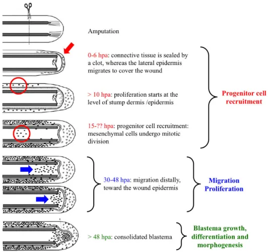
The first part of the fin regeneration process, from fin amputation to blastema formation, is schematized in Figure 8.
Figure 8: Schematic representation of the events occurring from fin amputation to blastema formation. After
amputation, the connective tissue is sealed by a clot and the lateral epidermal cells migrate to form the wound epithelium. Successively, proliferation starts first in the lateral epidermis and then also in the mesenchyme. These mesenchymal cycling cells continue to proliferate and then start to migrate toward the tip of the fin, where they form the blastema. Blastema cells proliferation and differentiation finally lead to the restoration of the amputated fin. Adapted from Santos-Ruiz et al., 2002.
1.2.3. The signaling pathways involved in the tail regeneration process
In the last years the signaling pathways involved in the blastema growth have been the objects of several studies; on the contrary, the early signals that could been involved in the activation of the regeneration module following the amputation/injury are not well known, even if there are several candidate signals.
The studies of these early signals have being conducted in different tissues and in different organisms, vertebrates and invertebrates. Since several years, it is known that amphibian limb and teleost fish fins regeneration processes start with the formation of the wound epithelium; before its maturation in a thick multilayer tissue, it has to secrete several factors that would be essential for the organization of the forming blastema. It has been shown that the rapid and
continuous elimination of the wound epithelium as soon as it forms impairs the progression of the regeneration process, in particularly affecting the blastema formation (Stocum and Cameron, 2011).
As already mentioned, the process of blastema formation shows similarities with the development of the embryonic bud. A number of factors involved in the development process have been identified also in the wound epithelium, both in amphibians and zebrafish (Suzuki et al., 2006; Yoshinari et al., 2009); these factors are expressed very early after injury and their role in blastema growth is well documented, but there are also some evidences suggesting their implication in the wound healing and the blastema formation processes. In the fish, for example,
fgf20a and wnt10a are expressed within the first six hours after amputation (Whitehead et al., 2005; Stoick-Cooper et al., 2007a); loss-of-function studies for both FGF and Wnt/β-catenin pathways showed that cells migrate to cover the wound but the formed wound epithelium is abnormal and incorrectly specified (Whitehead et al., 2005; Kawakami et al., 2006; Stoick-Cooper et al., 2007a); they are also involved in the process of blastema formation because their inhibition after wound healing leads to the non expression of msx gene and absence of blastema (Poss et al., 2000a; Stoick-Cooper et al., 2007a). The Wnt/β-catenin pathway seems to act upstream the FGF signaling because its inhibition induces the loss of fgf20a expression (Stoick-Cooper et al., 2007a); however, it is also possible that FGF signaling also induces Wnt/β-catenin pathway in a positive feed-back loop (Stoick-Cooper et al., 2007b). However, there are also other factors that inhibit blastema formation when overexpressed, as the chemokine stromal cell-derived factor (SDF-1) and Wnt5b (Dufourcq and Vriz, 2006; Stoick-Cooper et al., 2007a). Other potential signals for the induction of the regeneration process are the bioelectric signals. In planarians, the polarity of regeneration can be manipulated thanks to an electric field, because it has been demonstrated that the head is always formed at the cathode side (Carlson, 2007); moreover, different studies have shown that the diminution of β-catenin expression or the dysfunction of a Wnt agonist interfere on head regeneration (Gurley and Sanchez Alvarado, 2008; Petersen and Reddien, 2008), leading for the first time to find a relationship between an electric field and the signal transduction. Other studies conducted on the regeneration of the
Xenopus tadpole tail have demonstrated the implication of the V-ATPase H+ pump in the
regeneration process but not in the wound healing (Adams et al., 2007; Levin, 2007): the pump inhibition by concanamycin inhibits regeneration, whereas the expression of a pump insensible to the drug can rescue the inhibition. More recently, similar studies on zebrafish adult fin have shown that this H+ pump is necessary for the expression of the gene aldh1a2, responsible for
retinoic acid synthesis, and MAP Kinase Phosphatase 3 (MKP3): its inhibition blocks the blastema formation in the adult but, interestingly, has no effect on the larvae tail regeneration,