The relevance of covalent binding to mouse liver DNA to the
carcinogenic action of hexachlorocyclohexane isomers
Peter Sagelsdorff, Werner K. Lutz
1and Christian Schlatter
Institute of Toxicology, ETH and University of Zurich,
CH-8603 Schwerzenbach, Switzerland.
(Received on 20 April 1983; accepted on 15 July 1983)
Abstract
[
3H]HexachlorocycIohexane (HCH) was synthesized by
chlorination of [
3H]benzene prepared by catalytic tritiation of
benzene with tritiated water. The isomers of HCH were
separated by adsorption chromatography on silica gel. In
order to determine the covalent binding to DNA, [T-I]HCH
was administered to male mice by oral gavage, and liver DNA
was isolated via chromatin. The specific radioactivity of the
DNA was normalized by the dose administered and expressed
in the molar units of the Covalent binding index, CBI =
DNA damage/dose = (jimol bound HCH/mol DNA
nucleotide)/(mmol HCH administered/kg body weight). CBI
values of ~ 0.2 were found 10 h after the administration of
alpha- and gamma-HCH. Enzymatic digestion of the DNA
to the nucleosides and h.p.l.c. analysis revealed that - 4 0 %
of the radioactivity co-migrated with the natural nucleosides.
At elution volumes known to contain the more lipophilic
carcinogen-nucleoside adducts, —10% of the radioactivity
could be detected. The remaining 50% of the radioactivity
eluted with the front, representing a mixture of
oligonucleo-tide-HCH adducts and/or hydrophilic degradation products
which were strongly but not covalently associated with intact
DNA. Therefore, a true CBI of 0.02-0.1 must be expected
both for alpha- and gamma-HCH. This CBI is by a factor of
10
s—10
6below the value found with the strongest
DNA-binding carcinogens like aflatoxin B, or dimethylnitrosamine
and is unlikely to be decisive for the liver tumor induction in
mice because of the following additional findings: (i) Both
isomers gave rise to similar levels of DNA damage although
the alpha-isomer is a much more potent tumor inducer. This
similarity was seen not only at the time of maximum binding
but up to 10 days after oral administration; (ii) three mouse
strains with apparently different susceptibility to tumor
in-duction by gamma-HCH could not be distinguished with
respect to DNA binding; (Hi) the level of DNA binding of
alpha-HCH (CBI = 0.02-0.1) is more than three orders of
magnitude lower than would be expected if the mechanism of
tumor induction was by genotoxidty mediated by
DNA-binding. For a preliminary investigation on a potential
stimulatory effect on liver DNA replication and cell division,
[
14C]thymidine was administered i.p. 3.5 h before sacrifice of
the [
3H]HCH-treated mice. The alpha-isomer was found to
be more potent than the gamma-isomer in this respect. Taken
together, our data allow the conclusion that the
non-'To whom correspondence should be addressed.
'Abbreviations: HCH, hexachJorocydohexane; CBI, Covalent binding index; PBI, Protein binding index; II, Incorporation index; CIP, chloroform/ isoamyl alcohol/phenol ( 2 4 + 1 + 2 5 vol.); SDS, sodium dodecyl sulfate; HA, hydroxylapatite.
mutational processes must be more important for the
carcino-genicity of HCH.
Introduction
Hexachlorocyclohexane (HCH)* comprises of a group of
isomers of which the gamma-isomer, later called lindane, has
very useful pesticidal activity (1). HCH have become of great
public concern because the lindane batches used in the late
forties contained appreciable concentrations of alpha- and
beta-isomer. The alpha-isomer was found to induce liver
tumors in rats and mice (2), the beta-isomer was found to
have very low biodegradability and to be deposited in animal
fat. Although the lindane batches used since the fifties were at
least 99% pure gamma-isomer, a new discussion arose from
controversial findings of a liver tumor-inducing potential of
lindane itself.
Chemically-induced tumors are now thought to be the
result of a DNA damage succeeded by appropriate promotion
(3). Most chemicals exert their activity by covalent interaction
of a reactive metabolite with DNA in the target organ and are
therefore called genotoxic. The metabolism of HCH involves
the formation of olefins (1) and a subsequent epoxidation
could result in the generation of an electrophilic species.
Another group of tumor-enhancing agents, viz
co-carcinogens and promoters, do not themselves react with
DNA but apparently modulate one or several out of a variety
of biochemical and biological steps related to the process of
tumor formation. Such activities are also discussed for HCH.
For instance, alpha-HCH was found to enhance the
pro-liferation of putative preneoplastic cells in rat liver (4), and all
HCH isomers are known to be inducers of drug-metabolizing
enzymes (1), the alpha-isomer being more potent than
lin-dane.
It was the aim of this study to provide more information
about the mechanism of tumor induction by HCH. For this
reason it was examined whether the isomers of HCH can be
metabolized in vivo to reactive metabolites able to reach and
bind to liver DNA or whether the hepatocarcinogenicity is
rather due to non-genotoxic effects. It seemed especially
worthwhile to investigate whether the clear difference
bet-ween the alpha- and the gamma-isomer with respect to
biological effects and tumor induction (2) was reflected in
their ability to bind to DNA, and whether the apparent
dif-ference in susceptibility of different strains of mice to the
carcinogenicity of gamma-HCH (5 — 7) can be based upon
different levels of DNA binding.
Materials and Methods
Chemicals and apparatus
Reagents without specified distributor were of the highest purity available from Merck, Darmstadt, FRG. Hydroxylapatite (HA) (DNA-Grade, Bio-Gel HTP) was purchased from Bio-Rad, Richmond, CA, sodium dodecyl sulfate (SDS) from Sigma, St. Louis, MO, Nonidet P 40 (NP 40) and copper oxide (wire form) from BDH Chemicals Ltd., Poole, UK. Carrierfree pH]2 and
["CJthymidine with a sp. act. of 61 mCi/mmol were purchased from the Radiochemical Centre, Amersham, UK. Desoxyribonuclease I (E.C. 3.1.4.5.) from bovine pancreas, phosphodiesterase I (E.C. 3.1.4.1.) from Crotalus atrox venom and alkaline phosphatase III (E.C. 3.1.3.1.) from Escherichia
coli were obtained from Sigma, St. Louis, MO. Dialysis tubing (Visking type 20/32, mol. wt. exclusion at 12 0 0 0 - 1 4 000 daltons; diameter 17 mm) was from Union Carbide, Chicago, IL. The u.v. lamp was a Mineralight UVSL-58 50W from Ultra-Violet Products Inc., San Gabriel, CA.
Radioactivity measurements were carried out in 10 ml Insta-Gel (Packard Instruments, Downers Grove, IL) in a liquid scintillation counter, Packard Tri Carb 460 CD equipped with and calibrated for the automatic analysis of [3H]/["C] double-labelled samples.
The isomeric and radioactive purities of the isomers of HCH were deter-mined on a semipreparative gas chromatograph, Carlo Erba, Fractovap Linea 2200 (Carlo Erba, Rodana, Milano, Italy). The mass spectrum of HCH was recorded at the Institute of Organic Chemistry, ETH Zurich, Switzerland.
The h.p.l.c. analysis of the nucleosides were performed on a semiprepara-tive column (250 mm x 8 mm ID) equipped with two h.p.l.c. pumps, Kontron, LC Pump 410 (Kontron, Zurich, Switzerland), controlled by a Kontron Pro-grammer 200, for generating a linear gradient of two eluants.
Synthesis of ?H]HCH
[3H]Benzene was prepared by catalytic exchange tritiation of benzene on a
high-vacuum line. [3H]2O derived from combusting 8.8 Ci carrier-free [3H]2
at 600cC over 30 g copper oxide (wire form) was trapped under high vacuum
in 250 /U trifluoroacetic acid cooled with liquid nitrogen. This mixture was lyophilized into a break-seal ampoule containing 500 yX (430 mg, 5.5 mmol) benzene and 10 mg Platinum Black. The sealed ampoule was incubated for 6 days at 100°C.
PHJHCH was synthesized by chlorination of [3H]benzene (8). 16.5 mmol
chlorine gas were generated from oxidation of 33 mmol silver chloride with 12 g potassium dichromate in 100 ml concentrated sulfuric acid and trapped by cooling with liquid nitrogen. The [3H]benzene and chlorine were lyophiliz-ed into a 25 ml quartz round-bottom flask prefilllyophiliz-ed with 5 ml carbon tetra-chloride. The mixture was kept at - 25°C to - 30°C for 2 h under irradiation at 254 nm. Solvent and unreacted benzene were distilled off and the residue containing - 5 0 % of the radioactivity was extracted with petroleum ether (30—45°C boiling point) to bring the alpha-, gamma-, delta- and epsilon isomers into solution. The specific radioactivity of HCH was calculated from results derived in preliminary synthesis with a trace amount of radioactivity and was found to be — 1 Ci/mmol. The products were identified as HCH by mass spectrometry. The isomers were separated by chromatography on silica gel 60 according to Granger and Zwilling (9). The radiochemical purity of the isolated isomers was > 9 9 % , the isomeric purities were checked by semi-preparative gas chromatography. Column: OV 17, 2% methylphenylsih'cone, 200 cm; column temperature: 175°C; injection temperature: 210°C, N2
-pressure: 0.9 kg/cm2) and was found to be 98% (alpha-HCH). 92%
(gamma-HCH; 8% epsilon-HCH) and 95% (delta-(gamma-HCH; 5% epsilson-HCH). Beta-HCH was prepared by recrystallisation from a chloroform extract of the residue. The radiochemical purity was > 9 9 % , the isomeric purity was 96% (4% epsilon-HCH).
Animals and treatments
Young adult male mice of the strains NMRI, CF1, and C6B3F1 with weights ranging from 25 g to 40 g were obtained from Celamerck, Ingelheim, FRG. Laboratory chow (No. 21-343-7, Klingenthal MUhle AG, Basel, Switzerland) and tap water were provided ad libitum for one week for ac-climatisation. Administrations of HCH were carried out between 9.00 and 10.00 a.m. by oral gavage in polyethylene glycol 300 containing 5% ethanol. The animals received an i.p. injection of 5 - 1 5 pCx [14C]thymidine in 0.9%
NaCl 6.5 h later and were killed after another 3.5 h. For the determination of the time dependence of the DNA binding, the animals did not receive ["C]thymidine and were sacrificed 1, 3, 5 and 10 days after the [3H]HCH
ad-ministration.
Isolation of DNA and chromatin protein
Isolation of chromatin. (In the cold) Animals were bled by open heart puncture under ether anaesthesia. The livers were exised, minced and homogenized in a teflon Potter-Elvehjem-type homogenizer in 3 —4 volumes 75 mM NaCl, 10 mM EDTA, 10 mM Tris/HCl pH 7.8. Crude chromatin was prepared according to Yaneva and Dessev (10) with some modifications. After adding a solution of 2% (v/v) of the non-ionic detergent Nonidet P 40 up to a final concentration of 0.2% (v/v) the samples were incubated at 4CC
for 15 min. Chromatin was precipitated at 700 g for 5 min. The pellet was washed once with 75 mM NaCl, 10 mM EDTA, 10 mM Tris/HCl pH 7.8 and centrifuged for 5 min at 3500 g. This pellet contained about 2 —3 mg DNA and 20—30 mg protein/g liver.
Isolation of DNA. (Room temperature) DNA was isolated from chromatin according to Markov and Ivanov (11) with some modifications. The chromatin pellet was suspended in 25 ml lysing medium (8 M urea, 0.24 M sodium phosphate buffer p H 6.8, 10 mM EDTA, 1% (w/v) SDS) and homogenized on a Waring blender in a custom-made air-tight aluminium
vessel (46 mm diameter, 28 mm high) run at high speed for 30 s and then cooled in cold ethanol ( - 40°Q for 15 s. The blending-cooling procedure was repeated 4 times. After cautious transfer into a 50 ml polypropylene cen-trifuge tube the foam was broken by centrifugation for 3 min at 1000 g. 10 ml CIP (480 ml chloroform, 20 ml isoamyl alcohol, filled up to 1 liter under stir-ring with wanned liquid phenol) was added and proteins were extracted in the centrifuge tube under extensive shaking for 10 min on a shaking machine. The resulting suspension was separated into two layers by centrifugation for 15 min at 20 000 g. The CIP phase was saved for protein isolation, the super-natant aqueous layer was first decanted and then pipetted into a new poly-propylene centrifuge tube and was extracted once more with 10 ml CIP. The aqueous solution was extracted twice with 25 ml ether in a 250 ml separating funnel for the removal of trace amounts of phenol, was left standing over-night at room temperature and was applied to an HA column. Dry HA (1 g/g liver) of special batches tested for high absorptivity of DNA was suspended overnight in filtered MUP (8 M urea, 0.24 M sodium phosphate buffer pH 6.8), the slurry was swirled gently and was left untouched for 10 min. The fine particles were decanted. The remaining slurry was poured into 25 x 120 mm glass columns and the MUP was let run off. The aqueous nucleic acid solution was loaded on the column and the elution was monitored at 260 nm. Proteins were washed from the column with filtered MUP at a flow rate of 1 —2 ml/min by gravity until the transmission had returned to background value. To avoid a mixing of the eluants the column was let run dry before purging the urea from the column with two bed volumes 14 mM sodium phosphate buffer, pH 6.8. DNA was eluted with 0.48 M sodium phosphate buffer, pH 6.8, and - 2 0 ml of the DNA solution were collected. From here on, extreme caution is required not to use glassware, equipment or facilities which are also used for procedures involving high radioactivity levels. The sample was dialyz-ed at 4°C against 10 liter 0.2 M NaCl overnight. DNA was precipitatdialyz-ed by ad-ding 2 volumes ethanol and keeping at - 20°C for at least 12 h. The DNA was centrifuged for 20 min at 1000 g, the supernatant was decanted and DNA was dried in vacuo for 2 — 3 h. The highly purified DNA was dissolved in 10 mM MgCl2, 10 mM Tris/HCl, pH 7.0. The amount of DNA was
deter-mined on the basis of an absorbance of 20 at 260 nm for a solution of 1 mg DNA/ml. The yield of DNA was — 1 mg/g liver limited by the use of sub-optimal amounts of hydroxylapatite. The contamination of die DNA by pro-tein was <0.2% as derived from liver-DNA isolations from animals treated with L-[35S]methionine or with L-[3H]Iysine.
In a control experiment, DNA was isolated from the pooled livers of two mice treated with [3H]HCH and ["CJthymidine. After each purification step
DNA was precipitated from an aliquot of the aqueous solution by adding 2 volumes ethanol at - 20°C. After a centrifugation for 20 min at 1000 g, DNA was dried in vacuo and dissolved in deionized water. The amount of protein contaminating the crude DNA was measured by the method of Bensadoun and Weinstein (12), the amount of DNA was calculated from the specific [14C]activity. The specific [3H]activity of the DNA was calculated as (total
[^activity - [3H]activity of protein)/amount of DNA.
Isolation of chromatin protein. 1 ml of the first CIP extract from a DNA isolation of 10 g liver was shaken with ~ 5 ml 1% SDS in 14 mM sodium phosphate buffer, pH 6.8. Protein was precipitated with 25 ml acetone and washed 5 times by redissolving in 2 ml 1% SDS and acetone precipitation. The final protein sample in 1 % SDS was diluted with water to 0.1 % SDS, was precipitated by the addition of 2 volumes acetone and was stored at — 20°C overnight. After a centrifugation at 300 g, the supernatant was decanted and the protein residue was freed from acetone in vacuo for - 1 5 min. Protein was dissolved in 2 ml 1% SDS in 14 mM sodium phosphate buffer, pH 6.8, over-night and the solution was diluted with water to a final concentration of 1.4 mM sodium phosphate buffer. The amount of protein was determined with the Folin reagent. 1 —4 ml containing - 0 . 5 mg protein/ml were used for the liquid scintillation counting.
Isolation of HCH metabolites
The supernatant of the first acetone precipitation of chromatin protein from the CIP phase was dried in vacuo. About 75% of the radioactivity in the CIP phase could be dissolved in 10 mM MgCl2, 10 mM Tris/HCl, pH 7.0
and was loaded on a Lichrosorb R PU column also used for the analysis of
nudeosides by h.p.l.c. (see below).
Water-soluble metabolites were obtained from the aqueous solution after the first CIP extraction of chromatin homogenate. DNA was precipitaed by the addition of 2 volumes ethanol and the supernatant was dried in vacuo. After dissolving the residue in 10 mM MgCl2 10 mM Tris/HCl, pH 7.0 the
sample was analysed by h.p.l.c. H.p.l.c. analysis of the nucleosides
DNA ( 1 - 2 mg/ml) in 10 mM MgCl2, 10 mM Tris/HCl, pH 7.0 was
digested enzymatically by the method described by King et al. (13). The resulting deoxynudeosides were separated by h.p.l.c. on a Lichrosorb R Pl t
column (250 mm x 8 mm) with a distilled water/methanol gradient of 0 - 1 0 % methanol in 5 min, 10% methanol for 5 min and 10—100% methanol in 45 min at a flow rate of 3.5 ml/min. The optical density of the eluate was recorded at 254 nm. Fractions of 2 min were collected and the scin-tillation counting was performed after the addition of 10 ml Insta-Gd. To avoid phase separation between the scintillation cocktail and the eluate, the fractions 11 — 14 were diluted with 1 ml methanol. The retention times of the natural deoxynucleosides deoxycytosine, deoxyguanosine, thymidine and deoxyadenosine were 9 min, 11.5 min, 13 min and 18 min, respectively. Recovery of radioactivity eluted, as a fraction of the injected ranged between 90 and 110%, both for pH] and ["C].
Calculations and statistics
Determination of CBI. The radioactivity in the DNA after treatment of mice with pHJHCH was expressed after normalization to the dose ad-ministered:
£BI , _ d.p.m./mg DNA
d.p.m./kg body weight This value was converted to the molar units,
QQI _ >tmol chemical bound/mol DNA nucleotide mmol chemical applied/kg body weight
according to CBI = CBI' x 309 x 106 on the basis of an average mol. wt. of
309 g/mol DNA nucleotides (14).
Determination of Protein binding indices, PBI. The radioactivity in the chromatin protein after treatment of mice with [TiJHCH was expressed after normalization to the dose administered:
pBj , _ d.p.m./mg chromatin protein
d.p.m./kg body weight This value was converted to the molar units,
p g j _ /jnol chemical bound/mol amino acid mmol chemical appliedAg body weight
according to PBI = PBI'x HOx l ^ o n thebasisof amol. wt. of 110 g/mol for an average amino acid (15).
Determination of Incorporation indices, II. The radioactivity in the DNA after treatment of mice with ["CJthymidine was expressed after normalization to the dose administered:
. . , _ d.p.m./mg DNA d.p.m./kg body weight This value was converted to the molar units,
JJ _ /anol thymidine incorporated/mol DNA nucleotide mmol thymidine applied/kg body weight
according to II = II' x 309 x 106 on the basis of an average mol. wt. of 309
g/mol DNA nucleotides.
Calculation of standard deviation. The total variability (statistical coun-ting error and fluctuations due to vial, scintillation cocktail, counter, external radiation and composition of the sample) for the counting of a DNA sample containing little radioactivity was assumed to be equal to the variability of DNA samples isolated from untreated animals held together with the treated ones. On the basis of 33 background values compiled for 12 months, a respec-tive standard deviation of 1.89 c.p.m. ( = 1 S.D.) was calculated. The stan-dard deviation for a net radioactivity in a vial therefore was taken as: 1 S.D.
= VO-892 + (1.89A/33)2) = 1.92 c.p.m.
Limit of detection for radioactivity in nudeoside analysis. The total variability for each fraction of a h.p.l.c. analysis was calculated on a level of 1 standard deviation (1 S.D.) from 5 nudeoside analyses of control DNA digests. The mean background value for each fraction was calculated with an accuracy of 1 S.D.A/5. The maximum possible difference between sample and background radioactivity was determined on an interval of ± 2 S.D.
Results
Comparison of isomers
Table I compiles the radioactivities in mouse liver DNA
isolated 10 h after oral administration of a high radioactivity
dose of gamma- and alpha-[
3H]HCH. Under the assumption
that the radioactivity is due to DNA-bound HCH molecules,
the radioactivity can be expressed in the units of the CBI (14)
after normalization to the dose administered. The last line of
the Table I reveals the extremely low level of apparent DNA
binding (CBI around 0.2) and shows that the alpha-isomer
did not give rise to a higher CBI than the gamma-isomer
Table I. Specific activity of liver DNA of male NMRI mice, 10 h after oral
administration of gamma- and a l p h a - I ^ H C H .
Isomer gamma-HCH alpha-HCH
Animal wdght [g]; pool of 2 mice 66.2 70.2 68.2 72.7 66.7 70.3 Dose [mg/kg] 13.0 12.0 12.0 8.5 6.2 7.8 [mCi/kg] 44.9 41.5 41.5 27.5 21.2 26.8 DNA Sp. act. [d.p.m./mg] 89.9 77.1 75.8 39.4 30.6 25.7 ±1S.D. [CBI units] ± 1 S.D. Mean ± SEM ±6.2 ±7.3 ±6.6 ±4.8 ±4.8 ±4.1 0.28 0.26 0.25 0.20 0.20 0.13 ±0.02 ±0.03 ±0.02 ±0.02 ±0.03 ±0.02 0.26 ± 0.02 0.18 ± 0.03
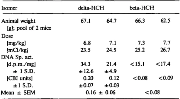
Table II. Specific activity of liver DNA of male NMRI mice, 10 h after oral
administration of delta- and beta-[3H]HCH.
Isomer delta-HCH beta-HCH
Animal weight [g]; pool of 2 mice Dose [mg/kg] [mCi/kg] DNA Sp. act. [d.p.m./mg] ± 1 S.D. [CBI units] ± 1 S.D. Mean ± SEM 67.1 6.8 23.5 34.3 ±12.6 0.20 ±0.07 0.16 64.7 7.1 24.5 21.4 ±4.9 0.12 ±0.03 ± 0.06 66.3 7.3 25.2 <15.1 <0.08 62.5 7.7 26.7 <17.4 <0.09 <0.08
although the former is a markedly more potent carcinogen.
This is the first indication to postulate that DNA binding
can-not be the decisive activity for the tumor-inducing potential
of the HCH's. This hypothesis is also supported by the
fding that the delta-isomer which has never been found to
in-duce tumors (16) also revealed a CBI of 0.16 (Table II). The
beta-isomer did not give rise to detectable DNA radioactivity
(Table II).
In order to check whether a difference between the
gamma-and the alpha-isomer might be found at later times after the
administration, the time dependence for the DNA binding
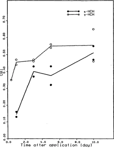
was investigated. Figure 1 shows that the liver-DNA
radio-activity reached the same plateau level of about CBI = 0.5
after 10 days. The time-dependent increase was faster in the
first three days with the gamma-isomer, in accordance with
the somewhat faster metabolism (17). It was also checked
whether the absorption from the gastro-intestinal tract of the
relatively high doses of chemical was different for the two
isomers. This was not the case because it was found that the
whole liver contained 2.1% and 2.2% of the radioactivity
dose of the gamma- and the alpha-isomer, respectively, one
day after the oral administration.
Comparison of mouse strains
An additional hint for whether DNA binding could be the
main mode of tumorigenic action of HCH should be
obtain-ed from studies with strains of mice that are of apparently
different susceptibility to liver tumor induction by the
gamma-isomer. The results given in Table III show that the
three strains used all gave rise to similar CBI values although
NMRI mice (CBI = 0.28) were found to be less susceptible to
the tumorigenic action of gamma-HCl than B6C3F1 mice
Table IV. Specific activity of liver chromatin protein of male NMRI mice,
10 h after oral administration
o.o 2 . 0 M.O 6 . 0 8 . 0 1 0 . 0
Time after application [day]
Fig. 1. Time dependence of the binding of alpha- and gamma-[3H]HCH to
mouse liver DNA. Groups of male NMRI mice were given p.o. — 30 mCi [3H]HCH/kg and two animals were sacrificed each after 10 h
(gamma-[3H]HCH only), 1, 3, 5 and 10 days.
Table m . Specific activity of liver DNA of male NMRI, CF1, and B6C3F1
mice, 10 h after oral administration of gamma-[3H]HCH. Strain Animal weight [g]; pool of 2 mice Dose [mg/kg] [mCi/kg] DNA Sp. art. [d.p.m./mg] ± 1 S.D. [CBI units] ± 1 S.D. Mean ± SEM NMRI 87.9 8.7 30.0 49.7 ±11.7 0.23 ±0.05 0.28 ± 91.7 16.7 57.7 138.4 ±11.6 0.33 ±0.03 0.04 CF1 73.5 21.2 73.0 122.6 ±11.5 0.23 ±0.02 0.26 ± 72.8 21.2 73.0 154.1 ±8.6 0.29 ±0.02 0.02 B6C3F1 67.4 23.0 79.3 91.8 ±9.6 0.16 ±0.02 0.17 64.9 21.6 74.3 92.7 ±12.8 0.17 ±0.02 ± 0.02
(CBI = 0.17) or CF1 mice (CBI = 0.26) ( 5 - 7 ) .
Contribution of contaminations to the DNA radioactivity
It was assumed above that the DNA radioactivity was due
entirely to D N A - H C H adducts. Other sources, such as
biosynthetic incorporation of radiolabelled breakdown
pro-ducts of HCH entering the pool of DNA precursors,
non-covalently bound HCH metabolites or protein
contamina-tions might contribute all or a fraction of the radioactivity
measured on the DNA. Any of these influences would make
our argument for a non-mutagenic mode of action of HCH
even stronger. A number of control experiments were
per-Isomer Animal weight [g]; pool of 2 mice Dose [mg/kg [mCi/kg] Protein Sp. act. [d.p.m./mg] [PBI units] Mean ± SEM gamma-HCH 66.2 13.0 44.9 6140 6.8 : 70.2 12.0 41.5 5170 6.2 5.6 ± 0.9 68.2 12.0 41.5 3150 3.8 alpha-HCH 72.7 8.5 27.5 1374 2.5 : 66.7 6.2 21.2 1015 2.4 '.3 ± 0.1 70.3 7.8 26.8 1126 2.1
formed to investigate some of the above-mentioned
contribu-tions.
Contamination by protein-bound HCH. The data given
in Table IV show that chromatin protein was also
radio-labelled, 10 h after [
3H]HCH administration. The specific
ac-tivity was 42- to 68-fold and 33- to 44-fold higher in protein as
compared with DNA for the gamma-, and the alpha-isomer,
respectively. Protein contamination of DNA was shown to be
lower than 0.2% as determined with radiolabelling of
chromatin protein in vivo with [
3H]lysine or [
35S]methionine.
Protein contaminations cannot therefore contribute
substan-tially to the radioactivity measured on the DNA.
Contamination of DNA by non-covalently bound HCH
metabolites. The control experiment where DNA was
precipitated at different steps of the purification procedure
showed that neither ether extraction nor dialysis resulted in
a reduction of the specific [
3H]activity of DNA. This means
that both lipophilic and hydrophilic metabolites had been
removed completely from the DNA during the entire isolation
procedure and that the DNA had been purified to constant
specific activity.
Nucleoside analysis
Separation of the deoxynucleosides by h.p.l.c. after
en-zymatic digestion of liver DNA of alpha- and gamma-[
3H]-HCH treated NMRI mice (10-h-point) revealed that
~30—40% of the radioactivity eluted together with the
op-tical density of the natural deoxynucleosides (Figure 2). This
radioactivity is therefore most probably due to biosynthetic
incorporation of breakdown products, for instance tritiated
water. At a later elution time, a small (~ 10% of the
radio-activity) but significant amount of radioactivity was detected.
This is the region known to contain the more lipophilic
nucleoside-carcinogen adducts. The formation of a
nucleoside-HCH adduct therefore is highly probable. Up to
50% of the radioactivity of the DNA eluted in the first two
fractions. This radioactivity can be due to DNA not
com-pletely degraded i.e., due to oligonucleotides which could still
carry HCH adducts.
The same relative distribution of radioactivity among the
three different elution regions was found with a duplicate
DNA sample isolated 10 h after the administration of
gamma-t^J-HCH and with a DNA pooled from 1-day,
3-day, and 5-day mice.
In order to further exclude the possibility that HCH
metabolites non-covalently bound to DNA could be
responsi-ble for the presumed adduct peak in fraction 16 and 18 for
the gamma- and the alpha-isomer, respectively, metabolites
isolated from the liver of a gamma-[
3H]HCH-treated mouse
0 5 10 15 20 25
Fraction Nr.
Fig. 2. Radioactivity profile of h.p.l.c. chromatograms of liver DNA of
[3H]HCH-treated NMRI-mice, enzymatically digested to deoxynucleosides. The shaded area covers the range of the limit of detection for radioactivity in one fraction, calculated on an interval of ± 2 S.D. Top, optical density pro-file, representing the natural deoxyribonucleosides in the order dC, dG, dT, dA; Center, DNA taken from a NMRI mouse, killed 10 h after oral ad-ministration of gamma-[3H]HCH; Bottom, DNA taken from an NMRI
mouse, killed 10 h after oral administration of alpha-f^JHCH. Similar pro-files were obtained from a DNA pooled from 1-, 3-, and 5-day-mice (gamma-isomer).
were analysed on the same h.p.l.c. system as used for the
nucleosides. Figure 3 shows that the hydrophilic metabolites
eluted in the first three fractions. Since this is the region which
also contained radioactivity in the h.p.l.c. analysis of the
DNA nucleoside it cannot be excluded that such metabolites
were closely but non-covalently associated with DNA. The
association must have been so strong that the dialysis did not
remove them and only upon enzymatic degradation of the
DNA were they released.
Lipophilic metabolites eluted at a retention time of
~ 50 min. No radioactivity could be detected at retention
times between 24 min and 48 min. These data indicate that
the radioactivity peaks eluting after ~ 35 min represent
HCH-deoxynucleoside-adducts and not metabolites set free during
the hydrolysis of the DNA.
True covalent DNA binding
The control experiments described above have revealed
that biosynthetic incorporation of radiolabel into DNA has
taken place and that hydrophilic metabolites might have been
strongly but non-covalently associated with DNA. For a
calculation of a true CBI, these contributions have to be
5 10 15 20
Fraction Nr.
Fig. 3. H.p.l.c. analysis of metabolites of gamma-[3H]HCH isolated from
mouse liver. The shaded area covers the range of the limit of detection for radioactivity in one fraction, calculated on an interval of ± 2 S.D. A, hydro-philic fraction; B, lipohydro-philic fraction.
deducted from the values given in Tables I — III. A reduction
by a factor of ~ 2 or 10 results, for the case where all
early-eluting radioactivity is regarded as oligonucleotide-HCH
ad-ducts or for the case where only the nucleoside-HCH adduct
peak is considered. On a most conservative approach,
therefore, a CBI of <0.1 results.
Protein binding
In our experiments on DNA binding we also determined
the level of non-extractable radioactivity in chromatin
pro-tein. This was done in a first place in order to determine
whether contamination of DNA with protein of high specific
radioactivity might simulate DNA binding. The
protein-binding values are also low on an absolute level upon
com-parison with standard carcinogens (15), and, there was again
no difference in protein binding between the two isomers
(Table IV) or between the different strains of mice (Table V).
Rate of DNA synthesis
The animals received, 3.5 h before sacrifice, an i.p.
injec-tion of [
14C]thymidine. The level of [
I4C]radioactivity on the
DNA was then used as an index for DNA synthesis. The
results summarized in Table VI show a tendency for the more
potent alpha-isomer to induce a higher rate of DNA
syn-thesis. It should be pointed out that these data were obtained
from the identical animals used for the determination of
DNA binding by [
3H]HCH (Table I), where the alpha-isomer
was even slightly less effective than the gamma-isomer.
Table V. Specific activity of liver chromatin protein of male NMRI, CF1
and B6C3F1 mice, 10 h after oral administration of gamma-[3H]HCH.
Strain NMRI CF1 B6C3F1
Animal weight fel; pool of 2 mice Dose [mg/kg] [mCi/kg] Protein Sp. act. [d.p.m./mg] [PBI units] Mean ± SEM 87.9 91.7 73.5 72.8 67.4 64.9 8.7 16.7 21.2 21.2 23.0 21.6 30.0 57.7 73.0 73.0 79.3 74.3 3300 7610 7910 9360 4560 4330 5.5 6.5 5.4 6.3 2.9 2.9 6.0 ± 0.5 5.8 ± 0.5 2.9 ± 0.02
Table VI. Incorporation of ["CJthymidine into liver DNA of male NMRI
mice, 3.5 h after i.p. injection, and 10 h after oral application of [3H]HCH.
Isomer gamma-HCH alpha-HCH
Animal weight [g]; pool of 2 mice Dose ["C]TdR 66.2 70.2 68.2 72.7 66.7 70.3 40.0 30.5 40.0 36.8 35.5 28.8 10.0 7.6 10.0 9.2 8.9 7.2 DNA Sp. act. [d.p.m./mg] Incorp. Index Mean ± SEM 184 149 181 284 180 180 2560 2710 2520 4280 2820 3460 2600 ± 60 3500 ± 400
Discussion
The previous section has provided good qualitative
evidence for a DNA-HCH adduct. A comparison among
strains and isomers makes it highly unlikely, however, that
this type of genotoxic activity is the decisive mode of
tumori-genic action. In addition, a quantitative analysis of the level
of DNA binding favours a non-genotoxic mode of
tumori-genic action. A quantitative correlation of CBI versus
carcinogenic potency expressed in TD50 units (i.e., the daily
dose estimated to induce a tumor in 50% of the animals
treated for life), has shown that CBI of the order of 10
3- 10
4are found with strong genotoxic carcinogens, of 100 for
moderate carcinogens and of 1 — 10 for weak carcinogens
(18). Since alpha-HCH has to be classified as a moderate
tumor-inducing agent with an approximate TD
Mvalue of 0.1
mmol/kg/day for mouse liver, a CBI for liver DNA of - 10
2would be required if its mode of action was by DNA binding.
The measured value of <0.1 is one thousand times lower.
Among the many possible mechanisms of tumorigenic
ac-tivity not related to DNA binding two aspects were amenable
to an assay within the present experimental set-up. Firstly,
binding to protein and the concomitant cytotoxicity might be
envisaged. Our results showed, however, that protein binding
cannot be an important contribution to the proposed
non-mutagenic mode of tumor induction.
Another possibility for a non-mutagenic mode of action,
the stimulation of cell division, was also tested simultaneously
with the determination of DNA binding. These results gave
some indication for a higher activity of the alpha-isomer
although the experimental set-up was not ideal for the
deter-mination of this activity. It has been shown in rats that the
in-duction of DNA synthesis after administration of various
tumor promoters is largest after a time period of ~20h
(19,20). In our experiments, we used mice and the interval
was only 6.5 h. Therefore, the borderline effect observed
with the alpha-isomer might have become more pronounced
if a time dependence had been investigated. Additional
evidence along these lines is available from two-stage
long-term carcinogenicity studies with rats where it has been shown
that alpha-HCH accelerated the manifestation of malignant
liver tumors after initiation of the carcinogenic process by a
single dose of diethylnitrosamine (21).
Chemical carcinogens are normally divided into two large
classes of mutagenic and non-mutagenic carcinogens (22). As
suggested by Radman and Kinsella (23) there may not be a
clear separation of these two groups of activities and it must
be expected that there are carcinogens acting on more than
only one level.
With HCH, we are faced with a situation where a minute
DNA binding was shown for the alpha- and the
gamma-isomer. Much additional information was given, however, to
show that this activity cannot be responsible alone for the
tumors induced and some hypotheses were presented and in
part supported by experimental data. For a risk evaluation in
man, a mutagenic risk by DNA binding after exposure to mg
amounts of lindane seems negligible. Although species
dif-ferences with respect to the formation of reactive metabolites
cannot be excluded we do have evidence that binding of
gamma-HCH to liver DNA in the rat is similar to the data
ob-tained in mice so that there are at least no indications for large
species differences with respect to metabolism. For the more
important events not related to DNA binding, an
extra-polation of animal data to man must be based upon the
elucidation of the specific mechanism of tumor induction by
HCH in the animal, and a test of whether similar activities are
found in man at dose levels that are by orders of magnitude
lower than those used in the long-term bioassays.
Acknowledgement
This study was supported by the 'Centre International d'Etudes du Lindane', C.I.E.L., Brussels, Belgium.
References
1. Ulmann,E. (1973), LINDAN, Monographie eines insektiziden Wirkstoffs, Verlag K. Schillinger, Freiburg im Breisgau, FRG.
2. IARC (1979), Some halogenated hydrocarbons, IARC Monographs on the Evaluation of the Carcinogenic Risk of Chemicals to Humans, Vol. 20, IARC, Lyon, France, pp. 195-257.
3. Weisburger.J.H. and Williams.G.M. (1981), The decision-point ap-proach for systematic carcinogen testing, Fd. Cosmet. Toxicol., 19, 561-566. 4. Schulte-Hermann.R., Ohde.C, SchuppIer.J. and Timmermann-Trosiener.I. (1981), Enhanced proliferation of putative preneoplastic cells in rat liver following treatment with the tumor promoters phenobarbital, hexa-chlorocyclohexane, steroid compounds and nafenopin, Cancer Res., 41, 2556-2562.
5. Thorpe,E. and WaIker,A.l.T. (1973), The toxicology of dieldrin (HEOD). II. Comparative long-term oral- toxicity studies in mice with dieldrin, DDT, phenobarbitone, beta-BHC and gamma-BHC, Fd. Cosmet. Toxicol., 11, 433-442.
6. National Cancer Institute (1977), Bioassay of lindane for possible car-cinogenicity, Carcinogenesis Technical Report Series, No. 14, DHEW Publication No. (NIH) 77-814, Public Health Service, National Institute of Health.
7. Weisse.l. and Herbst.M. (1977), Carcinogenicity study of lindane in the mouse, Toxicology, 7, 233-238.
8. Haider,K. and Jagnow.G. (1975), Abbau von ["C], [*H] und P Q ] -markiertem gamma-Hexachlorcyclohexan durch anaerobe Bodenmikro-organismen, Arch. Microbioi, 104, 113-121.
9. Granger.C. and Zwilling.J.P. (1950), Separation et dosage de quelques constituants des Hexachlorocyclohexanes par chromatographie d'adsorption sur silice, Bull. Soc. Chim., France, 17, 873-876.
chromatin from guerin ascites tumour and rat liver, Eur. J. Biochem., 66, 535-542.
11. Markov.G.G. and ivanov.I.G. (1974), Hydroxyapatite column chromatography in procedures for isolation of purified DNA, Anal. Biochem., 59, 555-563.
12. Bensadoun.A. and Weinstein.D. (1976), Assay of proteins in the presence of interfering materials, Anal. Biochem., 70, 241-249.
13. King.H.W.S., Osborne,M.R. and Brookes.P. (1979), The in vitro and in vivo reaction at the N'-position of guanine of the ultimate carcinogen deriv-ed from benzo[a]pyrene, Chem.-Biol. Interactions, 24, 345-353.
14. Lutz,W.K. (1979), In vivo covalent binding of organic chemicals to DNA as a quantitative indicator in the process of chemical carcinogenesis, Mutat. Res., 65, 289-356.
15. von Daniken.A. (1982), Anwendung der kovalenten Bindung an DNS und Protein in vivo als Kurzzeittest fur sehr schwache chemische Karzinogene, aufgezeigt am Beispiel von l-Cyano-2,3-epithiopropan, o-Chlor-benzyliden-malononitrol, Clofibrat, Fenofibrat und Methylbenzacridinen, Ph.D. Thesis, no. 6968, ETH Zurich.
16. Nagasaki.H., Tomii.S., Mega.T., Marugami.M. and Ito,N. (1972), Hepatocarcinogenic effect of alpha-, beta-, gamma- and delta-isomers of benzene hexachloride in mice, Gann, 63, 393.
17. Koransky.W., Portig.J., Vohland.H.W. and Klempau,I. (1964), Die Elimination von alpha- und gamma-Hexachlorcyclohexan und ihre Beeinflus-sung durch Enzyme der Lebermikrosomen, Naunyn-Schmiedebergs Arch. Exp. Pathol. Pharmakol., 247, 49-60.
18. Lutz.W.K. (1982), Constitutive and carcinogen-derived DNA binding as a basis for the assessment of potency of chemical carcinogens, in Snyder.R.R., Parke,D.V., Kocsis.J.J., JoUow.D.J., Gibson.C.G. and Witmer.C.M. (eds.), Biological Reactive Intermediates, D, Part B, Plenum, New York, pp. 1349-1365.
19. Hoffmann.V. and Schulte-Hermann.R. (1979), The regulative role of food consumption in the induction of rat liver cell proliferation by drugs and environmental pollutants, Arch. Toxicol., Suppl. 2, 457-461.
20. Schulte-Hermann.R. (1977), Two-stage control of cell proliferation in-duced in rat liver by alpha-hexachlorocyclohexane, Cancer Res., 37, 166-171. 21. Schulte-Hermann.R. and Parzefall.W. (1981), Failure to discriminate initiation from promotion of liver tumors in a long-term study with the phenobarbital-type inducer alpha-hexachlorocyclohexane and the role of sus-tained stimulation of hepatic growth and monooxygenases, Cancer Res., 41, 4140-4146.
22. Stott.W.T., Reitz,R.H., Schumann.A.M. and Watanabe.P.G. (1981), Genetic and nongenetic events in neoplasia, Fd. Cosmet. Toxicol., 19, 567-576.
23. Radman.M. and Kinsella.A.R. (1980), Chromosomal events in carcino-genic initiation and promotion: implications for carcinocarcino-genicity testing and cancer prevention strategies, in Montesano.R., Bartsch.H. and Tomatis,L. (eds.), Molecular and Cellular Aspects of Carcinogen Screening Tests, IARC Scientific Publications, No. 27 International Agency for Research on Cancer, Lyon, France, pp. 75-90.
![Fig. 2. Radioactivity profile of h.p.l.c. chromatograms of liver DNA of [ 3 H]HCH-treated NMRI-mice, enzymatically digested to deoxynucleosides.](https://thumb-eu.123doks.com/thumbv2/123doknet/14911983.659050/5.930.401.857.70.586/radioactivity-profile-chromatograms-liver-treated-enzymatically-digested-deoxynucleosides.webp)
![Table V. Specific activity of liver chromatin protein of male NMRI, CF1 and B6C3F1 mice, 10 h after oral administration of gamma-[ 3 H]HCH.](https://thumb-eu.123doks.com/thumbv2/123doknet/14911983.659050/6.936.75.465.107.278/table-specific-activity-liver-chromatin-protein-nmri-administration.webp)
