Dimerization of human 5-lipoxygenase
Ann-Kathrin H ä fner
1, Mihaela Cernescu
2,
Bettina Hofmann
1, Michael Ermisch
1, Michael H ö rnig
1,
Julia Metzner
1, Gisbert Schneider
3,
Bernhard Brutschy
2and Dieter Steinhilber
1,*
1
Institute of Pharmaceutical Chemistry/ZAFES , University
of Frankfurt, Max-von-Laue-Strasse 9, 60438 Frankfurt ,
Germany
2
Institute of Physical and Theoretical Chemistry , University
of Frankfurt, Max-von-Laue-Strasse 9, 60438 Frankfurt ,
Germany
3
ETH Zurich , Department of Chemistry and Applied
Biosciences, Institute of Pharmaceutical Sciences,
Wolfgang-Pauli-Strasse 10, CH-8093 Zurich , Switzerland
* Corresponding author
e-mail: [email protected]
Abstract
Human 5-lipoxygenase (5-LO) can form dimers as shown
here via native gel electrophoresis, gel fi ltration
chroma-tography and LILBID (laser induced liquid bead ion
des-orption) mass spectrometry. After glutathionylation of
5-LO by diamide/glutathione treatment, dimeric 5-LO was
no longer detectable and 5-LO almost exclusively exists in
the monomeric form which showed full catalytic activity.
Incubation of 5-LO with diamide alone led to a disulfi
de-bridged dimer and to oligomer formation which displays
a strongly reduced catalytic activity. The bioinformatic
analysis of the 5-LO surface for putative protein-protein
interaction domains and molecular modeling of the dimer
interface suggests a head to tail orientation of the dimer
which also explains the localization of previously reported
ATP binding sites. This interface domain was confi rmed by
the observation that 5-LO dimer formation and inhibition
of activity by diamide was largely prevented when four
cysteines (C159S, C300S, C416S, C418S) in this domain
were mutated to serines.
Keywords: diamide; 5-lipoxygenase; LILBID; leukotrienes;
molecular modeling.
Introduction
5-Lipoxygenase (5-LO) is the key enzyme in the formation
of leukotrienes (LTs) (Werz , 2002 ) which play an important
role in infl ammatory diseases like asthma and atherosclerosis
(Dahl é n et al., 1980 ; Funk , 2001 ). Recently, the 5-LO gene
was identifi ed as a regulator of leukemia stem cell
prolifera-tion in BCR-ABL-induced chronic myeloid leukemia (CML)
(Chen et al. , 2009 ). 5-LO catalyzes a two-step reaction, fi rst
the conversion of arachidonic acid (AA) into
5(S)-hydroper-oxy-6-trans-8,11,14-cis-eicosatetraenoic acid (5-HPETE),
second the conversion of 5-HPETE into the allylic epoxide
leukotriene A4 (LTA
4). This unstable epoxide can be further
metabolized to the biologically active LTB
4by LTA
4hydro-lase or to the cysteinyl leukotriene LTC
4by LTC
4synthase
(Shimizu et al. , 1986 ).
Mammalian 5-LO consists of 672
–
673 amino acids
(Matsumoto et al.
, 1988
). The 5-LO structure published
recently (Gilbert et al. , 2011 ) confi rms that human 5-LO
con-sists of an N-terminal regulatory C2-like domain (residues
1 – 114) and a C-terminal catalytic domain (residues 121 – 673)
which is mainly organized in
α -helices and contains the
non-heme iron in its catalytic center. The C2-like domain is
respon-sible for the association of 5-LO to the nuclear membrane
(Chen and Funk , 2001 ; Kulkarni et al. , 2002 ), for
diacylglyc-eride ( H ö rnig et al., 2005 ) and calcium binding (Hammarberg
et al. , 2000 ) and for the interaction with coactosin-like protein
(Rakonjac et al. , 2006 ).
In the cell, 5-LO activity can be regulated by many
factors. An increase in intracellular calcium leads to
trans-location of the enzyme from the cytosol to the nuclear
membrane (Peters -Golden and Brock, 2003 ), where 5-LO
co-localizes with 5-LO-activating protein (FLAP) and
cytosolic phospholipase A
2(cPLA
2) (Pouliot et al. , 1996 ).
An alternative modulation of 5-LO activity is
phospho-rylation at distinct serine residues. Phosphophospho-rylations at
Ser-271 by MAPKAP kinase 2 (Werz et al.
, 2000
) and
at Ser-663 by ERK2 (Werz et al. , 2002 ) have a
stimula-tory effect, whereas phosphorylation at Ser-523 by protein
kinase A (PKA) inhibits the activity (Luo et al. , 2004 ). In
most reviews, 5-LO is mentioned as a monomeric enzyme
(Rouzer and Samuelsson , 1985 ; R å dmark, 2002 ),
neverthe-less, it was previously shown that 5-LO from rat basophilic
leukemia cells (RBL-1) forms dimers in the presence of
calcium (Parker and Aykent , 1982 ). Recently, Shang et al.
analyzed dimerization and fl exibility of rabbit 12/15-LO
and human 12-LO by small angle X-ray scattering (SAXS)
(Shang et al. , 2011 ). The outcome of their study was that (i)
r12/15-LO is mainly monomeric, but dimerizes at higher
protein concentrations and with increasing protein fl
exibil-ity, and (ii) that human 12-LO is stable as a dimer confi
rm-ing their former studies for 12-LO (Aleem et al., 2008).
Moreover, studies of Zhang et al. provided an additional
hint for a 5-LO dimer (Zhang et al. , 2000 ). They performed
affi nity and photoaffi nity labeling experiments with 5
′ p
-fl
uorosulphonylbenzoyladenosine (FSBA) and
2-azido-ATP and identifi ed the amino acids 73 – 83 and 193 – 209 as
ATP-binding sites within the enzyme. The stoichiometry
suggested an equimolar ratio of ATP and 5-LO. This led us
to the assumption that both peptides must be nearby in the
tertiary structure forming one ATP binding site. Regarding
the crystal structure of Stable-5LOX, this could be only
true if 5-LO forms dimers/multimers, as one postulated
ATP binding site is located in the C2-like domain, the other
in the catalytic domain.
Herein, we describe the fi rst time that human recombinant
5-LO and 5-LO purifi ed from PMNL forms dimers.
Results
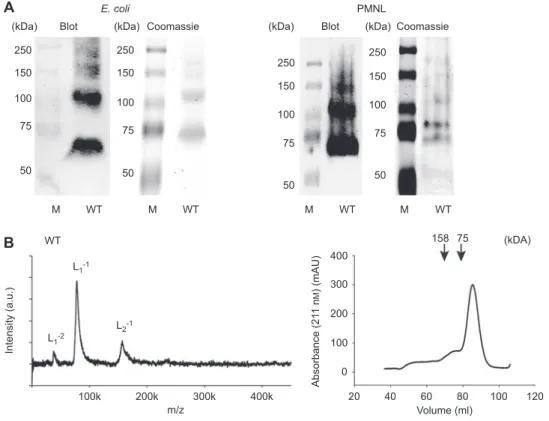
Analysis of 5-LO dimer formation by native gel electrophoresis, gel fi ltration and LILBID-MS
In order to examine the oligomeric state of 5-LO we fi rst
performed native gel electrophoresis. Both, recombinant
5-LO that was expressed in Escherichia coli as well as 5-LO
from PMNL were purifi ed via ATP-agarose and applied to a
native Tris-HCl PAGE. The resulting gel was analyzed via
Coomassie staining and Western blot. Two major 5-LO bands
were identifi ed which suggested that 5-LO may form dimers
(Figure 1 A).
In order to confi rm dimerization of 5-LO, LILBID-MS
and gel fi ltration chromatography was applied. The resulting
LILBID spectra of 5-LO WT showed monomeric as well as
dimeric and even a little trimeric enzyme (Figure 1 B).
For gel fi ltration, PB/EDTA with addition of 0.15 m up to
1 m NaCl was used. We observed an elution profi le consisting
of one large peak at an apparent MW of 30 kDa and some
smaller peaks which eluted earlier (Figure 1 B). The late
elu-tion of the 5-LO peak at an apparent MW of 30 kDa is
prob-ably due to unspecifi c interaction with the column material.
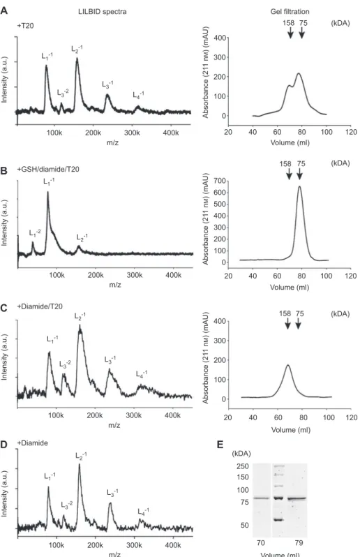
Repetition of the experiment in PBS/EDTA with addition of
0.5 % T20 to reduce this interaction led to the expected MW
of 75 kDa for the main peak (Figure 2 A) and to a smaller
peak at an apparent MW of about 160 kDa. SDS-PAGE and
Western blot analysis confi rmed that both peaks consist of
5-LO (Figure 2 E). Addition of ATP (1 m m ) or calcium (1 m m )
to the sample buffer and the mobile phase did not change the
5-LO elution profi le (data not shown).
In LILBID-MS and in gel fi ltration, the presence of T20
micelles led to a strong increase in 5-LO dimers indicating that
the interaction of 5-LO with micelles induces its dimerization
(compare Figures 1 B, 2 A and 3 ).
Modifi cation of 5-LO by GSH and diamide
Since 5-LO contains 10 cysteines on the protein surface we
wanted to investigate the effect of glutathionylation of the
cysteines and the associated changes in 5-LO surface
prop-erties on 5-LO dimerization (Figure 4 A). Glutathionylation
of 5-LO was performed by addition of GSH and diamide as
(kDa) 250 150 100 75 50 M WT WT 100k 200k 300k 400k 20 0 100 200 300 400 40 60 80 100 120 m/z Volume (ml) Intensity (a.u.) Absorbance (21 1 n M ) (mAU) L1-1 L1-2 L2-1 250 150 100 75 50 M WT 250 150 100 75 50 M WT 250 150 100 75 50 M WT 158 75 (kDa)
Blot Coomassie (kDa) (kDa)
(kDA) PMNL Blot Coomassie E. coli
A
B
Figure 1 Analysis of 5-LO WT w/o T20.
(A) Native gel electrophoresis of recombinant 5-LO WT and 5-LO from PMNL. 5-LO was detected with anti-5-LO antibody after blotting to a nitrocellulose membrane or with Coomassie blue staining. (B) LILBID spectra of 6.4 µ m 5-LO in 100 m m NH 4 HCO 3 and gel fi ltration chromatogram of 2 mg 5-LO in PB/EDTA + 0.5 m NaCl.
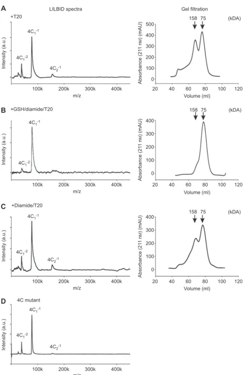
+T20 +GSH/diamide/T20 +Diamide/T20 +Diamide LILBID spectra
A
B
C
D
E
L1-1 L1-2 L1-1 L1-1 L1-1 L3-2 L3-2 L3-1 L3-1 L4-1 L4-1 L2-1 L2-1 L2-1 L2-1 L3-2 L3-1 L4-1 Intensity (a.u.) Intensity (a.u.) Intensity (a.u.) Intensity (a.u.) 100k 200k 300k 400k m/z 100k 200k 300k 400k m/z 100k 200k 300k 400k m/z 100k 200k 300k 400k m/z 20 400 158 158 75 75 300 200 100 0 0 100 200 300 400 500 600 700 40 60 80 100 120 Volume (ml) 20 40 60 80 100 120 Volume (ml) Absorbance (21 1 n M ) (mAU) Absorbance (21 1 n M ) (mAU) (kDA) 20 400 158 75 300 200 100 250 150 100 75 70 79 50 0 40 60 80 100 120 Volume (ml) Volume (ml) Absorbance (21 1 n M ) (mAU) (kDA) (kDA) (kDA) Gel filtrationFigure 2 LILBID spectra and gel fi ltration chromatograms of 5-LO WT in the presence of T20.
Arrows are marking the elution volumes of the reference proteins conalbumin (75 kDa) and aldolase (158 kDa). LILBID spectra of (A) 6.4 µ m 5-LO in 100 m m NH 4 HCO 3 + 0.1 % T20 and gel fi ltration of 2 mg 5-LO in PBS/EDTA + 0.5 % T20, (B) 6.4 µ m 5-LO incubated with 25 m m GSH and 2.78 m m diamide for 30 min at 37 ° C in PBS/EDTA + 0.1 % T20 followed by buffer exchange to 100 m m NH 4 HCO 3 + 0.1 % T20, (C) 6.4 µ m 5-LO incubated with 2.78 m m diamide for 10 min at 37 ° C in PBS/EDTA, afterwards the buffer was exchanged to 100 m m NH 4 HCO 3 + 0.1 % T20. The right panels shows the corresponding gel fi ltrations (mobile phase PBS/EDTA + 0.5 % T20) of 2 mg 5-LO incubated under the respec-tive conditions of the LILBID analysis. (D) LILBID spectrum of 6.4 µ m 5-LO that was preincubated with 2.78 m m diamide for 10 min at 37 ° C in PBS/EDTA followed by buffer exchange to 100 m m NH 4 HCO 3 . (E) SDS-PAGE analysis with Coomassie blue staining of gel fi ltration frac-tions of the 5-LO monomer (79 ml) and dimer (70 ml) peak from panel A.
oxidizing agent (Kosower and Kosower , 1995 ). To
investi-gate the infl uence of a glutathione modifi cation on the
oligo-meric state of the enzyme, 5-LO was incubated with GSH (10
m m ) plus diamide (1 m m ) for 30 min at 37
° C or diamide alone
(1 m m ) for 10 min at 37
° C.
Treatment of 5-LO with GSH and diamide led to the almost
exclusive formation of monomers in LILBID-MS and on gel
fi ltration in the presence of T20 (Figure 2 B). LILBID
mea-surements were performed in the presence of T20 (Figure
2 A – C), no T20 was added in Figure 2 D.
Obviously, treatment of 5-LO with GSH and diamide
pre-vents dimerization. To demonstrate glutathionylation of 5-LO
Untreated Diamide T20 Diamide/T20 GSH/diamide/T20 % of overall signal 0 20 40 60 80 100 Monomer Dimer Trimer Tetramer
Figure 3 Oligomer distribution of 5-LO WT under different condi-tions in LILBID-MS analysis. Distribution is shown as percentage of overall signal + SEM (n = 3).
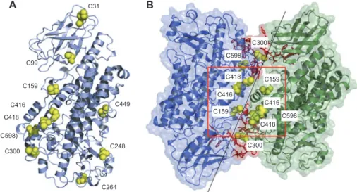
C31
A
B
C99 C159 C416 C418 (C598) C300 C264 C248 C449 C159 C416 C418 C598 C300 C159 C416 C598 C418 C300Figure 4 Protein-protein docking of 5-LO and cysteine residues in the putative dimer-interface and on the surface.
(A) Structure of human 5-LO with the surface exposed cysteines (yellow spheres). C598 is partially buried. (B) The most reasonable docking solution, exhibiting a head-to-tail orientation, in cartoon representation with semi-transparent surface. Highlighted are the two proposed ATP binding sites in red, together with the cysteines (yellow spheres) in the predicted dimer interaction interface. The cysteines in the red square (C159, C416, C418) might be suffi ciently close to allow for disulfi de bond formation.
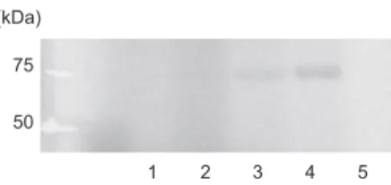
under these conditions, the enzyme was incubated with (
35S)-GSH in the presence of diamide. After separation by
SDS-PAGE, glutathionylation was detected with a phosphorimager.
Maximal glutathionylation of 5-LO was achieved when GSH
was combined with diamide and H
2O
2as oxidant to quench the
DTT contained in the (
35S)-GSH solution whereas addition of
GSH and diamide still leads to lower, but detectable
nylation (Figure 5 , lanes 3 and 4). To quantify the
glutathio-nylation of 5-LO, free cysteine residues were quantifi ed using
Ellman ’ s reagent and HPLC analysis (Chen et al. , 2008 ). We
found that treatment of 5-LO with GSH (10 m m ) and diamide
(1 m m ) leads to a reduction in the number of free cysteine
resi-dues from 10 in the unmodifi ed 5-LO to nine indicating that
one cysteine residue becomes glutathionylated.
Interestingly, treatment of 5-LO with diamide alone for 10
min at 37
° C (Figure 2 C) completely prevented monomer
for-mation on gel fi ltration and led to a peak that corresponded to
dimers. Elongation of the incubation time led to the
forma-tion of even higher molecular complexes. The formaforma-tion of
disulfi de bonded 5-LO dimers and oligomers as consequence
of the incubation with diamide can be seen in the LILBID
spectra both in the presence or absence of T20 (Figure 2 C
and D). Thus, oxidation of the enzyme and formation of
inter-molecular disulfi de bridges by diamide induced a shift of the
size distribution to higher masses concomitant with a
deple-tion of the monomer. For summary of oligomer distribudeple-tion in
LILBID-MS of 5-LO WT see Figure 3 .
Analysis of dimer formation in PMNL S100 by gel fi ltration
PMNL S100 was analyzed by gel fi ltration in PBS/EDTA.
5-LO activity as well as 5-LO protein (by Western blot) was
found in a broad range of fractions from about 60 to 90 ml
with the main activity eluting at 75 ml (Figure 6 ). Since the
gel fi ltration was performed without T20 in the elution buffer,
it can be assumed that there is a shift to a smaller MW due
to interaction with the column material, comparable to the
recombinant 5-LO in the same buffer (dotted line). Despite
this, 5-LO activity was also found in fractions that elute at an
apparent MW of the dimer indicating that at least a part of the
5-LO could exist as dimer in PMNL.
Molecular modeling and docking of 5-LO
To establish a model of a 5-LO dimer, we used the crystal
struc-ture of engineered human 5-LO {Protein Data Bank [PDB,
http://pdb.org, (Berman et al. , 2000 )], ID: 3o8y (Gilbert et al. ,
2011 )}. Gilbert et al. mutated or deleted several amino acids
to obtain the so-called ‘ Stable-5LOX ’ (Gilbert et al. , 2011 )
thereby enabling crystallization and structure determination.
To restore the WT enzyme, we performed in silico mutagenesis
Volume (ml) 20 40 60 80 100 120 5-LO products (ng) 0 500 1000 1500 2000 2500 Absorbance (211 n M ) (mAU) 0 200 400 600 800 1000 5-LO activity Absorbance (211 nM)
Figure 6 Gel fi ltration analysis of recombinant 5-LO and PMNL S100.
Gel fi ltration chromatography of 2 mg purifi ed 5-LO (dotted line) and of PMNL S100 (continuous line) from 2 × 10 9 cells using PBS/ EDTA as mobile phase. 5-LO in the PMNL S100 was recorded by determination of 5-LO activity in the fractions and detection of 5-LO by Western blot analysis with 5-LO antibody (marked with arrow; elution volume 60 – 90 ml).
(kDa) 75 50
1 2 3 4 5
Figure 5 SDS-PAGE analysis of ( 35S)-GSH incorporation into 5-LO.
Purifi ed 5-LO WT was incubated with ( 35S)-GSH and then sub-jected to SDS-PAGE analysis. GSH incorporation was detected by a Phosphorimager. Lane 1, 5-LO treated with 0.87 m m diamide, 2.5 µ Ci ( 35 S)-GSH; lane 2, 0.87 m m diamide, 0.87 m m H
2 O 2 , 2.5 µ Ci ( 35 S)-GSH; lane 3, 3.48 m m diamide, 2.5 µ Ci ( 35 S)-GSH; lane 4, 3.48 m m diamide, 0.87 m m H 2 O 2 , 2.5 µ Ci ( 35 S)-GSH; lane 5, 0.87 m m diamide; in all lanes: 8.7 m m GSH, 17.4 µ l 5-LO.
and constructed the completed structure of 5-LO WT. For this
structure, we predicted potential dimerization sites using four
independent software tools. All proposed interfaces (Figure 7 )
have one side of 5-LO in common, which is fl anked by the two
putative ATP binding sites K73-K83 and F193-K209 (Figure
7 A). The predicted interaction site was then used to evaluate
the proposed complex conformations by protein-protein
dock-ing performed with the protein dockdock-ing and clusterdock-ing
tech-nique ClusPro. ClusPro was the best performing server in the
last round of CAPRI (Critical Assessment of PRediction of
Interactions), a community wide experiment on the
compara-tive evaluation of protein-protein docking for structure
pre-diction ( http://www.ebi.ac.uk/msd-srv/capri/ ) (Janin , 2005 ).
Suggested docked conformations were fi ltered so that only
those complexes were retained whose dimerization site was in
agreement with the predicted interface region. Assuming that
both proposed ATP binding regions form a coherent ATP
bind-ing site, which is possible in a putative 5-LO dimer/multimer,
we considered only those complexes fulfi lling these criteria.
Among these complexes, we selected the number one ranked
docking solution as fi nal model (Figure 4 B). It exhibits
coher-ent ATP binding sites, matches the predicted interface regions,
and represents the biggest cluster of docking solutions;
there-fore it was ranked highest by the clustering algorithm. This
fi nal model constitutes the 5-LO subunits in a head-to-tail
ori-entation, where the C2-like domain of one subunit interacts
with the catalytic domain of the other. We found four cysteines
in the putative interface three of which (C159, C416, C418)
might be suffi ciently close to allow for intermolecular
dis-ulfi de bond formation (Figure 4 B), which might explain the
observed diamide-induced dimerization.
The reactivity of protein thiols depends on the pK
avalue and
the accessibility of the thiol, both of which can vary widely.
Although protein thiols typically have pK
avalues of 8.5–9,
these can largely vary depending on the local environment of
the cysteine residue (Gilbert , 1984 ; Simplicio et al. , 1998 ).
Therefore, we calculated the pK
avalues for the cysteines of
5-LO using the PROPKA program (Li et al. , 2005 ; Bas et al. ,
2008 ). All of the predicted interface cysteines have calculated
pK
avalues of around 9 in the protein environment (C159: 8.5;
C300: 9; C416: 8.5; C418: 9; C598: 9.3) which is in agreement
with possible glutathionylation/disulfi de-bond formation.
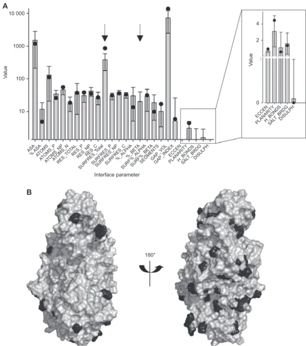
Evaluation of the predicted dimer interfaceWe compared the predicted 5-LO interface with interfaces
of 534 non-homologous homodimers. Therefore we used the
PROTORP server (Reynolds et al. , 2009 ), which analyzes
protein-protein complex formation by calculating a series of
physical and chemical parameters of the protein interaction
sites that contribute to the binding free energy of the
associa-tion. These parameters include size and shape, intermolecular
bonding, residue and atom composition, as well as secondary
structure contributions. The protein association parameters
for our dimer model were calculated and compared to those
observed in known homodimers (Figure 8 A; for
abbrevia-tions see supplementary Table S1
). For 24 out of the 26
com-puted parameters the values for the predicted 5-LO dimer are
within the mean
± SD of the known complexes. Deviations
from a ‘ typical ’ homodimer were detected for the total
num-ber of surface residues (858.0 instead of 380.5
± 202.6) and the
expected percentage of
β-sheets in the interface (20.2 ± 19.4)
as no
β-sheets are predicted for 5-LO.
Mutation of four cysteines prevents diamide-induced 5-LO dimerization
5-LO contains 13 cysteines and according to the restored
5-LO WT 10 of the cysteines are located on the protein
sur-face. Four of these cysteines are located close to each
other at a putative reactive site, which could be
impor-tant for the dimer formation in a head-to-tail conformation
(cf. Figure 4 ). In order to test the hypothesis that this interface is
involved in 5-LO dimer formation and that these four cysteines
are involved in diamide-induced 5-LO dimerization by
forma-tion of intermolecular disulfi de bridges, we designed a 4C mutant
in which four cysteines (C159S, C300S, C416S, C418S) were
replaced with serines. The resulting gel fi ltration chromatogram
of the 4C mutant in presence of T20 and after preincubation
with GSH and diamide was similar to the chromatogram of the
unmodifi ed and modifi ed WT (Figures 2 A, B, and 9 A, B). When
the 4C mutant was treated with diamide only, the majority of
the 4C 5-LO still eluted as monomer and only a minor fraction
eluted as dimer (Figure 9 C) whereas no monomer was
detect-able with the WT 5-LO under these conditions (Figure 2 C).
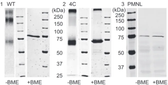
Formation of covalent dimers by diamide was analyzed with
SDS-PAGE without addition of
β -mercaptoethanol and detected
by Coomassie staining. As shown in Figure 10 , diamide-treated
WT 5-LO gives a main band at an apparent molecular weight
of about 150 kDa, whereas the main band of the 4C mutant
was detected at 75 kDa. After boiling of the protein samples
with
β -mercaptoethanol, only one band was detectable with
SDS-PAGE at 75 kDa with WT 5-LO as well as with the 4C
mutant indicating that the dimers a linked via disulfi de bridges.
Similarly, in PMNL only 5-LO monomers were observed when
5-LO was analyzed under denaturing conditions both in the
presence and absence or presence of
β -mercaptoethanol (Figure
10 ) indicating that the 5-LO dimers observed under
non-dena-turing conditions (Figure 1 A) are not covalently linked.
In LILBID-MS, the 4C mutant existed almost exclusively
as monomer, even in presence of T20 or diamide suggesting
that the four cysteines exchanged to serines in the 4C mutant
90°
PPI-Pred
E
C
D
Cons-PPISP SPPIDER ProMate
A
B
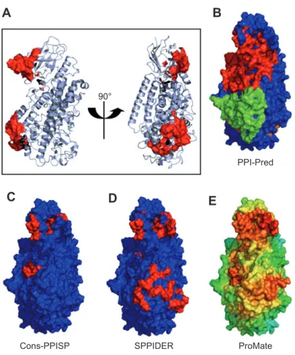
Figure 7 Prediction of protein-protein interaction sites of 5-LO.
(A) Cartoon representation of the tertiary structure of 5-LO. Postulated ATP binding sites are shown as red surface. The orientation of the right hand fi gure was kept for fi gures B-E, with the surface colored according to the predicted probability of a protein-protein interaction interface (red: highest probability, green: lower and blue: lowest probability): (B) PPI-Pred, (C) cons-PPISP, (D) SPPIDER, and (E) ProMate prediction.
10 000
A
B
4 2 0 ECCEN ECCEN ASA % ASAATO MS ATOMS_NPAT OMS_N RES_T OTA L RES_P RES_NPRES_C SURFRES_T otalSURFRES_PSURFRES_NPSURFRES_C %_ALPHA%_BET
A
SURF%_ALPHASURF%_BET A
SEGMENTSGAP_VOLGAP_INDEX ATOMS_P PLANARITY PLANARITY H_BONDS H_BONDS SAL T_BRDG SAL T_BRDG DISULPH DISULPH 1000 100 Va lu e Va lu e 10 Interface parameter 180°
Figure 8 Evaluation of the 5-LO protein-protein dimer interface.
(A) Interface parameters for the protein-protein interaction interface of the 5-LO dimer (black dots) were calculated using PROTORP and compared to the pre-calculated interface parameter distribution of the PROTORP homodimer dataset (grey bars). Data shown are mean values ± SD. Arrows indicate out-of-range values for 5-LO. Hydrogen-bond, salt bridge and disulfi de bond values were normalized to 100 Å 2 interface area. For abbreviations, see Table S1. (B) Surface representation of the predicted 5-LO dimer interface, which is conserved between rat and human 5-LO. The left panel presents the interface in the same orientation as in Figure 7, on the right panel the structure is rotated by approxi-mately 180 ° . The conserved surface is represented in gray, the amino acids that are not conserved are highlighted in black.
are mainly responsible for diamide-induced dimerization of
the 5-LO WT enzyme (Figure 9 ).
Functional studies on 5-LO WT and 4C mutant
5-LO WT and the 4C mutant show similar catalytic
activi-ties indicating that the mutations do not signifi cantly alter the
5-LO structure (Table 1
). When 5-LO was incubated in the
presence of T20, the specifi c activity was reduced from 534 to
86 ng/
µ g protein (Table 1) so that it retained only about 15 %
of its normal activity and activity loss increased with time
leading to almost no detectable products when 5-LO was kept
in a T20 containing buffer overnight before the activity was
determined. This activity loss explains the low specifi c
activ-ity of the enzyme after gel fi ltration overnight in the
pres-ence of T20 (about 4 ng 5-LO products per
µ g protein). When
freshly purifi ed 5-LO was treated with diamide for 10 min,
5-LO activity was reduced from 534 to 23 ng/
µ g protein which
corresponds to about 4 % of the activity of the control (Table
1). In contrast, diamide did not inhibit the 4C mutant which
is of interest since the reagent also failed to induce
dimeriza-tion of the 4C mutant (Figure 9 ). Also, the inhibitory effect
of T20 was less pronounced with the 4C mutant. Combined
GSH and diamide treatment led to similar activities as in the
+T20 LILBID spectra
A
B
C
D
4C1-1 4C1-1 4C1-1 4C1-1 4C2-1 4C2-1 4C1-2 4C1-2 4C1-2 4C1-2 4C2-1 Intensity (a.u.) 100k 200k 300k 400k m/z Intensity (a.u.) 100k 200k 300k 400k m/z Intensity (a.u.) 100k 200k 300k 400k m/z Intensity (a.u.) 100k 200k 300k 400k m/z 400 500 300 200 100 0 Absorbance (21 1 n M ) (mAU) 20 40 60 80 100 120 Volume (ml) 158 75 (kDA) 400 300 200 100 0 Absorbance (21 1 n M ) (mAU) 20 40 60 80 100 120 Volume (ml) 400 300 200 100 0 Absorbance (21 1 n M ) (mAU) 20 40 60 80 100 120 Volume (ml) 158 75 (kDA) 158 75 (kDA) Gel filtration +GSH/diamide/T20 +Diamide/T20 4C mutantFigure 9 LILBID spectra and gel fi ltration chromatograms of 4C mutant.
Arrows are marking the elution volumes of the reference proteins conalbumin (75 kDa) and aldolase (158 kDa). LILBID spectra of (A) 6 µ m 4C in 100 m m NH 4 HCO 3 + 0.1 % T20, (B) 6 µ m 4C mutant incubated with 25 m m GSH and 2.78 m m diamide for 30 min at 37 ° C in PBS/ EDTA + 0.1 % T20, afterwards the buffer was exchanged to 100 m m NH 4 HCO 3 + 0.1 % T20, (C) 6 µ m 4C mutant incubated with 2.78 m m diamide for 10 min at 37 ° C in PBS/EDTA, afterwards the buffer was exchanged to 100 m m NH 4 HCO 3 + 0.1 % T20 and (D) 6 µ m 4C mutant in 100 m m NH 4 HCO 3 . The right panels show the gel fi ltration analysis (mobile phase PBS/EDTA + 0.5 % T20) of 2 mg 4C mutant after incubation under the same conditions as for LILBID analysis.
250 150 100 75 50 37 25
-BME +BME -BME +BME -BME +BME
250 150 100 75 50 37 (kDa) (kDa) PMNL 3 2 1 WT 4C
Figure 10 SDS-PAGE analysis of WT 5-LO, 4C mutant and PMNL S100.
Coomassie staining with or without BME of either (1) WT 5-LO or (2) 4C mutant treated with 1 m m diamide and (3) WB of PMNL S100 with or without BME.
Table 1 Enzyme activity of 5-LO WT and the 4C mutant. WT 5-LO (ng products/ µ g protein) 4C 5-LO (ng products/ µ g protein) w/o 534 ± 7 436 ± 11 GSH/diamide 557 ± 5 442 ± 8 Diamide 23 ± 1 486 ± 19 T20 86 ± 3 299 ± 19 T20/GSH/ Diamide 72 ± 4 258 ± 3 T20/diamide 9 ± 2 299 ± 5
Purifi ed protein was either used without or with preincubation with 10 m m GSH and 1 m m diamide for 30 min at 37 ° C or with 1 m m diamide for 10 min at 37 ° C. Activity assays were performed in PBS/ EDTA with 2 m m Ca 2 + , 1 m m ATP and 20 µ m AA with or without 0.5 % T20. Enzyme activity is given as ng products per µ g protein ± SEM (n = 3).
untreated control in the WT enzyme as well as the 4C mutant,
suggesting that glutathionylation does not signifi cantly affect
5-LO activity (Table 1).
Discussion
In this work we investigated the dimerization of human
5-LO. The protein plays a key role in leukotriene formation in
infl ammation and host defense reactions (Samuelsson , 1983 ).
Although it was never studied in detail, 5-LO is generally
considered to be a monomeric enzyme. It catalyzes two
dif-ferent reaction steps, consisting of an initial oxygenation step
at C5 of arachidonic acid, followed by dehydration and
for-mation of leukotriene A
4. This led us to speculate that 5-LO
could form a dimeric complex, where one monomer of the
homodimer catalyzes the generation of 5-HPETE which is
then transferred to the other monomer for the formation of
LTA
4. Alternatively, for COX which catalyzes the initial steps
in prostaglandin biosynthesis by the oxygenation of
arachi-donic acid, Yuan et al. (2009) showed that COX forms dimers,
where one substrate molecule binds with high affi nity to one
COX site and facilitates the oxygenation of arachidonic acid
by the other catalytic partner (Yuan et al. , 2009 ). According to
these data we assumed that one monomer of the 5-LO could
be the catalytically active partner whereas the other monomer
has a regulatory function, e.g., via its pseudoperoxidase
activ-ity (Riendeau et al. , 1991 ).
By gel fi ltration, native PAGE and LILBID-MS
analy-sis, we found that 5-LO can form dimers and that dimer
formation is enhanced in the presence of T20 micelles.
Rechromatography of the monomeric peak leads again to
the typical monomer/dimer pattern indicating that there is
a dynamic balance between monomers and homodimers
(data not shown). Neither calcium nor ATP enhanced 5-LO
dimerization. 5-LO dimers were reported for rat 5-LO in the
presence of calcium, and 5-LO activity was attributed to the
dimeric form (Parker and Aykent , 1982 ). With the human
enzyme, we cannot confi rm this observation. In accordance
with Hammarberg et al. (Hammarberg and R å dmark, 1999 )
we found no evidence that Ca
2 +promotes 5-LO dimer
for-mation. This is supported by our diamide experiments.
Treatment of 5-LO with GSH/diamide leads to
glutathiony-lation of 5-LO which alters the surface properties of 5-LO
and prevents dimer formation as shown with LILBID-MS
and gel fi ltration. However these ‘ forced ’ 5-LO monomers
displayed similar catalytic activity as the untreated enzyme
suggesting that monomers seem to have similar catalytic
properties as the untreated enzyme consisting of monomers
and dimers (Table 1).
Though, different reaction kinetics for both quaternary
structures are conceivable, similarly to what was already
discussed for 12-LO (Aleem et al.
, 2008
) and 15-LO2
(Wecksler et al. , 2009 ). An impact of dimerization on the
reaction kinetics is presumable due to the observation that
the predicted dimer interface is located at the proposed
sub-strate and oxygen entry site of LOs (Ivanov et al. , 2010 ).
Although an entry channel to the active site is not directly
present in the crystal structure of 5-LO, substrate entry to
the catalytic site is expected via conformational changes by
amino acids of the so called ‘ FY-cork ’ in this region that
closes the active site (Gilbert et al. , 2011 ). However, due
to the dynamics of the monomer/dimer equilibrium and the
very low enzymatic activity of the covalently linked
dim-ers generated by diamide treatment, kinetic experiments on
monomers or dimers are diffi cult to perform. At present, we
cannot rule out that dimerization leads to a shift in the 5-LO
product pattern.
Addition of diamide to 5-LO (without GSH) yielded
mainly dimers and to a low extent even trimers or tetramers
depending on the incubation time as shown by gel fi ltration
and LILBID-MS. These 5-LO dimers were also detected by
SDS-PAGE (in the absence of
β -mercaptoethanol) whereas
the SDS-PAGE bands corresponded to the 5-LO monomer
when
β -mercaptoethanol was added indicating that diamide
induces 5-LO dimers in which both proteins are linked by
intermolecular disulfi de bridges. Interestingly, the covalently
linked 5-LO dimers displayed a rather low catalytic activity.
This could be explained by intermolecular disulfi de bonds that
are formed between both monomers and thus according to our
model block the entrance to the catalytic center or avoid a
conformational change that is necessary for catalytic activity.
For establishing a model of the 5-LO/5-LO interaction,
we used the just-deposited crystal structure of human 5-LO
[PDB ID: 3o8y (Gilbert et al. , 2011 )] and restored the WT
enzyme ( in silico mutation and insertions of the modifi ed
amino acids). 3o8y shows a crystallographic dimer, which
is not a thermodynamically stable complex (Shang et al. ,
2011
). Therefore, this assembly might solely rise from
crystal-packing. Again, (i) 3o8y was engineered which
might affect its oligomeric state and (ii) since our
observa-tions suggest a monomer-dimer equilibrium the
conforma-tion of the weak dimer might not be captured in the crystal
structure where the crystallization conditions can lead to
numerous non-physiological associations (Dafforn , 2007 ).
By using computational prediction algorithms for
protein-protein interaction sites, we established a model for a 5-LO
dimer (Figure 4 B). Protein-protein interfaces are
evolution-arily more conserved than the rest of the protein surface,
which has recently been verifi ed using a large protein
struc-tural dataset (Choi et al. , 2009 ). This might also be true
for weak dimers that are in equilibrium with the monomer
in solution (Dey et al. , 2010 ). Thus, we analyzed the
con-servation of the predicted dimer interface of human 5-LO
(Figure 8 B). The results suggest that the predicted interface
is, apart from the C2-like domain region and in contrast to
the rest of the surface, conserved between rat and human
5-LO. Predicted interface parameters support the feasibility
of the 5-LO dimer. In our model, four cysteines are located
in the interface domain, three of which (C159, C416, C418)
are in close spatial proximity suggesting disulfi de bridge
formation upon diamide treatment. In order to verify our
dimer model, we mutated all four cysteine residues and
found that mutation of these cysteines largely prevent
diamide-induced dimer and oligomer-formation. This is in
line with the observation that diamide treatment of the 4C
mutant does not inhibit its catalytic activity whereas the
WT-enzyme is strongly inhibited. Furthermore, since we
have demonstrated that the 4C mutant exists mostly as a
monomer in the presence of GSH and diamide, one could
speculate that GSH binds to C598, which is located in the
suggested dimer interface (cf. Figure 4 ), thereby
prevent-ing dimerization of 5-LO. Another argument supportprevent-ing our
dimer model and the head-to-tail orientation of the dimer
is the location of the ATP binding site, where two
other-wise distant tryptophans, which were previously labeled by
azido-ATP (Zhang et al. , 2000 ), are in close proximity to
each other (Figure 4 B). Such a model, where two ATP
mol-ecules are bound by the dimer, is in agreement with the
pre-viously reported equimolar ratio of 5-LO and ATP (Zhang
et al. , 2000 ). Although the experimental results correlate
well with the proposed dimer model, we are fully aware
that molecular modelling is prone to errors. Nevertheless,
as this is the fi rst suggested model of the putative dimer of
5-lipoxygenase, we hope that it will serve as a valid
work-ing tool for generatwork-ing hypotheses. The herein described
5-LO dimerization interface opens up novel opportunities
for the design of inhibitors. The feasibility of this approach,
designing protein-protein interaction inhibitors, has been
demonstrated recently (for review see Gonz á lez-Ruiz and
Gohlke, 2006 ; Grosdidier et al. , 2009 ). Up to now, there
are three main types of direct 5-LO inhibitors known: iron
ligand type, redox type, and non-redox type inhibitors.
(Ford-Hutchinson et al., 1994). Due to several short
com-ings, only one 5-LO inhibitor could reach the market to date,
the iron ligand type inhibitor zileuton (Carter et al. , 1991 ).
The interference with 5-LO function via modulation of the
protein-protein complex might open up a novel approach
to identify small molecule inhibitors to interfere with
LT-mediated diseases. It should also be mentioned that the
proposed model represents a dimeric complex only and that
higher oligomers might exhibit different conformations.
Interestingly, our LILBID-MS-data suggest that T20
micelles promote 5-LO dimerization (Figure 3 ). Assuming
that T20 micelles mimic cell membranes and thus provide
a lipophilic surface for 5-LO to interact with, it is
possi-ble that in stimulated intact cells, interaction of 5-LO with
the membrane after translocation from the cytosol induces
dimerization. Arachidonic acid, either supplied exogenously
or released by cPLA
2has been shown to induce 5-LO
translocation which could be blocked by a FLAP
inhibi-tor (Flamand et al. , 2006 ). 5-LO dimerization after 5-LO
translocation and interaction with FLAP is supported by a
previous observation where an 186 kDa 5-LO containing
complex was found in cross-linking experiments in Sf9
cells when FLAP was coexpressed but not in the absence
of FLAP (Plante et al. , 2006 ). Since FLAP is a
membrane-bound protein which is required for AA transfer after 5-LO
translocation to the nuclear membrane following cell
stimu-lation it might be possible that interaction of 5-LO with the
membrane and FLAP triggers dimerization and leukotriene
formation.
An interesting phenomenon is the increased dimerization
of the WT enzyme by T20 as shown by LILBID analysis
(Figure 2 A) which is accompanied with a 85 % decrease in
5-LO activity whereas the 4C mutant is only inhibited by 30 %
and does not show dimerization after short-term exposure to
T20 in LILBID analysis (Figure 9 D). This might give a hint
that 5-LO dimers show reduced catalytic activity but it might
on the other hand simply be due to the fact that the 4C mutant
is more resistant to detergent treatment.
From our data it is clear that the 5-LO monomer shows
full catalytic activity. Furthermore, our data show for the fi rst
time that leukotriene biosynthesis obviously does not require
5-LO dimerization. However, our study gives only limited
hints on the catalytic activity of the dimer. The low enzyme
activity observed after diamide-induced dimerization and in
the presence of T20 micelles could be related to the covalent
linkage of the dimers and to detergent effects, respectively.
The fact that T20 also inhibits enzyme activity of the
mono-meric GSH/diamide-treated 5-LO suggests that the inhibition
of 5-LO activity is not related to dimerization. In the native
enzyme, it is impossible to selectively detect the activity of
the dimer as gel fi ltration experiments with rechromato graphy
of the monomeric peak led to the usual monomer/dimer
dis-tribution again.
Taken together, we could show that 5-LO can form dimers
and that there is a dynamic equilibrium between the
mono-meric and dimono-meric form. Treatment of 5-LO with GSH and
diamide prevents dimerization whereas addition of diamide
alone leads to disulfi de-linked 5-LO dimers and oligomers.
The 5-LO monomer displayed similar catalytic properties as
the native enzyme suggesting that dimerization is not required
for leukotriene biosynthesis.
However, due to the nuclear localization of 5-LO and its
association with euchromatin or to its interaction with dicer,
other biological roles were proposed for 5-LO apart from
leukotriene biosynthesis, such as regulation of gene
expres-sion or miRNA processing, respectively (Woods et al. , 1995 ;
Dincbas -Renqvist et al., 2009 ). Thus, dimerization could be
of relevance for the proposed alternative 5-LO functions. In
our gel fi ltration experiments with PMNL supernatants, 5-LO
eluted in fractions corresponding to the monomer but the
enzyme was also detected in fractions corresponding to the
dimer and to higher molecular weights. Furthermore, purifi ed
5-LO from PMNL also formed dimers. A possible
explana-tion could be that a part of the 5-LO exists as dimer and/or in
a complex with other soluble proteins. It is known that 5-LO
in B-lymphocytes can be activated by treatment with diamide
(Jakobsson et al. , 1992 ). Thus, it is reasonable to assume that
in the presence of high intracellular GSH levels, diamide leads
to the glutathionylation of 5-LO. This could, for example,
pre-vent the interaction of 5-LO with an endogenous inhibitor.
Materials and methods
Site-directed mutagenesisSelected cysteine codons were mutated by using the QuikChange kit from Stratagene according to manufacturer ’ s protocol (for primer se-quence see Table 2 ). Mutations were confi rmed with the ABI PRISM dye terminator cycle-sequencing ready-reaction kit (PerkinElmer, Waltham, MA, USA), followed by analysis on a PRISM 377 se-quencer (Applied Biosystems, Carlsbad, CA, USA).
Expression and purifi cation of mutated and WT 5-LO
Recombinant 5-LO was expressed in E.coli BL21 (DE3) cells. The cells were transformed with pT3-5LO (kindly provided by Olof Rådmark), and preparation of the enzyme was performed as described previously (Fischer et al. , 2003 ). Further purifi ca-tion was performed by anion exchange column chromatography as described previously (Brungs et al. , 1995 ). In brief, the ATP-eluate (10 ml) was loaded on a ResourceQ 6 ml column (GE Healthcare, Uppsala, Sweden). Buffer A was phosphate-buffer
0.05 m m , pH 7.4 containing 1 m m EDTA, buffer B was buffer A plus 0.5 m NaCl. The elution of the 5-LO was performed in a gradient from 0 % to 100 % buffer B and the enzyme eluted at about 40 % buffer B. Either the partially purifi ed 5-LO obtained after ATP-agarose ( ∼ 90 % purity) or purifi ed 5-LO ( ∼ 98 % purity) was used for the experiments.
Isolation of PMNL from leukocyte concentrates
Blood bags were combined and diluted 1:1 (v:v) with PBS pH 7.4. To each 40 ml of diluted concentrates, 10 ml of dextrane-PBS solution (100 g/l) were added, mixed and sedimented for 30 min. Each 10 ml Nycoprep medium were overlaid with 40 ml of the supernatants and centrifuged at 800 g for 10 min at room temperature without deceler-ation. The supernatant was discarded and the pellet was resuspended in 50 ml ice-cold PBS pH 7.4. After re-centrifugation at 300 g for 10 min at room temperature with deceleration, the supernatant was discarded and the pellets were resuspended in 10 ml ice-cold water for lysis of erythrocytes. After 45 s, lysis was stopped by adding 40 ml PBS pH 7.4 at room temperature. The cells were centrifuged at 200 g for 10 min, washed with PBS pH 7.4 and lysed again. After re-centrifugation at 200 g for 10 min at room temperature, the pellet was resuspended in glucose/PBS medium (1 mg/ml).
Sample preparation and gel fi ltration of recombinant 5-LO
Modifi ed 5-LO was obtained by incubating 2 mg of 5-LO with GSH (10 m m ), GSSG (5 m m ) or GSH (10 m m ) plus diamide (1 m m ) for 30 min at 37 ° C or by incubation with diamide alone (1 m m ) for 10 min at 37 ° C. 5-LO was applied onto a 16/60 Superdex 200 pg column (GE Healthcare, Uppsala, Sweden) which was eluted with PBS containing 1 m m EDTA with or without addition of 0.5 % T20 at a fl ow rate of 1 ml/min. UV absorbance was recorded at 211 nm. To calibrate the column, the standard proteins conalbumin (75 kDa), aldolase (158 kDa) and ferritin (440 kDa monomer, 880 kDa dimer) were used for each buffer.
Sample preparation and gel fi ltration of PMNL S100
A total of 2 × 10 9 cells were resuspended in PBS/EDTA containing 60 µ g/ml soybean trypsin inhibitor, 0.4 m m PMSF and 10 µ g/ml leupeptin, homogenized by sonication (6 × 10 s) and centrifuged at 100 000 g for 70 min at 4 ° C. Afterwards, the resulting supernatant was applied to the gel fi ltration column and eluted with PBS/EDTA at a fl ow rate of 1 ml/min.
Radioactive glutathione binding assay
5-LO (2.5 µ m ) was incubated with GSH (8.7 m m ), ( 35 S)-GSH (2.5 µ Ci) and diamide (0.87 m m ) for 30 min at 37 ° C and then ana-lyzed via SDS-PAGE. ( 35 S)-GSH labeled proteins were detected using a Phosphorimager (Fuji FLA-3000, Fujifi lm, Düsseldorf, Germany).
Table 2 List and sequence of oligonucleotide primers used for construction of the 4C mutant.
Primer Sequence (5 ′ → 3 ′ )
Forward Reverse
C159S CGATGCCAAAAGCCACAAGGATTTACCCCG CGGGGTAAATCCTTGTGGCTTTTGGCATCG
C300S CAAAACAGACCCCAGCACACTCCAGTTCCTG CAGGAACTGGAGTGTGCTGGGGTCTGTTTTG
Determination of free cysteine residues using Ellman ’ s reagent and HPLC analysis
5-LO purifi ed by ion exchange chromatography was concentrated using Amicon Ultra-4 (10 kDa) columns (Millipore, Billerica, MA, USA), incubated with GSH and diamide as described above and subjected to the determination of thiols via HPLC quantifi cation as described previously (Chen et al. , 2008 ).
Determination of product formation of 5-LO
For determination of the activity of recombinant 5-LO and PMNL S100, 1 m m ATP was added to the resulting fractions of the gel fi ltra-tion, the samples were prewarmed for 30 s at 37 ° C and 2 m m CaCl 2 and 20 µ m AA were added to a fi nal incubation volume of 1 ml. The reaction was stopped after 10 min by the addition of 1 ml ice-cold methanol. The 5-LO products were extracted and analyzed by HPLC using PGB 1 as internal standard as described previously (Werz and Steinhilber , 1996 ).
SDS-PAGE and Western blot analysis
Samples derived from the gel fi ltration were mixed with 5 × SDS-PAGE loading buffer (250 m m Tris/HCl, pH 6.8, 5 m m EDTA, 50 % glycerol, 10 % SDS, 0.05 % BPB, 10 % BME) and heated for 5 min at 95 ° C. The proteins were separated according to the protocol of Lae mmli (Laemmli , 1970 ). When disulfi de bonds were investigated the samples where boiled without addition of BME. The resulting gels were either stained with Coomassie blue or analyzed by Western blot (Michel et al. , 2008 ). The antibodies were diluted in TBS plus 0.05 % FCS as follows: mouse 5-LO primary monoclonal antibody 1:1000 (produced in-house, binds to the catalytic domain), anti-mouse alkaline phosphatase-conjugated antibody 1:1000.
Laser induced liquid bead ion desorption (LILBID) measurement
The effect of diamide and glutathione were reassessed by a laser mass spectrometry (MS) method termed LILBID (laser induced liquid bead ion desorption). Details of the LILBID technique have been published elsewhere (Morgner et al. , 2006 ). Briefl y, aqueous micro droplets are transferred into vacuum where they are irradi-ated one by one by pulsed infrared laser radiation, with a wave-length corresponding to the water absorption ( λ = 3 µ m) leading to stretching vibration of the water molecules and transfer of energy to the droplets. Beyond a certain laser intensity threshold, the droplets ‘ explode ’ and preformed ions are ejected from the liquid into the gas phase where they are analyzed by time-of-fl ight mass spectrometry (TOF-MS). For LILBID analysis, the phosphate buffer of the 5-LO protein samples was exchanged for a 100 m m ammonium hydrogen-carbonate buffer pH 7.4 by utilizing Zeba Micro Spin Desalting Columns to avoid a high background signal that is caused by sodium ions contained in the phosphate buffer. In LILBID samples contain-ing T20, the detergent (0.1 % ) was added after the buffer exchange except in diamide/glutathione-treated 5-LO samples, T20 was added before buffer exchange to avoid binding of the 5-LO to the buffer exchange column.
In silico restoration of the structure of WT 5-LO
The crystal structure of engineered human 5-LO {Protein Data Bank [PDB, http://pdb.org, (Berman et al. , 2000 ) ID: 3o8y (Gilbert et al. ,
2011 ), chain B]} was subjected to in silico mutagenesis to restore the WT enzyme. For enabling crystallization and structure deter-mination, Gilbert et al. mutated or deleted several amino acids to obtain the so-called ‘ Stable-5LOX ’ (Gilbert et al. , 2011 ) which con-tains the following mutations: W13E, F14H, W75G, L76S, C240A, C561A, K 653 KK 655 → ENL, and ∆ P40 to D44GS. To restore the WT enzyme, we performed in silico mutagenesis of the modifi ed residues and construction of the missing segment using the software package MOE (version 2010.10; Chemical Computing Group, Montreal, QC, Canada), which uses rotamer libraries, loop prediction (Fechteler et al. , 1995 ), and energy minimization to construct the missing or modi-fi ed residues. Energy minimization was performed using the all-atom force fi eld AMBER99 (Wang et al. , 2000 ) with root mean square (RMS) gradient of 1.
Prediction of protein-protein interaction sites
Putative protein-protein interaction sites of 5-LO were indepen-dently calculated using ProMate 2.0 ( http://bioinfo.weizmann.ac.il/ promate/ ) (Neuvirth et al. , 2004 ), Cons-PPISP ( http://pipe.scs.fsu. edu/ppisp.html ) (Zhou and Shan , 2001 ; Chen and Zhou , 2005 ), PPI-Pred ( http://bmbpcu36.leeds.ac.uk/ppi_pred/ ) (Bradford and Westhead , 2005 ), and SPPIDER ( http://sppider.cchmc.org/ ) (Porollo and Meller , 2007 ). ProMate predicts the location of protein-protein binding sites in unbound proteins by extracting a set of surface patch-es for the query protein. Using the distributions of the propertipatch-es that have been found to distinguish binding from non-binding surfaces, the predictor evaluates the probability of each patch to appear in the interface. Cons-PPISP is a consensus neural network method that was trained on known structures of protein-protein complexes. The input to the neural network includes position-specifi c sequence pro-fi les and solvent accessibilities of each residue and its spatial neigh-bors. PPI-Pred calculates properties of the protein surface that allow for a distinction of protein-interfaces from the rest of the surface: hydrophobicity, residue interface propensity, electrostatic potential, solvent accessible surface area, surface topography (shape) and se-quence conservation. SPPIDER uses relative solvent accessibility fi ngerprints to discriminate between interacting and non-interacting sites.
Protein-protein docking
The protein docking and clustering technique ClusPro (Comeau et al. , 2004 ) was used to predict a model of the overall structure of the 5-LO dimer using the ‘ dimer ’ mode with balanced coeffi cients. One thousand low energy models were calculated and provided to the clustering algorithm, which clustered the results according to root mean square distance (RMSD). Clusters were ranked accord-ing to cluster size and the ten top rankaccord-ing clusters were retained. We manually evaluated these docking conformations combining the outcome of the protein-protein interaction site predictions and the experimental results.
Evaluation of the predicted dimer interface
The predicted dimer interface of 5-LO was compared with the inter-faces of known homodimers using the PROTORP server (Reynolds et al. , 2009 ), which calculates a set of 26 physicochemical param-eters exhibited by each protein interface. The dataset consists of 534 non-homologous homodimers. Values for the calculated parameters are given as mean ± SD. Hydrogen-bond, salt bridge and disulfi de bond values were normalized to 100 Å 2 interface area. The parameter ‘ bridging water molecule ’ was omitted.
Calculation of cysteine pK a values
pK a values of cysteine residues of the restored 5-LO WT structure were calculated using the PROPKA web interface 2.0 ( http://propka. ki.ku.dk/ ) (Li et al. , 2005 ; Bas et al. , 2008 ).
5-LO protein quantifi cation
The 5-LO protein concentration was determined by SDS-PAGE as described above with bovine serum albumin (BSA) as standard. Quantifi cation analysis was performed using an Odyssey ® Imaging System (Licor Biosciences, NE, USA).
Native gel electrophoresis
Native gel electrophoresis was performed as described previously (Betts et al. , 1999 ). In brief, the sample was mixed with 3 × sample buffer (0.6 ml 50 × running buffer, 3 ml glycerol (87 % ), 0.05 % BPB, and 10 ml purifi ed water) and applied to a native gel that consisted of a stacking gel (7 × stacking gel buffer: 0.5 m Tris, pH 6.8) and a resolving gel (4 × resolving gel buffer: 1.5 m Tris, pH 8.8). All solu-tions were equilibrated to 4 ° C and electrophoresis was performed in a cold room at 4 ° C in native running buffer (50 × running buffer: 248 m m Tris, 1.918 m glycine). Electrophoresis was started at 10 mA (300 V max) and increased up to 15 mA when the loading dye bromphenol blue entered the resolving gel. To investigate calcium dependence either 1 m m calcium or 1 m m EDTA was added to all buffers.
Acknowledgments
The study was supported by the Deutsche Forschungsgemeinschaft (FOR 784), the CEF and ECCPS Excellence Clusters.
References
Aleem, A.M., Jankun, J., Dignam, J.D., Walther, M., K ü hn, H., Svergun, D.I., and Skrzypczak-Jankun, E. (2008). Human platelet 12-lipoxygenase, new fi ndings about its activity, membrane bind-ing and low-resolution structure. J. Mol. Biol. 376 , 193 – 209. Aleem, A.M., Wells, L., Jankun, J., Walther, M., K ü hn, H., Reinartz,
J., and Skrzypczak-Jankun, E. (2009). Human platelet 12-lipoxygenase: naturally occurring Q261/R261 variants and N544L mutant show altered activity but unaffected substrate binding and membrane association behavior. Int. J. Mol. Med. 24 , 759 – 764.
Bas, D.C., Rogers, D.M., and Jensen, J.H. (2008). Very fast predic-tion and rapredic-tionalizapredic-tion of pKa values for protein-ligand com-plexes. Proteins 73 , 765 – 783.
Berman, H.M., Westbrook, J., Feng, Z., Gilliland, G., Bhat, T.N., Weissig, H., Shindyalov, I.N., and Bourne, P.E. (2000). The Protein Data Bank. Nucleic Acids Res. 28 , 235 – 242.
Betts, S., Speed, M., and King, J. (1999). Detection of early aggrega-tion intermediates by native gel electrophoresis and native west-ern blotting. Methods Enzymol. 309 , 333 – 350.
Bradford, J.R. and Westhead, D.R. (2005). Improved prediction of protein-protein binding sites using a support vector machines approach. Bioinformatics 21 , 1487 – 1494.
Brungs, M., R å dmark, O., Samuelsson, B., and Steinhilber, D. (1995). Sequential induction of 5-lipoxygenase gene expression
and activity in Mono Mac 6 cells by transforming growth factor β and 1,25-dihydroxyvitamin D3. Proc. Natl. Acad. Sci. USA 92 , 107 – 111.
Carter, G.W., Young, P.R., Albert, D.H., Bouska, J., Dyer, R., Bell, R.L., Summers, J.B., and Brooks, D.W. (1991). 5-lipoxygenase inhibi-tory activity of zileuton. J. Pharmacol. Exp. Ther. 256 , 929 – 937. Chen, H. and Zhou, H.X. (2005). Prediction of interface residues
in protein-protein complexes by a consensus neural network method: test against NMR data. Proteins 61 , 21 – 35.
Chen, W., Zhao, Y., Seefeldt, T., and Guan, X. (2008). Determination of thiols and disulfi des via HPLC quantifi cation of 5-thio-2-ni-trobenzoic acid. J. Pharm. Biomed. Anal. 48 , 1375 – 1380. Chen, X.S. and Funk, C.D. (2001). The N-terminal “ beta-barrel ”
domain of 5-lipoxygenase is essential for nuclear membrane translocation. J. Biol. Chem. 276 , 811 – 818.
Chen, Y., Hu, Y., Zhang, H., Peng, C., and Li, S. (2009). Loss of the Alox5 gene impairs leukemia stem cells and prevents chronic myeloid leukemia. Nat. Genet. 41 , 783 – 792.
Choi, Y.S., Yang, J.S., Choi, Y., Ryu, S.H., and Kim, S. (2009). Evolutionary conservation in multiple faces of protein interac-tion. Proteins 77 , 14 – 25.
Comeau, S.R., Gatchell, D.W., Vajda, S., and Camacho, C.J. (2004). ClusPro: a fully automated algorithm for protein-protein dock-ing. Nucleic Acids Res. 32 , W96 – W99.
Dafforn, T.R. (2007). So how do you know you have a macromolecu-lar complex ? Acta Crystallogr. D Biol. Crystallogr. 63 , 17 – 25. Dahl é n, S.E., Hedqvist, P., Hammarstr ö m, S., and Samuelsson, B.
(1980). Leukotrienes are potent constrictors of human bronchi. Nature 288 , 484 – 486.
Dey, S., Pal, A., Chakrabarti, P., and Janin, J. (2010). The subunit interfaces of weakly associated homodimeric proteins. J. Mol. Biol. 398 , 146 – 160.
Dincbas-Renqvist, V., P é pin, G., Rakonjac, M., Plante, I., Ouellet, D.L., Hermansson, A., Goulet, I., Doucet, J., Samuelsson, B., R å dmark, O., et al. (2009). Human Dicer C-terminus functions as a 5-lipoxygenase binding domain. Biochim. Biophys. Acta 1789 , 99 – 108.
Fechteler, T., Dengler, U., and Schomburg, D. (1995). Prediction of protein three-dimensional structures in insertion and deletion regions: a procedure for searching data bases of representative protein fragments using geometric scoring criteria. J. Mol. Biol. 253 , 114 – 131.
Fischer, L., Szellas, D., R å dmark, O., Steinhilber, D., and Werz, O. (2003). Phosphorylation- and stimulus-dependent inhibition of cellular 5-lipoxygenase activity by nonredox-type inhibitors. FASEB J. 17 , 949 – 951.
Flamand, N., Lefebre, J., Surette, M.E., Picard, S., and Borgeat, P. (2006). Arachidonic acid regulates the translocation of 5-lipoxy-genase to the nuclear membranes in human neutrophils. J. Biol. Chem. 281 , 129 – 136.
Ford-Hutchinson, A.W., Gresser, M., and Young, R.N. (1994). 5-Li-poxygenase. Annu. Rev. Biochem. 63 , 383 – 417.
Funk, C.D. (2001). Prostaglandins and leukotrienes: advances in eicosanoid biology. Science 294 , 1871 – 1875.
Gilbert, H.F. (1984). Redox control of enzyme activities by thiol/ disulfi de exchange. Methods Enzymol. 107 , 330 – 351.
Gilbert, N.C., Bartlett, S.G., Waight, M.T., Neau, D.B., Boeglin, W.E., Brash, A.R., and Newcomer, M.E. (2011). The structure of human 5-lipoxygenase. Science 331 , 217 – 219.
Gonz á lez-Ruiz, D. and Gohlke, H. (2006). Targeting protein-protein interactions with small molecules: challenges and perspectives for computational binding epitope detection and ligand fi nding. Curr. Med. Chem. 13 , 2607 – 2625.
Grosdidier, S., Totrov, M., and Fern á ndez-Recio, J. (2009). Computer applications for prediction of protein-protein interac-tions and rational drug design. Adv. Appl. Bioinform. Chem. 2 , 101 – 123.
Hammarberg, T. and R å dmark, O. (1999). 5-lipoxygenase binds cal-cium. Biochemistry 38 , 4441 – 4447.
Hammarberg, T., Provost, P., Persson, B., and R å dmark, O. (2000). The N-terminal domain of 5-lipoxygenase binds calcium and mediates calcium stimulation of enzyme activity. J. Biol. Chem. 275 , 38787 – 38793.
H ö rnig, C., Albert, D., Fischer, L., H ö rnig, M., R å dmark, O., Steinhilber, D., and Werz, O. (2005). 1-Oleoyl-2-acetylglycerol stimulates 5-lipoxygenase activity via a putative (phospho)lipid binding site within the N-terminal C2-like domain. J. Biol. Chem. 280 , 26913 – 26921.
Ivanov, I., Heydeck, D., Hofheinz, K., Roffeis, J., O ’ Donnell, V.B., Kuhn, H., and Walther, M. (2010). Molecular enzymology of lipoxygenases. Arch. Biochem. Biophys. 503 , 161 – 174.
Jakobsson, P.J., Steinhilber. D., Odlander, B., Rådmark, O., Claesson, H.E., and Samuelsson, B. (1992). Proc. Natl. Acad. Sci. USA 89, 3521–3525.
Janin, J. (2005). Assessing predictions of protein-protein interaction: the CAPRI experiment. Protein Sci. 14 , 278 – 283.
Kosower, N.S. and Kosower, E.M. (1995). Diamide: an oxidant probe for thiols. Methods Enzymol. 251 , 123 – 133.
Kulkarni, S., Das, S., Funk, C.D., Murray, D., and Cho, W. (2002). Molecular basis of the specifi c subcellular localization of the C2-like domain of 5-lipoxygenase. J. Biol. Chem. 277 ,
13167 – 13174.
Laemmli, U.K. (1970). Cleavage of structural proteins during the assembly of the head of bacteriophage T4. Nature 227 ,
680 – 685.
Li, H., Robertson, A.D., and Jensen, J.H. (2005). Very fast empirical prediction and rationalization of protein pKa values. Proteins 61 , 704 – 721.
Luo, M., Jones, S.M., Phare, S.M., Coffey, M.J., Peters-Golden, M., and Brock, T.G. (2004). Protein kinase A inhibits leukotriene synthesis by phosphorylation of 5-lipoxygenase on serine 523. J. Biol. Chem. 279 , 41512 – 41520.
Matsumoto, T., Funk, C.D., R å dmark, O., H ö ö g, J.O., J ö rnvall, H., and Samuelsson, B. (1988). Molecular cloning and amino acid sequence of human 5-lipoxygenase. Proc. Natl. Acad. Sci. USA 85 , 26 – 30.
Michel, A.A.Y., Steinhilber, D., and Werz, O. (2008). Development of a method for expression and purifi cation of the regulatory C2-like domain of human 5-lipoxygenase. Protein Expr. Purif. 59 , 110 – 116.
Morgner, N., Barth, H.D., and Brutschy, B. (2006). A new way to detect noncovalently bonded complexes of biomolecules from liquid micro-droplets by laser mass spectrometry. Austral. J. Chem. 59 , 109 – 114.
Neuvirth, H., Raz, R., and Schreiber, G. (2004). ProMate: a structure based prediction program to identify the location of protein-pro-tein binding sites. J. Mol. Biol. 338 , 181 – 199.
Parker, C.W. and Aykent, S. (1982). Calcium stimulation of the 5-lipoxygenase from RBL-1 cells. Biochem. Biophys. Res. Commun. 109 , 1011 – 1016.
Peters-Golden, M. and Brock, T.G. (2003). 5-lipoxygenase and FLAP. Prostaglandins Leukot. Essent. Fatty Acids 69 , 99 – 109. Plante, H., Picard, S., Mancini, J., and Borgeat, P. (2006).
5-Lipox-ygenase-activating protein homodimer in human neutrophils: evidence for a role in leukotriene biosynthesis. Biochem. J. 393
(Pt 1) , 211 – 218.
Porollo, A. and Meller, J. (2007). Prediction-based fi ngerprints of protein-protein interactions. Proteins 66 , 630 – 645.
Pouliot, M., McDonald, P.P., Krump, E., Mancini, J.A., McColl, S.R., Weech, P.K., and Borgeat, P. (1996). Colocalization of cyto-solic phospholipase A2, 5-lipoxygenase, and 5-lipoxygenase-ac-tivating protein at the nuclear membrane of A23187-stimulated human neutrophils. Eur. J. Biochem. 238 , 250 – 258.
R å dmark, O. (2002). Arachidonate 5-lipoxygenase. Prostaglandins Other Lipid Mediat. 68 – 69 , 211 – 234.
Rakonjac, M., Fischer, L., Provost, P., Werz, O., Steinhilber, D., Samuelsson, B., and R å dmark, O. (2006). Coactosin-like pro-tein supports 5-lipoxygenase enzyme activity and up-regulates leukotriene A4 production. Proc. Natl. Acad. Sci. USA 103 ,
13150 – 13155.
Reynolds, C., Damerell, D., and Jones, S. (2009). ProtorP: a pro-tein-protein interaction analysis server. Bioinformatics 25 ,
413 – 414.
Riendeau, D., Falgueyret, J.P., Guay, J., Ueda, N., and Yamamoto, S. (1991). Pseudoperoxidase activity of 5-lipoxygenase stimu-lated by potent benzofuranol and N-hydroxyurea inhibitors of the lipoxygenase reaction. Biochem. J. 274 (Pt 1) , 287 – 292. Rouzer, C.A. and Samuelsson, B. (1985). On the nature of the
5-li-poxygenase reaction in human leukocytes: enzyme purifi cation and requirement for multiple stimulatory factors. Proc. Natl. Acad. Sci. USA 82 , 6040 – 6044.
Samuelsson, B. (1983). Leukotrienes: mediators of immediate hyper-sensitivity reactions and infl ammation. Science 220 , 568 – 575. Shang, W., Ivanov, I., Svergun, D.I., Borbulevych, O.Y., Aleem,
A.M., Stehling, S., Jankun, J., Kuhn, H., and Skrzypczak-Jankun, E. (2011). Probing dimerization and structural fl exibility of mam-malian lipoxygenases by small-angle X-ray scattering. J. Mol. Biol. 409 , 654 – 668.
Shimizu, T., Izumi, T., Seyama, Y., Tadokoro, K., R å dmark, O., and Samuelsson, B. (1986). Characterization of leukotriene A4 synthase from murine mast cells: evidence for its identity to arachidonate 5-lipoxygenase. Proc. Natl. Acad. Sci. USA 83 ,
4175 – 4179.
Simplicio, P.D., Cacace, M.G., Lusini, L., Giannerini, F., Giustarini, D., and Rossi, R. (1998). Role of protein-SH groups in redox homeostasis – the erythrocyte as a model system. Arch. Biochem. Biophys. 355 , 145 – 152.
Wang, J., Cieplak, P., and Kollman, P.A. (2000). How well does a restrained electrostatic potential (RESP) model perform in cal-culating conformational energies of organic and biological mol-ecules ? J. Comput. Chem. 21 , 1049 – 1074.
Wecksler, A.T., Kenyon, V., Garcia, N.K., Deschamps, J.D., van der Donk, W.A., and Holman, T.R. (2009). Kinetic and structural investigations of the allosteric site in human epithelial 15-lipox-ygenase-2. Biochemistry 48 , 8721 – 8730.
Werz, O. (2002). 5-lipoxygenase: cellular biology and molecular pharmacology. Curr. Drug Targets Infl amm. Allergy 1 , 23 – 44. Werz, O. and Steinhilber, D. (1996). Selenium-dependent
peroxi-dases suppress 5-lipoxygenase activity in B-lymphocytes and immature myeloid cells. The presence of peroxidase-insensitive 5-lipoxygenase activity in differentiated myeloid cells. Eur. J. Biochem. 242 , 90 – 97.
Werz, O., Klemm, J., Samuelsson, B., and R å dmark, O. (2000). 5-lipoxygenase is phosphorylated by p38 kinase-depen-dent MAPKAP kinases. Proc. Natl. Acad. Sci. USA 97 ,
5261 – 5266.
Werz, O., B ü rkert, E., Fischer, L., Szellas, D., Dishart, D., Samuelsson, B., R å dmark, O., and Steinhilber, D. (2002). Extracellular signal-regulated kinases phosphorylate 5-lipoxygenase and stimulate
5-lipoxygenase product formation in leukocytes. FASEB J. 16 , 1441 – 1443.
Woods, J.W., Coffey, M.J., Brock, T.G., Singer, I.I., and Peters-Golden, M. (1995). 5-Lipoxygenase is located in the euchroma-tin of the nucleus in reseuchroma-ting human alveolar macrophages and translocates to the nuclear envelope upon cell activation. J. Clin. Invest. 95 , 2035 – 2046.
Yuan, C., Sidhu, R.S., Kuklev, D.V., Kado, Y., Wada, M., Song, I., and Smith, W.L. (2009). Cyclooxygenase allosterism, fatty acid-mediated cross-talk between monomers of cyclooxygenase homodimers. J. Biol. Chem. 284 , 10046 – 10055.
Zhang, Y.Y., Hammarberg, T., Rådmark, O., Samuelsson, B., Ng, C.F., Funk, C.D., and Loscalzo, J. (2000). Analysis of a nucle-otide-binding site of 5-lipoxygenase by affi nity labelling: bind-ing characteristics and amino acid sequences. Biochem. J. 351 , 697 – 707.
Zhou, H.X. and Shan, Y. (2001). Prediction of protein interaction sites from sequence profi le and residue neighbor list. Proteins 44 , 336 – 343.