Eur J Clin Microbiol Infect Dis (1998) 17:78-84
9 Springer-Verlag 1998
Article
Low Specificity of the Bacterial Index for the Diagnosis
of Bacterial Pneumonia by Bronchoalveolar Lavage
R. Speich, M. Hauser, T. Hess, J. Wrist, E. Grebski, F. H. Kayser, E. W. Russi
Abstract The bacterial index (BI) as defined by the sum of log~o colony-forming
units (cfu) of microorganisms per milliliter of bronchoalveolar lavage (BAL) fluid,
i.e., a multiplication of the single cfu/ml, has been used to distinguish between poly-
microbial pneumonia (BI___5) and colonization (BI<5). Since many false-positive
results are to be expected using this parameter, the diagnostic value of the BI was
studied prospectively by obtaining bacteriologic cultures of BAL fluid in 165 conse-
cutive unselected patients. In 27 cases the diagnosis of bacterial pneumonia was es-
tablished on clinical criteria. In 133 patients pneumonia could be excluded, and in
five patients the diagnosis remained unclear. Using a cut-off of _> 10 s cfu/ml B A L
fluid, sensitivity and specificity for the diagnosis of pneumonia were 33% (9/27) and
99% (132/133), respectively. Sensitivity was mainly influenced by prior treatment
with antibiotics, being 70% (7/10) in untreated and 12% (2/17) in treated patients,
Applying the BI methodology at a cut-off of _ 5, however, resulted in an unaccept-
ably high rate of 16 additional false-positive results, thus lowering the specificity to
87% (116/133; P<0.0001) while increasing the sensitivity to only 41% (11/27;
P = 0.77). In conclusion, given the high rate of false-positive results, the methodology
of the BI is of doubtful value for the diagnosis of bacterial pneumonia by BAL in an
unselected patient group. By applying the absolute number of cfu/ml BAL fluid,
however, positive bacteriologic cultures of BAL fluid are highly specific for the diag-
nosis of pneumonia. Their sensitivity is limited by previous antibiotic therapy.
Key words Pneumonia 9 Bronchoalveolar lavage 9 Diagnosis
Introduction
The diagnosis of bacterial pneumonia remains difficult.
Clinical findings such as fever, physical exam, leuko-
cytosis, or abnormal chest radiographs are often non-
specific or even misleading, especially in the ventilated
patient [1]. Microscopic examination and cultures of spu-
tum or tracheobronchial secretions retrieved by aspira-
R. Speich ([]), T. Hess, E. Grebski, E. W. Russi
Department of Internal Medicine, Zurich University Hospital,
Raemistrasse 100, CH-8091 Zurich, Switzerland
M. Hauser
Department of Medical Radiology, Zurich University Hospital,
Raemistrasse 100, CH-8091 Zurich, Switzerland
J. WSst, F. H. Kayser
Institute of Medical Microbiology, Zurich University Hospital,
Raemistrasse 100, CH-8091 Zurich, Switzerland
tion or bronchoscopy are nonspecific due to contam-
ination by flora from the upper respiratory tract [2],
Transtracheal aspirates also do not allow differentia-
tion between infection and colonization of the lower
respiratory tract, especially in patients with chronic
bronchitis [3]. Furthermore, transtracheal aspiration
cannot be performed in mechanically ventilated pa-
tients.
During recent years the reliability of bronchoalveolar
lavage (BAL) for the diagnosis of bacterial pneumonia
has been studied intensively. Kahn and Jones [4] and
Thorpe et al. [5] have demonstrated that quantitative
bacterial cultures of B A L fluid from nonventilated pa-
tients allow differentiation between colonization and
infection of the lower respiratory tract at a cut-off of
10 s colony-forming units (cfu) per milliliter of BAL
fluid. The sensitivity for detecting bacterial pneumonia
was reported to range from 87-100%, with specificity
r a n g i n g f r o m 7 0 - 1 0 0 % [4, 5]. T h e i m p o r t a n c e of B A L f o r t h e early r e c o g n i t i o n of v e n t i l a t o r - a s s o c i a t e d b a c t e - rial p n e u m o n i a has also b e e n d o c u m e n t e d [6, 7]. Lately, i n n o v a t i v e t e c h n i q u e s o f p e r f o r m i n g B A L , such as n o n b r o n c h o s c o p i c " b l i n d " [8, 9] a n d p r o t e c t e d B A L [10], h a v e b e e n described. F u r t h e r m o r e , we h a v e re- cently e v a l u a t e d a n o v e l b e d s i d e t e c h n i q u e f o r quanti- tative cultures of B A L fluid using dip slides [11]. T h e s e m e t h o d s will p r e s u m a b l y p r o p a g a t e t h e use o f B A L as an i m p o r t a n t diagnostic t o o l [12-17].
T o a c c o u n t f o r the fact that n o s o c o m i a l p n e u m o n i a is o f t e n d u e to p o l y m i c r o b i a l infection, J o h a n s o n et al. [6] p r o p o s e d the calculation o f a so-called b a c t e r i a l index ( B I ) by a d d i n g up t h e loglo c o n v e r t e d n u m b e r s o f colo- n y - f o r m i n g units of individual o r g a n i s m s p e r B A L spe- cimen. F o r instance, a B I o f 6 m a y r e p r e s e n t a single o r g a n i s m in a c o n c e n t r a t i o n of 106 cfu/ml B A L fluid o r m a y stand f o r the d e t e c t i o n of t h r e e d i f f e r e n t species, e a c h at a c o n c e n t r a t i o n o f 102 cfu/ml B A L fluid. Since a B I o f __ 5 is c o n s i d e r e d sensitive a n d specific f o r the di- agnosis o f a l o w e r r e s p i r a t o r y tract infection [6, 9], low c o n c e n t r a t i o n s of several b a c t e r i a l strains w o u l d e r r o n - e o u s l y suggest t h e diagnosis o f p n e u m o n i a . W e believe t h e c o n c e p t of t h e B I to lack a s o u n d m i c r o b i o l o g i c a l basis. M o r e o v e r , a c c o r d i n g to o u r e x p e r i e n c e , t h e use of t h e B I r e d u c e s the specificity o f B A L in t h e d i a g n o - sis o f b a c t e r i a l p n e u m o n i a . T h e r e f o r e , we d e c i d e d to study p r o s p e c t i v e l y the diagnostic value of t h e B I as in- t r o d u c e d b y J o h a n s o n et al. [6] a n d used b y o t h e r s [8, 9] c o m p a r e d to t h e a b s o l u t e n u m b e r o f t h e cfu/ml B A L fluid in an u n s e l e c t e d p a t i e n t g r o u p u n d e r g o i n g b r o n - c h o s c o p y f o r a v a r i e t y o f indications.
Patients and Methods
Patients. One hundred sixty-five patients (mean age, 45 years; range, 18-77 years; 55 women, 110 men) consecutively underwent flberoptic bronchoscopy with BAL for the following indications: evaluation of pulmonary complications during immunosuppres- sion (54 HIV-infected patients, 27 patients after solid organ trans- plantation, 18 patients with hematologic malignancies), suspected pneumonia in non-immunosuppressed patients (n =33), bilateral infiltrative lung disease of unknown origin (n = 22), and suspected bronchogenic carcinoma (n=11). Eight patients were lavaged twice, and each BAL was considered separately. Nine patients were intubated and mechanically ventilated, and 59 patients were treated with antibiotics prior to bronchoscopy.
Bronchoalveolar Lavage. The fiberoptic bronchoscope (Olym- pus type; Olympus Opticals, Switzerland) was inserted through a nostril in nonintubated patients and through an endotracheal tube via a sterile swivel adaptor (single-use 15 mm swivel adaptor; Portex, Hythe, UK) in ventilated patients. Nonintubated patients received 0.5 mg of atropine sulfate 15 rain before bronchoscopy. Intravenously administered hydrocodone 7.5-15 mg and flunitra- zepam 1-2 mg were used for additional sedation. The patients received 5-10 ml of nebulized 4% Iidocaine with 10 drops of sal- butamol followed by 10 ml of 1% lidocaine injected through the bronchoscope onto the vocal cords. Suctioning through the bron- choscope channel and injecting lidocaine onto the airways was avoided whenever possible. During the examination the patients were given supplemental oxygen 2-6 l/rain through the unused
nostril or a face mask. Intubated patients received additional se- dation with midazolam and morphine sulfate intravenously as re- quired. Before BAL was started, the ventilator settings were changed. The FIO2 was switched to 1.0 and the tidal volume was increased by 20%. The oxygen saturation was monitored by a transcutaneous pulse oximeter (Ohmeda Biox 3740 Pulse Oxi- meter; Ohmeda, USA) through a finger probe. The bronchoscope tip was wedged into the subsegmental bronchus leading to the area showing the most prominent infiltrations on the chest radio- graph. Four 50 ml aliquots of sterile isotonic saline were injected then gently hand-aspirated with a syringe. The recovery ranged from 40-80%. The BAL fluid was filtered through a double layer of sterile surgical gauze and pooled into a sterile graduate cylin- der. The BAL samples were immediately submitted for microbio- logic analysis.
Bacteriology. The quantitative bacterial cultures of the pooled BAL fluid were performed by plating 0.001 ml of the original spe- cimen with a calibrated loop according to a widely accepted standard [18] onto sheep blood agar, chocolate agar, CNA agar (blood agar containing colistin and nalixidic acid), and MacCon- key agar. After inoculation the plates were incubated at 37 ~ in 5% CO2; MacConkey agar was incubated aerobically without CO2. Culture plates were examined for growth after 24 and 48 h. Colonies with distinct morphologies were enumerated separately and the results were expressed as cfu/mI BAL fluid. Identification of organisms was performed according to standard recommenda- tions [19]. The BI was calculated according to Johanson et al. [6] for each of the BAL specimens by adding up the log10 of the con- centrations of the individual organisms per BAL specimen. A val- ue of _>5 was considered diagnostic for bacterial pneumonia [6, 9]. Furthermore, the absolute number of cfu/ml BAL fluid of the single microorganisms and the sum of cfu/ml per BAL sample were calculated for each patient, and a value _> 105 was consid- ered diagnostic for pneumonia [4, 5].
Clinical Variables. Bacterial pneumonia was diagnosed clinically if all of the following criteria were present: (i) fever >38.5 ~ or purulence of tracheobronchial secretions; (ii) a new or progres- sive localized infiltrate on chest radiographs; and (iii) improve- ment after adequate antimicrobial therapy. The diagnosis of pneumonia as well as the alternative diagnosis in the nonpneu- monia cases was made prospectively by the treating physicians, independent of the study team. The quantitative cultures were performed for the study purposes only.
Statistical Analysis. Frequencies and categories were compared with the use of Fisher's exact test. A P value of < 0.05 was consid- ered significant. Sensitivity and specificity were calculated accord- ing to standard formulas. Confidence intervals (CI) of 95% were calculated by standard methods [20]. The performance of the di- agnostic tests was determined by a receiver operating characteris- tic (ROC) curve analysis according to the recommendations of Hanley and McNeil [21]. Briefly, sensitivity and 1 - specificity are plotted at multiple cut points, and a curve is generated. The area under the curve (AUC) represents the diagnostic performance of the test relative to the diagnostic performance of a hypothetical perfect test (100% specificity; 100% sensitivity; AUC 1.0). By comparing the AUC of the various tests, it is possible to judge the performance of the tests as a diagnostic and screening tool. Most clinically useful diagnostic tests have an AUC of 0.8-0.9.
Results
I n 27 o f t h e 165 patients (16.4%), t h e diagnosis o f bac- terial p n e u m o n i a was established a c c o r d i n g to clinical criteria. T h e s e criteria w e r e n o t fulfilled in 133 cases (80.6%), and an alternative diagnosis was m a d e in m o s t instances: sepsis or f e v e r o f u n k n o w n origin with n o r -
80
mal chest radiograph (n=19);
Pneumocystis carinii
pneumonia in HIV-infected (n = 12) and non-HIV im-
munosuppressed patients (n = 12); nonspecific intersti-
tial pneumonia not treated with antibiotics (n=12);
vasculitis with pulmonary alveolar hemorrhage (n = 8);
pulmonary tuberculosis (n =7); pulmonary lymphoma
(n =7) and other neoplasias (n = 8); sarcoidosis (n = 6);
mucus plugging (n = 5); bronchiolitis obliterans (n = 4);
pulmonary embolism (n =4); left heart failure (n =3);
eosinophilic pneumonia (n =2); pneumonia due to
As-
pergillus fumigatus
(n=6),
Legionella pneumophila
(n=3), cytomegalovirus (n=3),
Nocardia asteroides
(n = 2),
Cryptococcus neoformans
(n = 2), or
MycopIas-
ma pneumoniae
( n = l ) ; herpetic tracheobronchitis
( n = l ) ; chemotherapy-induced lung diseases (n=2);
and chronic bronchitis with normal chest radiography
(n = 3). Five patients (3%) were lost to follow-up. They
were excluded from further analysis.
The distribution of the colony counts per milliliter of
BAL fluid in patients with and without pneumonia, re-
spectively, is shown in Figure 1A. In nine of the 27 pa-
tients with pneumonia, at least one bacterial strain at a
count of _> 105 cfu/ml BAL fluid was detected:
Haerno-
philus influenzae
(n=5),
Pseudornonas aeruginosa
(n=2), and viridans streptococci (n =2). Only in one
patient without pneumonia (Table 1, case 17) was
Streptococcus pneumoniae
at a concentration of _> 105
cfu/ml BAL fluid found. Thus, with regard to pneu-
monia diagnosed by clinical criteria (see Methods), a
threshold of 105 cfu/ml BAL fluid resulted in a sensitiv-
ity of 33% (9/27; CI, 23-57%) and a specificity of 99%
(132/133; C1, 98-100%). Lowering the cut-off point to
10 4cfu/ml BAL fluid increased the sensitivity to 59%
(16/27; C1, 49-84%; not significant compared to a cut-
off point of 105 cfu/ml, P=0.10) but significantly re-
duced the specificity to 82% (108/133; C1, 77-90%;
P<0.0001). Due to low case numbers, an analysis of
the various subgroups of underlying diseases was possi-
ble only in the 54 HIV-infected patients. Using the cri-
teria mentioned above, the sensitivity of BAL with re-
gard to pneumonia was 36% (4/11; CI, 27-54%; not sig-
nificant compared to the non-HIV group), and the spe-
cificity was 100% (43/43).
The low sensitivity of BAL for the diagnosis of bacteri-
al pneumonia in our study population with a relatively
low prevalence of pneumonia was mainly due to antim-
icrobial pretreatment. This important issue is illustrated
by the distribution of the colony counts per milliliter of
BAL fluid in patients with and without pneumonia, re-
spectively, who had not been trea(ed previously with
antibiotics (Figure 1B) compared to those who had
been receiving antimicrobial agents before BAL (Fig-
ure 1C). In patients without antibiotics prior to BAL,
the sensitivity for the diagnosis of pneumonia improved
to 70% (7/10; C1, 64-100%; P=0.067 compared to all
patients; P = 0.0037 compared to those pretreated with
antibiotics), and the specificity remained at 99% (94/95;
8o
to 6o Q. ~8 4 0E
2 0o
5O
tO 'E 4 0 .r"8
~.
2 0E=
10z
8O to "E 9 ~ 2 0 Q.."8
10 EZ
0 A 73 J - - <10 a B 4 6i
I _ _ L . _ _ _ <10a C 27 E3 no pneumonia [ ] pneumonia 3 5 1 - 9 . 9 x 1 0 a 1 - 9 . 9 x 1 0 4 ;~105 cfu/ml B A L C] no pneumonia I pneumonia 27 i 7 9 "r "r 1 - 9 . 9 x 1 0 a 1 - 9 . 9 x 1 0 4 ~10 ~ c f u / m l B A L 8 - - ] ~ 4 9 C3 no pneumonia [ ] pneumonia <10a 1 - 9 . 9 x 1 0 a 1 - 9 . 9 x 1 0 4 c f u / m l 8 A L 2 ~105Figure I Distribution of the sum of cfu/ml BAL fluid in individu-
al patients with and without bacterial pneumonia, considering A
all patients, B patients not receiving antibiotics before BAL, and
C patients treated with antibiotics at the time of BAL
C1, 98-100%). In contrast, compared to the patients not
pretreated with antibiotics, the sensitivity was signifi-
cantly reduced to 12% (2/17; CI, 8-39%; P=0.0037) in
patients receiving antibiotics at the time of BAL. Low-
ering the cut-off point in this patient group to 104 cfu/
ml BAL fluid increased the sensitivity to 35% (6/17; C1,
26-67%; NS, P=0.22) but decreased the specificity
from 100% (38/38; C1, 100-100%) to 92% (35/38; C1
88-100%; NS, P=0.24).
When a BI of _>5 was taken as a diagnostic criterion
for bacterial pneumonia, two further true-positive re-
sults were obtained (sensitivity 41%; C1, 31-66%;
P=0.77 compared to the sensitivity of the absolute
number of organisms at a cut-off point of 105 cfu/ml
BAL fluid). The use of the BI, however, resulted in 16
additional false-positive diagnoses. Therefore, specifici-
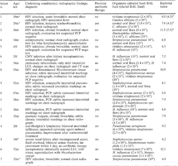
Table 1 Characteristics of patients without bacterial pneumonia exhibiting a bacterial index of - 5 Patient Age/ Underlying condition(s); radiographic findings; Previous
no. sex diagnosis antibiotic
therapy
Organisms cultured from BAL fluid (cfu/ml BAL fluid)
Bacterial index 1 39m a 2 29m a 3 36m 34m 43f 6 35f 7 34m 8 29m 9 52m 10 67m 11 38m [2 36m 13 35m 14 20f 15 18f d 16 52m 17 33m e
HIV infection, acute bronchitis; normal chest no radiograph; HIV-associated fever
HIV infection, herpetic tracheobronchitis; yes normal chest radiograph
HIV infection, chronic bronchitis; normal chest no radiograph; evaluation for suspected PCP
negative
asymptomatic; normal chest radiograph; evalua- no tion for hilar lymphadenopathy unremarkable
HIV infection, chronic bronchitis; normal chest no radiograph; evaluation for suspected PCP nega- tive
CMV infection after kidney transplantation; no normal chest radiograph
pulmonary tuberculosis; only mild interstitial no LUL changes on chest radiograph and CT scan HIV and disseminated Mycobacteriurn simiae no infection; subtle increased interstitial markings on chest radiograph; evaluation for suspected PCP negative
HIV infection, nonspecific interstitial pneumo- no nitis; subtle increased interstitial markings on
chest radiograph
HIV infection, PCP; subtle increased interstitial no markings on chest radiograph
HIV infection, PCP; subtle increased interstitial no markings on chest radiograph
HIV infection, PCP; subtle increased interstitiaI yes markings on chest radiograph
psoriasis vulgaris, chronic bronchitis; subtle no chronic interstitial markings on chest radio-
graph
non-Hodgkin's lymphoma; bilateral interstitial no infiltrates; suspected cytotoxic agent-induced
pneumonitis, improvement after corticosteroid treatment
severe renal failure after meningococcal sepsis, no fluid overload; bilateral acinar shadows; im-
provement within i day, no antibiotic therapy asymptomatic chronically scarred LUL infil- no trates; superinfecti0n with Mycobacterium
xenopi
HIV infection, bronchitiS; normal chest radio- no graph
viridans streptococci (2 x 10⁴), Candica albicans (5 x 10³) normal oral flora c (1.8 x 10⁴), C. albicans (2 x 10³) normal oral flora (6 x 10³), Haemophilus influenzae (5 x 10³), C. albicans (104) Streptococcus pneumoniae 103), normal oral flora (103 ) viridans streptococci (2 x 10³), H. influenzae (103) 8.0 (4.3) u 7.6 (4.3) u 11,5 (7.5) b 6,0 6.3
H. influenzae (103), normal oral 7.3 flora (2 x 10 4)
normal oral flora (1.4 x 10⁴), H, 7.4 influenzae (2 x 10³) Streptococcus pneumoniae 10.9 (4 x 10⁴), Staphylococcus aureus (2 x 10³), viridans streptococci (103 ) Staphyloccocus aureus 8.1 (1.4 x 10⁴), normal oral flora
(1o 4 )
Staphylococcus aureus (2 x }O3), 6.6 viridans streptococci (2 x 10 ")
Streptococcus pneumoniae 7.9 (2.4 x 10⁴), Staphylococcus epi-
dermidis (3 x 10³)
H. influenza (103), normal oral 6.6 flora (4 x 10³) Streptococcus pneumoniae 7.9 (5 x 10³), H. influenzae (1.5 x 10⁴) Pseudornonas aeruginosa 9.2 (6 • 104), viridans streptococci (2.3 x 10⁴) Staphylococcus aureus 6.2 (1.2 x 10³), Xanthomonas malto- phila (1.2 x 10³) viridans streptococci 3(.7 x 104), 12.1 H. influenzae (2 x 10 ), Strepto- coccus pneumoniae (1.1 x 10⁴) Streptococcus pneumoniae (106) 6.0
a Patients with a bacterial index of <5, if C. albicans isnot con- sidered
b Values of bacterial index without consideration 'of C. albicans are shown in parentheses : . . . . , c Normal oral flora defined as the growt h' of two or~ mo're of'the
following bacteria: viridans streptococci'; Neisseria spp., coryne- forms, coagulase-negative staphylococci' .. . . : ,
ty was significantly r e d u c e d f r o m 99 to 87% (116/133; C1, 8 3 - 9 4 % ; P < 0 . 0 0 0 1 ) . T h e clinical dat'a o f t h e pa- tients w i t h o u t p n e u m o n i a a n d with a B I Of _> 5 are dis- p l a y e d in T a b l e 1 ( T a b l e 1, patients 1-17).
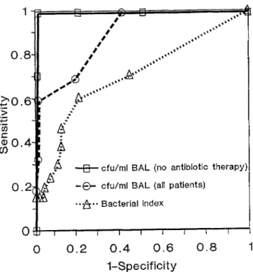
T h e p e r f o r m a n c e o f t h e d i f f e r e n t diagnostic p a r a m e t e r s is illustrated by t h e A U C of t h e R O C curve (Figure 2). W h e r e a s the A U C f o r t h e B I was o n l y 0.674, t h e A U C o f the s u m o f t h e cfu/mt B A L fluid p e r p a t i e n t r e a c h e d
d Mechanically ventilated patient
Patient with a bacterial index of -> 5 and an absolute number of cfu/ml BAL fluid of _ 105
CMV, cytomegalovirus; CT, computed tomography; HIV, human immunodeficiency virus; LUL, left upper lobe; PCP, Pneumocys- tis carinii pneumonia
0.879. T h e A U C of t h e s u m of cfu/ml B A L fluid includ- ing o n l y p a t i e n t s w i t h o u t p r e v i o u s antibiotic t r e a t m e n t was 0.99.
S o m e a u t h o r s e v e n include t h e c o l o n y c o u n t s o f fungal species in their calculations [8, 9]. H o w e v e r , this m i g h t n o t h a v e a s o u n d basis, since t h e r e is n o e v i d e n c e o f t h e diagnostic utility o f q u a n t i t a t i v e fungal cultures. T h e r e w e r e two patients in o u r g r o u p (Table 1, patients 1 a n d
[] c [] , ,
.,,..,"
II
Z
,,'"
O-all
;
,,,""
0 . 2
0
t 0,,, [] , cfu/ml BAt. (no antibiotic therapy',
~'~ - e - cfu/rnl BAL (all patients)
9 ,.~., Bacterial index
I t I I I t [ L
0 . 2 0 . 4 0 . 6 0 . 8 1
1 - S p e c i f i c i t y
Figure 2 Receiver operating characteristic (ROC) curve for the
bacterial index (AUC=0.674) for the sum of cfu/ml BAL fluid,
including all patients (AUC=0.879) and considering only pa-
tients not treated with antibiotics at the time of BAL
(AUC=0.99)
2) in whom the BI was lower than 5 when the fungal
colony counts were not included. However, even by ex-
cluding these two cases, the specificity of a BI of >_5
remained low at 89% (118/133; C1, 85-96%).
Discussion
The principal finding of this study is that using the BI at
a cut-off of _> 5 in an unselected patient group with a
relatively low prevalence of pneumonia (16.4%) results
in an inacceptably low specificity for the diagnosis of
bacterial pneumonia. Using the concept of the BI,
which is calculated by adding up the loglo of the con-
centrations of the individual organisms per milliliter of
B A L specimen (i.e., multiplying the numbers of colony-
forming units of individual organisms per milliliter of
BAL specimen), resulted in 17 false-positive results in
patients who decisively did not suffer from bacterial
pneumonia (Table 1). All these patients except case 17
(Streptococcus pneumoniae,
1 0 6cfu/ml BAL fluid)
would never have been diagnosed as having pneumonia
if the absolute number of organisms at a cut-off of 105
cfu/ml B A L fluid had been used. Hence, by applying
the BI methodology, the superb specificity of BAL for
the diagnosis of bacterial pneumonia was significantly
reduced from 99-87% ( P < 0.0001). The very low
power of diagnostic discrimination of the BI is illus-
trated by the small A U C in the R O C analysis (Figure
2).
The BI was introduced by Johanson et al. [6], who used
it in a baboon model of nosocomial pneumonia. They
demonstrated a very good correlation of the BI of BAL
fluid compared to the BI of quantitative lung tissue cul-
tures. The BI was created with the attempt to let spe-
cies present only in small numbers contribute to an es-
timation of the lungs' bacterial burden, especially dur-
ing prolonged mechanical ventilation [6], The hypothe-
sis behind the BI was that the presence of multiple spe-
cies in low concentrations indicated a marked impair-
ment in host defenses, a hypothesis that is still unprov-
en and basically untested. However, the study of Johan-
son et al. [6] became a landmark paper leading to a
widespread use of quantitative bacterial cultures of
BAL fluid for the diagnosis of bacterial nosocomial
pneumonia [10, 15, 22]. Unfortunately, the BI was used
rather uncritically by some subsequent authors [8, 91,
while others relied on the absolute count of cfu/ml
BAL fluid per single microorganism or the sum of cfu/
ml per B A L specimen at the cut-off point of 105 as an
indicator for pneumonia [4, 5, 10, 15, 161.
According to our findings, the concept of the BI, calcu-
lated by adding up the log~0 cfu/ml BAL fluid of the
individual microbial species per patient, thus multiply-
ing the number of cfu/ml of different organisms, has no
sound microbiological basis. In such a way, the pres-
ence of very low concentrations of several different co-
lonizing bacteria in an individual patient might lead to
a BI of >5. In particular, this may occur in immunosup-
pressed patients (such as in 12 of our study group, Ta-
ble 1). Why the lungs of these patients contain quite
high bacterial burdens is of great interest and should be
studied further. Although our series included mainly
nonventilated patients, we believe that our findings
also apply to intubated and mechanically ventilated pa-
tients. Since two-thirds of our patients were immuno-
compromised, it is not intelligible why the diagnostic
criteria for bacterial pneumonia, i.e. the amount of bac-
terial burden in the lung to cause invasive infection,
should not apply to patients with other types of host
defense impairment, such as mechanical ventilation in
an ICU setting.
We are well aware that our study lacks a gold standard
for the diagnosis of bacterial pneumonia. However,
while the clinical diagnosis of bacteria1 pneumonia re-
mains unreliable, especially in cases of nosocomiaI
pneumonia [1, 23], we are confident that none of the 17
patients listed in Table 1 had bacterial pneumonia. In
most cases the chest radiograph was normal or showed
only slightly increased interstitial markings, and in all
cases another diagnosis could be established. It was not
our intention to demonstrate the value of BAL for the
diagnosis of pneumonia but to prove the low specificity,
i.e. the high number of false~positive results, using the
methodology of the BI. Therefore, we decided to study
a consecutive heterogenous series of patients with a low
prevalence of pneumonia, including ventilated and
nonventilated as well as i m m u n o c o m p r o m i s e d and im- m u n o c o m p e t e n t patients.
O u r findings of a low specificity of the B I are s u p p o r t e d by a recent p a p e r investigating 27 mechanically venti- lated patients without clinical or radiographic evidence of p u l m o n a r y infection [24]. Analyzing the B A L data by means of the B I at a cut-off of 6 gave 23% false- positive results. T h e authors suggest that a B I of 8 would be the best threshold to get a low p e r c e n t a g e of false-positive results. This value is far higher than the B I cut-off of 5 or g r e a t e r used by m o s t o t h e r authors
Is, 91.
In contrast to the BI, the use of the absolute n u m b e r of cfu/ml B A L fluid of the individual organisms at the cut- off point of 105 was highly specific (99%) for the diag- nosis of bacterial p n e u m o n i a . On the o t h e r hand, the relatively low sensitivity (33%) of the cfu/ml B A L fluid was mainly due to a high rate of patients receiving an- timicrobial t h e r a p y prior to bronchoscopy. This is a m a - j o r p r o b l e m in the use of quantitative cultures in clini- cal practice and has b e e n addressed by others [22]. Considering m e r e l y the cases n o t t r e a t e d with antibio- tics prior to B A L , the sensitivity for the diagnosis of bacterial p n e u m o n i a increased to 70%, which is com- p a r a b l e to the findings of o t h e r studies [16]. This is also clearly d e m o n s t r a t e d by the large increase of the A U C in the R O C analysis (Figure 2). As m e n t i o n e d above, however, it must be r e m e m b e r e d that this study was not designed to assess the value of B A L for the diagno- sis of bacterial p n e u m o n i a , and that there was no gold standard test to p r o v e the true-positive rate, i.e. the sensitivity of the B A L cultures.
In conclusion, we h a v e shown that the use of the BI as a diagnostic p a r a m e t e r for bacterial p n e u m o n i a results in an u n a c c e p t a b l y low specificity. Thus, the concept of the B I is not applicable to clinical practice. Q u a n t i t a t i v e bacterial cultures of B A L fluid, however, m a y be a val- uable tool for the diagnosis of bacterial p n e u m o n i a w h e n the absolute a m o u n t of c o l o n y - f o r m i n g units of individual species p e r milliliter of B A L fluid is used at a cut-off point of 10 5, especially w h e n patients without prior antibiotics are considered.
Acknowledgement
The authors are indebted to the Zurich Lung Association, Zurich, Switzerland, for their support of the study.References
1. Andrews CP, Coalson J J, Smith JD, Johanson WG: Diagnosis of nosocomial pneumonia in acute, diffuse lung injury. Chest (1981) 80:254-258
2. Haas H, Morris JF, Samson S, Kilbourn JP, Kim PJ: Bacterial flora of the respiratory tract in chronic bronchitis: comparison of transtracheal, fiberbronchoscopic, and oropharyngeal sam- pling methods. American Review of Respiratory Diseases (1977) 116:41-47
3. Bartlett JG: Diagnostic accuracy of transtracheal aspiration: bacteriologic studies. American Review of Respiratory Dis- eases (1977) 115:777-782
4. Kahn FW, Jones JM: Diagnosing bacterial respiratory infec- tion by bronchoalveolar lavage. Journal of Infectious Dis- eases (1987) 155:862-869
5. Thorpe JE, Baughman RP, Frame PT, Wesseler TA, Staneck JL: Bronchoalveolar lavage for diagnosing acute bacterial pneumonia. Journal of Infectious Diseases (1987) 155:855- 861
6. Johanson WG, Seidenfeld JJ, Gomez P, de los Santos R, Coalson JJ: Bacteriologic diagnosis of nosocomial pneumonia following prolonged mechanical ventilation. American Re- view of Respiratory Diseases (1988) 137:259-264
7. Xaubet A, Torres A, Marco F, de la Bellacasa JP, Faus R, Augusti-Vidal A: Pulmonary infiltrates in immunocomprom- ised patients. Diagnostic value of telescoping plugged cathet- er and bronchoalveolar lavage. Chest (1989) 95:130-135 8. Gaussorgues P, Piperno D, Bachmann P, Boyer F, Jean G,
Gdrard M, Ldger P, Robert D: Comparison of nonbronchos- copic bronchoalveolar lavage to open lung biopsy for the bac- teriologic diagnosis of pulmonary infections in mechanically ventilated patients. Intensive Care Medicine (1989) 15:94- 98
9. Pugin J, Auckenthaler R, Mill N, Janssens JP, Lew D, Suter PM: Diagnosis of ventilator-associated pneumonia by bacteri- ologic analysis of bronchoscopic and nonbronchoscopic "blind" bronchoalveolar lavage. American Review of Respi- ratory Diseases (1991) 143 : 1121-1129
10. Meduri GU, Beats DH, Maijub AG, Baselski V: Protected bronchoalveolar lavage. A new bronchoscopic technique to retrieve uncontaminated distal airway secretions. American Review of Respiratory Diseases (1991) 143:855-864
11. Speich R, Wast J, Hess T, Kayser FH, Russi EW: Prospective evaluation of a semiquantitative dip slide method compared with quantitative bacterial cultures of BAL fluid. Chest (1996) 109:1423-1429
12. Chastre J, Fagon JY, Bornet Lecso M, Calvat S, Dombret MC, al Khani R, Basset F, Gibert C: Evaluation of bronchos- copic techniques for the diagnosis of nosocomial pneumonia. American Journal of Respiratory and Critical Care Medicine (1995) 152:231-240
13. Faling LJ: New advances in diagnosing nosocomial pneumon- ia in intubated patients. Part I. American Review of Respira- tory Diseases (1988) 137:253~55
14. Johanson WG: Ventilator-associated pneumonia. Light at the end of the tunnel? Chest (1990) 97:1026-1027
15. Meduri GU, Baselski V: The role of bronchoalveolar lavage in diagnosing nonopportunistic bacterial pneumonia. Chest (1991) 100:179-190
16. Meduri GU: Diagnosis and differential diagnosis of ventila- tor-associated pneumonia. Clinics in Chest Medicine (1995) 16:61-93
17. Torres A: Accuracy of diagnostic tools for the management of nosocomial respiratory infections in mechanically venti- lated patients. European Respiratory Journal (1991) 4:1010- 1019
18. Baselski VS, EI-Torky M, Coalson JJ, Griffin JP: The stand- ardization of criteria for processing and interpreting laborato- ry specimens in patients with suspected ventilator-associated pneumonia. Chest (1992) 102, Supplement 1:571-579 19. Baron EJ, Weissfeld AS, Fuselier PA, Brenner DJ: Classifica-
tion and identification of bacteria. In: Murray PR, Baron EJ, Pfaller MA, Tenover FC, Yolken RH (eds). Manual of clini- cal microbiology. American Society for Microbiology, Wash- ington, DC (1995) pp 249-264
20. Simon RS: Confidence intervals for reporting results of clini- cal trials. Annals of Internal Medicine (1986) 105:429-435 21. Hanley JA, McNeil B J: The meaning and use of the area un-
der a receiver operating characteristic (ROC) curve. Radiolo- gy (1982) 143:29-36
22. Meduri GU, Chastre J: The standardization of bronchoscopic techniques for ventilator-associated pneumollia. Chest (1992) 102, Supplement 1:557-564
23. Fagon JY, Chastre J, Hance AJ, Guiguet M, Trouillet JL, Do- mart Y, Pierre J, Gibert C: Detection of lung infection in ven- tilated patients: use of protected specimen brush and quanti- tative culture techniques in 147 patients. American Review of Respiratory Diseases (i988) 138:110-it6
24 Torres A, Martos A, de ~a Bellacasa JP, Ferrer M, Et-Ehiary M, Gonz~lez J, Gen4 A, Rodrfguez-Roisin R: Specificity of endotracheal aspiration, protected specimen brush, and bron- choalveolar lavage in mechanically ventilated patients. Amer- ican Review of Respiratory Diseases (1993) 147:952-957