Slow-stimulated multifocal ERG in high- and normal-tension glaucoma
Anja M. Palmowski-Wolfe
1,2, Reiner J. Allgayer
1, Bernhild Vernaleken
1, Andy Scho¨tzau
2& Klaus W. Ruprecht
11
University Eye Hospital, D-66421, Homburg/Saar, Germany; 2University Eye Hospital, CH 4053, Basel, Switzerland
Accepted: 6 February 2006
Key words: electroretinography, glaucoma, multifocal ERG, oscillatory potentials, slow stimulation
Abstract
Purpose:To study the ability and sensitivity of the slow stimulation multifocal ERG (mfERG) to detect glaucomatous damage. Methods: Right eyes of 20 patients with normal-tension glaucoma (NTG), 15 patients with high-tension glaucoma (HTG) and 15 healthy volunteers underwent testing with the mfERG (VERIS 4.1TM). The central 50 degrees of the retina were stimulated by 103 hexagons (m-sequence: 213-1, Lmax: 100 cd/m2, Lmin: 1 cd/m2, background: 50 cd/m2). Each m-sequence step was followed by 3 black frames (Lmax:<1 cd/m2). Five response averages of the first order response component (KI) were ana-lyzed: the central 7.5 degrees and the 4 adjoining quadrants. The amplitudes from the first minimum, N1, to the first maximum, P1, and from P1 to the second minimum, N2, were analyzed as well as the latencies of N1, P1, N2 and the latencies of 3 multifocal oscillatory potentials (mfOPs) with their maxima at about 73, 80 and 85 ms. Results: For each parameter the percentage of deviation from the mean of the control group was calculated. These values were then added for each individual to form a deviation index (DI). Seventeen patients (85.0%) with NTG and 3 patients (20.0%) with HTG showed a DI outside the normal range. The major changes were observed in the mfOPs of the NTG patients. MfOPs were then selectively filtered at 100–300 Hz and their scalar product was analyzed over an epoch of 68–105 ms. This confirmed that mfOPs differed significantly from the control in the central 7.5° and, for NTG, in the nasal field. With a logistic regression analysis the mfOPs had a sensitivity to differentiate 85% of the NTG patients and 73% of the HTG patients from normal. Conclusions: Under these conditions, the slow-stimulated mfERG can detect glaucomatous dysfunction in NTG (85.0%). The differences observed between NTG and HTG are in support of a different underlying pathomechanism.
Introduction
Open angle glaucoma (OAG) is the second lead-ing cause of vision loss worldwide [1]. As early therapeutic intervention may prevent progression and blindness, it is important to detect glaucoma at an early stage. Diagnosis is especially difficult in normal-tension glaucoma (NTG), where the intraocular pressure, which is one of the risk fac-tors for OAG is less than 22 mmHg, that is in the normal range.
The multifocal ERG (mfERG), which permits a topographic display of retinal function, has
shown promise in the investigation of OAG. It has been reported that the mfERG response con-tains a so called retinal component (RC) of pre-sumed outer retinal origin and an inner retinal contribution such as the optic nerve head compo-nent (ONHC), which is attributed mainly to the ganglion cell layer [2]. The ONHC and the RC differ in their luminance- and contrast-sensitivity [3]. As the ONHC saturates at about 60% contrast, whereas the RC tends to increase line-arly with increasing contrast, attempts have been made to increase the inner retinal contribution through decreasing the stimulus contrast.
ever, mfERGs at a low contrast (50%) were not sensitive enough to reliably detect retinal dys-function in individual patients with OAG [4, 5].
Recently it has been found, that naso-tempo-ral asymmetries in the oscillation rich contribu-tions to a special slow mfERG stimulus sequence are caused by the changes in the relative align-ment of the ONHC and the RC [6]. Therefore this stimulus holds promise in the investigation of glaucomatous functional damage. In this study we tested it’s sensitivity in different forms of open angle glaucoma.
Methods
The subjects consisted of 20 patients with differ-ent stages of normal-tension glaucoma (NTG), 15 patients with high-tension glaucoma (HTG) and a regulated intraocular pressure, as well as 15 heal-thy volunteers. Informed consent was obtained from all subjects after explaining the procedure. The Declaration of Helsinki was followed.
Inclusion criteria for both groups of glau-coma were the presence of glauglau-comatous visual field defects (octopus d32). For patients with normal-tension glaucoma the highest intraocular pressure (IOP), measured by Goldman applana-tion tonometry, was less than 22 mmHg and the cup disk ratio (CDR) was 0.5 or higher. For pa-tients with HTG the highest intraocular pressure (IOP) recorded on Goldman applanation tonom-etry, was over 22 mmHg. Other ocular diseases were excluded.
MfERGs were recorded of the right eyes using VERISTM. The mfERG signals were recorded monocularly with the help of a Burian-Allen bipo-lar contact lens electrode. The ground electrode was on the forehead. The pupils were dilated, the cornea was anesthetized. Refractive errors were corrected for best visual acuity at a viewing distance of 40 cm, the viewing distance was then adjusted to keep the image size constant [7].
During recording, the central 50 degrees of the retina were stimulated by 103 hexagons where each hexagon flickered according to a slow m-sequence stimulation. Figure 1 shows the stim-ulus sequence where each m-sequence step (M) with a luminance of either 100 or <1 cd/m2 was followed by 3 black frames (B) with a luminance <1 cd/m2. This four frame stimulus sequence
(MBBB) re-occurred every 53.3 ms. The length of the m-sequence was 213-1. Total recording time was 7 min 17 s. To enhance the signal-quality each recording was split into 16 or 32 cycles of about 27.29 or 13.65 s. Contaminated segments were dis-carded and re-recorded. The raw signals were fil-tered (10–300 Hz) and amplified (gain=100 000). 16 samples were obtained per display frame (sam-pling interval: 0.83 ms). An artifact elimination technique [8] was applied once. The first order response component (KI) was analyzed. For each location KI can be described as the difference between the mean local response to all the bright m-sequence stimuli and the mean focal response to the black m-sequence stimuli occurring in a stimulus cycle and taking into account the entire stimulus base interval (Figure 1).
Results
Table 1 summarizes the clinical information of the 20 NTG patients included in the study. Mean age was 50.8 years, mean Snellen visual acuity (VA) was 1.05. The mean CDR of the NTG pa-tients was 0.73. Mean visual field parameters (Octopus d32) were as follows, mean sensitivity: 20.50 dB, mean defect: 6.98 dB and loss vari-ance: 24.35 dB2.
Table 2 depicts the clinical data of the 15 HTG patients included in the study. Here, mean age was 58.0 years, mean VA was 0.87. The mean CDR of 0.71 compared well to that of the NTG group. Mean visual field parameters (Octo-pus d32) of the HTG patients were as follows, mean sensitivity: 17.81 dB, mean defect: 8.64 dB and loss variance: 33.31 dB2.
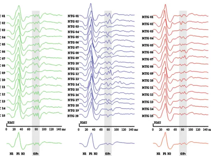
Figure 2 shows each subject’s overall response average for the control group (left), the NTG group (middle) and the HTG group (right). The mean overall response, is shown at the bottom.
Figure 1. This figure depicts the stimulus sequence applied (MBBB-Sequence). The luminance of the m-sequence step (M) was either 100 or <1 cd/m2, luminance of the interposed black frames (B) was <1 cd/m2. The background was set at
50 cd/m2. Each frame lasted 13.33 ms, resulting in a stimulus
The response to the MBBB stimulus consists of a first minimum, N1, followed by a maximum, P1, and then a second minimum, N2. Approximately one base interval later, the response average
contains 3 multifocal oscillatory potentials (mfOPs) with their peaks at about 73, 80 and 85 ms. A marked difference can be observed be-tween the mfOPs of the NTG-response average
Table 1. Characteristics of the NTG patients examined NTG ID Gender Age
[years]
Visual acuity log MAR
CDR
(cup disk ratio)
MS (mean sensitivity) [dB] MD (mean defect) [dB] LV (loss variance) [dB2] NTG 01 m 61 0.15 0.5 18.8 8.2 34.4 NTG 02 f 46 )0.1 0.9 19.8 7.5 19.5 NTG 03 f 40 0 0.8 23.0 4.5 7.3 NTG 04 f 62 )0.08 0.5 20.3 5.5 28.5 NTG 05 f 33 )0.1 0.8 26.1 2.0 6.5 NTG 06 f 52 )0.08 0.7 18.3 19.3 16.4 NTG 07 f 56 0.15 0.9 14.9 11.7 51.4 NTG 08 m 70 0 0.7 16.9 8.8 30.4 NTG 09 f 18 )0.1 0.8 26.0 2.9 11.0 NTG 10 f 29 )0.1 0.8 25.1 3.3 18.4 NTG 11 m 63 0 0.7 21.4 4.8 17.8 NTG 12 m 26 )0.08 0.5 23.2 5.4 10.4 NTG 13 f 65 0 0.75 11.9 14.1 80.8 NTG 14 m 63 0 0.5 16.8 9.3 57.3 NTG 15 f 65 0 0.8 22.0 3.9 21.1 NTG 16 f 35 0 0.75 23.0 5.0 17.5 NTG 17 f 53 0 0.7 24.9 1.8 6.6 NTG 18 m 50 0 0.65 24.1 2.9 9.1 NTG 19 f 64 0 0.9 18.9 7.2 22.0 NTG 20 f 65 0 0.9 14.6 11.4 20.6 Mean ± SD 50.80±15.53 )0.02±0.07 0.73±0.14 20.50±4.08 6.98±4.49 24.35±19.10
Table 2. Characteristics of the HTG patients examined HTG ID Gender Age [years] Visual acuity
log MAR CDR (cup disk ratio) MS (mean sensitivity) [dB] MD (mean defect) [dB] LV (loss variance) [dB2] HTG 01 m 49 )0.1 0.8 20.0 7.1 28.6 HTG 02 m 36 0 0.8 17.9 10 53.2 HTG 03 f 56 0 0.9 15.9 10.7 85.7 HTG 04 f 71 0 0.9 10.8 14.7 66.1 HTG 05 m 64 0 0.8 24.8 1.3 12.3 HTG 06 f 65 0 0.5 23.3 2.7 24.6 HTG 07 m 53 0 0.6 24.3 2.5 20.7 HTG 08 f 71 0 0.9 16.2 9.4 47.2 HTG 09 f 59 0.15 1.0 0.9 25.5 16.4 HTG 10 f 64 0.05 0.9 14.9 11.2 48.2 HTG 11 f 39 )0.08 0.1 25.1 2.6 18.9 HTG 12 f 68 )0.10 0.4 24.0 1.7 8.5 HTG 13 m 56 1.0 1.0 2.3 24.3 31.2 HTG 14 m 58 0 0.7 23.9 2.6 23.0 HTG 15 f 61 0 0.4 22.9 3.3 15.1 Mean ± SD 58.0±10.5 0.06±0.27 0.71±0.26 17.81±7.89 8.64±7.83 33.31±22.17
and the mfOPs of the control- or HTG-response average.
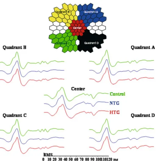
In order to take into consideration the naso-temporal variation of the mfOPs [6], five response averages were formed. Figure 3 (top) depicts these 5 response averages that consisted of the central 7.5 degrees (center) and four adjoining quadrants A–D. Quadrant A consti-tutes the response average from the upper tem-poral field, quadrant B from the upper nasal field, quadrant C from the lower nasal field and quadrant D from the lower temporal field.
Figure 3 (bottom) shows the resulting traces of the 5 response averages analyzed. For each re-sponse average, the rere-sponse of the control group is shown at the top, the middle trace
rep-resents the average of the NTG patients and the bottom trace the HTG patients. While the cen-tral response average shows mfOPs in NTG, HTG and in the control group, the mfOPs ap-pear diminished in all field quadrant averages of the NTG-group.
In every subject’s mfERG, the amplitudes of N1P1 and P1N2 were analyzed as well as the corresponding latencies of N1, P1, N2 for each of the five response averages. In addition, the latencies of the 3 mfOPs with their maxima at about 73, 80 and 85 ms were measured. A reliable measurement of mfOP latencies was pos-sible even in patients with NTG, as in an individ-ual’s group response averages the individual mfOP peaks were more clearly depicted than in
Figure 2. This figure shows each subject’s overall response average for the control group (left), the NTG group (middle) and the HTG group (right). The mean overall response, is shown at the bottom. The response to the MBBB stimulus consists of a first minimum, N1, followed by a maximum, P1, and then a second minimum, N2. One base interval later, 3 multifocal oscillatory potentials (mfOP) can be observed. In order to allow a better comparison of the waveforms, responses were normalized to have an equal root mean square (RMS). There is a marked difference between the mfOPs of the NTG-response average and the mfOPs of the control- or HTG-response average.
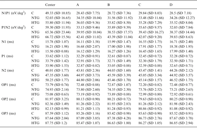
the average of the 20 NTG patients shown in Figures 2 and 3. Table 3 shows the mean ampli-tudes N1P1 and P1N2 and the latencies of N1, P1, N2 as well as the 3 mfOPs for each response average. The standard deviation expresses the high inter-individual variability which results in an overlap between the groups that precludes the observation of a significant difference.
In order to reduce the inter-individual vari-ability, the amplitudes of an individual’s response averages were normalized to the amplitudes of this individual’s overall response. For example, for each recording, the amplitude of N1P1 in quadrant A was divided by N1P1 of the overall
response of the same recording. For each param-eter (normalized amplitudes, latencies N1, P1, N2 and mfOP-latencies) the percentage of devia-tion from the mean of the control group was cal-culated. Adding these values resulted in a group deviation index for each of the five response averages. To obtain only one parameter that de-scribes the mfERG response, an individual’s 5 group deviation indices were added to form an overall deviation index.
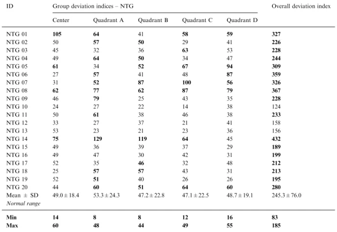
Table 4 shows the resulting group deviation indices and the overall deviation index for the 20 NTG patients, while Table 5 depicts these values for the 15 HTG patients. The patients’ data can
Figure 3. In the central 50 degrees responses of the central 7.5 degrees (Center) and the four adjoining quadrants (Quadrants A–D) were averaged as shown at the top. Quadrant A constitutes the upper temporal field, quadrant B the upper nasal field, quadrant C the lower nasal field and quadrant D the lower temporal field. Below the naso-temporal asymmetries of the respective mfERG re-sponses averages are shown (center and the four quadrants). For each response average, the response of the control group is shown at the top, the middle trace represents the average of the NTG patients and the bottom response of the HTG patients. In order to allow a better comparison of the waveforms, responses were normalized to have an equal root mean square (RMS). In the quad-rants the NTG-group again clearly differ in the range of the three mfOPs, that is between 70 and 90 ms.
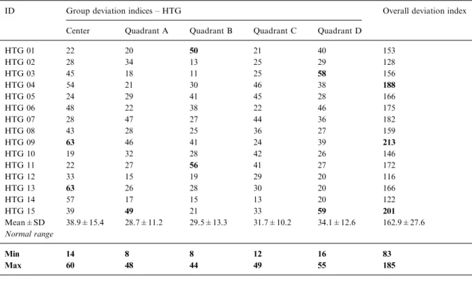
be compared to the range of normal which is shown in the lower two rows. In Tables 4 and 5 deviation indices outside the normal range are highlighted in black. An overall deviation index outside the range of the control group could be observed in 17 NTG patients but only in three HTG patients. This corresponds to a sensitivity of 85% for NTG and only 20% for HTG. Fig-ure 4 (left) shows a boxplot of the overall devia-tion index which graphically highlights these results.
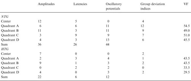
Table 6 shows how often the individual parameters were outside the normal range for each of the 5 response averages analyzed. Thus, these tables demonstrate which of the analyzed parameters (amplitudes, latencies or mfOP laten-cies) are most effected in glaucoma. Overall, the mfOP latencies differed most between NTG patients and the control group. However, these differences were only seen in the peripheral re-sponse averages, while in the central 7.5 degrees no glaucoma patient showed mfOP latencies out-side the normal range. The second column from
the right in Table 6 summarizes the number of patients that showed a group deviation index outside the range of norm for each group re-sponse average. Here the two upper quadrants (quadrants A and B) differed most. These chan-ges did not correlate with the chanchan-ges observed in the visual fields (Tables 6, rightmost column).
In order to appraise our results in a less examiner dependent manner, we selectively fil-tered the data at 100–300 Hz in order to isolate the mfOPs from the underlying response compo-nents. Over an epoch of 68–105 ms we formed the scalar product (SP), using the waveform of the respective group average as a template [8]. The average scalar product was calculated for each of the 5 groups. In addition to including information on latency, the scalar product also includes information on changes in amplitude. In contrast to absolute measurements of amplitude and latency, the SP measurement is less suscepti-ble to the influence of noise. In order to ensure a normalized distribution, the log of the SP val-ues was formed and an analysis of variance
Table 3. This table shows the mean amplitudes N1P1 and P1N2 and the latencies of N1, P1, N2 as well as the 3 mfOPs. The stan-dard deviation (SD) gives an indication of the inter-individual variability
Center A B C D N1P1 (nV/deg2) C 49.33 (SD 10.85) 28.43 (SD 7.75) 28.72 (SD 7.36) 29.84 (SD 8.03) 28.5 (SD 7.18) NTG 52.03 (SD 16.65) 34.35 (SD 18.04) 31.56 (SD 11.92) 33.48 (SD 11.66) 34.26 (SD 12.27) HTG 55.80 (SD 11.94) 36.03 (SD 9.36) 35.82 (SD 8.50) 35.28 (SD 7.29) 35.52 (SD 8.04) P1N2 (nV/deg2) C 59.30 (SD 11.93) 33.13 (SD 9.46) 35.89 (SD 9.39) 35.65 (SD 9.57) 32.03 (SD 7.84) NTG 65.36 (SD 23.44) 39.95 (SD 18.04) 38.53 (SD 17.57) 39.43 (SD 16.27) 38.37 (SD 14.44) HTG 66.72 (SD 15.56) 42.41 (SD 11.02) 43.39 (SD 11.04) 42.07 (SD 9.20) 39.83 (SD 8.63) N1 (ms) C 15.78 (SD 1.07) 16.11 (SD 1.58) 15.99 (SD 1.47) 16.55 (SD 1.75) 16.71 (SD 2.40) NTG 16.21 (SD 1.98) 16.68 (SD 2.07) 17.00 (SD 1.98) 17.91 (SD 1.77) 18.38 (SD 1.95) HTG 15.50 (SD 0.88) 16.12 (SD 1.29) 16.27 (SD 1.26) 16.45 (SD 1.65) 17.99 (SD 1.40) P1 (ms) C 33.62 (SD 1.12) 32.29 (SD 0.91) 32.61 (SD 0.87) 32.45 (SD 0.72) 32.28 (SD 0.72) NTG 33.79 (SD 1.42) 32.91 (SD 1.73) 32.71 (SD 1.49) 32.30 (SD 1.79) 32.50 (SD 1.71) HTG 33.90 (SD 1.53) 32.87 (SD 0.82) 33.05 (SD 0.88) 32.59 (SD 0.88) 32.65 (SD 0.71) N2 (ms) C 48.01 (SD 1.77) 43.81 (SD 2.36) 44.03 (SD 1.60) 43.47 (SD 1.41) 44.26 (SD 2.12) NTG 47.35 (SD 3.60) 44.97 (SD 3.73) 45.59 (SD 3.39) 45.05 (SD 3.34) 44.92 (SD 3.57) HTG 50.25 (SD 1.77) 44.80 (SD 2.06) 45.46 (SD 1.70) 45.14 (SD 1.57) 46.52 (SD 1.75) OP1 (ms) C 73.79 (SD 0.74) 72.48 (SD 0.88) 72.87 (SD 1.07) 72.20 (SD 0.82) 72.48 (SD 0.77) NTG 74.93 (SD 2.14) 73.80 (SD 2.60) 74.35 (SD 2.30) 73.76 (SD 2.52) 73.21 (SD 2.65) HTG 75.08 (SD 0.63) 73.19 (SD 0.92) 73.89 (SD 0.88) 72.99 (SD 0.80) 72.92 (SD 0.61) OP2 (ms) C 81.97 (SD 3.25) 80.13 (SD 0.90) 80.21 (SD 0.72) 79.63 (SD 0.82) 80.25 (SD 0.98) NTG 82.36 (SD 1.49) 81.26 (SD 2.22) 81.93 (SD 2.83) 81.26 (SD 2.12) 81.98 (SD 2.43) HTG 82.13 (SD 0.99) 81.21 (SD 1.13) 81.24 (SD 0.93) 80.86 (SD 0.92) 81.08 (SD 0.92) OP3 (ms) C 87.59 (SD 1.12) 85.26 (SD 1.16) 85.42 (SD 0.98) 85.63 (SD 0.90) 85.52 (SD 0.91) NTG 87.64 (SD 2.04) 87.09 (SD 3.85) 87.38 (SD 4.28) 86.73 (SD 2.76) 87.67 (SD 3.78) HTG 87.75 (SD 1.2) 85.47 (SD 1.07) 86.63 (SD 1.80) 86.27 (SD 1.05) 86.85 (SD 2.94)
(ANOVA) was performed. Age did not influence the results (p=0.95). To adjust for multiple testing, the Tukey test was performed as a post hoc test.
Figure 5 depicts the boxplots of the scalar product for each response average showing a re-duced SP in the mfOPs of glaucoma patients in all response averages. For patients with NTG, this reached a significance level in the nasal field (quadrant B, p=0.014, and quadrant C, p=0.001) as well as in the central response aver-age (p=0.022). HTG patients only differed sig-nificantly from the control group in the central response average (p=0.024).
In order to test for sensitivity, we then per-formed a stepwise logistic regression using SPSS. For NTG patients, quadrants C and A contained the most relevant parameters, allowing 85% of NTG patients to be differentiated from normal
(Table 7). For HTG patients, the central re-sponse average contained the most relevant parameters, allowing 73% of patients with HTG to be separated from normal (Table 8).
Discussion
A slow stimulation mfERG was applied in order to test it’s ability and sensitivity to detect glauco-matous damage in NTG and HTG. When an ‘overall deviation index’ was calculated, glauco-matous retinal dysfunction in this MBBB stimu-lus derived mfERG, could be detected with a sensitivity of 85.0% in NTG but only 20% in HTG. Major changes were observed in an induced component, the three mfOPs, with an average latency of 73, 80 and 86 ms.
Table 4. Group and overall deviation indices are shown for the 20 NTG patients (NTG 01 to NTG 20). Values outside the range of normal, which is shown in the two lower rows, are highlighted. The indices describe the deviation from the mean of the control group for the parameter analyzed (normalized amplitudes, latencies and mfOPs). Individual deviation indices were then added for each response average to form a group deviation index. In order to obtain a single measure that describes the mfERG, the group deviation indices were added to obtain an overall deviation index for each subject
ID Group deviation indices – NTG Overall deviation index Center Quadrant A Quadrant B Quadrant C Quadrant D
NTG 01 105 64 41 58 59 327 NTG 02 50 57 50 29 41 226 NTG 03 45 32 36 63 53 228 NTG 04 49 64 50 34 47 244 NTG 05 61 34 52 67 94 309 NTG 06 27 57 41 48 87 359 NTG 07 31 52 87 100 56 326 NTG 08 62 77 62 87 79 367 NTG 09 46 79 25 43 35 228 NTG 10 24 27 22 14 38 124 NTG 11 50 61 38 46 38 233 NTG 12 33 27 37 21 41 158 NTG 13 53 23 21 23 36 156 NTG 14 75 129 119 64 45 432 NTG 15 49 36 39 37 29 189 NTG 16 49 47 30 42 31 199 NTG 17 52 35 46 32 48 212 NTG 18 25 57 57 43 31 213 NTG 19 52 51 40 26 26 195 NTG 20 44 60 51 64 60 280 Mean ± SD 49.0±18.4 53.3±24.3 47.2±22.8 47.1±22.5 48.7±19.1 245.3±76.0 Normal range Min 14 8 8 12 16 83 Max 60 48 44 49 55 185
When these mfOPs were isolated by band-pass filtering at 100–300 Hz, the logSP of the glaucoma patients was lower than the logSP of
the control group in all response averages ana-lyzed. This reached significance level in the cen-tral 7.5° and for NTG patients also in the nasal
Table 5. Group and overall deviation indices of the 15 HTG patients (HTG 01 to HTG 15) are depicted in this table. As in Table 4, values outside the range of normal, which is shown in the two lower rows, are highlighted. The indices describe the deviation from the mean of the control group for the parameter analyzed (normalized amplitudes, latencies and mfOPs). Individual deviation indi-ces were then added for each response average to form a group deviation index. In order to obtain a single measure that describes the mfERG, the group deviation indices were added to obtain an overall deviation index for each subject
ID Group deviation indices – HTG Overall deviation index Center Quadrant A Quadrant B Quadrant C Quadrant D
HTG 01 22 20 50 21 40 153 HTG 02 28 34 13 25 29 128 HTG 03 45 18 11 25 58 156 HTG 04 54 21 30 46 38 188 HTG 05 24 29 41 45 28 166 HTG 06 48 22 38 22 46 175 HTG 07 28 47 27 44 36 182 HTG 08 43 28 25 36 27 159 HTG 09 63 46 41 24 39 213 HTG 10 19 32 28 42 26 146 HTG 11 22 27 56 41 27 172 HTG 12 33 15 19 29 20 116 HTG 13 63 26 28 30 20 166 HTG 14 57 17 15 13 20 122 HTG 15 39 49 21 33 59 201 Mean±SD 38.9±15.4 28.7±11.2 29.5±13.3 31.7±10.2 34.1±12.6 162.9±27.6 Normal range Min 14 8 8 12 16 83 Max 60 48 44 49 55 185
Figure 4. This figure shows the distribution of the overall deviation index. To the left boxplots of the overall deviation index are depicted. The whiskers (upper and lower horizontal bars) represent the range of values, the bold horizontal bar depicts the median. The box represents the interquartile interval, from the 25th to the 75th percentile. To the right a scatter plot of the overall devia-tion index versus age is shown. There was no significant influence of age on the overall deviadevia-tion index (Spearman Rank Test). Three of the 15 HTG patients (20%) and 17 of the 20 NTG patients showed an overall deviation index outside the norm, corre-sponding to a sensitivity of 85% for NTG. The three NTG patients (NTG 10, NTG 12 and NTG 13) with a deviation index inside the range of norm had a highest ever measured IOP of 21 mmHg, that is at the upper range of normal. Thus it cannot be ruled out, that these patients may actually constitute HTG patients, in whom a higher IOP was missed on previous IOP-profiles.
field quadrants. Using a stepwise logistic regres-sion on the logSP of the mfOPs, again NTG could be differentiated from normal with a sensitivity of 85% and HTG patients with a sen-sitivity of 73%.
Initial studies applying the mfERG to detect glaucomatous retinal dysfunction used fast stimu-lation recordings with high luminance and differ-ing contrast settdiffer-ings, [3]. While changes in the visual field parameters correlated with changes in
Table 6. For NTG (top) and HTG (below) patients, this table shows how often a deviation index was outside the range of normal. This information is shown for each parameter and each response average. NTG patients differed least from the control group in the central response average. The most sensitive parameters were the latencies of the mfOPs. The column on the right depicts the corresponding ranked visual field loss (VF) for the four quadrants, based on the probability plots (Octopus d32). Within the cen-tral 50 degrees, visual field loss was distributed evenly
Amplitudes Latencies Oscillatory potentials Group deviation indices VF NTG Center 12 5 0 4 Quadrant A 6 6 11 12 54.5 Quadrant B 11 3 11 9 49.0 Quadrant C 3 9 9 7 51.0 Quadrant D 4 3 13 6 45.5 Sum 36 26 44 HTG Center 7 0 0 2 Quadrant A 2 3 4 1 43.5 Quadrant B 9 1 3 2 43.5 Quadrant C 0 2 2 0 33.5 Quadrant D 4 0 3 2 29.5 Sum 22 6 12
Figure 5. This figure depicts the boxplots of the log scalar product of the mfOPs for the control group (N), the NTG and the HTG group. Each response average shows a reduced SP in the mfOPs of glaucoma patients. When compared to the control group, * depicts a difference at a significance level of p<0.05. The whiskers (upper and lower horizontal bars) represent the range of val-ues, the bold horizontal bar depicts the median. The box represents the interquartile interval, from the 25th to the 75th percentile.
the mfERG parameters [9], a considerable over-lap between the mfERG response parameters of glaucoma patients and a control group, pre-vented the reliable characterization of an individ-ual’s mfERG response as glaucomatous [4, 5, 10]. Recently, the sensitivity of the mfERG to de-tect inner retinal dysfunction in open angle glau-coma has been studied using global flash stimulation sequences, where for example, three bright flashes follow each m-sequence step regardless of it’s polarity. A response induced by the interposed bright flashes can only be seen in the presence of adaptive mechanisms which are generally attributed to the inner retina. With such a stimulation sequence, the changes in the relative contribution of the response to the sec-ond of three global flashes increased the sensitiv-ity to detect early retinal dysfunction in open angle glaucoma (OAG) to 50% [11]. When only one global flash was introduced into the m-sequence, changes in an induced oscillatory component increased the sensitivity of the mfERG in primary OAG patients to 88% [12].
The results of the mfERG may be compared to the pattern ERG (PERG), which has also been shown to detect glaucomatous dysfunction in 50% of glaucoma patients [13]. In the PERG [14] as well as in the mfERG [4] of glaucoma
pa-tients the ERG is affected more diffusely and thus the changes seen do not correspond too well with areas affected in the visual field [4, 14, 15]. This is in agreement with our results showing that the group deviation index or the mfOPs did not correlate well with the visual field quadrants affected.
In the PERG, there is a large inter-individual variability preventing characterization of individ-ual patients as glaucomatous when only the absolute amplitudes are analyzed. However, when the relative difference in the PERG re-sponse to different check sizes was studied, the overlap between OAG and control could be de-creased [16]. Under these circumstances, the sen-sitivity of the PERG to differentiate between primary OAG and a control increased to 82.7% [17]. The study by Pfeiffer and Bach [17] also included eyes with an intraocular pressure <21 mmHg in the presence of additional risk factors such as diabetes mellitus without retinop-athy or cardiovascular disease. However, HTG and NTG patients were not analyzed separately.
In the mfERG an induced component also becomes increasingly apparent, when the stimu-lus sequence is slowed down. This results in less overlap between the response to the initial m-sequence step and the response induced by the following m-sequence step.
In our study, the mfOPs follow the first re-sponse complex N1–P1–N2 by a latency of about one stimulus base interval. The calculation of the first order response component (Figure 1) shows, that a flash following the preceding m-sequence step by one stimulus base interval will only con-tribute to the first order response component in the presence of adaptation. This effect can be shown by shortening the stimulus base interval of the m-sequence stimulation from 53.3 to 13.3 ms by reducing the number of the inter-posed black frames. Under such conditions the mfOPs’ latencies will be shortened corresponding to the stimulus base interval until this complex contributes to N2 at a base interval of 13.3 ms. Thus, in analogy to the presence of a second or-der response component, the mfOPs constitute a nonlinear contribution to the first order response of the mfERG [6, 18, 19].
At a base interval of about 53.3 ms (three dark frames interposed after each m-sequence step, MBBB) the induced component, the mfOPs,
Table 7. For NTG patients, a stepwise logistic regression showed quadrants C and A to contain the most relevant parameters, allowing 85% of NTG patients to be differentiated from normal Group Classified as normal Classified as NTG Percent correctly classified Control 11 4 73.3 NTG 3 17 85 Overall percentage 80
Table 8. For HTG patients, a stepwise logistic regression showed the central response average to contain the most relevant parameters, allowing 73% of patients with HTG to be sepa-rated from normal
Group Classified as normal Classified as HTG Percent correctly classified Control 13 2 86.7 HTG 4 11 73.3 Overall percentage 80.0
shows a marked naso-temporal asymmetry [6]. This asymmetry may be attributed to the mis-alignment and partial cancellation of the retinal component with the ONHC in the nasal retina and the relative alignment and enhancement in the temporal retina [6]. Thus an impairment of mfOPs would be expected to be more easily seen in the temporal retina (nasal field) than in the nasal retina (temporal field) as well as in changes in the relation between nasal and temporal responses.
The oscillatory potentials of the photopic ERG receive a strong contribution from the in-ner retinal layers [20]. Glycine, GABA and TTX suppress the function of the inner retina and re-sult in reduced or missing oscillatory potentials of the photopic ERG [21]. In mfERG recordings these substances also affect nonlinear contribu-tions to the mfERG which under faster and brighter stimulation conditions are mainly apparent in higher order response components [22, 23]. Therefore the observation of major dif-ferences in the mfOPs points toward an inner retinal damage occurring in NTG, and also in HTG. In agreement with our results Turno-Krecika et al. [24] has reported the oscillatory potentials of the Ganzfeld ERG to be especially affected in NTG.
The three groups examined here differed in age (control: 39.5±10.7 years, NTG: 50.8±15.5 years and HTG: 58.0±10.5 years). However, there was no significant correlation be-tween age and the overall deviation index (Spear-man Rank Test, control: r=0.342, p=0.213; NTG: r=0.234, p=0.322; HTG: r=0.123, p=0.661). Figure 4 (right) shows a scatter plot of the overall deviation index versus age indicating that the influence of age on our findings, seems to be negligible. Also, age did not influence the results of the ANOVA when the logSP of the mfOPs were analyzed.
To our knowledge, this study reports the highest sensitivity of the mfERG to detect glau-comatous retinal dysfunction in patients with NTG. To a lesser degree, differences between NTG and HTG, have previously been observed in the fast stimulation mfERG obtained at a contrast of 50% [5]. The fact that the sensitivity of this stimulus differs between the two groups of glaucoma suggests that retinal dysfunction varies between NTG and HTG and is in support
of a differing underlying pathomechanism, that could possibly consist of differences in the neuro-vascular coupling: Flickering light is known to cause changes in retinal blood flow [25]. This coupling is affected in glaucoma. A recent study showed reduced vasodilatation following flicker stimulation in patients with glaucoma [26]. In this study, differences between HTG and NTG patients were not analyzed. In other areas of the body, differences in the vascular response of NTG and HTG patients have been described previously. For instance, a study by Gasser et al. reported a significantly reduced nail-fold capil-lary blood flow velocity in patients with normal-tension glaucoma. Cold provocation resulted in a capillary perfusion stop >12 s in 25 of 30 pa-tients with NTG but only 3 of 30 control sub-jects and 4 of 30 HTG patients [27]. Decreased blood flow velocities for NTG compared to HTG eyes have been reported in short posterior ciliary arteries, peak systolic and end diastolic velocities [28]. For the retina, a recent pilot study has also indicated that the flicker stimulation of the slow mfERG stimulus used in the present study may result in a reduced dilation of the retinal vessels that seems more apparent in NTG than in HTG [29].
Acknowledgement
This study was supported by DFG Grant Pa 609/2
References
1. Quigley HA. Number of people with glaucoma worldwide. Br J Ophthalmol 1996; 80(5): 389–93.
2. Sutter EE, Bearse MAJ. The optic nerve head component of the human ERG. Vis Res 1999; 39: 419–36.
3. Bearse MA, Sutter, EE. Contrast dependence of multifocal ERG components. In: America OSO, ed. Vision science and its applications. Washington DC: Optical Society of America, 1998: 24–7.
4. Hood DC, Greenstein VC, Holopigian K, Bauer R, Firoz B, Liebmann JM, Odel JG, Ritch R. An attempt to detect glaucomatous damage to the inner retina with the multifocal ERG. Invest Ophthalmol Vis Sci 2000; 41(6): 1570–79.
5. Palmowski AM, Allgayer R, Heinemann-Vernaleken B. The multifocal ERG in open angle glaucoma – a compar-ison of high and low contrast recordings in high- and
low-tension open angle glaucoma. Doc Ophthalmol 2000; 101: 35–49.
6. Bearse M, Shimada Y, Sutter EE. Distribution of oscilla-tory components in the central retina. Doc Ophthalmol 2000; 100: 185–205.
7. Palmowski AM, Berninger T, Allgayer R, Heinemann-Vernaleken B, Rudolph G. Effects of refractive blur on the multifocal electroretinogram. Doc Ophthalmol 1999; 99: 41–54.
8. Sutter EE, Tran D. The field topography of ERG components in man – I. The photopic luminance response. Vis Res 1992; 32(3): 433–46.
9. Palmowski AM, Ruprecht KW. Follow up in open angle glaucoma. A comparison of static perimetry and the fast stimulation mfERG. Doc Ophthalmol 2004; 108: 55– 60.
10. Hasegawa S, Takagi M, Usui T, Takada R, Abe H. Waveform changes of the first-order multifocal electroret-inogram in patients with glaucoma. Invest Ophthalmol Vis Sci 2000; 41(6): 1597–603.
11. Palmowski AM, Allgayer R, Heinemann-Vernaleken B, Ruprecht KW. Multifocal ERG (MF-ERG) with a special multiflash stimulation technique in open angle glaucoma. Ophthalmic Res 2002; 34: 83–9.
12. Fortune B, Bearse MAJ, Cioffi GA, Johnson CA. Selective loss of an oscillatory component from temporal retinal multifocal ERG responses in glaucoma. Invest Ophthalmol Vis Sci 2002; 43: 2638–47.
13. Korth M, Horn F, Storck B, Jonas J. The pattern-evoked electroretinogram (PERG): age-related alterations and changes in glaucoma. 1989; 227: 123–30.
14. Bach M, Birkner-Binder D, Pfeiffer N. In incipient glaucoma the pattern electroretinogram displays diffuse, retinal damage. Ophthalmologe 1993; 90(2): 128–31. 15. Neppert B, Breidenbach K, Dannheim F, Hellner KA.
Chronic open angle glaucoma: correlation of pattern electroretinography and visual field indices. Ophthalmo-loge 1996; 93(5): 539–43.
16. Bach M, Hiss P, Ro¨ver J. Check-size specific changes of pattern electroretinogram in patients with early open-angle glaucoma. Doc Ophthalmol 1988; 69: 315–22.
17. Pfeiffer N, Bach M. The pattern-electroretinogram in glaucoma and ocular hypertension. A cross-sectional and longitudinal study. Ger J Ophthalmol 1992; 1(1): 35– 40.
18. Palmowski AM. Multifocal stimulation techniques in ophthalmology – current knowledge and perspectives. Strabismus 2003; 11(4): 229–37.
19. Palmowski AM, Bearse MA, Sutter EE. Multifocal elec-troretinography in diabetic retinopathy. In: America OSO, ed. Vision science and its applications. Santa, Fe: Optical Society of America, 1996: 43–5.
20. Wachtmeister L. Oscillatory potentials in the retina: what do they reveal. Prog Retin Eye Res 1998; 17(4): 485–521. 21. Arndt C, Derambure P, Defoort-Dhellemmes S, Hache J.
Outer retinal dysfunction in patients treated with vigaba-trin. Neurology 1999; 52(6): 1201–5.
22. Horiguchi M, Suzuki S, Kondo M, Tanikawa A, Miyake Y. Effect of glutamate analogues and inhibitory neuro-transmitters on the electroretinograms elicited by random sequence stimuli in rabbits. Invest Ophthalmol Vis Sci 1998; 39(11): 2171–6.
23. Hood D, Frishman LS, Viswanathan S, Robson J, Ahmed J. Evidence for a ganglion cell contribution to the primate electroretinogram (ERG). Effects of TTX on the multifocal ERG in macaque. Vis Neurosci 1999; 96(3): 411–6. 24. Turno-Krecicka A, Nizankowska M, Zajac-Pytrus H,
Koziorowska M, Pelczar E, Robaczynska M. Flash elec-troretinography and pattern-type visual evoked potentials in early glaucoma. Klin-Oczna 1998; 100(5): 285–8. 25. Falsini B, Riva CE, Logean E. Flicker-evoked changes in
human optic nerve blood flow: relationship with retinal neural activity. Invest Ophthalmol Vis Sci 2002; 43(7): 2309–16.
26. Garhofer G, Zawinka C, Resch H, Huemer KH, Schmet-terer L, Dorner GT. Response of retinal vessel diameters to flicker stimulation in patients with early open angle glaucoma. J Glaucoma 2004; 13(4): 340–4.
27. Gasser P, Flammer J. Blood-cell velocity in the nailfold capillaries of patients with normal-tension and high-tension glaucoma. Am J Ophthalmol 1991; 111(5): 585–8. 28. Breil P, Krummenauer F, Schmitz S, Pfeiffer N. The relationship between retrobulbar blood flow velocity and glaucoma damage. An intra-individual comparison. Oph-thalmologe 2002; 99(8): 613–6.
29. Palmowski-Wolfe AM, Vilser W, Laak U, Mu¨ller D, Ruprecht KW. Retinal perfusion response to a multifocal m-sequence flicker stimulation. Ophthalmic Res 2005; 37(5): 250–4.
Address for correspondence: Priv.-Doz. Dr. Anja M. Palmowski-Wolfe, University Eye Hospital, Mittlere Strasse 91, CH 4053 Basel, Switzerland

![Table 2. Characteristics of the HTG patients examined HTG ID Gender Age [years] Visual acuity](https://thumb-eu.123doks.com/thumbv2/123doknet/14862140.635716/3.892.105.789.678.1000/table-characteristics-patients-examined-gender-years-visual-acuity.webp)
